Báo cáo Y học: ik3-1/Cables is a substrate for cyclin-dependent kinase 3 (cdk 3) pdf
Bạn đang xem bản rút gọn của tài liệu. Xem và tải ngay bản đầy đủ của tài liệu tại đây (1.32 MB, 7 trang )
ik3-1/Cables is a substrate for cyclin-dependent kinase 3 (cdk 3)
Tadanori Yamochi
1
, Kentaro Semba
2
, Keitaro Tsuji
1,3
, Kiyohisa Mizumoto
3
, Hiroko Sato
1
,
Yoshiharu Matsuura
4
, Ikuo Nishimoto
1
and Masaaki Matsuoka
1
1
Department of Pharmacology, KEIO University School of Medicine, Tokyo, Japan;
2
Department of Cellular and Molecular Biology, The
Institute of Medical Science, University of Tokyo, Japan;
3
Department of Biochemistry, School of Pharmaceutical Sciences, Kitasato
University, Tokyo, Japan;
4
Research Center for Emerging Infectious Diseases, Research Institute for Microbial Diseases, Osaka University,
Japan
p70
ik3-1
(a 70-kDa protein) contains a cyclin box, and binds
to p35
cdk3
in vivo and in vitro [Matsuoka, M., Matsuura, Y.,
Semba, K. & Nishimoto, I. (2000) Biochem. Biophys. Res.
Commun. 273, 442–447]. In spite of its structural similarity
to cyclins, p70
ik3-1
does not activate cyclin-dependent
kinase 3 (cdk3)-mediated phosphorylation of pRb, histone
H1, or the C-terminal domain of RNA polymerase II. Here,
we report that Ser274 of p70
ik3-1
is phosphorylated by cdk2
or cdk3 bound to cyclin A and to cyclin E in vitro. We also
found that in COS7 cells in which cyclin E and cdk3 were
ectopically overexpressed, the phosphorylation level of
Ser274 in coexpressed p70
ik3-1
is upregulated. We therefore
conclude that p70
ik3-1
is a substrate for cdk3-mediated
phosphorylation.
Keywords: cdk3; ik3-1; phosphorylation.
Mammalian G1 phase progression is regulated by G1 cyclin
and cyclin-dependent kinases (cdks). Cdk4 or cdk6 is
associated with D-type cyclins while cdk2 binds to cyclin E
or cyclin A to become an independent and essential kinase
[1,2]. Cdk3 is another putative G1 cdk, whose cyclin
partners have not been identified [3]. In vitro, cdk3 is an
active kinase in association with either cyclin E or cyclin A
[4,5]. In eukaryotes, overexpression of a dominant-negative
cdk3 induces G1 arrest, which is not rescued by upregu-
lation of wild-type cdk2, suggesting that the function of
cdk3 is distinct from that of cdk2 and independently essen-
tial for the mammalian G1– S transition [6]. Cdk3 partici-
pates in the G1 –S progression at least partially by binding to
E2F-1, E2F-2, or E2F-3 through DP-1 and by enhancing
their transcriptional activities [7].
To further understand the role of cdk3 in mammalian
G1–S transition, we searched for new molecules interacting
with p35
cdk3
and cloned ik3-1 (designated ik3-1 from an
interaction with cdk3) [8]. p70
ik3-1
seems to belong to the
cyclin family, as its C-terminal domain, composed of 124
amino acids, resembles the highly conserved cyclin box.
p70
ik3-1
also binds to p35
cdk3
in vivo and in vitro. The ik3-1
gene may belong to a multigene family and is highly
conserved during evolution. The expression pattern of ik3-1
suggests that it may work mainly in the G1 phase [8].
Parrallel with our findings, ik3-1 was also cloned
independently by Zukerberg et al. [9] as a putative adaptor
molecule connecting cdk5, a neuron-specific kinase, with
c-abl in neuronal cells, and hence named Cables by this
group. Cables enhances neurite growth in association with
cdk5 and c-abl. It should be noted that while cdk5 activity is
detected restrictedly in postmitotic neurons [10], ik3-1
(Cables) is nevertheless expressed ubiquitouly [8,9]. It is
thus tempting to investigate whether and how ik3-1
functions in non-neuronal cells.
In search of the functional relationship between ik3-1 and
cdk3 in self-replicating cells, we examined how ik3-1 could
modify cdk3 activity and whether ik3-1 could be a substrate
for cdk3-mediated phosphorylation. Here, we report that
ik3-1 is a novel substrate for cdk3/cyclin E and for cdk3/
cyclin A.
MATERIALS AND METHODS
Cell culture and transfection
Transient transfection to COS7 cells was performed with
lipofectAMINE PLUS
TM
reagents according to the manu-
facturer’s instructions (GibcoBRL). COS7 cells (80 –100%
confluency) in 100-mm dishes were incubated for 3 h with
precomplexed DNAs and the lipofectAMINE PLUS
TM
reagents. Unless specified, 7 mg of each DNA, 20–30 mLof
PLUS, and 15–25 mL of lipofectAMINE reagents were
used for each dish. At 40 –48 h after the start of transfection,
cells were harvested.
Plasmids and point mutations
pCMV–cdk3, pCMV–cdk3dn (dominant-negative cdk3),
pCMV–cdk2, pCMV–cdk2dn (dominant-negative
cdk2), pCMV–cyclin E, pCMV–cyclin A, and the backbone
vector (pCMV –neo-Bam) were as described previously [6–8].
pMF– ik3-1 and pMF–ik3-1DN were as described previously
[8]. ik3-1DN is the ik3-1 partial cDNA in which the
N-terminal 139 amino-acid region of ik3-1 is deleted.
The ik3-1 cDNA and the ik3-1
D
N cDNA were inserted
into pGEX (Pharmacia, UK) vectors for expression of
GST-tagged proteins in bacteria (GST–ik3-1 and GST–
ik3-1DN). A His-tagged p27
Kip1
plasmid, pET21a (1)mp27
Correspondence to M. Matsuoka: Department of Pharmacology, KEIO
University School of Medicine, 35 Shinanomachi, Shinjuku-ku, Tokyo
160-8582 Japan, Fax: 1 81 3 3359 8889, Tel.: 1 81 3 5363 3751,
E-mail:
(Received 17 April 2001, revised 2 July 2001, accepted 24 September
2001)
Abbreviations: cdk 3, cyclin-dependent kinase 3; DMEM, Dulbecco’s
modified Eagle’s medium; Tn5, Trichoplusia ni 5 cells; TLC, thin-layer
cellulose.
Eur. J. Biochem. 268, 6076–6082 (2001) q FEBS 2001
was the gift of A. Koff (Memorial Sloan Kettering Cancer
Center, NY, USA). GST–pRb and GST–cdk2 plasmids
were as described previously [11].
To replace Ser274 in ik3-1 cDNA with Thr or Ala,
mutagenic primers, 5
0
-TCTCCGGAGATGTCGAACACT
CTCAGGTACTCCCAGACCA-3
0
or 5
0
-TCTCCGGAGAT
GTCGAACACTCTCAGGTGCTCCCAGACCA-3
0
(corre-
sponding to amino-acid residues 264–277), were used for
PCR amplification.
Immunoprecipitation, immunoblotting, and metabolic
labeling
Rabbit polyclonal antibodies to cdk3 (Y-20) and cdk2 (M2)
were purchased from Santa Cruz Biotech (Santa Cruz, CA,
USA). An anti-FLAG mouse monoclonal antibody (M2)
was from Eastman Kodak.
Immunoprecipitation and immunoblotting procedures were
as described previously [8]. Briefly, cells were suspended at
5 Â 10
6
mL
21
in a NP40 lysis buffer (50 mM Hepes, 150 mM
NaCl, 1 mM EDTA, 1 mM dithiothreitol, 0.2% Nonidet P-40)
containing 2.5 mg
:
mL
21
of leupeptin, 5 mg
:
mL
21
of apro-
tinin, 20 m
M b-glycerophosphate, 0.2 mM phenymethane-
sulfonyl fluoride and 0.1 m
M orthovanadate and sonicated at
4 8C. The cleared supernatants were then incubated for 2 h
with the indicated antibodies and precipitated for 1 h with
20 mL of 1 : 1 slurry of protein G– Sepharose FF per ml
lysate at 4 8C. The washed immunoprecipitates were used
for further experiments. Immunoblotted signals were
visualized with an ECL detection kit (Amersham, UK).
For metabolic labeling, COS7 cells transfected with
indicated plasmids were washed twice and preincubated
with phosphate-free Dulbecco’s modified Eagle’s medium
(DMEM) supplemented with 10% dialyzed fetal bovine
serum for 1 h. Then cells were incubated in the same fresh
medium containing 0.3–0.5 mCi
:
mL
21
of [
32
P]orthophos-
phate (Amersham) for 5 h.
Baculovirus system
Cdk2, cyclin A, and cyclin E baculoviruses were as
described previously [11,12]. The recombinant baculovirus
encoding cdk3 was generated by homologous recombina-
tion as described previously, using the pAcYM-1 baculo-
viral vector with the cdk3 cDNA [13]. Trichoplusia ni (Tn)5
insect cells (1 Â 10
6
) grown in serum-free EX-CELL medium
(JRH Biosciences, Lenexa, KA, USA) were infected or
coinfected with indicated baculoviruses at a multiplicity of
infection of two. At 48 h after infection, cells were lysed
in 200 mL of the kinase buffer (50 m
M Hepes, 1 mM
dithiothreitol, 20 mM b-glycerophosphate, 10 mM MgCl
2
)
containing protease and phosphatase inhibitors for 1 h on
ice. The cleared supernatants were used for kinase assays.
Kinase assays
Immunoprecipitates were washed three times with the
NP40 lysis buffer and three times with the kinase buffer,
and then incubated in 15 mL of the kinase buffer contain-
ing 1 mg of GST-pRb, 1 mg of GST–ik3-1, 1 mg of GST –
ik3-1DN, or 5 mg of GST– cdk2 as substrates in the
presence of 25 m
M ATP and 0.5 mCi of [g-
32
P]ATP
(6000 Ci
:
mmol
21
) (Amersham) at 30 8C for 1 h [14]. For
kinase assays with insect-cell derived cyclin/cdks, 0.5 mL
of cleared lysates from Tn5 insect cells were used for each
lane. Phosphorylated substrates were visualized with FLA-
2000 (Fuji Film, Japan). Bacterially expressed proteins were
purified as described previously [14].
Two-dimensinal radioactive peptide mapping
Radioactive bands excised from dried gels were eluted in
50 m
M ammonium bicarbonate (pH 7.3) at 37 8C for 3 h.
The proteins in the supernatants were precipitated with 18%
trichloroacetic acid. Dried precipitates were dissolved in
50 m
M ammonium bicarbonate (pH 8.0) and incubated at
37 8C for 8 h in the presence of 20 mg (Tos-Phe-CH
2
-Cl)-
trypsin (Worthington Biochem.). After lyophilization,
digested phosphopeptides were electrophoresed with the
pH 1.9 system in the first dimension and then fractionated
by the ascending chromatography with the phospho-
chromatography buffer system in the second dimension
using a thin-layer cellulose (TLC) plate, as described pre-
viously [15]. For two-dimensional phosphoamino-acid
analysis, acid hydrolysis was performed by incubation of
purified phosphopeptides at 110 8C for 60 min with 6
M
HCl, followed by two-dimensional TLC electrophoresis as
described previously [15].
RESULTS
P70
ik3-1
is phosphorylated by cyclin A/cdk2, cyclin E/cdk2,
cyclin A/cdk3 or cyclin E/cdk3 which is produced in the
baculoviral system
We initially tested a hypothesis that p70
ik3-1
might be a
regulatory cyclin for p35
cdk3
. For this purpose, we co-
transfected COS7 cells with expression vectors for ik3-1 and
cdk3. A kinase assay with p35
cdk3
immunoprecipitated from
COS7 cells indicated that p70
ik3-1
did not activate the
cdk3-mediated phosphorylation of pRb, histone H1, or the
C-terminal domain of RNA polymerase II (data not shown).
To examine another possibility, namely that p70
ik3-1
is a
substrate for cdk3, we generated GST-tagged proteins of
ik3-1 in E. coli, and tested them as substrates for cdk3-
containing kinases produced in the baculoviral system. We
also tested whether p70
ik3-1
is phosphorylated by cdk2-
containing kinases. In order to reconstitute active kinases,
we coexpressed either cyclin A or cyclin E in association
with either cdk2 or cdk3 baculovirally in insect cells
(Fig. 1A, lanes 6–9 of each panel). In parallel, we expressed
each cyclin or each cdk separately as negative controls
(lanes 2–5). As described earlier [4], either cyclin A or E
was able to become a partner cyclin for cdk2 as well as cdk3
to phosphorylate pRb if reconstituted in the baculovirus
system (upper panel). Likewise, any of cyclin A/cdk3,
cyclin A/cdk2, cyclin E/cdk3, or cyclin E/cdk2, reconsti-
tuted in insect cells, phosphorylated GST–ik3-1 (middle
panel) but not GST–cdk2 (lower panel), indicating that
p70
ik3-1
is a potential substrate for these kinases.
P70
ik3-1
is phosphorylated by anti-cdk2
immunoprecipitates from COS7 cells
For further analysis, the endogenous cdk2 was immuno-
precipitated with anti-cdk2 Ig from COS7 cells and used for
q FEBS 2001 Cdk3-dependent phosphorylation of ik3-1 (Eur. J. Biochem. 268) 6077
kinase assays (Fig. 1B). On the grounds that both GST–pRb
and GST–ik3-1DN were phosphorylated by anti-cdk2
immunoprecipitates (lanes 2 and 6) and their phosphoryl-
ation was inhibited by coincubation with a cdk inhibitor,
p27
Kip1
, which was generated in E. coli (lanes 4 and 8),
p70
ik3-1
could again be considered as a potential substrate
for cdk2 in this system. If we used GST–ik3-1 instead of
GST– ik3-1DN, we obtained similar results (data not
shown). However, we could not assess whether p70
ik3-1
is
a substrate for cdk3 immunoprecipitated from COS7 cells
because expression of cdk3 is too low [6] and the
endogenous cdk3-mediated kinase activity could not be
detected even in the usual immunoprecipitation kinase assay
using pRB or histone H1 as substrates (Fig. 2A, lane 2).
P70
ik3-1
is phosphorylated by either cyclin A/cdk3 or
cyclin E/cdk3 reconstituted in COS7 cells
To obtain a sufficient amount of cdk3-mediated kinase
activity from COS7 cells, we ectopically expressed cdk3 in
association with either cyclin E or cyclin A by transient
transfection (Fig. 2A). Kinase assays indicated that pRb-
phosphorylating activity was prominently upregulated in the
anti-cdk3 immunoprecipitates from COS7 cells transfected
with pCMV–cdk3 in association with either the pCMV–
cyclin A or pCMV –cyclin E (lanes 3 and 4). In parallel,
GST–ik3-1-phosphorylating activity was also upregulated
(Fig. 2A, lanes 7 and 8) although the degree of upregulation
was relatively low. In order to exclude that this
phosphorylating activity originated from asocociated other
kinases, we performed a similar experiment using cdk3
dominant-negative form instead of wild-type cdk3
(Fig. 2B). If we compare lanes 3 and 4, we can recognize
that phosphorylating activity of ik3-1 increased only in
lysates from cells where cyclin A and wild-type cdk3, not
cdk3 dn, were expressed, supporting the theory that cyclin/
cdk3 actually phosphorylates ik3-1.
P70
ik3-1
is phosphorylated by either cyclin A/cdk3 or
cyclin E/cdk3
in vivo
Furthermore, to examine whether p70
ik3-1
is also phos-
phorylated by cdk3 in vivo, we transfected COS7 cells with
both pCMV–cyclin E and pCMV–cdk3 in association
with pMF –ik3-1, which were then metabolically labeled
with [
32
P]orthophosphate. By immunoprecipitation with the
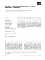
Fig. 1. p70
ik3-1
is phosphorylated by both p35
cdk3
and p33
cdk2
in
vitro. (A) Lysates from Tn5 insect cells not infected (lane 1), infected,
or coinfected with indicated baculoviruses were utilized for kinase
assays with GST–pRb (upper panel), GST–ik3-1 (middle panel), or
GST–cdk2 (lower panel) as substrates. A, E, k2, and k3 correspond to
the baculoviruses encoding cyclin A, cyclin E, cdk2, and cdk3.
Approximately 0.5 mg lysates were used for each reaction. (B) Lysates
from COS7 cells (1 Â 10
6
) were immunoprecipitated with the
nonimmune rabbit serum (N) and the anti-cdk2 Ig (k2) in the presence
or absence of 5 mg of BSA or the bacterially generated His-tagged
p27
Kip1
. Immunoprecipitates were then used for kinase assays with
GST–pRb (lanes 1–4) and GST–ik3-1DN (lanes 5–8) as substrates.
Because phosphorylation of GST–ik3-1DN and GST –ik3-1 by the
baculovirally generated cyclin/cdk kinases occurs in a similar fashion
(data not shown), we used GST–ik3-1DN as substrates in this
experiment.
Fig. 2. p70
ik3-1
is phosphorylated by cyclin/cdk3 reconstituted in
COS7 cells. (A) Lysates from cells (1 Â 10
6
) tranfected with pCMV–
cyclin A (lanes 1, 3, 5, and 7), pCMV–cyclin E (lanes 4 and 8), or the
backbone vector (lane 2 and 6) in association with either pCMV–cdk3
(indicated as 1) or the backbone vector ( –), were immunoprecipitated
with the nonimmune rabbit serum (N) and the anti-cdk3 Ig (k3).
Approximately 250 mg of lysates were contained in each immunopre-
cipitation. Immunoprecipitates were then used for kinase assays with
GST–pRb (lanes 1–4) and GST–ik3-1 (lanes 5–8) as substrates. (B)
Lysates from cells (1 Â 10
6
) tranfected with pCMV–cyclin A (lanes
2–5), or the backbone vector (lane 1) in association with either pCMV–
cdk3, pCMV–cdk3 dominant-negative form (cdk3 dn) or the backbone
vector (–), was immunoprecipitated with the nonimmune rabbit serum
(N) and the anti-cdk3 Ig (k3). Approximately 250 mg of lysates was
contained in each immunoprecipitation. Immunoprecipitates were then
used for kinase assays with GST –ik3-1 as substrates.
6078 T. Yamochi et al.(Eur. J. Biochem. 268) q FEBS 2001
anti-FLAG Ig, we obtained a larger amount of
32
P-labeled
FLAG–p70
ik3-1
from these cells (Fig. 3A, lane 3 of the
upper panel) than that from cells in which neither cdk3 nor
cyclin E was overexpressed (lane 2), or that from cells in
which both dominant-negative cdk3 and cyclin E were
overexpressed (lane 4). The lower panel of Fig. 3A
demonstrates that similar amounts of FLAG–p70
ik3-1
were
expressed in each transfection. If cyclin E was replaced with
cyclin A in the system, a similar result was obtained
(Fig. 3B). We could therefore conclude that p70
ik3-1
is a
substrate for cdk3-mediated phosphorylation. On the
contrary, however, the amount of labeled FLAG–p70
ik3-1
was not apparently increased in COS7 cells in which
cyclin E/cdk2 activity was potentiated by the cotransfection
of pCMV–cyclin E and pCMV– cdk2 (Fig. 3C). Currently,
we cannot therefore conclude that p70
ik3-1
is a substrate
for cdk2-mediated phosphorylation. As an answer to the
question of why the dominant-negative cdk3 did not
reduce phosphorylation of FLAG– p70
ik3-1
below the
normal state (compare lanes 2 and 4 of the upper panel in
Fig. 3A, and lanes 1 and 3 in Fig. 3B), we assume that other
types of kinases phosphorylate FLAG–p70
ik3-1
at different
sites (see Fig. 4) and render obscure the effect of the
dominant-negative cdk3 on its total phosphorylation.
Ser274 of ik3-1 is phosphorylated by cdk3
in vitro
Although there are no classical consensus sites (S/T-P-X-R/
K) for cdk-mediated phosphorylation in ik3-1, Ser274
followed by a P-R-P-K sequence resembles the site in p53
which is phosphorylated by cyclin A/cdk2 [16]. We
therefore asked whether this position is the phosphorylated
Fig. 3. p70
ik3-1
is phosphorylated by cyclin/cdk3 in vivo. (A–C)
COS7 cells (2 Â 10
6
) transfected with indicated vectors or the
backbone vectors, were labeled with [
32
P]orthophosphate. Cleared
lyasates were immunoprecipitated with the anti-FLAG Ig (F) or the
nonimmune rabbit serum (N). The same set of unlabeled transfected
cells (5 Â 10
5
) was harvested in parallel to estimate FLAG –p70
ik3-1
expression with sequential immunoprecipitation-immunoblotting with
anti-FLAG Ig in the lower panel of (A). E, A and dn indicate cyclin E,
cyclin A and a dominant-negative form of cdk.
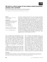
Fig. 4. Ser274 of p70
ik3-1
is phosphorylated by cyclin E/cdk3 in
vitro. Lysates from Tn5 insect cells not infected (lanes 1–3), or
coinfected with baculoviruses producing cyclin E and cdk3 (lanes 4–6)
were used for kinase assays with GST–ik3-1 (lanes 1 and 4), GST –
(S274T)ik3-1 (lanes 2 and 5), or GST–(S274A)ik3-1 (lanes 3 and 6) as
substrates. GST–ik3-1 and its mutant proteins eluted from lanes 4 – 6
were subject to digestion with trypsin and two-dimensional peptide
mapping. Spots A and B of peptide mapping for lane 4, and spots A
0
and
B
0
of peptide mapping for lane 5, were subject to two-dimensional
phosphoamino-acid analysis, as shown in the bottom panels.
Radioactive phosphopeptides were visualized with FLA 2000 Bioimage
Analyzer after five-day exposure (4 and 5), 10-day exposure (6).
Radioactive phosphoamino acids were visualized after 4-day exposure.
O indicates the origin of electrophoresis. W, S-T, and S-A indicate
GST–ik3-1, GST–(S274T)ik3-1, and GST–(S274A)ik3-1. S, T and Y
indicate phosphoserine, phosphothreonine and phosphotyrosine.
q FEBS 2001 Cdk3-dependent phosphorylation of ik3-1 (Eur. J. Biochem. 268) 6079
site in ik3-1. To this end, we produced mutant GST–ik3-1
proteins as substrates for in vitro kinase assays by replacing
Ser274 with threonine [(S274T)ik3-1] or alanine
[(S274A)ik3-1] using the site-directed mutagenesis tech-
nique. As expected, baculovirally generated cyclin E/cdk3
phosphorylated both wild-type GST–ik3-1 and GST–
(S274T)ik3-1 efficiently (Fig. 3, lanes 4 and 5) while it
phosphorylated GST–(S274A)ik3-1 to a much smaller
degree (lane 6), indicating that Ser274 is the main target for
cdk3-dependent phosphorylation. This was also true if the
baculovirally produced cyclin E/cdk2 or cyclin A/cdk3 as
well as anti-cdk2 immunoprecipitates from COS7 cells were
used as kinase source in place of the baculovirally produced
cyclin E/cdk3 (data not shown).
Furthermore, to analyze in detail, we eluted radioactive
GST–ik3-1 proteins from dried gels and digested them with
(Tos-Phe-CH
2
Cl)-trypsin, and then conducted two-dimen-
sional peptide mapping analysis (Fig. 4, middle panels). We
recognized two major phosphopeptide spots, A and B, with a
few spots with weaker radioactivity in the wild-type GST–
ik3-1 panel (middle left panel). Spot B in the wild-type
p70
ik3-1
panel was not observed in the S274A mutant panel
(middle right panel). At a glance, spot A seemed to exist in
the same mutant panel indicated as C (middle right panel).
However, if we estimate the relative radioactivity of spot A
or C against other spots, we could speculate that the phos-
phopeptide corresponding to spot C in the S274A mutant
seemed to represent one of the background phosphopeptides
that was normally hidden behind the spot corresponding to
A. This interpretation was also supported by the observation
that the migration pattern of spot C was similar to but
apparently not the same as that of phosphopeptide A (middle
left and right panels). Here we came to notice that both spot
A and spot B disappeared if Ser274 was replaced with
alanine. Regarding the relationship between spot A and spot
B, we have speculated that the phosphopeptides correspond-
ing to spot A and B arised by incomplete tryptic cleavage of
the Ser274-containing region. In fact, phosphoamino-acid
analysis of spots A and B indicated that both spots A and B
contained phosphoserine (Fig. 4, bottom left two panels).
Furthermore, we observed that re-digestion with trypsin of
the phosphopeptide purified from spot A gave rise to both
spot A and spot B by another two-dimensional peptide
mapping (data not shown), indicating that the assumption is
true. Spots A
0
and B
0
in the S274T mutant panel may
correspond to mutated phosphopeptides, in which threonine
substituted for serine was phosphorylated by cyclin E/cdk3,
and which therefore migrated a little differently from wild-
type ones (middle central panel). Phosphoamino-acid
analysis of spot A
0
and B
0
indicated that both spots A
0
and
B
0
contained phosphothreonine (Fig. 4, bottom right two
panels), strongly supporting that Ser274 is phosphorylated
by cyclin/cdk3.
Ser274 of ik3-1 also is phosphorylated by cdk3
in vivo
Next, we asked whether Ser274 of p70
ik3-1
is phosphorylated
by cyclin E/cdk3 intracellularly in mammalian cells. Another
in vivo labeling experiment for this purpose indicated that
in COS7 cells cotransfected with both pCMV–cyclin E and
pCMV–cdk3, the phosphorylation level of the wild-type
FLAG–p70
ik3-1
, but not that of FLAG– (S274A)p70
ik3-1
,is
upregulated as compared with control cells (Fig. 5, upper
panel, lanes 1 vs. 2, lanes 3 vs. 4). Purified FLAG–p70
ik3ÿ1
was then digested with trypsin and subjected to two-
dimensional peptide mapping. Here we observed that there
were several phosphopeptide spots including two spots
(indicated as A and B) that seemed to migrate similarly to
spots A and B in the wild-type GST–ik3-1 panel (compare
Fig. 4, lower panel 1 with Fig. 3, lower left panel). To
confirm that these spots A and B in the in vivo labeled
FLAG–p70
ik3-1
panel were the same as spots A and B in the
wild-type GST–ik3-1 panel, we mixed whole phosphopep-
tides generated by digestion with trypsin from the wild-type
FLAG–p70
ik3-1
labeled in vivo, and phosphopeptides
purified from spots A and B of in vitro labeled wild-type
GST–ik3-1 on a TLC plate (Fig. 3, the lowest panel). We
then conducted another two-dimensional peptide mapping
(Fig. 4, panel ‘mix’). We were able to see that the
Fig. 5. Ser274 of p70
ik3-1
is phosphorylated in vivo. COS7 cells
(2 Â 10
6
) transfected with indicated vectors were labeled with
[
32
P]orthophosphate. Cleared lysates were immunoprecipitated with
the anti-FLAG Ig. WT and S-A indicate pMF–ik3-1 for expression of
wild-type FLAG–p70
ik3-1
(lanes 1 and 2) and pMF–(S274A)ik3-1 for
expression of FLAG–(S274A)p70
ik3-1
(lanes 3 and 4), respectively.
They were cotranfected with pCMV–cyclin E and pCMV–cdk3 (lanes
2 and 4) or the backbone vectors (lanes 1 and 3). Wild-type FLAG–
p70
ik3-1
(lanes 1 and 2) and S274A mutant FLAG–p70
ik3-1
(lanes 4)
were eluted, digested with trypsin, and subjected to two-dimensional
peptide mapping (three middle panels). In vivo labeled wild-type
FLAG –p70
ik3-1
was purified from another gel, digested with trypsin,
mixed with one-fifth amount of phosphopeptides purified from spots A
and B of the GST–ik3-1 in the lower left panel of Fig. 4, and then
subjected to two-dimensional peptide mapping (panel ‘mix’).
Radioactive phosphopeptides were visualized by FLA 2000 Bioimage
Analyzer after 14-day exposure (1 and 2) and 21-day exposure (4). O
indicates the origin of electrophoresis.
6080 T. Yamochi et al.(Eur. J. Biochem. 268) q FEBS 2001
phosphopeptides, corresponding to spots A and B derived
from the in vivo labeled FLAG–p70
ik3-1
, comigrated in a
similar fashion with those from in vitro labeled GST–ik3-1,
indicating that the above assumption is true.
Moreover, if we estimate the radioactivity of phospho-
peptide spots A and B against those of the other spots, we can
recognize that the radioactivity of peptides corresponding to
spots A and B from FLAG–p70
ik3-1
in lane 2, are apparently
upregulated as compared with that from FLAG–p70
ik3-1
in
the lane 1 (Fig. 5, lower panels 1 vs. 2), suggesting that
cotransfected cyclin E/cdk3 increased the overall phos-
phorylation level of FLAG–p70
ik3-1
by increasing phos-
phorylation of Ser274. This fact confirms that Ser274 is the
major target residue phosphorylated by cdk3-mediated
kinase activity, not only in vitro but also in vivo.
Furthermore, to examine whether spots corresponding to
phosphopeptides A and B derived from FLAG–p70
ik3ÿ1
disappear if Ser274 is disrupted by site-directed mutagen-
esis, we eluted S274A mutant FLAG–(S274A)p70
ik3-1
from
the lane 4 gel of Fig. 5 and conducted two-dimensional
peptide mapping (Fig. 5, middle right panel 4). The A- and
B-corresponding spots disappeared while the D-correspond-
ing spot still existed. Unexpectedly, a weak spot corre-
sponding to spot E also seemed to disappear. We do not
know why this happened. However, we speculate that
phosphorylation giving rise to the spot E may occur only
when Ser274 is phosphorylated, or alternatively the S274A
mutation may induce a conformational change of the protein
blocking phosphorylation of the spot E-corresponding
peptide. We obtained similar results when we analyzed
S274A mutant FLAG–(S274A)p70
ik3-1
from the lane 3 gel
of Fig. 5 by two-dimensional peptide mapping (data not
shown). Thus, although we cannot completely exclude the
possibility that the S274A mutation induces a confor-
mational change blocking phosphorylation of non-Ser274
sites by cyclin/cdk3, we could assume that Ser274 is
phosphorylated by cyclin/cdk3 in vivo as it is in vitro.
Based on these data, we can consider that p70
ik3-1
is one
of the substrates for cdk3, while it is still possible that cdk3
indirectly increases phosphorylation of p70
ik3-1
through
some other unknown mechanism.
DISCUSSION
ik3-1 cDNA was originally cloned by its protein –protein
interaction with p35
cdk3
[8]. Both in vitro and in vivo,
p70
ik3-1
is considered to be a substrate for cdk3-dependent
phosphorylation. Furthermore, p70
ik3-1
could also be a sub-
strate for cyclin/cdk2 in vitro (Fig. 1). However, its inter-
action with p33
cdk2
is relatively weak [8], and in vivo
phosphorylation of p70
ik3-1
was not apparently enhanced by
overexpression of cyclin/cdk2 (Fig. 3C), suggesting that the
ik3-1-mediated pathway is mainly regulated by cdk3. This
result reminds us of the foregoing observation that the
dominant-negative cdk3-mediated G1 arrest is not com-
pletely rescued by wild type cdk2 in human osteosarcoma
cells [6], indicating that the function of cdk3 is at least
partially distinct from that of cdk2 in G1 progression.
Intriguingly, ik3-1 has no classical cdk sites (S/T-P-X-R/K)
that are phosphorylated by cyclin/cdk2 or cyclin B/cdc2.
Instead, it contains a S-P-R-P-K sequence at residues 274–278
that resembles the S-P-Q-P-K-K sequence of human p53,
which is phosphorylated by cyclin A/cdk2 [16]. Site-directed
mutagenesis procedures and the two-dimensional peptide
mapping analysis have established that Ser274 in p70
ik3-1
is
phosphorylated by both cdk3/cyclin A and cdk3/cyclin E in
vitro (Fig. 4). The same residue is also phosphorylated by both
cdk3/cyclin A and cdk3/E in vivo (Fig. 5). Analysis of ik3-1
amino-acid sequence indicates that ik3-1 has a putative ZRXL
(Z and X are typically basic) motif that would allow it to be a
substrate by cyclin/cdk3 as well as cyclin/cdk2 through binding
to cyclin, leading to speculation that binding of the cyclin
subunit, but not cdk3, to ik3-1 might be required for
phosphorylation of ik3-1. The fact that cyclin/cdk2 could
phosphorylate ik3-1 in vitro supports this assumption.
In postmitotic neurons, ik3-1 or Cables [9] may enhance
neurite growth by potentiating c-abl-mediated tyrosine
phosphorylation of cdk5. Tyr14-phosphorylated cdk5 is
more active in vitro, and ik3-1 is phosphorylated by p35/
cdk5 in vitro [9]. In spite of this observation, it still remains
to be clarified how ik3-1 functions in non-neuronal cells
because ik3-1 is basically expressed ubiquitously and cdk5
is inactive in non-neuronal cells [10]. In COS7 cells, cdk5
activity is also undetectable even after ik3-1 is over-
expressed (M. Matsuoka, unpublished observation).
Accordingly, we can conclude that in COS7 cells, Ser274
in p70
ik3-1
is phosphorylated by endogenous kinases other
than cdk5 (Fig. 4), at least one of which is cdk3 as shown in
this work. Currently, however, the question of how ik3-1
function is modified by the cdk3-mediated phosphorylation
of Ser274 remains to be addressed.
One of the major issues in the cell-cycle field is how G1
cyclin/cdks accelerates the mammalian G1 –S progression
and commits to DNA replication. To address this question,
the substrates and target molecules need to be clarified. So
far, it has been shown that cyclin E/cdk2 phosphorylates
pRb, which is also the sole known substrate for cyclin
D/cdk4 or cyclin D/cdk6 at this time. Hyperphosphorylating
pRb and upregulating free E2F, both cyclin E/cdk2 and
cyclin D/cdk4 or cyclin D/cdk6 co-operatively promote the
transcription of various genes, including the cyclin E gene
necessary for G1– S transition [1,2]. Other candidate sub-
strates for cyclin E/cdk2 include NPAT [17], components of
the premRNA splicing machinery [18] and Id2 [19], and
more are emerging. In this respect, the functional analysis of
ik3-1, a candidate target for cdk3, will contribute to the
further understanding of cdk3 function in the mammalian
G1–S transition which is distinct from the cdk2 function in
self-replicating cells.
ACKNOWLEDGEMENTS
We are indebted to Tomo Yoshida, Kazumi Nishihara, Kouichi
Tsuchiya, Fusano Igarashi and Dovie Wylie for expert technical
assistance; Drs Jiyong Zhao, S. van den Heuvel, Ed Harlow, Andrew
Koff, Charles J. Sherr, Hiroshi Hirai, Makoto Nakanishi and Hitoshi
Matsushime for providing us with plasmids and baculoviruses. This
work is supported in part by grant from the Ministry of Education,
Culture, Sports, Science, and Technology of Japan, the Organization for
Pharmaceutical Safety and Research and KEIO University Special
Grant-in-Aid for Innovative Collaborative Research Projects.
REFERENCES
1. Sherr, C.J. (1993) Mammalian G1 cyclins. Cell 73, 1059–1065.
2. Resnitzky, D. & Reed, S.I. (1995) Different roles for cyclins D1 and
q FEBS 2001 Cdk3-dependent phosphorylation of ik3-1 (Eur. J. Biochem. 268) 6081
E in regulation of the G1-to-S transition. Mol. Cell. Biol. 15,
3453–3469.
3. Meyerson, M., Enders, G.H., Wu, C.L., Su, L.K., Gorka, C.,
Nelson, C., Harlow, E. & Tsai, L.H. (1992) A family of human
cdc2-related protein kinases. EMBO J. 11, 2909–2917.
4. Harper, J.W., Elledge, S.J., Keyomarsi, K., Dynlacht, B., Tsai, L.H.,
Zhang, P., Dobrowolski, S., Bai, C., Connell-Crowley, L., Swindell,
E., Fox, M.P. & Wei, N. (1995) Inhibition of cyclin-dependent
kinases by p21. Mol. Biol. Cell 6, 387–400.
5. Meikrantz, W. & Schlegel, R. (1996) Suppression of apoptosis by
dominant negative mutants of cyclin-dependent protein kinases.
J. Biol. Chem. 271, 10205–10209.
6. van den Heuvel, S. & Harlow, E. (1993) Distinct roles for cyclin-
dependent kinases in cell cycle control. Science 262, 2050–2054.
7. Hofmann, F. & Livingston, D.M. (1996) Differential effects of cdk2
and cdk3 on the control of pRb and E2F function during G1 exit.
Genes Dev. 10, 851–861.
8. Matsuoka, M., Matsuura, Y., Semba, K. & Nishimoto, I. (2000)
Molecular cloning of a cyclin-like protein associated with cyclin-
dependent kinase 3 (cdk 3) in vivo. Biochem. Biophys. Res.
Commun. 273, 442–447.
9. Zukerberg, L.R., Patrick, G.N., Nikolic, M., Humbert, S., Wu, C.L.,
Lanier, L.M., Gertler, F.B., Vidal, M., Van Etten, R.A. & Tsai, L.H.
(2000) Cables links Cdk5 and c-Abl and facilitates Cdk5 tyrosine
phosphorylation, kinase upregulation, and neurite outgrowth.
Neuron 26, 633–646.
10. Lew, J. & Wang, J.H. (1995) Neuronal cdc2-like kinase. Trends
Biochem. Sci. 20, 33 –37.
11. Matsuoka, M., Tani, K. & Asano, S. (1998) Interferon-alpha-
induced G1 phase arrest through up-regulated expression of CDK
inhibitors, p19Ink4D and p21Cip1 in mouse macrophages.
Oncogene 16, 2075–2086.
12. Kato, J., Matsushime, H., Hiebert, S.W., Ewen, M.E. & Sherr, C.J.
(1993) Direct binding of cyclin D to the retinoblastoma gene
product (pRb) and pRb phosphorylation by the cyclin D-dependent
kinase CDK4. Genes Dev. 7, 331–342.
13. Matsuura, Y., Possee, R.D., Overton, H.A. & Bishop, D.H. (1987)
Baculovirus expression vectors: the requirements for high level
expression of proteins, including glycoproteins. J. Gen. Virol. 68,
1233–1250.
14. Matsuoka, M., Kato, J., Fisher, R.P., Morgan, D.O. & Sherr, C.J.
(1994) Activation of cyclin-dependent kinase 4 (cdk4) by mouse
MO15-associated kinase. Mol. Cell. Biol. 14, 7265–7275.
15. Boyle, W.J., van der Geer, P. & Hunter, T. (1991) Phosphopeptide
mapping and phosphoamino acid analysis by two-dimensional
separation on thin-layer cellulose plates. Methods Enzymol. 201,
110–149.
16. Wang, Y. & Prives, C. (1995) Increased and altered DNA binding of
human p53 by S and G2/M but not G1 cyclin-dependent kinases.
Nature 376, 88–91.
17. Zhao, J., Dynlacht, B., Imai, T., Hori, T. & Harlow, E. (1998)
Expression of NPAT, a novel substrate of cyclin E-CDK2, promotes
S-phase entry. Genes Dev. 12, 456–461.
18. Seghezzi, W., Chua, K., Shanahan, F., Gozani, O., Reed, R. & Lees,
E. (1998) Cyclin E associates with components of the pre-mRNA
splicing machinery in mammalian cells. Mol. Cell. Biol. 18,
4526–4536.
19. Hara, E., Hall, M. & Peters, G. (1997) Cdk2-dependent phos-
phorylation of Id2 modulates activity of E2A-related transcription
factors. EMBO J. 15, 332–342.
6082 T. Yamochi et al.(Eur. J. Biochem. 268) q FEBS 2001