Báo cáo y học: "Are chest compressions safe for the patient reconstructed with sternal plates? Evaluating the safety of cardiopulmonary resuscitation using a human cadaveric model" docx
Bạn đang xem bản rút gọn của tài liệu. Xem và tải ngay bản đầy đủ của tài liệu tại đây (758.22 KB, 4 trang )
RESEARC H ARTIC L E Open Access
Are chest compressions safe for the patient
reconstructed with sternal plates? Evaluating the
safety of cardiopulmonary resuscitation using a
human cadaveric model
Douglas R McKay
1*
, Hosam F Fawzy
2
, Kathryn M McKay
3
, Romy Nitsch
4
, James L Mahoney
5
Abstract
Background: Plate and screw fixation is a recent addition to the sternal wound treatment armamentarium.
Patients undergoing cardiac and major vascular surgery have a higher risk of postoperative arrest than other
elective patients. Those who undergo sternotomy for either cardiac or major vascular procedures are at a higher
risk of postoperative arrest. Sternal plate design allows quick access to the mediastinum facilitating open cardiac
massage, but chest compressions are the mainstay of re-establishing cardiac output in the event of arrest. The
response of sternal plates and the chest wall to compressions when plated has not been studied. The safety of
performing this maneuver is unknown. This study intends to demonstrate compressions are safe after sternal
plating.
Methods: We investigated the effect of chest compressions on the plated sternum using a human cadaveric
model. Cadavers were plated, an arrest was simulated, and an experienced physician performed a simulated
resuscitation. Intrathoracic pressure was monitored throughout to ensure the plates encountered an appropriate
degree of force. The hardware and viscera were evaluated for failure and trauma respectively.
Results: No hardware failure or obvious visceral trauma was observed. Rib fractures beyond the boundaries of the
plates were noted but the incidence was comparable to control and to the fracture incidence after resuscitation
previously cited in the literature.
Conclusions: From this work we believe chest compressions are safe for the patient with sternal plates when
proper plating technique is used. We advocate the use of this life-saving maneuver as part of an ACLS resuscitation
in the event of an arrest for rapidly re-establishing circulation.
Background
Chest compress ions are a cornerstone of cardiopulmon-
ary resuscitation. Recent work confirms the importance
of early compressions to improve survival [1]. Oxygen is
present in the blood up to ten minutes after arrest; re-
establishing circulation of this blood via sternal com-
pressions is the most important step of the ABCs early
in resuscitation [2].
Sternal wound dehiscence after median sternotomy
can be a devastating complication. The mainstay of
treatment has been a ggressive debridement followed by
flap closure. This diminishes mechanical c hest wall
integrity. A new advance, sternal repair with plate and
screw fixation, can obviate the complications of persis-
tent sternal instability. These include chro nic pain, para-
doxical chest wall motion, and decreased pulmonary
function [3]. The modality is safe when used appropri-
ately and confers the advantages of early extubation,
tension-free repair and simple soft tissue advancements
in lieu of more complicated flaps whilst restoring
mechanical stability [4].
Cardiac or major vascular surgery places patients at a
higher risk for perioperative cardiac events, and the
* Correspondence:
1
Department of Surgery, Queen’s University, Kingston, Ontario, Canada
Full list of author information is available at the end of the article
McKay et al. Journal of Cardiothoracic Surgery 2010, 5:64
/>© 2010 McKay et al; licensee BioMed Ce ntral Ltd. This is an Open Access article distributed under the terms of the Creative C ommons
Attribution Licens e ( nses/by/2.0), which pe rmits unrestricted use, distribution, and reproduct ion in
any medium, provided the original work is properly cited.
subset whose wounds dehisce are typically at higher risk
on the basis of medical comorbidity [5,6]. Some of this
population will require perioperative resuscitation. The
response of sternal plates and the plated chest wall to
compressions has not been studied. Potential hypothe-
sized pitfalls include hardware failure or skeletal and
visceral trauma.
To determine the safety of performing this potentially
life-saving maneuver, we designed an experiment to
study the effects of chest compressio ns on sternal hard-
ware and the thorax. We studied these outcomes using
a human cadaveric model while monitoring intrathor-
acic pressure during a simulated resuscitation.
Methods
Institutional Review Board ethics approval was applied
for and granted for this study by the University of Tor-
onto Ethics Review Office, protocol reference # 18535.
Compressions were performed on an un-pla ted cada-
ver to serve as control. Intrathoracic pressures were
monitor ed in the control with the intrathoracic pressure
monitoring system detailed below, placed inferior to the
sternum through an incision in the diaphragm. No ster-
notomy was performed on the control experiment. The
anterior thorax was exposed and checked for fracture.
Observations were documented. In the experimental
group, a midline sternotomy was performed on five
fresh frozen cadavers. Bilateral composite myocutaneous
pectoralis major flaps were elevated exposing the ante-
rior thorax for plating.
A digital manometer that records pressure within a
closed system at appropriate range and intervals was
selected (Reed PM9100®, Alaron Instruments, Newmar-
ket, ON). A 250 cc silicone bladder measuring 10 cm
across was connected via fill tube and intravenous tub-
ing to t he manometer to create a closed system. This
bladder was then seated immediately deep to the infer-
ior third of the sternum and distended with air to con-
form to the cavity in which it was placed. (Mentor
Corp., Santa Barbara, CA). The manometer was con-
nected via RS232 cable to a laptop and configured to
display real-time intrathoracic pressure while simulta-
neously recording absolute values, both in mmHg, every
two seconds (SW-U801 for Windows®, Alaron Instru-
ments, Newmarket, ON, Canada).
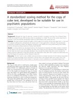
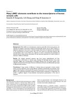
The sternum was reduced and held with forceps. The
cadaver was plated using three rib plates combined with
asinglemanubrialplate(SeeFig.1).Ribplateswere
placed on the second, third and fourth ribs (Titanium
Sternal Fixation System, Synthes, USA). Holes were
drilled using the system guide. A depth gauge was used
to select the appropriate screw length. Our intent was
that screws would catch the deep cortex without signifi-
cantly breaching the cortex.
The incisions were closed in a layered fashion. Vicryl
2.0 sutures (Johnson & Johnson, Piscataway, NJ) were
used for the deep layer and the skin was closed with
skin staples (3 M, St. Paul, MN). The manometer was
zeroed. A physician trained and experienced in perform-
ing c ardiopulmonary resuscitation carried out compres-
sions for a total of five minutes at a rate of 60 to
80 compressions per minute, on both the control and
cadaver specimens. Intrathoracic pressures were displayed
to the physician performing the resuscitation and chest
compressions were maintaine d at a depth that generated
minimum peak intrathoracic pressures of 60 mmHg.
The incisions were opened and the hardware and
thorax were examined for trauma. Observations were
recorded and photo-documented. An oscillating saw was
used to completely excise the anterior tho rax. The deep
surfaces of the skeletal thorax and the viscera were
examined for trauma. Th e plates and screws wer e
removed. Each screw was removed from the plates and
each plate was disassembled. Each screw, pin and plate
was examined for damage or failure.
Results
The plating mechanism was visually evaluated for
damage and checked for functional compromise. No
screw, pin or plate damage, or failure was noted. All pins
and screws were removed with ease. All plates easily dis-
engaged at the midline; there was no compromise of the
mediastinal access mechanism secondary to the sustained
compressions. No obvious pleural or visceral damage was
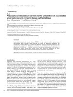
noted. No rib fractures were noted in the plated zone.
Rib fractures were noted in all cadavers beyond the limits
of the sternal plates. (See Fig. 2) Two fractures were
noted in a control specimen after an identical compres-
sion sequence (See Table 1). We were unable to physi-
cally generate a force that fractured the hardware.
Figure 1 Plated sternum.
McKay et al. Journal of Cardiothoracic Surgery 2010, 5:64
/>Page 2 of 4
Discussion
Predicting the risk of perioperative cardiac events is a
complicated science. Patient risk estimates are based
on a number of known risk factors. Cardiac and major
vascular surgery places a patient at a higher risk for
perioperative cardiac events and is highest for coronary
artery surgery [7]. Those who dehisce sternotomy
wounds may do so as the result of medical comorbid-
ity. It follows that plated patients are more likely to
arrest and require resuscitation. The safety of perform-
ing chest compressions in this group merits
investigation.
Chest compressions are a traumatic procedure. Rib
fracture is the most common c omplication. In a recent
review, Hoke et al. summarize the literature on skeletal
injury as a result of chest compression and discover a
spectrum of fracture incidence in resuscitated adults
ranging from 12.9% to 96.6% [8]. The most common
complication of rib fractures is pain; pain may inhibit
deep breathing, which may increase the risk of at electa-
sis or pneumonia. Despite the potential morbidity, they
emphasize that the value of chest compressions out-
weigh the risk of skeletal damage and conclude that the
risk of fracture should not deter an adequate and
appropriate cardiopulmonary resuscitation in the event
of arrest.
In our model, the plates appear to bolster the chest
wall and prevent fracture immediately deep to the plated
thorax. Our fracture incidence is higher than in the lit-
erature but consistent with our control. The observed
fractures were all significantly beyond the plates and
predominantly immediately lateral to the plates on the
plated ribs. The incidence of rib fracture may be higher
when compressions are performed on the plated ster-
num. This high incidence we observed may be a result
of the frailty of the elderly cadaveric model, when com-
pared to the documented incidence in the living.
One hypothesized source of morbidity was hardware
failure and i ts potential to damage underlying viscera
under dynamic compression. No hardware fracture or
loosening was noted for either plates or screws. All were
removed and examined individually. Both appear cap-
able of enduring the dynamic stresses and absolute pres-
sures encountered during resuscitation. We were unable
to physically apply forces via compressions that resulted
in hardware failure.
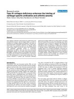
When the anterior thorax was removed and the cada-
ver examined, no obvious visceral trauma was noted
(see Fig. 3). The screw depth appeared appropriate;
none sat proud. Screws protruding from the inferior
cortex may cause significant damage. We cannot over-
emphasize the importance of proper screw selection
when plating. At our institution we use preoperative CT
scanning and measure and m ap absolute rib depth to
ensure appropriate screw selection.
In the living, the adequacy of chest compressions has
been measured via end-tidal CO
2
levels, depth of com-
pression and intra-tho racic pressure measurement. End
tidal CO
2
is the most commonly used modality. The
Figure 2 Rib fractures after resuscitation.
Table 1 Rib fracture incidence and position relative to
plated sternum; comparison between control and
cadaveric specimens
Specimen Number of fractures Location of fractures
Control 2 lateral, xyphoid
Cadaver 1 1 inferolateral
Cadaver 2 2 lateral, xyphoid
Cadaver 3 2 lateral
Cadaver 4 3 lateral
Cadaver 5 1 lateral
Figure 3 Elevation and examination of deep sternal cortex and
viscera.
McKay et al. Journal of Cardiothoracic Surgery 2010, 5:64
/>Page 3 of 4
cadaveric model is most amenable to intrathoracic pres-
sure measurement.
Peak aortic compression pressures of 61 ± 22 mmHg
have been measured via cook catheter during resuscita-
tions in humans when performed by individuals experi-
enced in c ardiopulmonary resuscitation [9]. In order to
ensure the plated sternum experienced an appropriate
and adequate compressive force, a pressure manometer
was attached to a closed bladder, and was inserted
immediately deep to the ster num. The absolute pressure
in this closed system was recorded every 2 seconds dur-
ing resuscitation and displayed to the physician per-
forming the compressions via digital readout. The
compression depth was maintained to create a mini-
mum peak intrathoracic pressure of 60 mmHg to accu-
rately simulate mechanical forces experienced during
the r esuscitation. The maximum recorded pressure was
87 mmHg. There i s a potential for slight inaccuraci es in
the absolute pressure measurements recorded.
This model has limitations. The distensible nature and
elasticity of the s ilicone shell, fill tube, and intravenous
tubing have the potential to alter pressure readings. Pre-
sumably this would result in a reading that was lower
than the absolute pressure at peak and during decom-
pression. Either scenario would mean the hardw are was
experiencing higher pressures than recorded. The sili-
cone shell when distended and placed deep to the ster-
num has the potenti al to damage the under lying viscera
but may also be protective.
The fresh frozen cadaveric model may not mimic the
dynamics in the living. The frailty of the frozen and
thawed cadavers may mislead us with regard to the true
fracture incidence. We were unable to procure fresh
cadavers; the use of fresh cadavers could significantly
improve this study. The cost of procuring and preparing
the cadavers limited the number of specimens used in
the study and the power may be inadequate.
If screws are protruding deep to the deep cortex, com-
pressions have the potential to inflict significant damage.
Perioper ative hypocoagulation may exace rbate potential
complications. The risk of excessive screw length caus-
ing trauma during compressions may justify a post-
operative CT. A significant breech of the deep cortex
may modify recommendations to ward staff in the event
of an arrest. One patient has a rrested and undergone
chest compressions after plating without adverse clinical
sequelae.
Conclusions
Based on our work with this human cadaveric model we
believe chest compressions are safe in the plated ster-
numintheeventofarrestwiththecaveatthatappro-
priate screw length must be chosen. Chest compressions
can be used to immediately re-establish blood flow and
temporize until the chest m ay be re-o pened according
to the accepted algorithm for resuscitation after cardiac
surgery. No hardware failure was observed. Rib fracture
incidence beyond plates was higher than in the literature
but comparable to control. Skeletal injury is well docu-
mented after chest compressions but fracture should
not deter first re sponders from using chest compres-
sions to re- establish circulation. This is also true for the
plated patient.
Acknowledgements
This research was funded by an independent Resident Trauma Research
Grant from the AO group of North America. The plating systems used in the
experiment were donated by Synthes, USA.
Author details
1
Department of Surgery, Queen’s University, Kingston, Ontario, Canada.
2
Department of Cardiac Surgery, University of Toronto, Toronto, Ontario,
Canada.
3
Oceanworks International, Burnaby, British Columbia, Canada.
4
Queen’s University, Department of Obstetrics and Gynecology, Kingston,
Ontario, Canada.
5
Department of Plastic Surgery, University of Toronto,
Toronto, Ontario, Canada.
Authors’ contributions
DRM wrote the grant and applied for funding, coordinated and executed
the cadaveric study, analyzed the results and wrote the manuscript. HSM
performed the sternotomy and plated the cadavers. KMM designed the
pressure monitoring device used in the study. RN participated in the
execution of the cadaveric resuscitation and plating study. JLM conceived of
the study, participated in its execution and reviewed and edited the
manuscript. All authors read and approved the final manuscript.
Competing interests
The authors declare that they have no competing interests.
Received: 14 December 2009 Accepted: 18 August 2010
Published: 18 August 2010
References
1. Wik L, et al: Delaying defibrillation to give basic cardiopulmonary
resuscitation by patients with out of hospital ventricular fibrillation: a
randomized trial. JAMA 2003, 289:1389-95.
2. Kern KB: Cardiopulmonary resuscitation without ventilation. Crit Care Med
2000, N186-9.
3. Yuen JC, Zhou AT, Serafin D, Gerogiade DS: Long-term sequelae following
median sternotomy wound infection and flap reconstruction. Ann Plast
Surg 1995, 35:585-589.
4. Cicilioni OJ, Stieg FH, Papanicolaou G: Sternal wound reconstruction with
transverse plate fixation. Plast Reconstr Surg 2005, 115(5):1297-30.
5. Detsky AS, Abrams HB, McLaughlin JR, et al: Predicting cardiac
complications in patients undergoing non-cardiac surgery. J Gen Intern
Med 1986, 1(4):211-9.
6. Goldman L, Caldera DL, Nussbaum SR, et al: Multifactorial index of cardiac
risk in noncardiac surgical procedures. N Engl J Med 1977, 297(16):845-50.
7. Tuman KJ: Perioperative myocardial infarction. Semin Thorac Cardiovasc
Surg 1991, 3(1):47-52.
8. Hoke RS, Chamberlain D: Skeletal chest injuries secondary to
cardiopulmonary resuscitation. Resuscitation 2004, 63(3):327-38.
9. Paradis NA, Martin GB, Goetting MG, et al: Simultaneous aortic, jugular
bulb, and right atrial pressures during cardiopulmonary resuscitation in
humans. Insights into mechanisms. Circulation 1989, 80:361-68.
doi:10.1186/1749-8090-5-64
Cite this article as: McKay et al.: Are chest compressions safe for the
patient reconstructed with sternal plates? Evaluating the safety of
cardiopulmonary resuscitation using a human cadaveric model. Journal
of Cardiothoracic Surgery 2010 5:64.
McKay et al. Journal of Cardiothoracic Surgery 2010, 5:64
/>Page 4 of 4