Báo cáo y học: "An alternative surgical approach to subclavian and innominate stenosis: a case series" ppsx
Bạn đang xem bản rút gọn của tài liệu. Xem và tải ngay bản đầy đủ của tài liệu tại đây (686.35 KB, 4 trang )
CAS E REP O R T Open Access
An alternative surgical approach to subclavian
and innominate stenosis: a case series
Amina Khalil
1*
, Samer AM Nashef
2
Abstract
We report three cases of symptomatic stenosis of the great vessels or supra-aortic trunks successfully treated surgi-
cally with aorto-subclavian and aorto-innominate bypass. Two were performed via manubriotomy and a third case
via standard median sternotomy because of concomitant coronary revascularisation. There was complete sympto-
matic relief on follow-up, and radiological imaging confirmed good flow in the grafts and post-stenotic arteries.
Background
Like other arteries, the innominate, left comm on carotid
and subclavian arteries or supra-aortic trunks (SATs)
can be affected by atherosclerosis. Many patients with
SAT disease are asymptomatic, but some may present
with symptoms of cerebral or limb ischaemia. The use
of endovascular intervention for SAT occlusive disease
is increasing but open surgical reconstruction remains
an effective treatment option with good long term
results. Although the cervical approach for the treat-
ment of SAT disease has proven to be a good surgical
option over the years, a transthoracic approach can pro-
vide durable results particularly when the disease pro-
cess affects all three trunks or involves long segments
[1]. The morbidit y associated with the transthoracic
route may be reduced by using a less invasive approach
such as manubriotomy. A short summary of clinical pre-
sentation, the surgical technique employed and the out-
comes forms the basis of the present case series.
Case 1
A 64-year-old male presented with frequent episodes o f
dizziness after myocardial infarction. Ambulatory 24-
hour cardiac monitoring showed periods of asystole, and
a dual chamber pacemaker was implanted. The patient
remained symptomatic with the same frequency of dizzy
spells and reported syncopa l episodes precipita ted by
left arm exertion. Contrast-enhanced spiral compu-
terised tomography (CT) revealed disease at the origin
of all great vessels , with an irregul ar 50% stenosis at the
origin of innomina te artery, a 70% stenosis at the origin
of the right subclavian and a 30% stenosis of at the ori-
gin of left common carotid artery. The first 15-mm seg-
ment of the left subclavian artery proximal to the origin
of left vertebral artery was totally occluded. The dist al
left subclavian filled by retrograde flow through the ipsi-
lateral vertebral artery (subclavian steal syndrome).
At operation, the s kin was incised above the clavicle
from the left mid-clavicular point to the suprasternal
notch and the incision extended vertically downwards
towards the manubriosternal junction. This was fol-
lowed by a vertical manub rioto my extending laterally to
the left, stopping short of the internal mammary pedicle.
A self-retaining retractor was used to separate the two
halves of the manubrium and to elevate the ster nal edge
on the left side, giving good ac cess to both the ascend-
ing aorta and the distal subclavian artery. Under full
heparinisation, the artery was clamped 3 cm distal to
the occlusion and a polytetrafluoroethylene graft was
anastomosed end-to-side beyond the occlusion using 5/
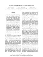
0 monofilament polypropylene. The graft was trimmed
to size and anastomosed to the ascending aorta using a
partial occlusion clamp (Fig 1). Heparin was reversed
and the incision closed over a small suction drain with a
figure-of-8 single sternal wire and sta ndard soft-tissue
closure. The patient made an uneventful recovery.
Repeat CT at 2 weeks demonstrated good antegrade fill-
ing of the distal left subclavian and vertebral artery from
the aorto-subclavian graft. The patient became comple-
tely symptom-free at clinical evaluation one year follow-
ing surgery.
* Correspondence:
1
Department of Cardiothoracic Surgery, John Radcliffe Hospital, Headley
Way, Headington, Oxford OX3 9DU, UK
Full list of author information is available at the end of the article
Khalil and Nashef Journal of Cardiothoracic Surgery 2010, 5:73
/>© 2010 Khalil and Nashef; licensee BioMed Central Ltd. This is an Open Access articl e distributed under the terms of the Creati ve
Commons Attribution License (http://c reativecommons.org/licenses/by/2.0), which permits unrestricted use, distribution, and
reproduction in any medium, provided the original work is properly cited.
Case 2
A 49-year-old female smoker presented with a two-year
history of intermittent diplopia, dizziness and ataxia. On
physical examination there was a diminished left radial
pulse and a bruit was audible in the left supraclavicular
region. Contrast-enhanced spiral volumetric CT images
showed patchy calcification at the origin and along the
course of all the great vessels. The first 10 mm segment
of the left subcl avian was completely occluded with ret-
rograde filling of the left subclavian and vertebral
arteries. She underwent the same procedure as Case 1
and made an uneventful recovery. Post-operative CT
showed good antegrade flow through the graft to distal
left subclavian artery. The patient remained symptom-
free at follow up review.
Case 3
A 68-year-old female presented with a 3-year history of
progressively worsening a ngina. She then devel oped
intermittent diplopia and subsequently complained of
exertional right arm pain. Angiography showed triple
vessel coronary artery disease and an occluded right
innominate artery. Doppler ultrasound showed
intermittent flow reversal in the right common carotid
artery and retrograde flow in the right vertebral artery
(subclavian and carotid steal). At operation, a standard
median sternotomy was performed with a small exten-
sion of the incision into the neck. The innominate artery
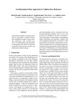
was clamped with a single partial occlusion clamp distal
to the lesion and a 5 mm Goretex graft sutured to it
under full heparinization (Fig 2). This was followed by
standard triple coronary artery bypass grafting. The aor-
tic cross clamp was removed and the innominate graft
was attached to the aorta in a similar fashion to the
proximal coronary anastomosis.
Postoperative magnetic resonance imaging showed a
patent aorto-innominate bypass with good antegrade
flow in the right carotid and subclavian arteries ( Fig 3).
The patient had an uneventful recovery with comple te
resolution of all symptoms (angina, diplopia and exer-
tional arm pain) on follow-up.
Discussion
Subclinical aortic atherosclerosis may start as early as
the second decade of life [2] and the commonest disease
in the aorta and the SATs is atherosclerotic in causa-
tion. Lesions develop principally in high shear stress
regions, which is in the zone of flow separat ion and is
associated with whirlpools that form near the lateral
wall of bifurcations. Plaques are relatively uncommon in
ascending aorta but more common in arch and descend-
ing thoracic aorta [3]. Hypertension, diabetes mellitus,
cigarette smoking, dyslipideamia and genetic
Figure 1 Aorto-subclavian Bypass Graft.
Figure 2 Aorto-innominate Bypass Graft.
Khalil and Nashef Journal of Cardiothoracic Surgery 2010, 5:73
/>Page 2 of 4
preponderance are common risk factors for developing
atherosclerosis and in combination have a greater than
additive effect. The majority of patient with SAT steno-
sis are asymptomatic, but some patients may present
with symptoms of vertebrobasilar ischaemia including
episodes of dizziness, diplopia, ataxia, vertigo, limb clau-
dication, paraesthesia and steal syndrome. Physical
examination may reveal diminished pulse and decreased
blood pressure (> 20 mmHg reduction compared to the
normal side) in the affected limb. The subclavian steal
syndrome is one of the best recogn ised presentations of
SAT stenosis. It is more common on the left side, per-
haps due to the acute angle at the origin of the left sub-
clavian artery which may result in accelerated
atherosclerosis from increased turbulence. SAT stenosis
can be diagnosed by digital subtraction angiography,
duplex scanning, contrast enhanced spiral CT, magnetic
resonance imaging and arch aortography.
The concept of extra-anatomic bypass was first intro-
duced in 1952 by Freeman and Leeds [4], when they
used superficial femoral artery to carry blood from one
femoral artery to other, and this procedure has now
become a widely used and accepted method of r evascu-
larisation. The physiologic basis of extra-anatomic
bypass reveals that inflow in the donor artery is the key
factor that determines the haemodynamic effects of
extra-anatomic bypass. If the inflow in the donor artery
is below a critical level of 60%, it may be insufficient to
supply adequate blood flow simultaneously to both the
distal segment of the donor artery and the bypass graft.
Moreover, the capacity of the donor artery to provide
increased blood flow on demand may be compromised
because of atherosclerosis oriatrogenicstenosisatthe
site of anastomosis. To ensure a good result the donor
artery should be free of disease and every precaution
should be taken to a void anastomotic stenosis [5]. The
increased flow demand following the extra-anatomic
bypass is met by increased flow in donor artery proxi-
mal to the anastomosis and the flow remai ns essentially
unaffected by changes in the outflow and hypotension.
The only factor that leads to the phenomeno n of vascu-
lar steal is restriction or obstruction of inflow in donor
artery.
Endovascular techniques are increasingly used in the
treatment of occlusive S AT disease because they are less
invasive, may be performed under local anaesthesi a and
are associated with shorter hospital stay. The vascular
patency rates reported in different studies are variable and
there are no randomised trials comparing endovascular
and open surgical approaches. The innominate artery may
present as a challenging SAT lesion for interventional
endovascular therapists, due to its larger diameter, and
short length between its origin and its bifurcation and
between the bifurcation and the take-off of vertebral artery
[6]. In addition it is sometimes difficult to negotiate a very
tight stenosis or occluded lesion through an endovascular
approach and the long term benefits of these therapies are
uncertain. Modarai et al. [7] reported a better patency and
lower complication rat e related to extra-ana tomic bypass
for SAT disease as compared to percutaneous endovascu-
lar intervention. In this series of 76 patients, with a mean
follow up of 5 years, the extra-anatomic graft patency was
97% with no complications against 82% patency for the
endovascular intervent ion with angioplasty with a rate of
complication of 11%.
In the past, atherosclerotic SAT stenosis was treated
with anatomic bypass between aortic arch and innomi-
nate, carotid and subclavian arterie s. Graft p atency was
good but perioperative mortality and stroke rates were
high [8]. This led to the introduction of safer extra-ana-
tomic approaches. The most commonly used open pro-
ceduresforSATstenosisinvolve a cervical approach,
which is ideally suited for single trunk disease that
involves either subclavian or common carotid arteries
[1]. The procedure may include endarterectomy, bypass
grafting from ipsilateral carotid artery or the subcuta-
neous crossover axillo-axillary bypass and transposition
of the subclavian to carotid artery. Subclavian transposi-
tion has the benefit of a single anastomosis and elimi-
nates the potential thrombotic and infection risks
associated with the use of prosthetic grafts or saphenous
veins [9]
.
In comparison to the cervical approach, the transthor-
acic approach may carry a relatively higher morbidity
but the results may be more durable in atherosclerotic
disease involving SATs [1].
Figure 3 Grafted Right Brachiocephalic Stenosis.
Khalil and Nashef Journal of Cardiothoracic Surgery 2010, 5:73
/>Page 3 of 4
Transthoracic direct aorto -subclavian and aorto-inno-
minate bypass have advantages in selected patients,
including those with atherosclerosis which spares the
ascending aorta, those isolated supra aortic trunks ste-
nosis involving long vessel segments and those with dis -
ease affecting potential donor arteries such as the
ipsilateral or contralateral carotid. Thus aorto-SAT
bypass can be used safely in patients with concomitant
carotid disease with reduced cerebral risks, avoids
endarterectomy and its attendant thrombotic risks and
provides a more physiological blood flow pattern than
axillo-axillary or carotid-subclavian bypass.
Manubriotomy is a small, cosmetically acceptable inci-
sion which is well tolerated and less painful than stan-
dard median sternotomy. This approach gives excellent
access to ascending aorta, arch and proximal supra aor-
tic trunk. Manubriotomy, being a less invasive techni-
que, can be used safely in patients with reduced
cardiopulmonary reserve, but caution should be taken in
previous mediastinal surgery or irradiation.
In patients with concomitant coronary artery disease
and vertebrobasilar ischaemia, both pathologies can be
dealt with simultaneously using a standard median ster-
notomy, avo iding the risks associated with seco nd
operation. Proximal ascending aortic anastomosis is a
commonly performed procedure in coronary artery sur-
gery and the technique is well established. Atherosclero-
sis is a progress ive disease and if one SAT stenosis is
present, future stenosis may develop in the feeding ves-
sel after carotid-subclavian bypass and axillo-axillary
bypass. Using the ascending aorta eliminates this risk.
Patients who have or will ha ve coronary revascularisa-
tion using an internal mammary artery as a conduit pre-
sent a special problem in subclavian and innominate
stenosis, as functionally the vascular segment between
the origin of subclavian artery and the coronary artery
becomes part of coronary circulation [10]. In such
patients, the myocardium may be dependent on subcla-
vian flow and care should be taken to ensure that what-
ever procedure is carried out, satisfactory antegrade flow
to the relevant subclavian artery can be assured f or the
long term or alternatives should be sought for the inter-
nal mammary graft to prevent a coronary-subclavian
steal syndrome and myocardial ischaemia.
Conclusion
In conclusion, SAT stenosis is an uncommon c ondition
which may be treated safely and effectively using aorto-
subclavian and aorto-innominate bypass in selected
patients.
Consent
A written informed consent was obtained from the
patient for the publication of this case report and
accompanying images. A copy of the written consent is
available for review by the editor-in-Chief of the journal.
Abbreviation
SAT: Supra Aortic Trunk.
Acknowledgements
I am grateful to Dr Nick Screaton (Consultant Cardiothoracic Radiologist,
Radiology Clinical Director, Department of Radiology, Papworth Hospital,
Papworth Everard Cambridge CB23 3RE) for providing me with the post
operative MRI imaging for the manuscript.
Author details
1
Department of Cardiothoracic Surgery, John Radcliffe Hospital, Headley
Way, Headington, Oxford OX3 9DU, UK.
2
Papworth Hospital, Cambridge
CB23 3RE, UK.
Authors’ contributions
AK did the literature review, drafted the manuscript, drew the illustration
and also cared for the patient in peri-operative period. SAM is the operating
surgeon, helped in the critical appraisal and final approval of draft. Both
authors have read and approved the final manuscript.
Competing interests
The authors declare that they have no competing interests.
Received: 19 January 2010 Accepted: 23 September 2010
Published: 23 September 2010
References
1. Berguer R, Morasch MD, Kline RA, Kazmer A, Friedland MS: Cervical
reconstruction of the supra-aortic trunk: A 16-year experience. Journal of
Vascular Surgery 1999, 29(2):239-248.
2. Oalmann OP, Strongn JP, Tracey RE, Malcom GT: Atherosclerosis in youth:
are hypertension and other coronary heart disease risk factors already at
work. Paediatric Nephrology 1997, 11:99-107.
3. Agmon Y, Khandheria BK, Meissner I, Petterson TM, O’Fallon WM,
Weibers DO, Christianson TJH, McConnell JP, Whisnant JP, Seward JB,
Tajik AJ: C-Reactive Protein and Atherosclerosis of the Thoracic Aorta, A
Population-Based Transesophageal. Echocardiographic Study. Arch Intern
Med 2004, 164:1781-1787.
4. Freeman NE, Leeds FH: Operation on large arteries. Application of recent
advances. Calif Med 1952, 77:229.
5. Criado FJ: Perspectives in Vascular Surgery and Endovascular Therapy.
Perspect\vasc Surg Endovasc Ther 2007, 19:231.
6. Shin CS, Chaudhry AG: The physiologic Basis of the Extra-Anatomic
Bypass. Vasc Endovascular Surg 1980, 14:217.
7. Modarai B, Ali T, Dourado R, Reidy JF, Taylor PR, Burnaud KG: Comparison
of the extra-anatomic bypass with angioplasty for atherosclerotic
disease of the supra-aortic trunk. British Journal of Surgery 2004,
91:1453-1457.
8. Vogt DP, Hertzer Nr, O’Hara PJ, Bevan EG: Braciocephalic artery
reconstruction. Ann Surg 1982, 196:541-552.
9. Kretschmer G, Teleky B, Marosi L, Wagner O, Wunderlich M, Kamel F, et al:
Obliteration of proximal subclavian artery: to bypass or the anastomose?
J Cardiovasc Surg (Torino) 1991, 32:334-339.
10. Cinar B, Enc Y, Kosem M, Bakir I, Goskil O, Kurc E, Cicek S, Eren E: Carotid-
Subclavian Bypass in Occlusive Disease of Subclavian Artery: More
Important Today than Before. Tohoku J Exp Med 2004, 204:53-62.
doi:10.1186/1749-8090-5-73
Cite this article as: Khalil and Nashef: An alternative surgical approach
to subclavian and innominate stenosis: a case series. Journal of
Cardiothoracic Surgery 2010 5:73.
Khalil and Nashef Journal of Cardiothoracic Surgery 2010, 5:73
/>Page 4 of 4