Báo cáo y học: "Primary leiomyosarcoma of the right atrium: a case report and literature updat" ppsx
Bạn đang xem bản rút gọn của tài liệu. Xem và tải ngay bản đầy đủ của tài liệu tại đây (718.84 KB, 4 trang )
CAS E REP O R T Open Access
Primary leiomyosarcoma of the right atrium:
a case report and literature update
Haralabos Parissis
1*
, Mohamad Taukeer Akbar
2
, Vincent Young
3
Abstract
Leiomyosarcoma of the right atrium is a very rare cardiac tumor. Various combinations of treatments including
resection or transplant surgery and Chemotherapy have been advocated. We report a case of a man who
presented with pulmonary embolism secondary to right atrial leiomyosarcoma. He was managed by excision of
the tumor and reconstruction of the right atrium with autologous pericardium. Postoperatively tumor dissemina-
tion was controlled with adjuvant chemotherapy.
A vigorous attempt aiming at tumor clearance followed by adjuvant multimodality therapy along with a tumor
surveillance program may improve survival.
Introduction
Primary cardiac malignancies (PCM) are rare. The preva-
lence of primary cardiac malignancies has been estimated
at only 0.001% - 0.28% [1]. Primary cardiac tumors are
detected in 1 in a 1000 autopsies and PCM are found in
only about 0.0017% of autopsies [2,3]. Metastatic cardiac
tumors are a 100-fold more common than primary
lesions. The majority of Primary Cardiac tumors are
benign (with half of them being myxomas) [4] and
approximately 25% of primary cardiac neoplasms are
malignant. Among malignant primary cardiac tumors,
the most reported are those histopathologically consid-
ered as undifferenti ated, followed by angiosarcomas with
leiomyosarcomas being rare. Due to delayed presentation
there is infrequently, a systemic spread at the time of
diagnosis. As a result management of this condition is
difficult and controversial.
Case Report
We present a case of a 36 year old male who was admitted
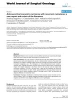
with recent onset of shortness of breath. CT pulmonary
angiogram demonstrated large right sided pulmonary
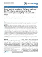
emboli (Figure 1). Moreover, a filling defect was noticed in
the right atrium (Figure 2). The defect appeared to be
lobulated, irregular, of low attenuation and arising from
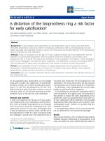
the free atrial wall. On transthoracic echocardiography
(Figure 3) the mass was demonstrated to be extending
through the tricuspid valve. A presumptive diagnosis of
right atrial myxoma with complicati ng pulmonary embo-
lism was made. Urgent surgery was arranged. At the time
of surgery the right atrial appendage was noted to be very
congested and “angry looking” . Total cardiopulmonary
bypass was established using aortic and bi-caval cannula-
tion. The right atrial cavity was found to be replaced by a
friable tumor which had “fronds like” appearance (Figure
4). The mass was extending through the tricuspid valve to
the right ventricle. A sample of the tumor was subjected
to frozen section examination which suggested the diagno-
sis of Leiomyosarcoma. The entire free wall of right atrium
was excised extending from and to the origin of vena
cavae. Anteriorly the incision was carried forward up to
the atrioventricular groove, taking care to preserve the
right coronary artery while ensuring macroscopic clear-
ance of tumor. The resection margins were submitted for
histological examination and were subsequently proven to
be tumor free. The right atrium was reconstr ucted using
autologous pericardium (Figure 5). Bilateral pulmonary
embolectomy was also performed. Histological examina-
tion of tumor confirmed the frozen section findings. On
the cut surface, the tumor had a whirled white appearance,
with focal brown areas. T he microscopic examination
revealed the presence of a spindle cell tumor, forming fas-
cicles orientated at right angles. The study revealed the
morphological aspect characteristic to leiomyosarcoma.
Although resection margins were clear the margin
width was deemed to be inadequate. After recovery
* Correspondence:
1
Cardiothoracic Department, Royal Victoria Hospital, Grosvernor Rd, Belfast,
BT12 6BA, Northern Ireland
Full list of author information is available at the end of the article
Parissis et al. Journal of Cardiothoracic Surgery 2010, 5:80
/>© 2010 Parissis et al; licensee BioMed Central Ltd. This is an Open Access article distributed under the terms of the Creative Commons
Attribution License ( censes/by/2.0), which permits unrestricted use, distribution, and reproduction in
any medium, provided the original work is properly cited.
from surgery the patient was submitted to chemother-
apy with Doxirubicin and Isofomaide. A tumor surveil-
lance plan involving serial echocardiograms is planned.
Discussion
Of the few hundred cases of malignant tumors of the
heart reported, most have been based on autopsy se r-
ies. Moreover, throughout the last 30 years literature
from a 100 plus articl es on cardiac neoplasms, only
few publications are reporting on primary cardiac
malignancies [5].
Cardiac sarcomas are the second most common type
of primary cardia c neoplasms with Leiomyosarcom as to
consist of 8% of cardiac sarcomas [6,7]. As per Kim et al
[8] angiosarcomas and unclassified sarcomas are the
most common sarcomas of the heart accounting for
76%of the cases, with leiomyosarcomas being a minority.
Thereisawideageandsizerangewithaslightfemale
predilection. As per Zhang et al [6] the sarcoma arises
in the atria/pulmonary vessels in 74% of the cases, and
in the ventricles, mitral valve, and epi/pericardium in
14%, 3.7% and 7.4% of the cases correspon dingly. There
is probably a slight left side predilection. In contrary,
predominantly right side distribution is given in o ther
reports [8] with right atrial involvement being 58% and
left atrial 25% of the cases.
Leiomyosarcoma favors the left atriu m and most likely
originates from pulmonary vein s mooth muscles and
present as a left atrial tumor. Affected patients typically
present i n the 4th decade of life which is slightly
younger than the average ag e at presentation for cardiac
sarcoma patients. Unlike angiosarcomas hemorrhage is
unusual. Those tumors are likely to involve the mitral
valve and extend into the pulmonary veins and therefore
present with pulmonary congestion. Macroscopically the
tumor appears as gelatinous mass and maybe multiple
in up to 30% of the cases [9].
Figure 1 Three dimensional reconstruction of Computerized
Tomogram Pulmonary Angiogram demonstrating large filling
defect in the branches of right pulmonary artery.
Figure 2 Intravenous contrast enhanced Computerized
Tomogram demonstrating right atrial wall tumor which
appears to be lobulated, irregular and of low attenuation.
Figure 3 Transthoracic Echocardiogram demonstrating right
atrial tumor extending from the free wall to the tricuspid
valve and protruding through it to the right ventricle.
Figure 4 Right atrial tumor resection.
Parissis et al. Journal of Cardiothoracic Surgery 2010, 5:80
/>Page 2 of 4
In our case report the site of origin of the tumor was
the right atrium which is rare. The presentation was
consistent with thromboembolic phenomenon com-
monly associated with this tumor site. Other possible
presentations include progressive or sudden right sided
cardiac failure on the account of tricuspid valve block-
age by the tumor or paroxysmal atrial arrhythmias.
Broadly speaking, cardiac tumors produce a large variety
of symptoms through any of 4 mechanisms. Their mass
can obstruct intracardiac blood flow or int erfere with
valve function. Local invasion can lead to arrhythmias
or pericardial effusions with tamponade. Bits of tumor
can embolize, causing systemic def icits when the tumors
are on the left side of the heart. Finally, the tumors may
cause systemic or constitutional symptoms.
Echocardiographic imaging is the most sensitive ima-
ging technique with ability to identify tumors as small as
3 mm. However, soft-tissue characterization remains lim-
ited compared with that achieved with computed tomo-
graphy (CT) and magnetic resonance (MR ) imaging, and
myocardial disease such as tumor infiltration is not
clearly depicted [9]. On the other hand with MRI or con-
trast enhanced CT the tumor has to be around 1 cm in
size before becoming detectable.
In this case CT scan raised the suspicion of intracardiac
tumor by depicting a low attenuation filling defect and
echocardiography confirmed the diagnosis. A preopera-
tive tissue diagnosis was not attempted due to the emer-
gent presentation. However atypical appearance of the
right atrium and of the tumor raised the suspicion of
malignancy and frozen section examination was confir-
matory. It has been recommended that all atrial tumors
should be subjected to frozen section examination in
order to ensure optimum surgical resection.
According to Mayer et al [1 0] half the patients with
cardiac sarcomas, are presented with high g rade tumors
and distant metastases: lungs 35.7%, lymph nodes 14.2%,
and liver 7.14%. Tumor spread from primary cardiac
sarcoma to the bone is very rare and has a poor prog-
nosis. Only six cases have been reported in the literature
[11]. Furthermore, from the patients that are deemed
suitable for surgery, complete macroscopic resection is
only possible in 33% [12].
Operative mortality has been reported to be high at 8.3%
with an overall actuarial survival of 14% at 24 months after
resection [13]. Likewise other groups [8,12] have reported
poor prognosis with a median survival time of 25 months
after diagnosis.
As per Burke et al [14], the survival rate on univaria te
analysis was more favorable for patients with tumors
located on the left side of the heart, without necrosis,
with a low mitotic count, and without metastasis at
diagnosis. By multivar iate analysis, a low level of mitotic
activity and any therapy were the only significant factors
affecting survival rate. Furthermor e tumor grade, unlike
histological type, appears to be prognostically important
in cardiac sarcoma [6].
The optimum treatment of Leiomyosarcoma is not
known. Of the several reports in the literature, patients
subjected to multimodality tre atment including heart
transplantation (The most common cause of death is local
recurrence of the tumors in 50% of the cases [12]) have
longer survival.
We adopted a strategy that would ensure local control of
the tumor by surgical resection and address systemic
spread by adjuvant chemotherapy. Tumor shrinkage can
be achieved by chemotherapy prior to surgery in non
emergency setting. Given the high risk of tumor recur-
rence we plan to follow-up the patient with serial echocar-
diographic scans with the view to further surgical or
chemotherapeutic intervention aimed at early treatment of
recurrence.
Conclusion
In conclusion PCM are rare and will always pose a diag-
nostic dilemma. Nevertheless, atypical presentation of sus-
pected “atrial myxoma” should raise the possibility of rare
atrial tumors. Unfortunately, almost half of those tumors
have metastasized at presentation, up to 30% could be
multifocal and the rest may be amenable to surgery. All
such tumors should be subjected to frozen section exami-
nation intraoperatively.
Surgery carries a high mortality and the overall long
survival was only achieved in patients who survived the
initial surgery well.
Consent
Written informed consent was obtained from the patient
for publication of this case report and accompanying
images. A copy of the written consent is available for
the review by the Editor-in-Chief of this journal.
Figure 5 Right atrial reconstruction with autologous pericardium.
Parissis et al. Journal of Cardiothoracic Surgery 2010, 5:80
/>Page 3 of 4
Author details
1
Cardiothoracic Department, Royal Victoria Hospital, Grosvernor Rd, Belfast,
BT12 6BA, Northern Ireland.
2
Cardiothoracic Department, Basildon & Thurrock
University Hospital NHS FT, Essex, UK.
3
Cardiothoracic Department, St James
Hospital, Dublin 8, Dublin, Ireland.
Authors’ contributions
HP conceived of the study and wrote the manuscript with the help of MTA.
VY overlooked the progress of the manuscript and advised on valuable
points. All authors read and approved the final manuscript.
Competing interests
The authors declare that they have no competing interests.
Received: 10 June 2010 Accepted: 12 October 2010
Published: 12 October 2010
References
1. Engberding R, Daniel WG, Erbel R: Diagnosis of heart tumours by
transoesophageal echocardiography: a multicentre study in 154
patients. Eur Heart J 1993, 14:1223-8, [Medline].
2. Basso C, Valente M, Poletti A, Casarotto D, Thiene G: Surgical pathology of
primary cardiac and pericardial tumors. Eur J Cardiothorac Surg 1997,
12(5):730-7, discussion 737-8.
3. Castillo JG, Silvay G: Characterization and management of cardiac tumors.
Cardiothorac Vasc Anesth 2010, 14(1):6-20.
4. Patel J, Sheppard MN: Pathological study of primary cardiac and
pericardial tumors in a specialist UK Centre: surgical and autopsy series.
Cardiovasc Pathol 2009.
5. Neragi-Miandoab S, Kim J, Vlahakes GJ: Malignant tumours of the heart: a
review of tumour type, diagnosis and therapy. Clin Oncol (R Coll Radiol)
2007, 19(10):748-56.
6. Zhang PJ, Brooks JS, Goldblum JR, Yoder B, Seethala R, Pawel B, Gorman JH,
Gorman RC, Huang JH, Acker M, Narula N: Primary cardiac sarcomas: a
clinicopathologic analysis of a series with follow-up information in 17
patients and emphasis on long-term survival. Hum Pathol 2008,
39(9):1385-95.
7. Vander Salm TJ: Unusual primary tumors of the heart. Semin Thorac
Cardiovasc Surg 12(2):89-100.
8. Kim CH, Dancer JY, Coffey D, Zhai QJ, Reardon M, Ayala AG, Ro JY:
Clinicopathologic study of 24 patients with primary cardiac sarcomas: a
10-year single institution experience. Hum Pathol 2008, 39(6):933-8.
9. Araoz P, Eklund H, Welch T, Breen J: CT and MR imaging of primary
cardiac malignancies. RadioGraphics 1999, 19:1421-1434.
10. Mayer F, Aebert H, Rudert M, Königsrainer A, Horger M, Kanz L, Bamberg M,
Ziemer G, Hartmann JT: Primary malignant sarcomas of the heart and
great vessels in adult patients– a single-center experience. Oncologist
2007, 12(9):1134-42.
11. Strina C, Zannoni M, Parolin V, Cetto GL, Zuliani S: metastases from
primary cardiac sarcoma: case report. Tumori 2009, 95(2):251-3.
12. Donsbeck AV, Ranchere D, Coindre JM, Le Gall F, Cordier JF, Loire R:
Primary cardiac sarcomas: an immunohistochemical and grading study
with long-term follow-up of 24 cases. Histopathology 1999, 34(4):295-304.
13. Putnam JB Jr, Sweeney MS, Colon R, Lanza LA, Frazier OH, Cooley DA,
cardiac sarcomas: Ann Thorac Surg 1991, 51(6):906-10.
14. Burke AP, Cowan D, Virmani R: Primary sarcomas of the heart. Cancer
1992, 69(2):387-95.
doi:10.1186/1749-8090-5-80
Cite this article as: Parissis et al.: Primary leiomyosarcoma of the right
atrium: a case report and literature update. Journal of Cardiothoracic
Surgery 2010 5:80.
Submit your next manuscript to BioMed Central
and take full advantage of:
• Convenient online submission
• Thorough peer review
• No space constraints or color figure charges
• Immediate publication on acceptance
• Inclusion in PubMed, CAS, Scopus and Google Scholar
• Research which is freely available for redistribution
Submit your manuscript at
www.biomedcentral.com/submit
Parissis et al. Journal of Cardiothoracic Surgery 2010, 5:80
/>Page 4 of 4