Báo cáo y học: " Surgical resection of a renal cell carcinoma involving the" ppt
Bạn đang xem bản rút gọn của tài liệu. Xem và tải ngay bản đầy đủ của tài liệu tại đây (1.4 MB, 6 trang )
RESEARC H ARTIC LE Open Access
Surgical resection of a renal cell carcinoma
involving the inferior vena cava: the role of the
cardiothoracic surgeon
Haralabos Parissis
1*
, Mohammad Taukeer Akbar
2
, Michael Tolan
3
, Vincent Young
3
Abstract
Background: The techniques for the resection of renal tumors with IVC extension are based on the experience of
individual units. We attempt to provide a logical approach of the surgical strategies in a stepwise fashion.
Methods: Over 6-years 9 patients with renal cell carcinoma invading the IVC, underwent surgery. There were 6
males. The extension was at level IV in 4 and III in 5 cases. CPB used in 8 and hypothermia and circulatory arrest in
all patients with level IV disease. The results and an algorithm of the plan of action, as per level of extension are
presented.
Results: Plan of action: For level I-II disease: No Cardiothoracic involvement, For level III: Cardiopulmonary Bypass
(CPB) & control of the cavo-atrial junction. For level IV: use of brief periods of Circulatory Arrest & repair of the
Cavotomy with a pericardial patch. Postoperative morbidity: prolonged ICU stay, 3 patients (33.3%); tracheostomy,
1 (11.1%); Sepsis, 2 (22.2% ); CVA 1, (11.1%). Mortality: 2 patients (22.2%)
Conclusions: Total clearance of the IVC from an adherent tumor is important, therefore extensive level IV disease
presents a surgical challenge.
We recommend CPB for level III and brief periods of Total Circul atory Arrest (TCA) for level IV disease.
Background
Inferior Vena Cava (IVC) involvement in patients under-
going surgery for renal cell carcinoma (RCC) is rare
(4-8%) [1]. The overall 5 year survival following success-
ful resection can be up to 40 - 50% [2,3], therefore one
should not preclude surgical therapy in this group of
patients [4].
The level of the IVC involvement as defined in the lit-
erature [1,3,4], d ictates the surgical strategies and man-
dates the development of a plan of action that should be
safe, reproducible and reliable.
Favorable outcome in patients with non-metastatic
renal carcinoma and IVC involvement correlates with
complete clearance of the IVC from tumor-thrombus.
This principle sometimes can only be achieved following
an optimal exposure of the infra & supra hepatic IVC
concomitantly with clearance of the IVC -right atrial
junction. Furthermore prevention of tumor disruption
and pulmonary embolism has to be considered during
thrombectomy & manipulation of the diseased cava.
The guidelines regarding the vario us techniques for
the resection of RCC with IVC extension are very scat-
tered in the literature. In this article we attempt to pro-
vide a systematic approach of the cardiothoracic surgical
strategies in a stepwise fashion.
Methods
Over 6-years 9 patients with RCC invading the IVC,
underwent surgery. There were 6 males. The extension
was at level IV in four(4) and III in five(5) cases. Cardio
Pulmonary Byp ass was used in eight(8) patients and
hypothermia and circulatory arrest in all patients with
level IV disease. Abdominal MRI (Figure 1) is useful to
determine the extent of IVC involvement with tumo r/
thrombus. Peri-operative Trans-Oesophageal Echo
(Figure 2) provides information’s regarding the amount
of adherence, supra-hepatic extension and mobility of the
tumour. Multidisciplinary approach is needed. Metastatic
* Correspondence:
1
Royal Victoria Hospital, Grosvernor Rd, Belfast, BT12 6BA, Northern Ireland
Full list of author information is available at the end of the article
Parissis et al. Journal of Cardiothoracic Surgery 2010, 5:103
/>© 2010 Parissis et al; licensee BioMed Central Ltd. This is an Open Access article distributed under the terms of the Creative Commons
Attribution License (http:// creativecomm ons.org/licenses/by/2.0), which permits unrestricted use, distri bution, and reproduction in
any medium, provided the or iginal work is properly cited.
dis ease is a contraindicat ion for surgical therapy and has
to be ruled out. The patients characteristics are p resent
in appendix 1.
Surgical Approach
Mobilisation of the affected kidney with retroperitoneal
lymphadenectomy is performed first. For level I-II dis-
ease cardiothoracic involvement is not necessary. Lim-
ited cavotomy with the brief use of an intermittent
Caval clamp above and below the lesion is usually ade-
quate. The need for cardiac surgical involvement is
usually contemplated when the tumor/thrombus is
extending up to level III. We favour a standard midline
laparotomy and assessment of resectability o f the renal
tumour.
Following sternotomy, institution of CPB is achieved
using a split venous cannula: Superior Vena Cava &
Right femoral vein. Control of the cavo-atrial junction is
considered in order to avoid tumour embolization.
Bulky disease extending into the right atrium may be
better controlled by splitting the diaphragm through the
central tendon towards the IVC. This manoeuvre,
enables extension of the Right atrial incision towards
the IVC for direct resection of severely adhere tumours
(ie. Patient number 3).
The porta hepatis is dissected so that the liver blood
supply could be briefly interrupted (Pringle manoeuvre:
occlusion of blood inflow to the liver) during cavotomy
to further facilitate bloodless surgical field. Furthermore,
by applying a cross clamp on the sub-diaphragmatic
aorta during caval extirpation of the tumour, bloodless
operative conditions could be achieved.
Level IV involvement presents a challenge; the disease
extends into the RA with various degrees of infiltration
and adherence into the wall of IVC. Under those cir-
cumstances the use of Total Circulatory Arrest (TCA)
has become the centre of an argument. The patho-
physiological sequelae of the use of TCA are balanced
against the risk of a suboptimal tumour clearance. We,
like others believe that with such extension of the dis-
ease the wall of the IVC is infiltrated by tumour and
unless a complet e bloodless field is instituted, only by
blunt dissections, it is impossible to achieve complete
clearance.
Therefore for level IV e xtension of the tumour or for
suspected “suboptimal thrombectomy” for level III dis-
ease we advocate brief period of TCA. During the cool-
ing period in an arrested heart the RA is opened and
tumour mobilization around the ostium of the IVC is
carried out. Endarterectomy knifes further facilitate opti-
mal extirpation of the tumour by negotiating anatomical
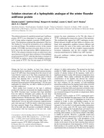
Figure 1 MRI images of a level IV disease.
Figure 2 Echo images of tumor extending into the IVC.
Parissis et al. Journal of Cardiothoracic Surgery 2010, 5:103
/>Page 2 of 6
planes of excision. During TCA the cava is incised up to
10 cm cephalad in a longitudinal fashion taking care to
include with the specimen the origin of the renal vein
which is usua lly involved with the tumour. Clearance of
the luminal deposits of the IVC using sharp and blunt
dissections could be then carried out under direct
vision. Having mobilised the tumour proximally at the
IVC- RA junction, final extraction is usually achieved in
continuity with the nephrectomy specimen (Figure 3).
Furthermore, tumour embolization to the lungs is
avoided. This process provides a controlled bloodless
environment for facilitation of complete tumour clear-
ance (Figure 4). Always the cavotomy is repaired with
the use of a pericardial patch (Figure 5), in order to
avoid narrowing of the cava. An algorithm of the plan
of action, as per level of extens ion is depicted in appen-
dix 2.
Results
Outcome
During th e beginning of t his program, Venove nous
bypass was used in one patient (number 7) with level III
disease. However the technique was deem cumbersome
and unsatisfactory, mainly due to excessive blood in the
surgical field, resulting in suboptimal exposure.
Cardio Pulmonary Bypass was used in eight(8) patients
and hypothermia and circulatory arrest in all patients
with level IV disease.
The operative time range f rom 3 hours 5 2 minutes to
9 hours 36 minutes. Estimated blood loss was 1850 mL
(range 950 to 3800 mL). Blood and blood product
requirement was high (7 out of nine patients). The aver-
age blood transfusion was 2 units of red Blood Cells
(range between 1 and 4 Units). Blood products were
used in all four patients following hypothermia and cir-
culatory arrest. Cell-s aving techniques used routinely in
our institution.
Transient inotropic support by means of Dopamine
and Noradrenaline was used in 5 patients. Average
intensive care unit length of stay was 19 days (range, 1
to 164 days). In three (3) patients (33.3%) the ICU stay
was prolonged. Furthermore one (1) patient required a
tracheostomy (11.1%). Two patients developed septice-
mia (one MRSA positive) and one patient develop a
CVA. Two patients died; one from septicaemia post-
operative day 55 and one from multiple organ failure
post operative day 164. The mean size of the renal mass
was 5.2 cm (range, 3.5 to 11.2 cm). Histological exami-
nation showed renal cell carcinoma of clear type in 8
patients and papillary type in 1 patient. Lymph node
metastasis was detected in 2 patients.
Two of the discharged patients were lost to follow up.
Of the remaining five patients, 2 ha ve had tumor recur-
rence and one had pulmonary metastasis at 2 years, on
follow up chest X Ray. Those 3 patients were referred
for adjuvant chemotherapy. The cumulative postopera-
tive follow-up of the remaining two patients was 45
+/-11 months. They were alive at the last follow up and
free of recurrence.
Figure 3 Renal cell carcinoma invad ing the upper pole of the
kidney with tumor propagating into the IVC.
Figure 4 Direct removal of the tumor mass.
Figure 5 Closure of the IVC with a pericardial patch.
Parissis et al. Journal of Cardiothoracic Surgery 2010, 5:103
/>Page 3 of 6
Discussion
Metastasis has occurred in 34.6% of the patients with
RCC and luminar propagation of the tumor into the
IVC [5]. Furthermore, as per thesameauthors,micro-
metastasis is taken place in 11.1% of those patients.
Therefore, only half of the patients with level II I-IV dis-
ease would be free of distal spread and subsequently
would benefit from an operation. Palliative resection to
control polycythemia and parane oplastic syndromes in
patients with metastatic disease, is questionable.
Level I and II is probably the commonest entity occur-
ring in 60-65% of the cases and usually treated by local
resection. According to Lubahn et al [6] approximately
50% of the patients with renal tumors involving the
IVC, warrant cardiothoracic involvement. Furthermore
the overal l incidence of extensive IVC disease involving
the right atrium according to Bissada et al [5] & Herma-
nek et al [7] is around 27.7%.
It has been postulated that the involvement of the IVC
in RCC is generally not a vascular invasion by the malig-
nancy [8]; one could argue however, that following
removal of the thrombus-tumor from the IVC, invari-
ably, an area is found that indicates sub-endothel ial
invasion. In addition, in 12.9% of the patients in Bissada
et al series [5] the IVC wall was invaded by tumor.
Suprahepatic extension of the tumor (level III disease)
poses a challenge, especially when the tumor is densely
adhering to the Venus wall or when the h epatic veins
contain propagating segments of tumor. Budd-Chiari
syndrome, is an extreme form of hepatic venous stasis
resulting from occlusion of the major hepatic veins or
the supra- hepatic IVC from various malignant causes,
with renal cell carcinoma being the most common. A
hepatic vein obstruction that causes Budd-Chiari syn-
drome, is an adverse feature. Under such conditions,
bleeding diathesis is accelerated; this is due to Liver
congestion with reduce “ synthetic function” and also
portal hypertension with the development of port a-caval
collaterals.
Generally for level III disease some institutions [9]
favo r cavotomy without the use of CPB [10] or with the
use of venous-venous bypass [11,6]. The latter group in
a large series of patients concluded that the need for
invasive cardiovascular procedures increased the risk of
perioperative complications. The advantages of using
veno-venous bypass are restoration of hemodynamic
instability during venal clamping and the fact that there
is no need for systemic heparinization. However one
wouldarguethatwithoutCPBandpossiblywithout
additional maneuvers to reduce the venus return (such
as Pringle maneuver, clamping of the abdominal aorta,
the superior mesenteric artery or the contralateral renal
artery) bloodless field cannot be achieved during cavot-
omy; furthermore the imposed hemodynamic instability
at the time, has another adverse impact: the surgeon is
“pushed ” to complete the extirpation of the thrombus
against the time. That can rather lead to de-bulking of
the tumor. It could also lead to dislodgment of tumor
material and subsequent pulmonary embolism.
Table 1 Patients’ characteristics
Sex Pre-Op Creatinine Hgt
(cm)
Weight
(kg)
Euroscore Operation-Findings CPB
(min)
Cross
Clamp
Time
(min)
m 175 182 85 4 left kidney tumor Level IV 111 43
m 132 182 90 7 Lt Kidney tumor Level III 51 17
f 108 154 60 7 right renal tumor Level IV 101 37
m 124 178 76 5 right renal tumor, Level III 22 0
f 79 166 76 3 right renal tumor, Level III 36 0
m 144 183 80 4 Right kidney tumor Level IV 89 19
m 104 170 106 2 right renal tumor, Level III 0 0
f 103 155 72.5 5 left kidney tumor Level IV 75 25
m 86 180 66 2 left renal tumor, Level III 13 0
Table 2 Surgical steps as per level of IVC involvement by
tumor
Surgical steps - IVC involvement
↓
Level I-II (60% of the cases) No cardiothoracic involvement/
Cardiothoracic “back up” only
↓
Level III & IV disease mandates Cardiothoracic involvement
↓↓
LEVEL III (12-15% of the cases)
LEVEL IV (25% of the cases)
CPB, Pringle manoeuvre and if necessary
Always use of CPB and brief period of cross clamp of sub-
diaphragmatic aorta TCA
If suboptimal thrombectomy, then brief TCA
Parissis et al. Journal of Cardiothoracic Surgery 2010, 5:103
/>Page 4 of 6
Therefore, for level III disease, besides CPB we would
also favor the approach reported by Chowdhury et al
[12] whereby in termittent cross clamp of the sub-dia-
phragmatic aorta is applied. This brief maneuver would
further optimize the conditions for a bloodless surgical
field.
In the situation where the IVC is fully occluded by the
tumor in level III disease, then probably the patient may
tolerate clamping of the IVC at the junction with the
RA (under TOE guidance) without significant hemody-
namic compromise. Under those circumstances, one
could debate that CPB is not necessary. Nevertheless,
one should bear in mind the theoretical risk, that de-
balking of the tumor increases the incidence of local
recurrence.
Five patients in our series had level III disease (Three
patients had Right side RCC). Venovenou s bypass was
used in one patient. The tumor w as removed satisfac-
tory, however hemodynamic instability and access was
deemed cumbersome. Complications with Venovenous
bypass [6] and difficulty in accessing the hepatic veins
and suprahepatic cava lead us to abandoning this
procedure.
For level IV disease with tumor extensi on in the right
atrium controversy still exists as regarding the need for
Total Circulatory Arrest (TCA). So sa et al [13] has
reported a poor survival for patients with level IV
disease. Cerwinka et al [14] advocates excision of supra-
diaphragmatic tumors off pump with no TCA. In
contrary, Chiappini et al [15] and Mazzola et al [16],
clai m that the use of TCA provides a safe technique for
removing the tumor thrombus in a bloodless field, and
has good early and long-term results. We, like others
[17] believe that when the tumor thrombus is invading
the caval wall or reaches the right atrium -ventricle then
TCA becomes a necessity. We reckon that this approach
has improved the safety and efficacy of a diffi cult surgi-
cal undertaking by facilitating controlled dissection, pro-
viding a bloodless field, and reducing the risk of tumor
embolization. The high postoperative morbidity reported
by various groups [13,15] is reflecting the preoperative
compromise health status of this group of patients a nd
possibly the use of circulatory arrest. According to
Cooper et al [18] the use of TCA increases up to 40%
the risk of complications and also adds up, on the peri-
operative mortality. Furthermo re as per Schimmer et al
[17] the risk of bleeding (at least theoretically) could be
exponentially higher due to: 1) profound hypothermia
itself 2) extended bypass ti me as a result of cooling-
rewarming,and3)thefactthatthosepatientshave
undergone extensive retroperitoneal dissections and
have accessory high pressure venous collaterals due to
the IVC obstruction.
For all those reasons aforementioned, a single institu-
tional approach [19] advocates in selected cases o f renal
cell carcinoma with level IV IVC extension, resection of
the tumor without sternotomy, CBP, or DHCA. This
technique however has limitations ([19] Invited
commentary).
The need for extensive surgery with relative good out-
come has been outlined from various groups. According
to Tanaka et al [2] and Yazici and associates [20] the
length of tumor extension is not an incremental risk
factor for adverse survival. Likewise Chiappini et al, [15]
states that the tumor extension into the IVC to what-
ever degree is not associated with an adverse prognosis,
provided a complete resection is advocated [21].
Complete resection of the entire tumor is mandatory
for a reas onable attempt at a long survival, as demon-
strated by Nesbitt and colleagues [9] and Hatcher and
colleagues [22], where no patients with incomplete local
resection survived to 5 years. Following the same princi-
plewefavor“Controlled Cavotomy” whereby the inter-
ior of the IVC can be adequately inspected in a
bloodless surgical environment.
Finally, survival is also associated with the tumor char-
acteristics (grade of tumor cells) and lymph node invol-
vement [2]. Throughout the literature the overall 5 year
survival is been reported to be between 40 to 50% over-
all [3,23,18,24].
Five patients in our series were followed up. There
was lymph node involvement at the initial specimen of
the two pa tients, that had local recurren ces at 2 years.
Of the remaining 3 patients, one h ad pulmonary metas-
tasis at 2 year s, and 2 pat ients were alive at 4 years and
free of recurrence.
Conclusions
In summary, RCC with advance IVC involvement poses
a surgical challenge. During this report we eluded on
the pros and cons of the various approaches. In keeping
with the principles for local clearance one should con-
sider: multidisciplinary approach with proper pre-opera-
tive evaluation of the extension of the tumor, optimal
control of hemodynamic conditions during c avotomy,
ability to visually assess the extent of the tumor inva-
sion, avoidance of tumor fragmentation and emboliza-
tion and repair of the IVC without narrowing of the
vessel.
Finally in this paper, although the number of patients
reported is small, we have attempted t o provide a clear
strategy for tackling a difficult and unusual entity.
Consent
Written informed consent was obtained from the
patients for publication of the series and accompanying
Parissis et al. Journal of Cardiothoracic Surgery 2010, 5:103
/>Page 5 of 6
images. A copy of the written consent is available for
the review by the Editor-in-Chief of this journal.
Appendix 1: Patients’ characteristics.
Appendix 2: Surgical steps as per level of IVC
involvement by tumor.
Author details
1
Royal Victoria Hospital, Grosvernor Rd, Belfast, BT12 6BA, Northern Ireland.
2
Essex Cardiothoracic Center, Basildon & Thurrock University Hospital, Essex,
UK.
3
Cardiothoracic Department, St James Hospital, Dublin, Ireland.
Authors’ contributions
HP conceived of the study and wrote the manuscript with the help of MTA.
MT made valid corrections, VY organized and overlooked the progress of the
manuscript and advised on valuable points. All authors read and approved
the final manuscript.
Competing interests
The authors declare that they have no competing interests.
Received: 6 April 2010 Accepted: 5 November 2010
Published: 5 November 2010
References
1. Babu SC, Mianoni T, Shah PM, Goyal A, Choudhury M, Eshghi M,
Moggio RA, Sarabu MR, Lafaro RJ: Malignant renal tumor with extension
to the inferior vena cava. Am J Surg 1998, 176(2):137-9.
2. Tanaka M, Fujimoto K, Okajima E, Tanaka N, Yoshida K, Hirao Y: Prognostic
factors of renal cell carcinoma with extension into inferior vena cava. Int
J Urol 2008, 15(5):394-8.
3. Belis JA, Livinson ME, Pae WE: Complete radical nephrectomy and vena
caval thrombectomy during circulatory arrest. J Urol 2000, 163:434-436.
4. Skinner DG, Pritchett TR, Lieskovsky G, Boyd SD, Stiles QR: Vena caval
involvement by renal cell carcinoma. Ann Surg 1989, 210:387-392.
5. Bissada NK, Yakout HH, Babanouri A, Elsalamony T, Fahmy W, Gunham M,
Hull GW, Chaudhary UB: Long-term experience with management of
renal cell carcinoma involving the inferior vena cava. Urology 2003,
61(1):89-92.
6. Lubahn J, Sagalowsky A, Rosenbaum D, Dikmen E, Bhojani R, Paul M,
Dolmatch B, Josephs S, Benaim E, Levinson B, Wait M, Ring W, DiMaio M:
Contemporary techniques and safety of cardiovascular procedures in
the surgical management of renal cell carcinoma with tumor thrombus.
J Thorac Cardiovasc Surg 2006, 131:1289-1295.
7. Hermanek P, Schrott KM: Evaluation of the new tumor, nodes and
metastases classification of renal cell carcinoma. J Urol 1990, 144:238-242.
8. Kalkat M, Abedin A, Rooney S, Doherty A, Faroqui M, Wallace M, Graham T:
Renal Tumors with cavo-atrial extension: surgical management and
outcome. Interac Cardiov & Thorac Surgery 2008, 7(6):981-5.
9. Nesbitt JC, Soltero ER, Dinney CPN, Walsh GL, Schrump DS, Swanson DA,
Pisters LL, Willis KD, Putnam JB Jr: Surgical management of renal cell
carcinoma with inferior vena cava tumor thrombus. Ann Thorac Surg
1997, 63:1592-1600.
10. Langenburg SE, Blackbourne LH, Sperling JW, Buchanan SA, Mauney MC,
Kron IL, Tribble CG: Management of renal tumors involving the inferior
vena cava. J Vasc Surg 1994, 20(3):385-8.
11. Belgrano E, Liguori G, Trombetta C, Siracusano S, Bucci S, Zingone B:
Modified pump-driven venous bypass in surgery for renal cell carcinoma
(RCC) involving the inferior vena cava (IVC). World J Urol 2002, 20(1):56-8.
12. Chowdhury U, Mishra A, Seth A, Dogra P, Honnakere J, Subramaniam G,
Malhotra A, et al: Novel Techniques for Tumor Thrombectomy for Renal
Cell Carcinoma With Intraatrial Tumor Thrombus. Ann Thorac Surg 2007,
83:1731-1736.
13. Sosa RE, Muecke EC, Vaughan ED, McCarron JP Jr: Renal cell carcinoma
extending into the inferior vena cava: the prognostic significance of the
level of vena caval involvement. J Urol 1984, 132:1097-1100.
14. Cerwinka WH, Ciancio G, Salerno TA, Soloway MS: Renal cell cancer with
invasive atrial tumour thrombus excised off-pump. Urology
2005, 661319-
e9-11.
15. Chiappini B, Savini C, Marinelli G, Suarez SM, Di Eusanio M, Fiorani V,
Pierangeli A: Cavoatrial tumor thrombus: single-stage surgical approach
with profound hypothermia and circulatory arrest, including a review of
the literature. J Thorac Surg 2002, 124:684-688.
16. Mazzola A, Gregorini R, Villani C, Colantonio L, Giancola R, Gravina G,
Vicentini C: Cavoatrial Tumor Thrombectomy With Systemic Circulatory
Arrest and Antegrade Cerebral Perfusion. Ann Thorac Surg 2007,
83:1564-1565.
17. Schimmer C, Hillig F, Riedmiller H, Elert O: Surgical treatment of renal cell
carcinoma with intravascular extension. Interactive Cardiovascular and
Thoracic Surgery 2004, 3:395-397.
18. Cooper WA, Duarte IG, Thourani VH, et al: Hypothermic circulatory arrest
causes multisystem vascular endothelial dysfunction and apoptosis. Ann
Thorac Surg 2000, 69:696-703.
19. Ciancio G, Shirodkar S, Soloway M, Livingstone A, Barron M, Salerno T:
Renal Carcinoma with Supradiaphragmatic tumor thrombus: Avoiding
sternotomy and Cardiopulmonary bypass. Ann Thorac Surg 2010,
89:505-11.
20. Yazici S, Inci K, Bilen CY, Gudeloglu A, Akdogan B, Ertoy D, Kaynaroglu V,
Demircin M, Ozen H: Renal cell carcinoma with inferior vena cava
thrombus: The Hacettepe experience. Urol Oncol 2009.
21. Dedeilias P, Koletsis E, Rousakis AG, Kouerinis I, Zaragkas S, Grigorakis A,
Leivaditis V, Malovrouvas D, Apostolakis E: Deep hypothermia and
circulatory arrest in the surgical management of renal tumors with
cavoatrial extension. J Card Surg 2009, 24(6):617-23, Epub 2009 Sep 2.
22. Hatcher PA, Anderson EE, Paulson DF, Carson CC, Robert-son JE: Surgical
management and prognosis of renal cell carcinoma invading the vena
cava. J Urol 1991, 145:20-3.
23. Terakawa T, Miyake H, Takenaka A, Hara I, Fujisawa M: Clinical outcome of
surgical management for patients with renal cell carcinoma involving
the inferior vena cava. Int J Urol 2007, 14(9):781-4.
24. Wotkowicz C, Wszolek MF, Libertino JA: Resection of renal tumors
invading the vena cava. Clin North Am 2008, 35(4):657-71, viii.
doi:10.1186/1749-8090-5-103
Cite this article as: Parissis et al.: Surgical resection of a renal cell
carcinoma involving the inferior vena cava: the role of the
cardiothoracic surgeon. Journal of Cardiothoracic Surgery 2010 5:103.
Submit your next manuscript to BioMed Central
and take full advantage of:
• Convenient online submission
• Thorough peer review
• No space constraints or color figure charges
• Immediate publication on acceptance
• Inclusion in PubMed, CAS, Scopus and Google Scholar
• Research which is freely available for redistribution
Submit your manuscript at
www.biomedcentral.com/submit
Parissis et al. Journal of Cardiothoracic Surgery 2010, 5:103
/>Page 6 of 6