Báo cáo y học: "Demonstration of the histopathological and immunohistochemical effects of a novel hemostatic agent, ankaferd blood stopper, on vascular tissue in a rat aortic bleeding mode" pps
Bạn đang xem bản rút gọn của tài liệu. Xem và tải ngay bản đầy đủ của tài liệu tại đây (1007.69 KB, 7 trang )
RESEARC H ARTIC LE Open Access
Demonstration of the histopathological and
immunohistochemical effects of a novel
hemostatic agent, ankaferd blood stopper, on
vascular tissue in a rat aortic bleeding model
Ozer Kandemir
1*
, Mustafa Buyukates
1
, Nilufer Onak Kandemir
2
, Erol Aktunc
3
, Aylin Ege Gul
4
, Sanser Gul
5
,
S Akin Turan
1
Abstract
Background: Ankaferd Blood Stopper® (ABS) is a folkloric medicinal plant extract used as a hemostatic agent in
traditional Turkish medicine. This experimental study investigated the histopathological and immunohistochemical
effects of ABS on vascular tissue in a rat model of aortic bleeding.
Methods: Four groups of 11 Wistar albino rats were used. The abdominal aortas of the rats were wounded; an
ABS-soaked tampon was applied to rats in Groups 1 and 3, and a plain gauze tampon was applied to rats in
Groups 2 and 4 until the bleeding stopped. The bleeding time was recorded. Immediately following sacrificing, the
arteriotomy sites from Groups 1 and 2 were removed. The abdominal incisions in Groups 3 and 4 were closed
following hemostasis. On Day 7 of the study, Group 3 and 4 rats were sacrificed and the abdominal aorta
arteriotomy sites were removed for histopathologica l and immunohistochemical evaluation.
Results: The mean bleeding time in 15 animals in Groups 2 and 4 was 4.9 ± 0.6 s, and in 22 animals in Groups 1
and 3 was 3.1 ± 0.6 s. Distal aortic occlusion was not observed on either Day 1 or 7 in any group. Significantly
more widespread and dense endothelial nitric oxide synthase (eNOS) staining was observed in Group 1 animals
than Group 2. On Days 1 and 7 after application of ABS, histopathological changes, consisting of necrosis,
inflammation, and endothelial cell loss, in the rat abdominal aortas did not differ between Groups 1 and 2. The
basophilic discoloration in the ABS group on the operation day was a result of a foreign body reaction and
hemosiderin-loaded histiocyte accumulation, which occurred on Day 7.
Conclusions: In this study, hemostasis was successfully achieved with ABS in rat abdominal aortas. No
histopathological change was found in the rat abdominal aortas between the ABS and control groups on Days 1
and 7. Further studies on the long-term effects of foreign body reactions and hemosiderin-loaded histiocyte
accumulation are required.
Background
Impaired tissue integrity and uncontrollable hemorrhage
are important causes of morbidity and mortality, espe-
cially in the presence of coagulopathies [1]. Various
hemostatic agents have been developed to achieve suffi-
cient hemostasis [2,3]. In cardiovascular surgery,
bleeding from anastomos is sitesisusuallycontrolled
with pressure or additional suturing techniques. Occa-
sionally, these techniques may be insufficient, requiring
tis sue adhesives as supportive agents [4,5]. Additionally,
blind suturing for blood oozing from sutured vascular
segments may impair the quality of anastomosis.
To preserve the quality of anastomosis, adjuvant topical
hemostatic agents are favored in cardiac and vascular sur-
gery. However, topical hemostatic agents may have disad-
vantages, such as limited efficacy, limited availability,
* Correspondence:
1
Department of Cardiovascular Surgery, Zonguldak Karaelmas University,
Zonguldak, Turkey
Full list of author information is available at the end of the article
Kandemir et al. Journal of Cardiothoracic Surgery 2010, 5:110
/>© 2010 Kandemir et al; licensee BioMed Cent ral Ltd. This is an Open Access article distribu ted under the terms of the Crea tive
Commons Attribution License ( which permits unrestricted use, distribution, and
reproduction in any medium, provided the original work is properly cite d.
limited vascular biol ogical c ompatibility, expensiveness,
and risk of infection as a result of the requirement for
human blood for commercial production of collagen,
thrombin, and prothrombin [6]. S urgeons should also be
trained in the use of hemostatic agents, such as fibrin
glues.
Ankaferd Blood Stopper® (ABS) is a folkloric medicinal
plant extract used as a hemostatic agent in traditional
Turkish medicine [7]. The use of this product
was approved by the Ministry of Health, Turkey, on
October 26, 2007.
In a recent literature search, we found no study on the
histopathological and immunohistochemical effects of
ABS on vascular tissue. In this experimental study, we
investigated the effects of ABS on vascular tissue in a
rat model of aortic bleeding.
Methods
Wistar albino (WA) rats were used to demonstrate the
vascular histopathological and immunohistochemical
changes following the application of ABS (Trend Tekno-
loji Ilac AS, Istanbul, Turkey) on the abdominal aorta.
The experimental procedure was approved by the
Committee for Animal Research at Zonguldak Karaelmas
University School of Medicine. All animal studies con-
formed w ith the anima l experiment guidelines of the
Committee for Humane Care. All animals received care
in compliance with the “Principles of Laboratory Animal
Care” formulated by the National Society for Medical
Reseacrh and “Guide for the Care and the Use of Labora-
tory Animlas” prepared by the US Natinoal Academy of
Sciences and published by the US Natinoal Institute of
Health (NIH Publications, No:80-23)
Animals
Male adult WA rats (Zonguldak Karaelmas University
Laboratories, Zonguldak, Turkey), weighing 250-300 g,
were maintained on a 12/12-h light/dark cycle and fed ad
libitum. All animals were housed in individual cages in a
temperature-controlled environment (20 ± 2°C). The rats
were randomly assigned into ABS and control groups.
Surgical procedure
All animals were anesthetized with intramuscularly
administered ketamine hydrochloride (75 mg/kg). Post-
operative analgesia was provided by 1-2 mg/mL parace-
tamol added to the drinking water. The abdominal aorta
was accessed surgically by a midline abdominal incision
using sterile technique. The retroperitoneum was
explored and the aorta was exposed. The abdominal
aorta was wounded just proximal to the iliac bifurcation
using an iris blade. ABS solution (1 mL) in a glass vial
was poured on a gauze tampon through a syringe. Either
an ABS-soaked or plain gauze tampon was applied to
the vascular wound, and the bleeding time was
recorded. In case of insufficient hemostasis using either
of the tampons, an 8/0 Prolene suture (Prodek, Sutures
Ltd, UK) was used t o provide hemostasis. Aortic sam-
pling was performed in all rats to search for immediate
and Day-7 postoperative histopathological changes in
vascular tissues as a result of ABS.
Bleeding assay
The duration of bleeding was measured using a chron-
ometer and defined as the time from wounding until
the time bleeding stopped.
Animal groups
The abdominal aortas of the animals were wounded.
ABS- soak ed tampons were applied in Group 1 (n =11),
and plain gauze tampons were applied in Group 2 (n =
11) until the bleeding stopped. All of these animals were
sacrificed by cervical dislocation on the operation day.
The abdominal aortas of Groups 3 (n = 11) and 4 (n =
11) were wounded, and hemostasis was prov ided with
ABS-soaked tampons in Group 3 and plain gauze tam-
pons in Group 4. The abdominal incisions in these two
groups were closed following hemostasis. They were
kept alive for 7 days and fed ad libitum.OnDay7after
the operation, all Group 3 and 4 animals were sacrificed
by cervical dislocation. Immediately following sacrifice,
the arteriotomy sites from all 44 animals were removed
en bloc with a safety margin of 1 mm of untouched aor-
tic vascular tissue both distal and proximal to the
wound site.
Histopathological procedure
All specimens were fixed in 10% phosphate-buffered for-
maldehyde solutio n for 24 h at room temperature. Each
specimen was cut into three sections: the proximal,
intact part of the aorta, the wounded part of the aorta,
and the distal, intact part of the aorta.
Following the dehydration process using graded etha-
nols, specimens were embedded in paraffin blocks and
cut into 5-μm-thick sections to be mounted on glass
slides. Sections were then deparaffinized with xylene
and counterstained with hematoxylin and eosin (H&E),
iron blue, and Elastic van Gieson (EVG). EVG staining
was performed to identify the external and internal elas-
tic lamina. Iron blue staining was performed to identify
hemosiderin. All of the sections were examined in 10
random fields at ×40 magnification using a light micro-
scope. Blinded light microscopic examinations were per-
formed by two of the coauthors (NOK, AEG).
Histopathological grading of the specimens
Light microscopic findings were graded semi-quantita-
tively from 0 (no histopathological change) to +3 (severe
Kandemir et al. Journal of Cardiothoracic Surgery 2010, 5:110
/>Page 2 of 7
histopathological change). This histopathological grading
was performed for vascular (endothelial cell loss, inflam-
matory reaction, medial necrosis, fibrin plug formation,
and erythrocyte aggregation) and perivascular (inflam-
matory reaction, hemosiderin-loaded histiocytes, and
granulation tissue formation) connective tissue reactions
in the specimens.
Immunohistochemical procedure
In immunohistochemical surveys, anti-CD31 was used
to monitor vascular endothelial cells, and anti-eNOS
antibodies were used to determine eNOS expression of
endothelial cells. For immunohistochemical studies,
immunostaining was performed according to th e avidin-
biotin-peroxidase (BSA-DAB) complex technique. Paraf-
fin sections were coll ected on slides, deparaffinized, and
dehydra ted. Endogenous peroxidase activity was blocked
using a 3% hydrogen peroxide solution for 10 min. To
enhance staining, heat-induced epitope retrieval was
performed. Primary antibodies against CD31 (rabbit
monoclonal, JC70, Dako, Copenhagen, Denmark) and
endothelial nitric oxide synthase (eNOS; rabbit polyclo-
nal, RR-1711-R7, Neomarkers; Lab Vision, Fremont, CA,
USA) were used. The sections were incubated with pri-
mary antisera (including CD31 or eNOS) for 1 h at
room temperature. After washing in phosphate-buff ered
saline, the tissues were incubated with biotin-conjugated
secondary antibody and then a streptavidin-biotin sys-
tem for 30 min at room temperature. The reactions
were visualized using diaminobenzidine tetrahydrochlor-
ide. The sections were counterstained using hematoxy-
lin, then cleared and mounted.
Controls and grading of the immunostaining
Appropriate positive (placenta, capillary endothelium for
CD31 and eNOS) and negative (omitted primary anti-
body) controls were evaluated simultaneously in all
cases. All cytoplasmic s taining was recorded as positive
for eNOS and CD31. The extent and intensity of eNOS
reactions were semi-quantitatively evaluated using a
four-level grading system. Grade 0 was no apparent
reaction product. Focal and minimal staining intensity
was graded 1, and the most prominent staining reaction
covering nearly the whole area of the specimen was clas-
sified as 3. Grade 2 was intermediate between 1 and 3.
Statistical analysis
Statistical analyses were carried out using the SPSS soft-
ware (v. 11.0 for Windows; SPSS Inc.; Chicago, IL). All
values are expressed as means ± SD. P-values less than
0.05 were deemed to be statistically significant. Group
compa risons were made by one-way analysis of variance
(Kruskall-Wallis) followed, in cases of significance, by
the Mann-Whitney U test.
Results
Bleeding did not stop in four of the Group 2 animals
and in three of the Group 4 animals, for a total of seven
in the plain gauze tampon groups. The ABS-soaked
gauze tampon stopped bleeding in all Group-1 and -3
animals.
The mean bleeding time in 15 animals with the gauze
tamponinGroups2and4was4.9±0.6s,andin22
animals in the ABS-soaked tampon Groups 1 and 3 was
3.1 ± 0.6 s. The mean bleeding time in the ABS-applied
groups was 36.7% shorter than that of the plain gauze
tampon groups, producing a significantly shorter dura-
tion of bleeding in the ABS groups (p = 0.0001).
Distal aortic occlusion was not observed on Days 1
or 7 after the operation in any group.
Comparisons of the histopathological changes in
Group 1 and 2 animals are depicted in Table 1. Necrosis
was absent, and the intensity of the inflammatory reac-
tion together with endothelial cell loss did not differ sig-
nificantly between the groups.
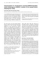
Fibrin plug formation and erythrocyte aggregation at
the arteriotomy site were more prominent in Group 1
than in Group 2. In the ABS tampon groups, a micro-
scopically evident basophilic discoloration in the perivas-
cular tissue was observed (Figure 1).
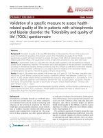
Significantly more widespread and dense eNOS stain-
ing was observed in Group 1 animals than Group 2
(Figure 2). The immunostaining of the unaffected vascu-
lar segments in ABS tampon and plain gauze tampon
groups did not differ significantly for eNOS expression
(Tabl e 1). Comparisons of the histopatho logical changes
Table 1 Light microscopical and immunohistochemical findings in the rats abdominal aorta on the operation day
Group I (ABS) Group II (Plain gauze) P
Histopathological changes
Inflammatory reaction 1.0 ± 0.5 1.0 ± 0.6 0.7
Necrosis none none N/A
Endothelial cell loss 1.0 ± 0.3 1.1 ± 0.4 0.5
Fibrin plug formation/Erythrocyte aggregation 1.5 ± 0.6 0.18 ± 0.4 0.0001
Immunohistochemical changes
e-NOS staining 1.9 ± 0.7 1.1 ± 0.4 0.0001
Kandemir et al. Journal of Cardiothoracic Surgery 2010, 5:110
/>Page 3 of 7
on D ay 7 of the operation are depicted in Table 2.
There was no necrosis adjacent to the intimal and
endothelial regeneration in either group.
The microscopically evident basophilic discoloration in
the ABS group on the operation day was a result of a
foreign body reaction (Figure 3A-B) and hemosiderin-
loaded histiocyte accumulation on Day 7 after the
operation (Figure 4A-B). Immunostaining with CD-31
showed an intact endothelial cell lining, and eNOS
stai ning did not differ among groups on Day 7 after the
operation.
Discussion
Hemorrhage from anastomosis sites can usually be man-
age d by additional sutures or light pressur e. If adeq uate
hemostasis cannot be achieved, various hemostatic
agents may be used. The ideal hemostatic agent should
be easy to use, require min imal training, show an effect
within minutes, be effective in both arterial and venous
bleeding, be non-toxic, and be anaphylactic [7]. Cur-
rently, no hemostatic agent possesses all of these
characteristics.
ABS is a novel topical hemostatic agent that consists
of various folkloric medicinal plant extracts (Thymus
vulgaris 0.1 mg, Vitis vivifera 0.16 mg, Glycyrrhiza
glabra 0.18 mg, Alpina officinarum 0.14 mg, and Urtica
dioica 0.12 mg). Each of these plants has vascular
actions and some effect on the hemat ologi cal system. T.
vulgaris has anti-oxidative effects, such as prevention of
lipid peroxidation [8]. V. vivifera has anti-atherosclerotic
effects [9]. G. glabra decreases vascular endothelial
growth factor production and cytokine-induced neovas-
cularization [10]. A. officinarum inhibits nitric oxide
production [11].
The hemostatic mechanism of ABS is effected by fibri-
nogen-erythrocyte agglutination, resulting in the forma-
tion of an encapsulated protein network that stimulates
erythrocyte aggregation. This encapsulated p rotein net-
work occurs very rapidl y, in less than 1 s [12]. The ABS
network might cover the entire physiological hemostat ic
process without affecting any individual clotting factor.
Göker at al. demonstrate d that coagulation factors II, V,
VII, VIII, IX, X, XI, and XIII were not affected, and that
plasma fibrinogen activity as well as total protein, albu-
min, and globulin levels were decreased by the addition
of ABS to plasma [6,7]. These results showed that nor-
mal hemostat ic elements were spared during the forma-
tion of the protein network. Thus, ABS might be useful
in patients with antithrombotic drug-induced primary or
secondary hemostatic abnormalities [13,14]. Cipil et al.
Figure 1 Fibrin plug formation and erythrocyte aggregation at the arteriotomy site were more prominent in Group 1 (A) than in
Group 2 (B). In the ABS tampon groups, microscopically evident basophilic discoloration in perivascular tissue was observed (A) (H&E, ×400).
Figure 2 A significantly more widespread and dense eNOS staining was observed in Group 1 (A) animals compared with Group 2 (B)
(Immunohistochemistry, eNOS, ×400).
Kandemir et al. Journal of Cardiothoracic Surgery 2010, 5:110
/>Page 4 of 7
demonstrated that ABS also had hemostatic effects in
animals pretreated with warfarin. The bleeding time was
reduced to 44% with ABS treatment [13].
Karakaya et al. demonstrated that ABS significantly
reduced blood loss and death in experimental rat liver
laceration [15]. Also, Dogan et al. used ABS for coron-
ary artery bypass surgery patients. They sprayed 4-8 mL
of ABS solution to bypass suture lines and the bleeding
area. They indicated that patients who had used ABS
required no revisions [16]. Our study revealed that fibrin
plug formation and erythrocyte aggregation at the arter-
iotomy site were more prominent in Group 1 than in
Group 2, and that bleeding time was 4.9 ± 0.6 s versus
3.1 ± 0.6 s in the ABS and control groups. Thus, ABS
reduced bleeding time by 36.7% compared with the con-
trol group. In clinical experiments, ABS has been suc-
cessfully used to control upper gastrointestinal bleeding
[17,18], acute anterior epistaxis [19], and bleeding due
to solitary rectal ulcers [20].
Although studies regarding the hemostatic effects
and mechanism of ABS are available, there is no
reported study regarding histopathological effects on
vascular tissue. Negative effects of tissue topical agents
used in anastomoses in cardiovascular surgery can
influence the patency of grafts in both the short- and
long-term.
Necrosis in vascular tissues, inflammatory reaction,
and endothelial cell loss are important, particularly in
terms of graft patency. Intimal hyperplasia can cause
aneurysm and thrombus formation [1]. In our study, on
Days 1 and 7 post-ABS application, histopathological
changes in the rat abdominal aorta did not differ
between Group 1 and 2 with regard to necrosis, inflam-
matory reaction, or endothelial cell loss.
After application of ABS, brown-colored changes
occurred around the tissue [21]. We believe that the
encapsulated protein network caused these changes. In
the ABS tampon groups, a microscopically evident baso-
philic discoloration in the perivascular tissue was
observed on the operation day and was caused by for-
eign body reaction and hemosiderin- loaded histiocyte
accumulation. This status could be explained by the for-
mation of the encapsulated protein networ k, causing
delayed degradation of erythrocytes. The long-term clin-
ical outcomes of this reaction must be clarified in pro-
spective experimental studies.
U. dioica, one of medicinal plant extracts in ABS,
causes vasodilatation by inducing nitric oxide produc-
tion by the endothelium [22]. Significantly more wide-
spread and dense eNOS staining was observed in Group
1 animals compared with Group 2. An increased eNOS
level around arteriotomy areas in the early stages
Table 2 Light microscopical and immunohistochemical findings in the rats abdominal aorta on the 7
th
day of the
operation
Group III (ABS) Group IV (Plain gauze) P
Histopathological changes
Necrosis none none N/A
Endothelial cell egeneration all all N/A
Foreign body reaction 2.0 ± 0.7 1.1 ± 0.4 0.006
Hemosiderin loaded hystiocyte 1.8 ± 0.6 0.8 ± 0.6 0.001
Immunohistochemical changes
e-NOS staining 0.8 ± 0.4 0.7 ± 0.4 0.6
Figure 3 Foreign body reaction on Day 7 after the operation in Groups 1 (A) and 2 (B) (H&E, ×400).
Kandemir et al. Journal of Cardiothoracic Surgery 2010, 5:110
/>Page 5 of 7
consistently stopped the bleeding in vitro without
impairing tissue oxygenation or microcirculation of ABS.
The advantages of ABS when compared with other
products that are readily available include effectiveness,
ease of application, and no requirement for technical
skills. However, as t he product is relatively new, a lim-
ited amount of data is available related to long-term
side effects and toxicity [1,21].
A limitation of this study is that only acute and early-
stage effects of ABS were evaluated. Long-term anasto-
mosis patency effects must be evaluated in further stu-
dies. Additional studies are required regarding possible
effects of ABS on vascular tissues over a period longer
than 7 days.
Conclusions
This is the first reported study evaluating the histo-
pathological and immunohistochemical effects of ABS
on vascular structure. In this study, hemostasis was suc-
cessfully achieved using ABS on rat abdominal aortas.
No histopathological change in rat abdominal aortas
between ABS and control groups on Days 1 and 7 was
found. Further prospective studies are also required
regarding long-term effects of foreign body reaction and
hemosiderin-loaded histiocyte accumulation.
Author details
1
Department of Cardiovascular Surgery, Zonguldak Karaelmas University,
Zonguldak, Turkey.
2
Department of Pathology, Zonguldak Karaelmas
University, Zonguldak, Turkey.
3
Department of Family Medicine, Zonguldak
Karaelmas University, Zonguldak, Turkey.
4
Department of Pathology, Dr.Lutfu
Kirdar Research and Training Hospital, Istanbul, Turkey.
5
Department of
Neurosurgery, Zonguldak Karaelmas University, Zonguldak, Turkey.
Authors’ contributions
OK: Acquisition, analysis and interpretation of data, surgical procedure,
drafting of manuscript. MB, SAT: study design. NOK, AEG: performed
microscopic and immunohistochemical evaluation and drafted the
manuscript. EA: drafting of manuscript, design of the study. SG:
interpretation of data, surgical procedure.
All authors have read and approved the final manuscript.
Competing interests
The authors declare that they have no competing interests.
Received: 28 August 2010 Accepted: 14 November 2010
Published: 14 November 2010
References
1. Kaplan M, Bozkurt S, Kut MS, Kullu S, Demirtas MM: Histopathological
effects of ethyl 2-cyanoacrylate tissue adhesive following surgical
application: an experimental study. Eur J Cardiothorac Surg 2004,
25(2):167-72.
2. Björses K, Holst J: Various local hemostatic agents with different modes
of action; an in vivo comparative randomized vascular surgical
experimental study. Eur J Vasc Endovasc Surg 2007, 33(3):363-70, Epub
2006 Nov 28.
3. Unlü Y, Vural U, Koçak H, Ceviz M, Becit N, Akbulut O: Comparison of the
topical haemostatic agents for the prevention of suture hole bleeding.
An experimental study. Eur J Vasc Endovasc Surg 2002, 23(5):441-4.
4. Kheirabadi BS, Pearson R, Rudnicka K, Somwaru L, MacPhee M, Drohan W,
Tuthill D: Development of an animal model for assessment of the
hemostatic efficacy of fibrin sealant in vascular surgery. JSurgRes2001,
100(1):84-92.
5. Werker PM, Kon M: Review of facilitated approaches to vascular
anastomosis surgery. Ann Thorac Surg 1997, 63(6 Suppl):S122-7.
6. Bilgili H, Kosar A, Kurt M, Onal IK, Goker H, Captug O, Shorbagi A, Turgut M,
Kekilli M, Kurt OK, Kirazli S, Aksu S, Haznedaroglu IC: Hemostatic efficacy of
Ankaferd Blood Stopper in a swine bleeding model. Med Princ Pract 2009,
18(3):165-9, Epub 2009 Apr 6.
7. Göker H, Haznedaroglu IC, Ercetin S, Kirazli S, Akman U, Ozturk Y, Firat HC:
Haemostatic actions of the folkloric medicinal plant extract Ankaferd
Blood Stopper®. J Int Med Res 2008, 36:163-170.
8. Lee SJ, Umano K, Shibamato T, Lee KG: Identification of volatile
components in basil (Ocimum basilicum L.) and thyme leaves (Thymus
vulgaris L.) and their antioxidant properties. Food Chem 2007, 91:131-137.
9. Yamakoshi J, Kataoka S, Koga T, Ariga T: Proanthocyanidin-rich extracts
form grape seede attenutaee the development of aortic atherosclerosis
in cholesterol-fed rabbits. Atherosclerosis 1999, 142:139-149.
10. Ramakrishna MK, Salimath BP: Angiogenic and proliferative effects if the
cytokine VEGF in Ehrlich ascites tumor cells is inhibited by Glycyrrhiza
glabra. Int Immunopharmacol 2006, 6:494-498.
11. Matsuda H, Ando S, Kato T, Morikawa T, Yoshikawa M: Inhibitors from the
rhizomes of Alpina officinarum on production of nitric oxide in
lipopolysaccharide-activated macrophages and the structural
requirements of diarrylheptanoids for the activity. Bioorg med Chem 2006,
14:138-142.
12. Haznedaroglu BZ, Haznedaroglu IC, Walker SL, Bilgili H, Goker H, Kosar A,
Aktas A, Captug O, Kurt M, Ozdemir O, Kirazli S, Firat HC: Ultrastructural
and Morphological Analyses of the In Vitro and In Vivo Hemostatic
Effects of Ankaferd Blood Stopper. Clin Appl Thromb Hemost 2010,
6(4):446-453.
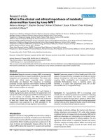
Figure 4 In the ABS tampon groups, prominent hemosiderin-loaded histiocyte accumulation in perivascular tissue was observed (A-B)
(A; H&E, B; iron blue, ×400).
Kandemir et al. Journal of Cardiothoracic Surgery 2010, 5:110
/>Page 6 of 7
13. Cipil HS, Kosar A, Kaya A, Uz B, Haznedaroglu IC, Goker H, Ozdemir O,
Koroglu M, Kirazli S, Firat HC: In vivo hemostatic effect of the medicinal
plant extract Ankaferd Blood Stopper in rats pretreated with warfarin.
Clin Appl Thromb hemost 2009, 15:270-276.
14. Kosar A, Cipil HS, Kaya A, Uz B, Haznedaroglu IC, Goker H, Ozdemir O,
Ercetin S, Kirazli S, Firat HC: The efficacy of Ankaferd Blood Stopper in
antithrombotic drug-induced primary and secondary hemostatic
abnormalities of a rat-bleeding model. Blood Coagul Fibrinolysis 2009,
20(3):185-90.
15. Karakaya K, Ucan HB, Tascilar O, Emre AU, Cakmak GK, Irkorucu O,
Ankarali H, Comert M: Evaluation of a new hemostatic agent Ankaferd
Blood Stopper in experimental liver laceration. J Invest Surg 2009,
22:201-206.
16. Dogan OF, Ozyurda U, Uymaz OK, Ercetin S, Haznedaroglu I: New
anticoagulant for CABG surgery. 4th Clinical Vascular Biology Congress,
2008, Antalya, Turkey .
17. Kurt M, Onal I, Akdogan M, Kekilli M, Arhan M, Sayilir A, Oztas E,
Haznedaroglu I: Ankaferd Blood Stopper for controlling gastrointestinal
bleeding due to distinct benign lesions refractory to conventional
antihemorrhagic measures. Can J Gastroenterol 2010, 24(6):380-4.
18. Kurt M, Disibeyaz S, Akdogan M, Sasmaz N, Aksu S, Haznedaroglu IC:
Endoscopic application of ankaferd blood stopper as a novel
experimental treatment modality for upper gastrointestinal bleeding: a
case report. Am J Gastroenterol 2008, 103(8):2156-8.
19. Teker AM, Korkut AY, Kahya V, Gedikli O: Prospective, randomized,
controlled clinical trial of Ankaferd Blood Stopper in patients with acute
anterior epistaxis. Eur Arch Otorhinolaryngol 2010, 267(9):1377-1381.
20. Ibis M, Kurt M, Onal IK, Haznedaroglu IC: Successful management of
bleeding due to solitary rectal ulcer via topical application of Ankaferd
blood stopper. J Altern Complement Med 2008, 14(9):1073-4.
21. Tokgöz H, Karakaya K, Hanci V, Abdusoglu M, Erol B, Turksoy O, Akduman B,
Mungan NA: Protective value of a folkloric medicinal plant extract
against mortality and hemorrhage in a life-threatining renal trauma
model. Urology 2010, 75:1515.e9-1515.e14.
22. Testai L, Chericoni S, Calderone V, Nencioni G, Nieri P, Morelli I, Martinotti E:
Cardiovascular effects of Urtica dioica L. (Urticaceae) roots extracts: in
vitro and in vivo pharmacological studies. J Ethnopharmacol 2002,
81(1):105-9.
doi:10.1186/1749-8090-5-110
Cite this article as: Kandemir et al.: Demonstration of the
histopathological and immunohistochemical effects of a novel
hemostatic agent, ankaferd blood stopper, on vascular tissue in a rat
aortic bleeding model. Journal of Cardiothoracic Surgery 2010 5:110.
Submit your next manuscript to BioMed Central
and take full advantage of:
• Convenient online submission
• Thorough peer review
• No space constraints or color figure charges
• Immediate publication on acceptance
• Inclusion in PubMed, CAS, Scopus and Google Scholar
• Research which is freely available for redistribution
Submit your manuscript at
www.biomedcentral.com/submit
Kandemir et al. Journal of Cardiothoracic Surgery 2010, 5:110
/>Page 7 of 7