Báo cáo y học: "Does left atrial volume affect exercise capacity of heart transplant recipients" docx
Bạn đang xem bản rút gọn của tài liệu. Xem và tải ngay bản đầy đủ của tài liệu tại đây (482.83 KB, 7 trang )
RESEA R C H ART I C L E Open Access
Does left atrial volume affect exercise capacity of
heart transplant recipients?
Mohammad Abdul-Waheed
1
, Mian Yousuf
1
, Stephanie J Kelly
2
, Ross Arena
3,4
, Jun Ying
5
, Tehmina Naz
1
,
Stephanie H Dunlap
1
, Yukitaka Shizukuda
1,6*
Abstract
Background: Heart transplant (HT) recipients demo nstrate limited exercise capacity compared to normal patients,
very likely for multiple reasons. In this study we hypothesized that left atrial volume (LAV), which is known to
predict exercise capacity in patients with various cardiac pathologies including heart failure and hypertrophic
cardiomyopathy is associated with limited exercise capacity of HT recipients.
Methods: We analyzed 50 patients [age 57 ±2 (SEM), 12 females] who had a post-HT echocardiography and
cardiopulmonary exercise test (CPX) within 9 weeks time at clinic follow up. The change in LAV (ΔLAV) was also
computed as the difference in LAV from the preceding one-year to the study echocardiogram. Correlations among
the measured parameters were assessed with a Pearson’s correlation analysis.
Results: LAV (n = 50) and ΔLAV (n = 40) indexed to body surface area were 40.6 ± 11.5 ml·m
-2
and
1.9 ± 8.5 ml·m
-2·
year
-1
, data are mean ± SD, respectively. Indexed LAV and ΔLAV were both significantly correlated
with the ventilatory efficiency, assessed by the VE/VCO
2
slope (r = 0.300, p = 0.038; r = 0.484, p = 0.002,
respectively). LAV showed a significant correlation with peak oxygen consumption (r = -0.328, p = 0.020).
Conclusions: Although our study is limited by a retrospective study design and relatively small number of patients,
our findings suggest that enlarged LAV and increasing change in LAV is associated with the diminished exercise
capacity in HT recipients and warrants further inves tigation to better elucidate this relationship.
Introduction
The exercise capacity of heart transplant (HT) recipients
is reportedly 30 to 40% lower than age/sex matched
apparently healthy individuals [1-4]. Mechanisms for
this limitation are sugge sted to be multifactorial. Dener-
vation, altered response to catecholamines, tissue
damage due to rejection episodes, general decondition-
ing associated with heart failure prior to HT, and long-
term use of immunosuppressant drugs have all been
proposed, but conclusive data for each mechanism is
lacking [2]. R enlund et al. have reported that although
longer donor heart ischemic time and frequent rejection
have no effect, elevated resting pulmonary vascular
resistance inhibits exercise capacity [2]. Similarly, animal
models of heart denervation bot h with chemicals [5,6]
and HT [7] show no indication of a decrease in cardiac
function during exercise due to denervation. Therefore,
the factors, which limit exercise capacity of HT recipi-
ents, remain undefined.
Recently, increased left atrial volume (LAV) has been
reported to predict diminished exercise capacity in
patients with hear t failure [8] and hypertrophic non-
obstructive cardiomyopathy [9]. One proposed mechanism
is that expanded LAV could be a reflection of chronic left
ventricular (LV) diastolic dysfunction, either at rest or dur-
ing exercise, which may in turn impair exercise capacity
[8,9]. Another possible aspect of altered left atrial function
[10,11] in HT recipients is that suboptimal active contrac-
tion in a presence of dilated left atrium and the surgical
scar of the anastomosis between native and donor atrium
in post-transplant may diminish left ventricle preload and
thus further limit exercise capacity caused by LA enlarge-
ment itself. Therefore, we hypothesized that increased
LAV is associated with diminished exercise capacity in HT
recipients, and used echocardiography and cardiopulmon-
ary exercise testing (CPX) to evaluate their relationship.
* Correspondence:
1
Division of Cardiovascular Diseases, Department of Internal Medicine
University of Cincinnati, Cincinnati, Ohio, USA
Full list of author information is available at the end of the article
Abdul-Waheed et al. Journal of Cardiothoracic Surgery 2010, 5:113
/>© 2010 Abdul-Waheed et al; licensee BioMed Central Ltd. This is an Open Access article distributed under the terms of the Creative
Commons Attribution License ( which permits unrestricted use, distribution, and
reproduction in any medium, provided the original work is properly cited.
Design and Methods
Study population
This clinical protocol was approved by the Institutional
Review Board and was consistent with the principles of
the Declaration of Helsinki [12]. Due to the retrospective
nature of the study, waiver of consent was approved.
Patients with heart failure who underwent post HT clini-
cal follow up were i ncluded when the following conditions
were met: 1) Post HT follow up was performed in our
institution, 2) Baseline post-HT echocardiography was
performed within 9 weeks of post transplant CPX, 3) No
more than mild mitral regurgitation during baseline echo-
cardiograph, 4) No clinically significant myocardial ische-
mia with stress testing at the time of study entry, 5)
Normal sinus rhythm, 6) No clinically significant active
transplant rejection at the time of study entry, and 7) No
prescription of b-adrenergic receptor blocker at the time
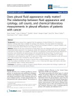
of CPX. The study design for the present investigation is
illustrated in Figure 1. Fifty out of a potential 108 patients
who visited our clinic for a post HT follow up between
1998 and 2007 met the inclusion criteria. Among them,
48 patients received HT at our institution and 2 patients
received HT at an outsi de hospital. Among the patients
studied, 45 patients received standard right atrial anasto-
mosis and 3 re ceived bicaval anastomosis. The type of
right sided anastomosis could not be determined in two
cases. All cases received standard left atrial cuff anastomo-
sis. In 40 cases, echocardiography at one year prior to the
baseline echocardiogram was available to calculate the
change in the LAV. By the study design, CPX was not
performed to evaluate a change in exercise capacity dur-
ing this one year interval to calcula te the change in the
LAV. The time duration after HT to the echocardiogra-
phy conjunction for the CPX analysis was within 2 years
in 11 patients, between 2 years and 5 years in 18 patients,
and more than 5 years for the remaining patients.
Echocardiographic measurements
The patients were imaged with multifrequency transducers
with center frequencies of 2.5 or 3.5 MHz (ATL HDL
1000, Philips Medical system, Bothell , Washington, USA,
iE33, Philips Medical System, Bothell, Washington, USA,
Vivid 7 GE Healthcare system, Milwaukee, Wisconsin,
USA).Briefly,inallcasespulmonaryveinsandtheLA
appendage were excluded from planim etric analysis. The
outline of the atrial endocardium was traced at the end of
ventricular systole at the point of maximum LA dimen-
sion. Studies were recorded digitally and stored in the
Camtronics Imaging system (Emageon Camtronics system,
Birmingham, Alabama, USA). Left atrial volume measure-
ments were performed off-line on digital loops using a
Digisonics review station (version 3.2 software, Digisonics
Inc. Houston, Texas, USA) as previous ly reported by our
group [9,13,14]. LAV were measured using the hand four
chamber views at end systole [9,13,14]. We used this
method over the area-length method recommended by
the American Society of Echocardiography [15] to calcu-
late LAV because our method is based by fewer geometric
assumptions than the area-length method. In our preli-
minary study, the interobserver variability of non-indexed
LAV was 13.5 ± 2.0% volume, n = 19 and intraobserver
variability was 8.8 ± 1.5% volume, n = 23 (values are
mean ± SEM). These findings were typical noted for volu-
metric measurements based on 2-dimensional echocardio-
graphy [15]. The one-year change in LAV (ΔLAV) was
computed as a difference between left atrial volume mea-
surements i n the same patient one year apart. Additionally,
left ventricular volume and ejection fraction were calcu-
lated from apical 4 and 2 chamber views using the biplane
Simpson method [15]. Left ventricular diastolic function
was assessed in all patients using pulsed Doppler peak E, A
velocities, and E/A of mitral inflow as previously described
[16]. The tissue Doppler imaging of lateral mitral annulus
was also performed to measure peak diastolic E’ velocity
and E/E’ ratio was calculated to assess left ventricular dia-
stolic function as previously described [17]. The studies
were blinded and measured by a single r eader (Y.S.).
Cardiopulmonary Exercise Testing
Exercise tests were performed on a treadmill using a
ramping protocol, which is appropriate for patients with a
diminished aerobic capacity [18-20]. Briefly, the starting
speed and grade were 27 m·min
-1
and 0% respectively.
After 2 min of exercise the speed plateaued at 64 m·min
-1
then the grade was increased by 0.5% every 15 seconds.
Throughout the test, ECG, symptoms, blood pressure, and
respiratory gas analysis were recorded. Ventilatory expired
gas analysis was performed by a metabolic cart (Med-
graphics Ultima, Medgraphics, St. Paul, Minnesota, USA)
[21,22]. The oxygen and carbon dioxide sensors were cali-
brated prior to each test using gases with known oxygen,
nitrogen, and carbon dioxide concentrations. Test termi-
nat ion criteria consis ted followed American Heart Asso-
ciation/American College of Cardiology guidelines [23].
Oxygen consumption, VO
2
(ml·kg
-1·
min
-1
), Carbon diox-
ide production, VCO
2
(L·min
-1
), and minute ventilation,
VE (L·min
-1
) were collected throughout the exercise test.
Peak VO
2
was expressed as the highest 30-second average
value obtained during the last stage of the exercise test.
Peak respiratory exchange ratio (RER) was the highest 30-
second averaged value during the last stage of the exercise
test. Ven tilatory efficiency was assessed by the VE/VCO
2
slope as previously reported with higher values (steeper
VE to VCO
2
relation ship, norma l < 30) reflect limited
exercise capacity and abnormal cardiopulmonary physiol-
ogy [9,13,24].
Abdul-Waheed et al. Journal of Cardiothoracic Surgery 2010, 5:113
/>Page 2 of 7
Statistical Analysis
Data are presented mean ± SD. for measurements. The
relationship between both LAV and ΔLAV and CPX
variables were analyzed by a Pearson correlation test.
The correlation between CPX variables and time since
HT w as also assesse d. Exercise parameters b etween the
patients with positive and negative values of indexed
ΔLAV were compared with an unpaired Student t-test.
All tests were two-sided and analyses with a p-value <
0.05 were considered statistically significant.
Results
Patients’ characteristics
Among the patients investigated, most were asympto-
matic [36 patients (72%) were NYHA class I] and
although 48% of the patients had a history of histologi-
cal-determined transplant tissue rejection in the past, all
were subclinical with less than International Society for
Heart and Lung Transplantation grade II (Table 1). The
etiology of heart failure resulted in HT was non
ischemic in 22 patients, ischemic in 27 patients, and
combined non ischemic and ischemic in 1 patient. Base-
line echocardiography showed that the patients had nor-
mal left ventricular systolic and diastolic function
demonstrated by normal peak E tissue velocity of the
mitral annulus (Table 2). The estimation of left atrial
pressure, E/E’ [17,25], was also within the normal range
for this group. The average of left atrial volume indexed
to body surface areas was significantly larger than nor-
mative values (indexed left atrial volume < 34 ml·m
-2
)
[9], reflecting typical HT morphology and 32 patients
(64%) demonstrated indexed atrial volume > 34 ml·m
-2
.
The indexed ΔLAV was 1.9 ± 8.5 ml·m
-2·
year
-1
,indicat-
ing a relatively small increase i n the LAV over the one
year observation period in this cohort. In our popula-
tion, the average baseline systolic blood pressure was
T
ime
Heart Transplant
Baseline
Echocardiography
CPX
Preceding
Echocardiography
One year
Average 4.7 years
Δ
ΔΔ
ΔLAV
LAV
Figure 1 Study design. The study design is shown. Left atrial volume (LAV) was calcula ted from baseline echocardiography and the volume
change in LAV (ΔLAV) was calculated from the baseline LAV subtracted that at the preceding one year. CPX = cardiopulmonary stress test.
Abdul-Waheed et al. Journal of Cardiothoracic Surgery 2010, 5:113
/>Page 3 of 7
125 ± 18 mmHg and the baseline diastolic blood pres-
sure was 78 ± 11 mmHg. Only 4 subjects demonstrated
clinically significant hypertension (systolic blood pres-
sure > 150 mmHg or diastolic blood pressure > 95
mmHg). In addition, no significant correlation was
noted between baseline blood pressures and parameters
of exercise capacity.
Relationship between LAV and ΔLAV and exercise
test characteristics
All exercise parameters were significantly augmented
during exercise in these patients (Table 3), with the
exception of diastolic blood pressure. Neither the VE/
VCO
2
slope (r = -0.012, p = 0.934) nor peak VO
2
(r =
0.010, p = 0.487) correlated with duration post HT, indi-
cating that changes in CPX parameters are not time
dependent in this group. However, these findings did
notprecludeatimedependenceofCPXparametersat
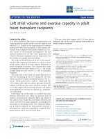
an individual level. A significant correlation was noted
between both absolute LAV and ΔLAV and the VE/
VCO
2
slope (Figure 2). When the patients were classi-
fied according to positive and negative values of indexed
ΔLAV, those with positive ΔLAV (increasing LA size
over one year) showed a significantly higher VE/VCO
2
slope as compared with those with negative values
(40.2 ± 6.5 vs. 33.6 ± 5.0, p = 0.003). Left atrial volume
correlated with peak VO
2
(r = -0.328, p = 0.020) while
the correlation with ΔLAV was not significant
(r = 0.079, p = 0.616 for those not i ndexed, r = 0.006,
p = 0.971 for those indexed).
Discussion
The results of the present study demonstrate that in this
cohort of HT patients, abnormalities in the exercise
response is modest but significantly correlated with both
the magnitude of baseline post-HT LAV, as well as posi-
tive change in LAV over one year’stime(ΔLAV), as
reflected by thei r relationship with ventilatory efficiency
(i.e. the VE/VCO
2
slope). Thus, the association of
increased LAV with an abnormal exercise response pre-
sents a possibility that left atrial remodeling may be a
surrogate for factors limiting the physiologic response to
exertion in HT recipients.
It has been proposed that increasing LAV reflects
chronic changes in left ventricular diastolic function
[26]; therefore, left ventricular diastolic dysfunction may
play a role in the pathophysiologic mechanisms that
reduce exercise capacity in several different cardiac
populations. Although our study population did not
show abnormal baseline left ventricular diastolic func-
tion parameters with echocardiography, it is possible
that this is still a mechanism related to limited exercise
capacity with larger LAV, in part because left ventricular
diastolic dysfunction frequently may only become evi-
dent during exercise while re maining undetected in stu-
dies done at r est [27,28]. Only 4 patients (8%) in the
current study demonstrated elevated baseline blood
pressure; however, 58% of ou r patients had a history of
hypertension. Thus, our study population may be sus-
ceptible to exercise-induced left ventricular diastolic
Table 1 Baseline Characteristics
Variables N = 50
Age 57 ± 14
Gender (female) 12 (24%)
Body surface area (m
2
/kg) 2.0 ± 0.2
Time after transplant (years) 4.7 ± 3.3
NYHA class 1.4 ± 0.6
Histological rejection 24 (48%)
Hypertension 29 (58%)
Diabetes 20 (40%)
Data are mean ± SD.
Table 2 Echocardigraphic measurements
Variables
Left ventricular ejection fraction (%) 67 ± 7
Left ventricular end diastolic volume (ml) 68 ± 19
Indexed Left ventricular end diastolic volume (ml/m
2
)34±9
Left atrial volume (ml) 83.5 ± 23.7
Indexed-left atrial volume (ml/m
2
) 40.6 ± 11.5
Change in left atrial volume (ml/year) 3.9 ± 17.6
Indexed-change in left atrial volume (ml/year/m
2
) 1.9 ± 8.5
Mitral inflow peak diastolic E velocity (cm/sec) 85.0 ± 23.1
Mitral inflow peak diastolic A velocity (cm/sec) 41.3 ± 13.5
Mitral valve inflow E/A 2.3 ± 1.1
Peak diastolic E velocity of lateral mitral annulus 13.8 ± 3.7
E/E’ 6.8 ± 3.3
E = diastolic early filling. A = diastolic atrial contraction. E/A = ratio of peak E
velocity to A velocity of mitral inflow. E/E’ = ratio of peak E mitral inflow
velocity of peak E velocity of lateral mitral annulus. Data are mean ± SD.
n = 50 except change in left atrial volume (n = 40).
Table 3 Exercise measurements
Variables N = 50
Baseline heat rate (bpm) 89 ± 14
Baseline systolic blood pressure (mmHg) 125 ± 18
Baseline diastolic blood pressure (mmHg) 78 ± 11
Baseline pressure rate product (bpm·mmHg·10
3
) 1.09 ± 0.20
Peak exercise heart rate (bpm) 134 ± 18*
Peak exercise systolic blood pressure (mmHg) 161 ± 27*
Peak exercise diastolic blood pressure (mmHg) 81 ± 14
Peak exercise pressure rate product (bpm·mmHg·10
3
) 2.16 ± 0.49*
Peak respiratory exchange ratio 1.13 ± 0.09
Peak exercise oxygen consumption (ml O
2·
min
-1·
kg
-1
) 17.7 ± 6.0
Peak exercise VE/VCO
2
slope 38.7 ± 7.5
Data are mean ± SD. *P < 0.01 vs. baseline measurements. bpm denotes beat
per minute. The comparison of measurements between at baseline and at
peak exercise was performed with a paired Student t-test.
Abdul-Waheed et al. Journal of Cardiothoracic Surgery 2010, 5:113
/>Page 4 of 7
dysfunction. In this regard, a future study using exercise
echocardiography to assess exercise left ventricular dia-
stolic function in this population could be quite
revealing.
The dilatation of LAV might be also in part related to
the surgical scar of the left atrial anastomosis. The sur-
gical scar between the native and the donor atrium may
impede correct left atrial pump function and therefore,
the left atrium may subsequently dilate to increase the
reservoir capacity as a compe nsatory mechanism, which
in turn theoretically would maintain left atrial output in
the presence of impaired atrial pump function.
Following HT, an enlarged left atrium is considered to
be a typical and clinically insignificant finding during
any post-transplant echocardiography. This fact often
leads to an under-appreciation of how left atrial enlarge-
ment may play a role in transplanted heart function.
Thus, increases in left atrium size in HT patients, as
well as in other cardiac disease patients [9,13], may be
an important surrogate for significant loss of atrial func-
tion or worsening of left ventricular diastolic function,
and furthermore, such functional deterioration may on ly
appear during exercise. For example, as a possible atrial
structure-function mechanism, consider that in an
enlarged left atrium with preserved wall compliance but
without compensatory augmentation of active atrial con-
traction - as would be the case after HT - with exercise
there may be pooli ng of intra-atrial venous return; such
pooling could lead to a significant restriction of left ven-
tricular preload during the period of increased cardiac
demand, and therefore in turn limit the patient’ sexer-
cise capac ity. Thus, improved functional capacity in HT
recipients with total orthotopic HT using both bicaval
and pulmonary vein anastomosis, as compared to tradi-
tional orthotopic HT technique, may be in part related
to reduction of left atrial size [29]. This hypothesized
mechanism might be investigated by assessing left atrial
volume and function and exercise capacity in our HT
population using exercise echocardiography. Our study
for the first time suggests that both indicators - larger
absolute LAV and an increase in LAV following HT -
may be early warning signs of declining exercise capacity
in this population.
The correlation between ΔLAVandCPXmeasuresof
peak aerobic capacity was considerably weaker than the
correlation with ventilatory efficiency in the present
AB
P = 0.038
R = 0.300
60
P = 0.002
R = 0.484
o
pe
60
l
ope
50
V
E/VCO
2
sl
o
50
V
E
/
V
CO
2
s
l
40
V
40
V
30
30
20
020406080
20
-30 -20 -10 0 10 20
Indexed-LA volume
(
ml·m
-2
)
Indexed-
'
LA Volume
(
ml·m
-2
·
y
ea
r
-1
)
Figure 2 Relationship between left atrial volume and ventilatory efficiency. The linear correlation between left atrial (LA) volume in panel
A or yearly change in LA volume (ΔLA) volume with ventilatory efficiency (VE/VCO
2
slope) in panel B is shown. The correlation was analyzed
with the Pearson product moment correlation.
Abdul-Waheed et al. Journal of Cardiothoracic Surgery 2010, 5:113
/>Page 5 of 7
study. Previous work in patients with non-obstructive
hypertrophic cardiomyopathy has also found that the
linkage between LAV and ventilatory efficiency was
stronger compared to that found between LAV and VO
2
at peak exercise [9,13]. Other investigations in patients
with heart failure rather consistently demonstrate that
the relationship between various markers of cardiovas-
cular pathophysiology (b-type natriuretic peptide, pul-
monary vascular pressures, pulmonary diffusion
capacity, e tc) and ventilatory efficiency is stronger than
the correlation found with peak VO
2
[30]. A primary
reason for the present and past correlation difference
may be t he reliance that a tr ue peak VO
2
response has
on maximal subject effort, a prerequisite that is not
required for attainment of a physiologically valid mea-
sure of ventilatory efficiency.
The retrospective nature of this study and relatively
small sample size are the primary limitations of the pre-
sent investigation. While the demonstrated correlation
of LAV and exercise capacity holds potential clinical sig-
nificance, the relationships presented in the present
study are numerically relatively modest, indicating that
additional factors are likely associated with the CPX
response in patients undergoing HT or LAV may be a
surrogate for factors that affect exercise capacity rather
than a primary determinant. To further strengthen our
findings, a prospective study addressing these issues in a
larger HT cohort is required. It is also possible that new
echocardographic parameters obtained from emerging
technology, such as strain/strain rate asse ssment [31],
or more accurate assessment of LAV with other
imaging modality may better correlate with exercise
performance.
Conclusion
In conclusion, our study sho ws that increa sing LAV is
significantly associated with the limited exercis e capacity
of HT recipients. Further investigation to evaluate t he
relationship between LAV and exercise capacity in the
HT population is therefore warranted.
Acknowledgements
We appreciate Stantosh Likki, MD, Division of Cardiovascular Diseases,
Department of Internal Medicine, University of Cincinnati, Cincinnati, Ohio,
USA, for assistance collecting data. We thank Allan Harrelson, DO, PhD,
Division of Cardiovascular Medicine, Oregon Health Science & University,
Oregon, USA, for critical reading of the manuscript.
Author details
1
Division of Cardiovascular Diseases, Department of Internal Medicine
University of Cincinnati, Cincinnati, Ohio, USA.
2
UC Health, Cincinnati Ohio,
USA.
3
Department of Physiology and Physical Therapy, Virginia
Commonwealth University, Richmond, Virginia, USA.
4
Department of Internal
Medicine, Virginia Commonwealth University, Richmond, Virginia, USA.
5
Department of Public Health Sciences, University of Cincinnati, Cincinnati,
Ohio, USA.
6
Cincinnati Veterans Affairs Medical Center, Cincinnati, Ohio, USA.
Authors’ contributions
MAW carried out collection of data, data analysis, and editing the
manuscript. MY participated in study design, collection of data, and editing
the manuscript. SJK participated in collection of data, editing the
manuscript. RA participated in study design and editing the manuscript. JY
participated in study design and editing the manuscript. NT participated in
study design and editing the manuscript. SHD participated in study design
and editing the manuscript. YS carried out study design and coordination,
collection of data, data analysis, and drafting the manuscript. All authors
read and approved the final manuscript.
Competing interests
The authors declare that they have no competing interests.
Received: 31 July 2010 Accepted: 17 November 2010
Published: 17 November 2010
References
1. Savin WM, Haskell WL, Schroeder JS, Stinson EB: Cardiorespiratory
responses of cardiac transplant patients to graded, symptom-limited
exercise. Circulation 1980, 62(1):55-60.
2. Renlund DG, Taylor DO, Ensley RD, O’Connell JB, Gilbert EM, Bristow MR,
Ma H, Yanowitz FG: Exercise capacity after heart transplantation:
influence of donor and recipient characteristics. J Heart Lung Transplant
1996, 15:16-24.
3. Labovitz AJ, Drimmer AM, McBride LR, Pennington DG, Willman VL,
Miller LW: Exercise capacity during the first year after cardiac
transplantation. Am J Cardiol 1989, 64(10):642-645.
4. Kavanagh T, Yacoub MH, Mertens DJ, Kennedy J, Campbell RB, Sawyer P:
Cardiorespiratory responses to exercise training after orthotopic cardiac
transplantation. Circulation 1988, 77(1):162-171.
5. Donald DE, Shepherd JT: Response to Exercise in Dogs with Cardiac
Denervation. Am J Physiol 1963, 205:393-400.
6. Donald DE, Shepherd JT: Initial Cardiovascular Adjustment to Exercise in
Dogs with Chronic Cardiac Denervation. Am J Physiol 1964,
207:1325-1329.
7. Daggett WM, Willman VL, Cooper T, Hanlon CR: Work capacity and
efficiency of the autotransplanted heart. Circulation 1967, 35(Suppl 4):
I96-I104.
8. Donal E, Raud-Raynier P, De Place C, Gervais R, Rosier A, Roulaud M,
Ingels A, Carre F, Daubert JC, Denjean A: Resting echocardiographic
assessments of left atrial function and filling pressure interest in the
understanding of exercise capacity in patients with chronic congestive
heart failure. J Am Soc Echocardiogr 2008, 21(6):703-710.
9. Sachdev V, Shizukuda Y, Brenneman CL, Birdsall CW, Waclawiw MA, Arai AE,
Mohiddin SA, Tripodi D, Fananapazir L, Plehn JF: Left atrial volumetric
remodeling is predictive of functional capacity in nonobstructive
hypertrophic cardiomyopathy. Am Heart J 2005, 149(4):730-736.
10. Stefanadis C, Dernellis J, Toutouzas P: A clinical appraisal of left atrial
function. Eur Heart J 2001, 22(1):22-36.
11. Garcia MJ: Left ventricular filling. Heart Fail Clin 2008, 4(1):47-56.
12. World Medical Association: World Medical Association declaration of
Helsinki. Recommendations guiding physicians in biomedical research
involving human subjects. JAMA 1997, 277(11):925-926.
13. Shizukuda Y, Sachdev V, Fananapazir L, Tripodi D, Mohiddin SA, Arai AE,
Waclawiw MA, Plehn JF: Is functional capacity related to left atrial
contractile function in nonobstructive hypertrophic cardiomyopathy?
Congest Heart Fail 2005, 11(5):234-240.
14. Shizukuda Y, Bolan CD, Tripodi DJ, Yau YY, Nguyen TT, Botello G,
Sachdev V, Sidenko S, Ernst I, Waclawiw MA, Leitman SF, Rosing DR:
Significance of left atrial contractile function in asymptomatic subjects
with hereditary hemochromatosis. Am J Cardiol
2006, 98(7):954-959.
15. Lang RM, Bierig M, Devereux RB, Flachskampf FA, Foster E, Pellikka PA,
Picard MH, Roman MJ, Seward J, Shanewise JS, Solomon SD, Spencer KT,
Sutton MS, Stewart WJ: Recommendations for chamber quantification: a
report from the American Society of Echocardiography’s Guidelines and
Standards Committee and the Chamber Quantification Writing Group,
developed in conjunction with the European Association of
Echocardiography, a branch of the European Society of Cardiology. JAm
Soc Echocardiogr 2005, 18(12):1440-1463.
Abdul-Waheed et al. Journal of Cardiothoracic Surgery 2010, 5:113
/>Page 6 of 7
16. Quinones MA, Otto CM, Stoddard M, Waggoner A, Zoghbi WA:
Recommendations for quantification of Doppler echocardiography: a
report from the Doppler Quantification Task Force of the Nomenclature
and Standards Committee of the American Society of Echocardiography.
J Am Soc Echocardiogr 2002, 15(2):167-184.
17. Nagueh SF, Middleton KJ, Kopelen HA, Zoghbi WA, Quinones MA: Doppler
tissue imaging: a noninvasive technique for evaluation of left ventricular
relaxation and estimation of filling pressures. J Am Coll Cardiol 1997,
30(6):1527-1533.
18. Wasserman K: Testing regulation of ventilation with exercise. Chest 1976,
70(Suppl 1):173-178.
19. Wasserman K, Zhang YY, Gitt A, Belardinelli R, Koike A, Lubarsky L,
Agostoni PG: Lung function and exercise gas exchange in chronic heart
failure. Circulation 1997, 96(7):2221-2227.
20. Arena R, Myers J, Williams MA, Gulati M, Kligfield P, Balady GJ, Collins E,
Fletcher G: Assessment of functional capacity in clinical and research
settings: a scientific statement from the American Heart Association
Committee on Exercise, Rehabilitation, and Prevention of the Council on
Clinical Cardiology and the Council on Cardiovascular Nursing.
Circulation 2007, 116(3):329-343.
21. Weber KT, Janicki JS, McElroy PA: Determination of aerobic capacity and
the severity of chronic cardiac and circulatory failure. Circulation 1987,
76(6):VI40-VI45.
22. Simonton CA, Higginbotham MB, Cobb FR: The ventilatory threshold:
quantitative analysis of reproducibility and relation to arterial lactate
concentration in normal subjects and in patients with chronic
congestive heart failure. Am J Cardiol 1988, 62(1):100-107.
23. Gibbons RJ, Balady GJ, Bricker JT, Chaitman BR, Fletcher GF, Froelicher VF,
Mark DB, McCallister BD, Mooss AN, O’Reilly MG, Winters WL, Gibbons RJ,
Antman EM, Alpert JS, Faxon DP, Fuster V, Gregoratos G, Hiratzka LF,
Jacobs AK, Russell RO, Smith SC: ACC/AHA 2002 guideline update for
exercise testing: summary article. A report of the American College of
Cardiology/American Heart Association Task Force on Practice
Guidelines (Committee to Update the 1997 Exercise Testing Guidelines).
J Am Coll Cardiol 2002, 40(8):1531-1540.
24. Shizukuda Y, Bolan CD, Tripodi DJ, Yau YY, Smith KP, Arena R,
Waclawiw MA, Leitman SF, Rosing DR: Exercise capacity of cardiac
asymptomatic hereditary hemochromatosis subjects. Med Sci Sports Exerc
2007, 39(1):3-7.
25. Nagueh SF, Lakkis NM, Middleton KJ, Spencer WH, Zoghbi WA,
Quinones MA: Doppler estimation of left ventricular filling pressures in
patients with hypertrophic cardiomyopathy. Circulation 1999,
99(2):254-261.
26. Rossi A, Cicoira M, Zanolla L, Sandrini R, Golia G, Zardini P, Enriquez-
Sarano M: Determinants and prognostic value of left atrial volume in
patients with dilated cardiomyopathy. J Am Coll Cardiol 2002, 40(8) :1425.
27. Ha JW, Oh JK, Pellikka PA, Ommen SR, Stussy VL, Bailey KR, Seward JB,
Tajik AJ: Diastolic stress echocardiography: a novel noninvasive
diagnostic test for diastolic dysfunction using supine bicycle exercise
Doppler echocardiography. J Am Soc Echocardiogr 2005, 18(1):63-68.
28. Ha JW, Lee HC, Kang ES, Ahn CM, Kim JM, Ahn JA, Lee SW, Choi EY, Rim SJ,
Oh JK, Chung N: Abnormal left ventricular longitudinal functional reserve
in patients with diabetes mellitus: implication for detecting subclinical
myocardial dysfunction using exercise tissue Doppler echocardiography.
Heart 2007, 93(12):1571-1576.
29. Magliato KE, Trento A:
Heart transplantation–surgical results. Heart Fail Rev
2001, 6(3):213-219.
30. Arena R, Myers J, Guazzi M: The clinical and research applications of
aerobic capacity and ventilatory efficiency in heart failure: an evidence-
based review. Heart Fail Rev 2008, 13(2):245-269.
31. Sachdev V, Aletras AH, Padmanabhan S, Sidenko S, Rao YN, Brenneman CL,
Shizukuda Y, Lie GR, Vincent PS, Waclawiw MA, Arai AE: Myocardial strain
decreases with increasing transmurality of infarction: a Doppler
echocardiographic and magnetic resonance correlation study. J Am Soc
Echocardiogr 2006, 19(1):34-39.
doi:10.1186/1749-8090-5-113
Cite this article as: Abdul-Waheed et al.: Does left atrial volume affect
exercise capacity of heart transplant recipients?. Journal of Cardiothoracic
Surgery 2010 5:113.
Submit your next manuscript to BioMed Central
and take full advantage of:
• Convenient online submission
• Thorough peer review
• No space constraints or color figure charges
• Immediate publication on acceptance
• Inclusion in PubMed, CAS, Scopus and Google Scholar
• Research which is freely available for redistribution
Submit your manuscript at
www.biomedcentral.com/submit
Abdul-Waheed et al. Journal of Cardiothoracic Surgery 2010, 5:113
/>Page 7 of 7