Báo cáo y học: "Primary cardiac osteosarcoma in a 42-year-old woman" pptx
Bạn đang xem bản rút gọn của tài liệu. Xem và tải ngay bản đầy đủ của tài liệu tại đây (621.83 KB, 2 trang )
CAS E REP O R T Open Access
Primary cardiac osteosarcoma in a
42-year-old woman
Honghe Luo
1
, Yiyan Lei
1
, Chunhua Su
1
, Lie Cai
2
, Tao Wang
3
, Jianyong Zou
1
, Zhenguang Chen
1*†
Abstract
We describe here a 42-year-old woman who was admitted to hospital with a pedunculated mass in her left atrium.
She was diagnosed with a primary cardiac osteosarcoma with special immunohistochemical characteristics. Echo-
cardiography and computed tomography can be used to differentiate cardiac osteosarcomas from routine intracar-
diac tumors. The patient was treated by surgical removal of the mass. Two years later, she has shown no evidence
of disease recurrence. We discuss primary osteosarcomas in the cardiac cavity and their management.
Introduction
Although osteosarcoma is a common tumor of the skeletal
system, primary cardiac osteosarcoma is an extremely rare
malignant disease wit h no nspecific symptoms, making
early diagnosis a challenge. We describe here a 42-year-
old woman with a primary cardiac osteosarcoma, which
was surgically removed by cardiopulmonary bypass.
Two years later, she has shown no evidenc e of tumor
recurrence.
Case report
A 42-yea r-old woman was admitted to our hospital com-
plaining of chest pain, shortness of breath and weight
loss. Physical examination revealed an extra systolic mur-
mur at the cardiac apex, with NYHA stage III. An elec-
trocardiogram revealed sinus bradycardia, and
echocardiography showed a pedunculated mass in her
left atrium with weak aortic and mitral valve insuffi-
ciency, similar to myxoma (Figure 1). Computed tomo-
graphy revealed a mass, 65 × 20 × 20 mm in size and
attached to th e posterior wall of the left atrium, witho ut
calcification or pericardial effusion. The patient was diag-
nosed with a primary cardiac tumor and was referred for
surgical removal of the mass. During surgery, a tumor
measuring 50 × 20 × 20 mm was found, with a stalk
attached to the posterior wall of the left atrium and near
the orifice of the left pulmonary vein. The mass was
removed and a partial endocardiectomy was performed.
Pathological examination of the tumor showed that the
malignant cells were irregularly osteoid without polygo-
nal to stellate shapes. The tumor cells were strongly
stained with antibodies to the osteoclast marker CD68
and vimentin, but we re weakly stained with antibodie s to
CK,EMA,S-100,andCD34(Figure1).Basedonthese
histological and immunohistochemical findings, the final
diagnosis was primary cardiac osteosarcoma [1,2]. At
present, 2 years after surgical removal of the tumor, the
patient remains healthy with no evidence of tumor
recurrence.
Discussion
Most primary cardiac tumors are myxomas, and only a
very small proportion of these cardiac tumors (< 0.28%)
are malignant [3]. Only a few isolated cases of primary
cardiacosteosarcomahavebeen reported, making the
etiology of these tumors unclear [1-5]. To our knowl-
edge, therefore, primary cardiac o steosa rcomas are rare
and difficult to diagnose.
The symptoms of primary cardiac osteosarcoma have
been described as protean, with obstruction and heart
failure being the primary manifestations [1,3]. On echo-
cardiography, cardiac osteosarcom as often show asym-
metrical internal echoes, and computed tomography has
shown the calcification of cardiac osteosarcomas. Cer-
tain features (e.g., a broad base of attachment or origin
at a site other than the atrial septum) help differentiate
these tumors from left atrial myxomas [6]. However, the
tumor in our patient presented as a soft symmetrical
parenchymal tumor, the presence of calcification did not
* Correspondence:
† Contribu ted equally
1
Department of Thoracic Surgery, The First Affiliated Hospital, Sun Yat-sen
University, Guangzhou (510080), Guangdong, People’s Republic of China
Full list of author information is available at the end of the article
Luo et al. Journal of Cardiothoracic Surgery 2010, 5:120
/>© 2010 L uo et al; licens ee BioMed Central Ltd. This is an Open Access article distributed unde r the terms of the Creative Commons
Attribution License ( which permits unrestrict ed use, distribution, and reproduction in
any medium, provided the original work is properly cited.
seem useful in diffe rentiating atrial osteosarcoma from
myxoma.
Cardiopulmonary bypass is essential for removing the
primary cardiac osteosarcoma. We c hose a right angle
type superio r vena cava tube t o avoid crushing the
tumor in our patient. The mass was removed, along
with at least 5 mm of the surrounding endocardium.
Because of the risks of tumor fragmentation and emboli-
zation, vigorous manipulation should be avoided during
surgical treatment.
In brief, we have shown that, although rare, primary
cardiac osteosarcoma should be included in the differen-
tial diagnosis of patients with neoplasms in the cardiac
cavity.
Consent
Written informed consent was obtained from the patient
for publication of this case report and accompanying
images. A copy of the written consent is available for
review by the Editor-in-Chief of this journal.
Acknowledgements
This study was supported by grants Key Scientific and Technological Projects
of Guangdong Province (No. 2008B030301311, and 2008B030301341).
Author details
1
Department of Thoracic Surgery, The First Affiliated Hospital, Sun Yat-sen
University, Guangzhou (510080), Guangdong, People’s Republic of China.
2
Department of Rehabilitation, The First Affiliated Hospital, Sun Yat-sen
University, Guangzhou (510080), Guangdong, People’s Republic of China.
3
Center for Stem Cell Biology and Tissue Engineering, Sun Yat-sen University,
Key Laboratory for Stem Cells and Tissue Engineering, Ministry of Education,
Guangzhou (510080), Guangdong, People’s Republic of China.
Authors’ contributions
HL and ZC conceived the study and drafted the manuscript. YL, CS and LC
managed the histopathological analysis of tumor sample and participated in
the manuscript preparation. TW participated in the figure preparation. All
authors read and approved the final manuscript.
Competing interests
The authors declare that they have no competing interests.
Received: 21 July 2010 Accepted: 27 November 2010
Published: 27 November 2010
References
1. Takeuchi I, Kawaguchi T, Kimura Y, Kojima J, Shimamura H, Shimizu N,
Izumi T: Primary cardiac osteosarcoma in a young man with severe
congestive heart failure. Intern Med 2007, 46(10):649-51.
2. Sogabe O, Ohya T: Right ventricular failure due to primary right ventricle
osteosarcoma. Gen Thorac Cardiovasc Surg 2007, 55(1):19-22.
3. Vander Salm TJ: Unusual primary tumors of the heart. Semin Thorac
Cardiovasc Surg 2000, 12:89-100.
4. Lurito KJ, Martin T, Cordes T: Right atrial primary cardiac osteosarcoma.
Pediatr Cardiol 2002, 23:462-5.
5. Kocak H, Karapolat S, Gündogdu C, Bozkurt E, Unlü Y: Primary cardiac
osteosarcoma in a pregnant woman. Heart Vessels 2006, 21(1):56-8.
6. Araoz PA, Eklund HE, Welch TJ, Breen JF: CT and MR imaging of primary
cardiac malignancies. Radiographics 1999, 19(6):1421-34.
doi:10.1186/1749-8090-5-120
Cite this article as: Luo et al.: Primary cardiac osteosarcoma in a 42-
year-old woman. Journal of Cardiothoracic Surgery 2010 5:120.
Submit your next manuscript to BioMed Central
and take full advantage of:
• Convenient online submission
• Thorough peer review
• No space constraints or color figure charges
• Immediate publication on acceptance
• Inclusion in PubMed, CAS, Scopus and Google Scholar
• Research which is freely available for redistribution
Submit your manuscript at
www.biomedcentral.com/submit
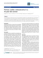
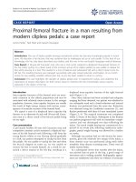
Figure 1 Characteristic of the primary cardiac osteosar coma in
our patient.(A) Echocardiography results, showing a mass in the
left atrium with accelerated color flow across the mass, suggesting a
hemodynamically significant obstruction. The mitral valve area was
2.5 cm
2
.(B) Histopathologic examination, showing that,
microscopically, the tumor was composed of a uniform population
of large atypical cells with prominent nucleoli and an osteogenic
sarcomatous element. Original magnification ×400; (C-F)
Immunohistochemical results, showing that the tumor was strongly
stained with antibodies to vimentin (C) and CD68 (E), weakly
stained with antibodies to CD34 staining (D), and completely
negative for S100 (F). Original magnification ×400. Bar, 100 μm.
Luo et al. Journal of Cardiothoracic Surgery 2010, 5:120
/>Page 2 of 2