Báo cáo y học: "Surgical management of pulmonary inflammatory pseudotumors: A single center experience" pot
Bạn đang xem bản rút gọn của tài liệu. Xem và tải ngay bản đầy đủ của tài liệu tại đây (625.44 KB, 6 trang )
RESEARC H ARTIC L E Open Access
Surgical management of pulmonary inflammatory
pseudotumors: A single center experience
Baldassare Mondello, Salvatore Lentini
*
, Mario Barone, Pietro Barresi, Francesco Monaco, Dario Familiari,
Annunziata La Rocca, Michele Sibilio, Ignazio Eduardo Acri, Antonio David, Maurizio Monaco
Abstract
Background: The pulmonary inflammatory pseudotumor (PIP) is a rare disease. It is still debated whether it
represents an inflammatory lesion characterized by uncontrolled cell growth or a true neoplasm. PIP is
characterized by a cellular polymorphism.
Methods: We retrospectively analyzed 8 patients with PIP treated by surgery between 2001 and 2009. Preoperative
thoracic computed tomography (CT) scan was performed in all cases. All patients underwent preoperative
bronchoscopy with washing and brushing and/or transbronchial biopsy and preoperative cytology examination
Results: There were 5 men and 3 women, aged between 38 and 69 years (mean of 58 years). 3 patients (37%)
were asymptomatic. The others had symptoms characterized by chest pain, shortness of breath and persistent
cough or hemoptysis. 5 patients had neutrophilic leucocytosis. CT scan demonstrated solitary nodules (maximum
diameter <3 cm) in 5 patients (62%) and lung masse s (maximum diameter >3 cm) in 3 patients (37%). In 2 patients
there were signs of pleural infiltration. Distant lesions were excluded in all cases. A preoperative histology
examination failed to reach a definitive diagnosis in all patients. At surgery, we performed two lobectomies, one
segmentectomy and five wedge resections, these being performed with videothoracoscopy (VATS), except for one
patient where open surgery was used. Complete tumor resection was obtained in all patients. According to the
Matsubara classification, there were 2 cases of organizing pneumonia, 5 cases of fibrous histiocytoma and one case
of lymphoplasmacytoma. All patients were discharged alive from hospital between 4 and 7 days after surgery. At
follow-up CT scan performed annually (range 11 to 112 months) (mean 58 months), there were no residual lesions,
neither local nor distant recurrences.
Conclusions: PIP is a rare disease. Many synonyms have been used for this disease, usually in relation to the most
represented cell type. The true incidence is unclear. Preoperative diagnosis is difficult to reach, despite performing
a bronchoscopy or a transparietal needle aspiration. Different classifications have been proposed for PIP. Either
medical, radiation or surgical therapy has been used for PIP. Whenever possible, surgery should be considered the
standard treatment. Complete surgical resection is advocated to prevent recurrence.
Background
Pulmonary inflammatory pseudotumor (PIP) is a rare
disease and it is still debated whether it re presents an
inflammatory lesion characterizedbyuncontrolledcell
growth or a true neoplasm, as recently suggested [1,2].
PIP is characterized by a cellular polymorphism, but
trans-bronchial and trans-thoracic biopsies are often
inconclusive for diagnosis [3]. Surgery is often use ful for
both treatment and diagnosis [4]. Complete resection is
considered essential to prevent relapses [5].
In this study, the authors report their experience on
the management of patients presenting with PIP.
Methods
Data prospectively entered into the registry of our surgi-
cal thoracic unit we re analyzed. We retrospectively ana-
lyzed 8 patients with PIP treated by surgery between
2001 and 2009.
* Correspondence:
Thoracic Surgery Unit, Cardiovascular and Thoracic Department, Policlinic
University Hospital, University of Messina, Italy
Mondello et al. Journal of Cardiothoracic Surgery 2011, 6:18
/>© 2011 Mondello et al; licensee BioMed Central Ltd. This is an Open Access article distributed under the terms of the Creative
Commons Attribution License ( which permits unrestricted use, distribut ion, and
reproduction in any medium, provided the original work is properly cited.
Preoperative symptoms, concomitant disease and
abnormal blood test results for all patients in the study
group were recorded.
Preoperative thoracic computed tomography (CT)
scans were performed in all cases and extended to the
abdomen and skull. In the three most recently treated
patients, a Fluorodeoxyglucose (
18
F) Positron emission
tomography (FDG-PET) scan was performed as well.
All patients underwent preoperative bronchoscopy
with washing and brushing and/or transbronchial biopsy
and preoperative cytology examination. All patients
underwent surgery either by thoracotomy or by video
ass isted thoracoscopy (VATS) after lung function study.
An intraoperative frozen section histology study was
performed in all cases.
Histology study
For a definitive histology study the following procedures
were used. The surgical specimens were fixed in 10%
formalin solution, embedded in paraffin, cut into sec-
tions of 4 μm, stained with hematoxylin and eosin and
then subjected to conventional histology.
For immunohistochemical techni ques we used antibo-
dies against vimentin, cytokeratins, desmin, smooth
muscle actin and epithelial membrane antigens.
Ultrastructural study by electron microscopy was per-
formed after histology sections were fixed with 2.5% glu-
taraldehyde solution, post-fixed with osmic tetroxide
and embedded in an epoxidic resin.
Patients were regularly seen at our out-patient clinic
for postoperative follow-up with CT scan performed
annually to rule out recurrences.
Results
Between January 2001 and December 2009, we treated 8
patients affected by PIP, 5 men and 3 women, aged
between 38 and 69 years (mean of 58 years).
Three patients (37%) were asymptomatic and lung
nodules on radiological examination were occasionally
detected. 5 patients were admitted to our surgical unit
for symptoms characterized by chest pain, shortness of
breath and persistent cough or hemoptysis, despite anti-
biotic and anti-inflammatory therapy.
Three patients had concomitant diseases at the time of
hospital presentation: One had arterial hypertension,
one had chronic obstructive pulmonary disease and one
had viral hepatitis. This last patient had also had pre-
vious heart surgery for endocarditis. A fourth patient
had a previous history of surgery for ovarian cancer.
Blood tests performed on admission were normal in 3
patients. The remaining 5 patients had neutrophilic leuco-
cytosis without other non-specific signs of inflammation.
Computed tomography (CT) scan demonstrated soli-
tary nodules (maximum diameter <3 cm) in 5 patients
(62%) and lung masses (maximum diameter >3 cm) in 3
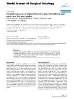
patients (37%). In 6 patients the CT scan showed find-
ings of parenchymal tumors without signs of infiltration;
in the other 2 patients there were signs of pleural infil-
tration (Figure 1). Distant lesions were excluded in all
cases.
The 3 patients who underwent FDG-PET scan had a
focus of activity with SUV (Standardized Uptake Value)
values between 6.2 and 9.8.
Preoperative bronchoscopy was negative in 7 patie nts.
In 1 patient we found bleeding from the right basal pyr-
amid and bronchial brushing cytology was falsely posi-
tive for carcinoma with a finding of atypical e pithelial
cells, lymphocytes and histiocytes. In 3 patients with
peripheral nodules we performed US-guided percuta-
neous needle aspiration. A preoperative histology exami-
nation failed to reach a definitive diagnosis in all
patients.
At surgery, we performed two lobectomies, one seg-
mentectomy and f ive wedge resections, these being per-
formed with videothoracoscopy (VATS), except for one
patient where open surgery was used. Complete tumor
resection was obtained in all patients. The maximum
tumor diameter w as between 2.5 and 5 cm, with the
gross appearance of a well c ircumscribed mass without
a fibrous capsule. Microscopic results were characterized
by a collection of inflammator y mesenchymal cells (his-
tiocytes, plasma cells, lymphocytes and spindle cells).
Intrapulmonary and mediastinal lymph nodes were
found in all cases free from invasion.
According to the Matsubara classification, microscopic
examination revealed 2 cases of organizing pneumonia
(Figure 2), 5 cases of fibrous histiocytoma (Figure 3) and
one case of lymphoplasmacytoma.
The postoperative course was uneventful in all cases.
The length of hospital stay was between 4 and 7 days.
At follow-up CT scan performed annually (range 11 to
112 months) (mean 58 months), there were no residual
lesions, neither local nor distant recurrences One
patient died of unrelated disease 23 months after sur-
gery. Two patients escaped from follow-up.
Discussion
Although inflammatory pseudotumo rs may develop in
different organs, such as brain and liver, the lung is the
preferred site [1,4]. PIP is a rare disease with a reported
incidence b etween 0.04 to 1.2% of all lung cancers [4].
Many synonyms have been used for th is disease, usually
in relation to the most represented cell type: plas ma cell
granuloma, inflammatory myofibro blastic tumor, fibrox-
antoma, histiocytoma, or pseudoneoplastic pneumonia
[4]. The true incidence is unclear, as well as the clinical
history in some cases and the response to different
therapies [4]. Still nowadays we discuss the nature of
Mondello et al. Journal of Cardiothoracic Surgery 2011, 6:18
/>Page 2 of 6
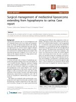
Figure 2 Hist ology study at l ow and high magnification of
lung pseudotumor: “organizing pneumonia” type following
Matzubara classification. There are areas of necrosis with
inflammatory infiltration of macrophages and lymphocytes.
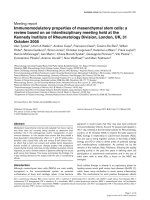
Figure 1 Computed tomography in patients wi th pulmonary
inflammatory pseudotumor: A) Non-calcified right lower lobe
lung tumor with irregular margins and pleural bridging. B) Non-
calcified right lower lobe lung tumor with adjacent pleural
thickening. C) Tumor with partial internal cavitation and pleural
infiltration.
Mondello et al. Journal of Cardiothoracic Surgery 2011, 6:18
/>Page 3 of 6
this lesion: inflammatory or neoplastic [6]. According to
some authors PIP represents a non-neoplastic process
characterized by the uncontrolled growth of inflamma-
tory cells [4]. The exact etiology of this inflammatory
reaction is unknown and many hypotheses have been
raised. The hypothesis of an immune disorder s eems to
prevail: i.e., a response to viral infection such as to
human herpes virus 8 or an antigen-antibody reaction
[7]. According to others, PIP represents a true neo-
plasm, benign or of low-grade malignancy, in considera-
tion of the slow and localized growth [8,9]. This opinion
is supported by the detection of cases of PIP with local
aggressiveness and infiltration of pulmonary vessels,
heart, chest wall , vertebrae and diaphragm, or by detec-
tion of cases with distant metastasis or multicentre dis-
ease [4,10-12]. Recently discovered cytogenetic
abnormalities on chromosome 2p23 would support a
neoplastic etiology for this disease [13-15]. PIP is more
common in young adults and does not show sex predi-
lection [9].
Patients may remain asymptomatic in 30 to 70% of
cases, the disease being occasionally detected on chest
radiological examination performed for other reasons
[3]. When symptoms occur they are represented by
cough, fever, hemoptysis, weight loss, chest pain and
respiratory infections due to endobronchial growth or
mediastinal invasion [3].
There are no specific radiological signs for PIP. Radi-
ological examination may show the appearance of soli-
tary nodules or masses that may present as either
calcified and well demarcated, with no evidence of
malignancy, or with irregular contours [16]. C omputed
tomography (CT) usually shows single nodules or single
masses, and multiple locations in only 5% of cases [17].
Agrons et al. reported signs of hilar, mediastinal or
bronchial infiltration in 16% of the examined cases [17].
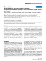
Figure 3 Histology study at low and high magnification of lung pseudotumor: “ fibrous histiocytoma” type followi ng Matzubara
classification. There is a nodular area with large amount of histiocytes, lymphocytes, plasma cells and ialin fibrous connective tissue.
Mondello et al. Journal of Cardiothoracic Surgery 2011, 6:18
/>Page 4 of 6
Fluorodeoxyglu cose (
18
F) Positron emission tomogra-
phy (FDG-PE T) scan shows an uptake similar to that of
malignant tumors [18]. We believe the use of FDG-PET
scan is also useful for the study of mediastinal lymph
nodes.
As often see n in previously reported series, also in our
experience we did not reach a preoperative diagnosis
with certainty, despite performing a bronchoscopy in all
cases and a transparietal needle aspiration in 3 cases.
Frozen sections from transbronchial or transparietal
biopsy are often dif ficult to interpret, giving an uncer-
tain diagnosis [3,6]. Due to the large number of inflam-
matory cells and fibroblast proliferation, differential
diagnosis would include conditions such as fibrohistio-
cystic neoplasm, plasmocytomas, Hodgkin’s sclerosing
lymphoma, primary lung cancer or sarcoma, or mediast-
inal fibrosis [3,6,19]. However, frozen sections are
usually able to rule out malignancies [3,6]. For the rea-
sons mentioned, surgery would be reco mmended not
only as treatment but also to reach a definitive diagnosis
[4,20,21].
Different classifications have been proposed for PIP.
According to Cerfolio, PIP is histologically classified
into tw o types with respect to its local invasiveness. The
first type, called non-invasive PIP, mostly presenting in
asymptomatic patients, appears as a small lesion without
invasiveness of blood vessels or adjacent structures and
usually easily resectable with a wedge resection [4]. The
second type of Cerfolio classification is called invasive
PIP and is usually diagnosed in younger patients, with
symptoms such as fever, fatigue and weight loss. In
these cases, the PIP usually has a larger size and may
present with chest wall or mediastinal invasion, requir-
ing lobectomy or pneumonectomy for a complete surgi-
cal resection. Invasive PIP may macroscopically appear
as lesions infiltrating tissue planes and histologically
characterized by nuclear atypia and frequent mitosis [4].
Matsubara et al, reporting on their expe rience, distin-
guish three subtypes of PIP according to clinicopathol o-
gical characteristics: 1) organizing pneumonia type
(44%), fibrous histiocytoma type (44%), and lymphoplas-
macytic type (12%) [6]. These authors with their classifi-
cation consider most likely an inflammatory genesis for
PIP [6].
Colby et al instead classify PIP into a fibrohistiocytic
subtype and a plasma cell granuloma subtype [22].
A recent classification of the World Health Associa-
tion (WHA) clas sifies the PIP into three main histologic
patterns: 1) myxoid vascular, 2) compact cord cell and
3) hypocellular fibrous. The three different patterns may
coexist in the same lesion [23].
Neither the Matsubara nor the WHA classification
seems to have a prognostic value [5].
Either medical, radiation or surgical therapy has been
used for PIP.
Corticosteroid therapy has been proposed in the case
of inoperable patients, for concurrent cardio-respiratory
diseases, for unresectable lesions or in case of recur-
rences [24,25]. The reported results are extremely vari-
able, ranging from ineffectiveness to complete disease
regression [25,26].
Radiation therapy is usually reserved for cases of
aggressive PIP, or after incomplete excision, or for post-
operative recurre nce or for patients at high surgical risk
[27,28]. The alternative roles of radiation therapy or
chemot herapy v ersus surgery is controversial [3,4,21,28].
It is a shared opinion that whenever possible, surgery
should be considered the standard treatment [2]. Even
in recurrence, whenever possible, s urgical resection is
advocated, allowing even in these cases a longer disease-
free interval [4].
Complete surgical resection is advocate d to prevent
recurrence. Prognosis after surgical radical resection is
usually excellent [4,9,21]. Long-term follow-up is still
required due t o the possibility of local or distant recur-
rence, even after several years [4,24].
Whenever possible, wedge resection should be consid-
ered the first line treatment. It would allow saving of
lung parenchyma and intraoperative histological exami-
nation to exclude malignancy [2,5]. If needed, lobectomy
or pneumonectomy would b e performed to ensure radi-
cal resection, or if diagnosis of malignancy may not
excluded. En bloc resection may be needed in cases of
chest wall invasion or main bronchus, pericardium or
diaphragm involvement [2].
A colle ction of case studies report ed a survival rate at
5 and 10 years, respectively, of 91 and 77%, with values
similar to those of a low-grade malignant neoplasm [29].
We should also mention that the possibility of a trans-
formation of PIP to sarcoma has been described [30,31],
as well as the occurrence of aggressive forms with unfa-
vorable outcome [32,33]. It is likely that P IP includes a
complex of diseases ranging from benign fibroistioci-
toma to malignant forms, thus explaining the large clini-
cal outcome variability reported in the literature [10].
Conclusions
Inflammatory pseudotumor of the lung is a rare disease,
histologically characterized by the presence of myofibro-
blasts and chronic inflammatory cells, such as plasma
cells, lymphocytes and histio cytes. Whenever possible
surgical resection represents the treatment of choice.
Major resections are sometimes needed due to the
tumorsizeorlocalinvasiveness. Complete resection is
advocated to prevent recurrence. Long term follow-up is
needed.
Mondello et al. Journal of Cardiothoracic Surgery 2011, 6:18
/>Page 5 of 6
Consent
Written informed consent was obtained from patients
for publication of this report and accompanying images.
A copy of the written consent is available for review by
the Editor in chief of this journal.
Authors’ contributions
All authors: 1. have made substantial contributions to conception and
design, or acquisition of data, or analysis and interpretation of data; 2. have
been involved in drafting the manuscript or revisiting it critically for
important intellectual content; 3. have given final approval of the version to
be published.
Competing interests
The authors declare that they have no competing interests.
Received: 18 January 2011 Accepted: 23 February 2011
Published: 23 February 2011
References
1. Chan YF, White J, Brash H: Metachronous pulmonary and cerebral
infiammatory pseudotumor in a child. Pediatr Pathol 1994, 14:805-15.
2. Fabre D, Fadel E, Singhai S, de Montpreville V, Mussot S, Mercier O,
Chataigner O, Dartevelle PG: Complete resection of pulmonary
inflammatory pseudotumors has excellent long-term prognosis. J Thorac
Cardiovasc Surg 2009, 137:435-440.
3. Copin MC, Gosselin BH, Ribet ME: Plasma cell granuloma of the lung:
difficulties in diagnosis and prognosis. Ann Thorac Surg 1996, 61:1477-82.
4. Cerfolio RJ, Allen MS, Nascimento AG, Deschamps C, Trastek VF, Miller DL,
Pairolero PC: Inflammatory pseudotumors of the lung. Ann Thorac Surg
1999, 67:933-6.
5. Melloni G, Carretta A, Ciriaco P, Arrigoni G, Fieschi S, Rizzo N, Bonacina E,
Augello G, Belloni PA, Zannini P: Inflammatory pseudotumor of the lung
in adults. Ann Thorac Surg 2005, 79:426-32.
6. Matsubara O, Tan-Liu NS, Kenney RM, Mark EJ: Inflammatory
pseudotumors of the lung: progression from organizing pneumonia to
fibrous histiocytoma or to plasma cell granulomas in 32 cases. Hum
Pathol 1988, 19:807-14.
7. Gomez-Roman JJ, Sanchez-Velasco P, Ocejo-Vinyals G, Niceto EH, Leyva-
Cobian F, Val-Bernal JF: Human herpesvirus-8 genes are expressed in
pulmonary inflammatory myofibroblastic tumor (inflammatory
pseudotumor). Am J Surg Pathol 2001, 25:624-9.
8. Snyder CS, Dell’Aquila M, Haghighi P, Baergen RN, Suh YK, Yi ES: Clonal
changes in inflammatory pseudotumor of the lung: a case report. Cancer
1995, 76:1545-9.
9. Dehner LP: The enigmatic inflammatory pseudotumors: the current state
of our understanding, or misunderstanding. J Pathol 2000, 192:277-9.
10. Gal AA, Koss MN, McCarthy WF, Hochhzer L: Prognostic factors in
pulmonary fibrohistiocytic lesions. Cancer 1994, 73:1817-24.
11. Berman M, Georghiou GP, Schonfeld T, Feinmesser M, Horev G, Vidne BA,
Saute M: Pulmonary inflammatory myofibroblastic tumor invading the
left atrium. Ann Thorac Surg 2003, 76:601-3.
12. Maier HC, Sommers SC: Recurrent and metastatic pulmonary fibrous
histiocytoma/plasma cell granuloma in child. Chest 1987, 60:1073-6.
13. Yousem SA, Shaw H, Cieply K: Involvement of 2p23 in pulmonary
inflammatory pseudotumors. Hum Pathol 2001, 32:428-33.
14. Chan JKC, Cheuk W, Shimuzu M:
Anaplastic lymphoma kinase expression
in inflammatory pseudotumors. Am J Surg Pathol 2001, 25:761-8.
15. Lawrence B, Perez-Atayde A, Hibbard MK, Rubin BP, Dal Cin P, Pinkus GS,
Xiao S, Yi ES, Fletcher CD, Fletcher JA: TPM3-ALK and TPM4-ALK
oncogenes in inflammatory myofibroblastic tumors. Am J Pathol 2000,
157:377-84.
16. Ishida T, Oka T, Nishino T, Tateishi M, Mitsudomi T, Sugimachi K:
Inflammatory pseudotumor of the lung in adults: radiographic and
clinicopathological analysis. Ann Thorac Surg 1989, 48:90-95.
17. Agrons GA, Rosado-de-Christenson ML, Kirejczyk WM, Conran RM,
Stocker JT: Pulmonary inflammatory pseudotumor: radiologic features.
Radiology 1998, 206:511-518.
18. Slosman DO, Spiliopoulos A, Keller A, Lemoine R, Besse F, Couson F,
Townsend D, Rochat T: Quantitative metabolic PET imaging of a plasma
cell granuloma. J Thorac Imaging 1994, 9:116-119.
19. Takeda S, Onishi Y, Kawamura T, Maeda H: Clinical spectrum of pulmonary
inflammatory myofibroblastic tumor. Interact Cardio Vasc Thorac Surg
2008, 7:629-633.
20. Coffin CM, Watterson J, Priest JR, Dehner LP: Extrapulmonary inflammatory
myofibroblastic tumor (inflammatory pseudotumor). A clinicopathologic
and immunohistochemical study of 84 cases. Am J Surg Pathol 1995,
19:859-72.
21. Sakurai H, Hasegawa T, Watanabe S, Suzuki K, Asamura H, Tsuchiya R:
Inflammatory myofibroblastic tumor of the lung. European Journal of
Cardio-thoracic Surgery 2004, 25:155-159.
22. Colby TV, Koss MN, Travis WD: Tumors of the lower respiratory tract
Washington DC: Armed Forced Institue of Pathology; 1995, 327-352.
23. Coffin CM, Fletcher JA: Inflammatory myofibroblastic tumor. In World
Health Organization classification of tumor: pathology and genetics, tumor of
soft tissue and bone. Edited by: Fletcher CDM, Unni KK, Mertens F. Lyon:
IARC Press; 2002:91-3.
24. Urschel JD, Horan TA, Unruh HW: Plasma cell granuloma of the lung. J
Thorac Cardiovasc Surg 1992, 4:870-5.
25. Doski JJ, Priebe CJ Jr, Driessnack M, Smith T, Kane P, Romero J:
Corticostyeroids in the management of unresected plasma cell
granuloma (inflammatory pseudotumor) of the lung. J Pediatr Surg 1991,
26:1064-66.
26. Bando T, Fujimura M, Noda Y, Hirose J, Ohta G, Mitsuda T: Pulmonary
plasma cell granuloma improves with corticosteroid therapy. Chest 1994,
105:1574-5.
27. Smahi AM, Kabiri H, Rhissassi J, Achir A, Al Aziz AS, Messlout A,
Benosman A: Pseudotumor inflammatoire du poumon. Etude
anatomoclinicque d’une observation. Chirurgie 1999, 124:73-6.
28. Imperato JP, Folkman J, Sagerman RH, Cassady JR: Treatment of plasma
cell granuloma of the lung with radiation therapy. A report of two cases
and a review of the literature. Cancer
1986, 57:2127-2129.
29. Hussain SF, Salahuddin N, Khan A, Memon SS, Fatimi SH, Ahmed R: The
insidious onset of dyspnea and right lung collapse in a 35-year-old man.
Chest 2005, 127:1844-7.
30. Coffin CM, Watterson J, Priest JR, Dehner LP: Extrapulmonary inflammatory
myofibroblastic tumor (inflammatory pseudotumor). A clinicopathologic
and immunohistochemical study of 84 cases. Am J Surg Pathol 1995,
19:859-72.
31. Donner LR, Trompler RA, White R: Progression of inflammatory
myofibroblastic tumor (inflammatory pseudotumor) or soft tissue into
sarcoma after several recurrences. Hum Pathol 1996, 27:1095-8.
32. Girard F, Kambouchner M, Maugendre S, Naccache JM, De Meyer-Cristiani R,
Battesti JP, Delaval P, Valeyre D: Inflammatory pseudotumors of the lung
with severe course. Rev Mal Respir 2001, 18:541-4.
33. Kato S, Kondo K, Teramoto T, Harada T, Ikeda H, Hara K, Nagata Y: A case
report of inflammatory pseudotumor of the lung: rapid recurrence
appearing as multiple lung nodules. Ann Thorac Cardiovasc Surg 2002,
8:224-7.
doi:10.1186/1749-8090-6-18
Cite this article as: Mondello et al.: Surgical management of pulmonary
inflammatory pseudotumors: A single center experience. Journal of
Cardiothoracic Surgery 2011 6:18.
Mondello et al. Journal of Cardiothoracic Surgery 2011, 6:18
/>Page 6 of 6