Báo cáo y học: " Androgens as therapy for androgen receptorpositive castration-resistant prostate cancer" ppt
Bạn đang xem bản rút gọn của tài liệu. Xem và tải ngay bản đầy đủ của tài liệu tại đây (1.82 MB, 12 trang )
Androgens as therapy for androgen receptor-
positive castration-resistant prostate cancer
Chuu et al.
Chuu et al. Journal of Biomedical Science 2011, 18:63
(23 August 2011)
REVIE W Open Access
Androgens as therapy for androgen receptor-
positive castration-resistant prostate cancer
Chih-Pin Chuu
1,2*
, John M Kokontis
3
, Richard A Hiipakka
3
, Junichi Fukuchi
4
, Hui-Ping Lin
1,2
, Ching-Yu Lin
1,2
,
Chiech Huo
1,2,5
and Liang-Cheng Su
1,2
Abstract
Prostate cancer is the most frequently diagnosed non-cutaneous tumor of men in Western countries. While surgery
is often successful for organ-confined prostate cancer, androgen ablation therapy is the primary treatment for
metastatic prostate cancer. However, this therapy is associated with several undesired side-effects, including
increased risk of cardiovascular diseases. Shortening the period of androgen ablation therapy may benefit prostate
cancer patients. Intermittent Androgen Deprivation therapy improves quality of life, reduces toxicity and medical
costs, and delays disease progression in some patients. Cell culture and xenograft studies using androgen receptor
(AR)-positive castration-resistant human prostate cancers cells (LNCaP, ARCaP, and PC-3 cells over-expressing AR)
suggest that androgens may suppress the growth of AR-rich prostate cancer cells. Androgens cause growth
inhibition and G1 cell cycle arrest in these cells by regulating c-Myc, Skp2, and p27
Kip
via AR. Higher dosages of
testosterone cause greater growth inhibition of relapsed tumors. Manipulating androgen/AR signaling may
therefore be a potential therapy for AR-positive advanced prostate cancer.
Introduction
In 1941, Huggins and Hodges reported that androgen
ablation therapy causes regression of primary and meta-
static prostate cancer [1]. Approximately 20-40% of
patients treated with radical prostatectomy will have
tumor recurrence and elevation of serum prostate-specific
antigen (PSA) [2]. Primary metastatic sites for prostate
cancer include bones and lymph nodes. More than 80% of
patients who die from prostate cancer develop bone
metastases [3-5]. Androgen ablation therapy is provided to
patients who develop recurrent or metastatic prostate
tumors. However, 80-90% of the patients who receive
androgen ablation therapy ultimately develop recurrent
castrate-resistant tumors 12-33 months after androgen
ablation therapy. The median overall survival of patients
after tumor relapse is 1-2 years [6,7]. Several long-term
studies have failed to show that androgen ablation therapy
provides a disease-specific survival advantage in patients
[6]. Androgen ablation therapy is associated with unde-
sired side-effects that impair the patient’s quality of life as
well as increased risk of diabetes and cardiovascular
diseases [6]. Therefore, shortening the period of androgen
ablation therapy may protect the patients.
Androgens and Androgen Receptor in Prostate Cancer
Androgens are male sex hormone and include several ster-
oids, such as testosterone, dehydroepiandrosterone,
androstenedione, androstenediol, androsterone, and dihy-
drotestosterone (DHT). 90-95% of androgens are pro-
duced by the testes, while some androgens are produced
in the adrenal glands. Testosterone is the main circulating
androgen in human body, while DHT is a m ore potent
androgen that has 5-fold higher affinity for the androgen
receptor (AR) than does testosterone [7-9]. When testos-
terone enters prostate cel ls, 90% is converted to dihydro-
testosterone (DHT) by the enzyme 5a-reductase [9].
The average serum tes tosterone level declines with age
and elderly men usually have the conditi on as partial
androgen deficiency. It decreases from approximately
620-670 ng/dl at age 25-44 to 470-520 ng/dl at age 65-84
[10]. A low serum testosterone level is associated with an
increased risk of prostate cancer [11], and prostate
tumors arising in a low testosterone environment appear
to be more aggressive [12]. A retrospective review of 117
patients by Hoffman et al. revealed that patients with
low (150 ng/dl) free testosterone have an increased
* Correspondence:
1
Institute of Cellular and System Medicine, National Health Research
Institutes, Miaoli, Taiwan
Full list of author information is available at the end of the article
Chuu et al. Journal of Biomedical Science 2011, 18:63
/>© 2011 Chuu et al; licensee BioMed Central Ltd. This is an Open Access article distributed under the terms of the Creative Commons
Attribution License (http://creative commons.org/licenses/by/2.0), which permits unrestricted use, distributio n, and reproduction in
any medium, provided the original work is prope rly cited.
percentage of biopsies with cancer present (43% versus
22%, p = 0.013) as well as an increased incidence of a
biopsy with Glea son score of 8 or greater (7 of 64 versus
0 of 48, p = 0.025) [13]. These observations suggest that
patients with prostate cancer and low free testosterone
have more extensive disease, and low serum free testos-
terone may be a marker for more aggressive disease [13].
Androgen receptor (AR), an androgen-activated tran-
scription factor, belongs to the nuclear receptor superfam-
ily. Binding of DHT to the androgen receptor (AR)
induces dissociation of AR from h eat-shock proteins
(HSPs) and stimulates AR p hosphorylation [14]. AR
dimerizes, translo cates into the nu cleus, and binds to
androgen-response elements (ARE) in the promoter
regions of target genes [14]. Co-activators and co-repres-
sors also bind the AR complex, facilitating or preventing
transcription of AR target genes. Activation or repression
of target genes regulates growth, survival, and the produc-
tion of prostate-specific antigen (PSA) in prostate cells
[15,16].
Based on gene microarray studies of seven different
human prostate cancer xenograft models, an increase of
AR mRNA w as the only change consistently assoc iated
with the development of the castration-resistant pheno-
type [17]. Increase in AR mRNA and protein is both
necessary and sufficient to convert prostate cancer growth
from a hormone-sensitive to a hormone-refractory stage,
and is dependent on a functional ligand-binding domain
[16,17]. Elevated AR expression in hormone-refractory
prostate cancer cells or recurrent hormone-refractory
tumors is observed in our progression model [15,18-22]
and by several o ther groups [ 17,23-35]. Re cent studies
revea led that although androgen deprivation therapy sig-
nificantly reduced serum testosterone concentrations,
levels of testosterone and dihydrotestosterone occur in
recurrent prostate cancer tissue are sufficient to stimulate
AR transcription, PSA secretion, and tumor growth. These
observations suggested that prostate cancer cells may sur-
vive androgen deprivation therapies by increasing intra-
crine androgen synthesis within the prostate [36,37].
Androgen Ablation Therapy
Androgen ablation therapy, using luteinizing hormone-
releasing hormone agonists (LH-RH) (also known as gona-
dotropin-releasing hormone, GnRH) or bilateral orchiect-
omy, has b ecome a primary treatment for metastatic
prostate cancer [6]. More than 80% of men with advanced
prostate cancers respond to androgen ablation therapy,
resulting in tumor shrinkage and reduction of serum PSA
[6]. Anti-androgens are frequently used in conjunction
with androgen ablation therapy as a combined androgen
blockade to improve therapeutic outcome. Most pat ients
experience an initial rapid decline in PSA followed by a
slower decline to the nadir. The initial rapid decrease in
PSA results from the cessation of androgen-regulated PSA
synthesis and apoptosis of prostate cancer cells, while the
ongoing slower decline perhaps reflects decreasing tumor
volume [38]. A nti-androgen finasteride prevents and
delays the a ppearance of prostate cancer observed in a
prevention trial with 18,882 men, however, tumors of
higher Gleason grade (7-10) were more common in the
finasteride group (37%) than in the placebo control group
(22%) [39].
In addition, androgen deprivation therapy is associated
with several undesired side-effects, including sexual dys-
function, osteoporosis and bone fractures, hot flashes, fati-
gue, gynecomastia, anemia, depression, cognitive
dysfunction, increased risk of diabetes, and cardiovascular
diseases [6,40-42]. Androgen deprivation therapy using
LH-RH agonists increases risk of incident diabetes, inci-
dent coronary heart disease, myocardial infarction, sudden
cardiac death, and stroke [43-45]. Combined androgen
blockade (LH-RH agonists treatment plus oral anti-andro-
gens) is associated with increased risk of incident coronary
heart disease [42]. Orchiectomy is associa ted with coron-
ary heart disease a nd myocardial infarction [42]. There-
fore, shortening the period of androgen ablation therapy
may be beneficial for some prostate cancer patients.
Intermittent Androgen Deprivation Therapy
Clinical and ba sic studies have shown that in comparison
with continuous androgen ablation (CAB) therapy,
Intermittent Androgen Deprivation (IAD) therapy
substantially pro longs the time to development of castra-
tion-resistant prostate canc er [39,46-48]. Intermittent
Androgen Deprivation t herapy is a s trategy to periodi-
cally perform and terminate the androgen ablation ther-
apy, ther efore patients in “off-androgen ablation therapy”
periods may decrease undesired side effects and improve
quality of life.
The growth of Shionogi mammary carcinoma is stimu-
lated by androgens and was the first experimental model
to test IAD therapy. Hormone-dependent Shionogi mam-
mary carcinoma become androgen ablation-resistant fol-
lowing IAD therapy using cycles of transplantation into
intact male mice followed by c astration [49]. However,
IAD delayed the recurrence time of Shionogi tumor
growth from 51 days to 147 days [46]. Five to six c ycles
of IAD therapy de lays the progression of LNCaP prostate
xenografts tow ards androgen ablation-resistance. IAD
prolongs the time to andro gen ablation-resistance of PSA
gene regulation from an average of 26 days to 77 days
compared to continuous androgen ablation (CAB) [47].
By 15 weeks post-castration, serum PSA l evels increase
7-fold above pre-castrate levels in CAB-treated mice
compared to a 1.9-fold increase in IAD-treated mice [47].
In a Canadian Prospective Trial, Bruchovsky et al.
showed that IAD therapy causes repeated differentiation
Chuu et al. Journal of Biomedical Science 2011, 18:63
/>Page 2 of 11
of prostate tumors with recovery of apopto tic potential,
inhibition of tumor growth after rapid restoration of
serum testosterone, and restraint of tumor growth by
subnormal levels of serum testosterone [43]. Pether
et al. reported in a clinical trial of 102 patients that
there is a trend toward extended times to progression
and death compared to CAB treatment, and growth of
advanced prostate tumors is delayed in ~50% patients
treated with IAD [45]. They concluded that IAD is a
viable treatment option for men with prostate c ancer
which affords an improved quality of life when the
patient is off therapy and with reduced toxicity and
costs [43-45].
Androgenic Suppression of Advanced Prostate Cancer
Cells in Vitro
The delay of progression toward and rogen-independency
in IAD treatment might be related to the suppressive
effect of androgen on AR-positive hormone-r efractory
prostate cancer cells that is observed in the LNCaP and
other prostate cancer cell models. LNCaP i s one of the
most commonly used cell lines for prostate cancer
research and was derived from a human lymph node
metastatic lesion of prostate adenocarcinoma [49,50].
LNCaP cells express AR and PSA. To establish relapsed
androgen-ablation resistant prostate canc er cells that
mimic the clinical situation in which prostate cancer
recurs during androgen deprivation, we cultured andro-
gen-sensitive LNCaP 104-S cells in androgen-depleted
conditions in vitro [19,20]. After 20 passages (3 months)
in androgen-depleted media supplemented with dextran-
coated charcoal-stripped fetal bovine serum, most
LNCaP 104-S cells undergo cell cycle arrest. After 60-80
passages ( 8-11 months), cells called 104-R1 cells emerge
that grow much more rapidly in the absence of androgen.
After 120-150 passages (16-20 months) in androgen-
depleted me dium, 104-R1 cells give rise to cells called
104-R2 c ells, that proliferate in the absence of androgen
at a rate comparable to the proliferation rate o f 104-S
cells grown in media with androgen [19,20].
During the transition of 104-S cells to 104-R 1 and 104-
R2 cells, AR mRNA and protein levels increase. AR tran-
scriptional activity also increases several fold [15,18-20,51].
Proliferation of 104-R1 and 104-R2 cells is not dependent
on androgen (i.e. hormone-refractory) but is unexpectedly
suppressed by physiological concentrations of androgen
both in vitro and in vivo [15,18-22,51]. When 104-R1 or
104-R2 cells are incubated for several weeks in a high con-
centration of R1881 (20 nM, approximately equivalent to
200 nM DHT), cells adapt after a period of growth arrest
to grow at a rate equivalent to the parental 104-R1 or 104-
R2 cells [20,51]. The adapted cells derived from 104-R1
called R1Ad cells, wh ich grow optimally in 10 nM R1881
[26]. R2Ad cells, which derived from 104-R2 cells under
androgen treatment, grow androgen-insensitively [51].
R1Ad and R2Ad cells have dramatically reduced levels of
AR, which suggests that elevated AR expression is respon-
sible for the repressive effect of androgen in 104-R1 and
104-R2 cells.
To further mimic the clinical situation of combined
androgen deprivation and an ti-androgen therapy, LNCaP
104-S cells we re incubated with 5 μM Casodex in andro-
gen-depleted medium. After four weeks, Casodex-resistant
colonies (CDXR cells) appear at low frequency (1 in 1.4 ×
10
5
) as most of the cells appear to undergo senescent cell
death [21]. Like 104-R1 and 104-R2 cells, CDXR cells have
increased AR expression and activity and are repressed by
androgen [21]. Unlike 104-R1 cells, CDXR cells grown in
10 nM R1881 undergo apoptotic cell death starting 6 to
8 days after R1881 exposure. However, 1 in 1.9 × 10
3
cells
form colonies of a ndrogen-insensitive cells that are not
repressed by R1881 or Casodex . Th ese sublines, desig-
nated IS cells, show greatly reduced AR expression [27].
Unlike R1Ad cells, the growth of IS cells is not stimulated
by R1881. IS cells are mor e similar to R2Ad cells. During
progression from 104-R1 to 104-R2 stages, the cells appear
to pass a point where cells can no longer recover respon-
siveness to androgen, but instead progress to androgen
insensitivity [52]. Dire ct progression of 104-S cells to the
CDXR stage by selection in anti-androgen seems to bypass
this intermediate 10 4-R1 stage and speed up the diseases
progression. Stimulation of prostate cancer disease pro-
gression by antiandrogen treatment is also observed in
clinical trials. Bales et al. compared the effect of bicaluta-
mide (50 m g daily) to surgical or medical castration in
three randomized trials involving more than 1000 patients
and fo und that treatment with bicalutamide resulted in a
statistically significant shorter time to treatment failure,
time to progression, and median survival compared to cas-
tration (hazard ratios 1.59, 1.62, and 1.44, respectively)
[53].
An androgen-suppressive phenotype of hormone-
refractory LNCaP cells has been observed by several
other groups [20,38,54-56]. Elevated AR is observed in
hormone-refractory LNCaP cells [32,57, 58]. In one study,
the most optimal concentration of androgen for prolif-
eration of cells at intermediate stage shift s from 0.01 nM
R1881 to 0.001 nM R1881 [57]. The proliferation of the
late stage hormone-refractory LNCaP cells is suppressed
by androgen [57].
LNCaP cel ls express a mutant AR (T87 7A) that dis-
plays relaxed ligand binding specificity [20,59]. However,
androgenic suppression is not limited to LN CaP cells.
ARCaP is a n AR-positive, tumorigenic, and highly meta-
static cell line derived from the ascites fluid of a patient
with advanced metast atic disease. Proliferati on of ARCaP
cells is suppresse d by androgen [60]. ARCaP cells engi-
neered to overexpress AR have a b iphasic androgenic
Chuu et al. Journal of Biomedical Science 2011, 18:63
/>Page 3 of 11
response, the cells are stimulated by low concentration of
androgen (0.1-10 nM R1881), but suppressed by high
concentration of androgen (100-1000 nM R1881) [61].
MDA PCa 2b-hr cells were generated in vit ro from bone
metastasis-derived, hormone-dependent MDA PCa 2b
human prostate cancer cells after 35 weeks of culture in
androgen-depleted medium. MDA PCa 2b-hr express 3-
fold higher AR protein and proliferation of MDA PCa
2b-hr is stimulated by 3.5 nM testosterone or physiologi-
cal concentrations of adrenal androgens but is inhibited
by higher concentration s of testosterone or bical utam ide
[31]. PC-3 is a commonly used AR-negative human pros-
tate cancer cell line established from a bone-derived
metastasis [50]. Physiological conce ntrations of DHT
cause growth inhibition, G1 cell cycle arrest, and apopto-
sis in PC-3 cells overexpressing full length wild-type AR
[62-64]. Much evidence therefore exists for AR function-
ing as a ligand-dependent tumorsuppressorinprostate
cancer cells when it is expressed at high levels and is fully
activated.
Androgenic Suppression of Advanced Prostate Cancer
Cells in Vivo
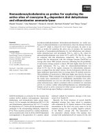
Castration causes regression of 104-S xenografts,
but tumors begin to regrow after 8 weeks as androgen
ablation-resistant relapsed tumors called 104-Rrel with
elevated AR mRNA and protein expression [18]. Low
serum levels of testosterone (130 ± 60 ng/dl) stop growth
of 104-Rrel tumors but tumo r growth resumes in about
4 weeks. High serum levels of testosterone (2970 ±
495 ng/dl), which is approximately 5-fold higher than nor-
mal levels, cause regression of 104-Rrel tumors. However,
104-Rrel cells adapt to androgen and relapse after 4 weeks
as androgen-stimulated 104-Radp tumors [18] (Figure 1).
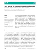
Growth of the LNCaP 104-R1 tumors is also suppressed
by androgen, but tumors adapt to androgenic suppression
and relapse as androgen-stimulated R1Ad tumors in 5-6
weeks [15] (Figure 2A, B). Growth of these tumors is sti-
mulated by testosterone and removal of testosterone
totally stopped the tumor growth [15,18]. Both 104-Radp
and R1Ad tumors express very little AR and PSA mRNA
and protein or serum PSA level (Figure 2C, D), simi lar to
R1Ad cells in cell culture [15,18,20]. Xenograft of CDXR
cells, which are also derived from 104-S cells, behave dif-
ferently under androgen suppression compared to 104-R1
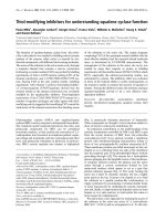
xenografts. Both early and late treatment with androgen
causes regression of CDXR tumors. Approximately 70% of
tumors regress completely and the rest of the tumors
relapse after 60-90 days o f treatment [27]. The relapsed
tumors show diminished expression of AR and no longer
require androgen for growth, essentially identical to the
behavior of IS3 cells that emerged after androgen exposure
in vitro [21]. It is worthwhile noting that 100% of 104-R1
tumor treated with testostero ne relapse in 4-5 weeks,
while only 30% of CDXR tumors and 70% of R2Ad tumors
relapse after 9-13 and 4-5 weeks, respectively, after testos-
terone treatment [15,21,51 ] (Figure 3). This is probably
Figure 1 Progression of hormone-dependent LNCaP 104-S
tumors to androgen-ablation-resistant 104-Rrel tumors, and
androgenic growth suppression of 104-Rrel tumors. (A) Mice
were injected subcutaneously with hormone-dependent 104-S cells.
After allowing tumors to grow for 7 weeks, mice were separated
into control (filled circles, 14 mice with 19 tumors) and castration
groups (open circles, 24 mice with 36 tumors) and the time was
designated as week 1 [18]. (B) Mice in the castrated group in (A) at
the 14
th
week were separated into 3 groups including a control
group (open circles, 6 mice with 9 tumors), a low dosage
testosterone treatment group that received a subcutaneous implant
of a 20 mg Testosterone/cholesterol (1:9) pellet (filled squares, 9
mice with 12 tumors), and a high-dosage testosterone treatment
group that received a subcutaneous implant of a 20 mg pure
Testosterone pellet (filled circles, 10 mice with 12 tumors) [18].
Tumor volumes are expressed as the mean + standard error.
Chuu et al. Journal of Biomedical Science 2011, 18:63
/>Page 4 of 11
Figure 2 Progression and regression of LNCaP 104-R1 tumor xenografts in nude mice treated with testosterone. (A) LNCaP 104-R1
tumor xenografts in castrated male nude mice were allowed to grow until they reached an average volume of 300 mm
3
on the 58th day. On
the 67th day, mice were separated into a control group (open circles) and a treatment group (filled circles). The treatment group received a
subcutaneous implant of a 20 mg testosterone pellet. The mice in the control group were implanted with a 20 mg testosterone pellet on day
121. Open circles represent tumor in mice without testosterone, while filled circles and filled squares represent tumors in mice with testosterone.
Tumor volumes are expressed as the mean ± standard error [15]. (B) For mice carrying adapted R1Ad tumors from (A), testosterone pellets were
removed from 5 mice (10 tumors). Their tumor growth was compared with tumors in mice bearing testosterone pellets (5 mice with 10 tumors)
[15]. (C) PSA, AR, and actin protein levels in 104-S tumor (in intact mice), 104-R1-T tumors, R1Ad-1+T tumors, and R1Ad-T were assayed by
Western blot [15]. (D) Serum PSA level of mice with 104-S tumors (in intact mice), 104-R1-T tumors, 104-R1+T tumors, R1Ad+T tumors, R1Ad-T
tumors was determined by ELISA [15].
Chuu et al. Journal of Biomedical Science 2011, 18:63
/>Page 5 of 11
due to the slower proliferation rate of CDXR cells and the
apoptosis induced in CDXR cells but not 104-R1 cells by
androgen [20,21]. Regression and relapse after androgen
treatment of LNCaP xenograft is also observed by another
group [64] and ARCaP xenograft [65]. AR overexpression
decreases adhesion, invasion, and migration ability of
ARCaP cells in vitro, as well as reduces ARCaP tumor
growth in athymic mice [61].
Molecular Mechanism of Androgenic Suppression
The anti-and rogen Casodex, unlike flutamide and cypro-
terone acetate, does not exhibit agonist activity and acts as
a true antiandrogen in the LNCaP 104-S, 104-R1, 104-R2
cell lines [66,67]. Casod ex does not affect proliferation of
104-R1 and 104-R2 cells but blocks androgenic repression
of growth as well as androgenic induction of PSA [68],
suggesting that the growth inhibition caused by androgen
treatment is via AR. Knockdown of AR expression in
CDXR3 cells by shRNA, either constitutive or conditional,
relieves androgenic repression of growth and does not
affect cell growth in the absence of androgen [21]. Retro-
viral overexpression of AR in IS2 and IS3 cells, on the
other hand, restores the androgen-repressed phenotype in
these cells [21]. R2Ad cells show similar beha vior com-
pared to CDXR cells [51]. Conditional overexpression of
AR in 104-S cells causes androgen-induced growth repres-
sion and does not confer hormone-refractory growth [21].
These observations confirm that androgen causes growth
inhibition via AR.
Flow cytometric analysis of androgen-treated cells
reveals that androgen treatment of hormone-dependent
LNCaP FGC [54] or LNCaP 104-S cells [20] relieves a G1
arrest induced by androgen deprivation. Conversely,
R1881 induces G1 arrest in 104-R1 and 104-R2 cells
beginning after about 24 hours of exposure [20] (Figure 4)
as well as other LNCaP model [55,58]. Casodex blocks the
effect of androgen in all cell lines. Expression of known
cdk inhibitors (p15, p16, p18, p19, and p21
waf1/cip1
,
p27
Kip1
,p57
Kip2
) has been examined in 104-S, 104-R1, and
104-R2 cells treated with or deprived of androgen. p21
waf1/
cip1
and p27
Kip1
levels are induced by androgen in 104-R1
and 104-R2 cells [20,51] (Figure 4). p21
waf1/cip1
is induced
transiently in 104-R1 cells only, while p27
Kip1
is induced
persistently about 3-fold in both 104-R1 and 104-R2 cells
[20,51]. Similar results have been obtained with the CDXR
sublines [27]. In c ontrast, expression of p21
waf1/cip1
and
p27
Kip1
is repressed by androgen in 104-S cells. Androgens
regulate expression of the F-box protein Skp2 that binds
phosphorylated p27
Kip1
[59,60,69] leading to its ubiquiti-
nation and proteolysis. Androgen down-regulates Skp2 in
104-R1, 104-R2 (Figure 4) [51] and CDXR ce lls, which
leads to accumulation of p27
Kip1
. Androgen treatment
down-regulates c-Myc mRNA and protein expression in
hours in 104-R1 and 104-R2 cells (Figure 4) [51], and
Figure 3 Regression and relapse of LNCaP CDXR-3 tumor
xenografts in nude mice treated with testosterone LNCaP CDXR
tumor xenografts in castrated male nude mice were allowed to
grow until they reached an average volume of 400 mm
3
on the
38th day. All mice carrying tumors received a subcutaneous implant of
a 20 mg testosterone pellet. The mice in the control group were
implanted with a 20 mg testosterone pellet either at an early stage
(50 days after inoculation, 7 tumors) (A) or late stage (92 days after
inoculation, 7 tumors) (B) [27]. Open triangles represent tumors relapsed,
while open squares represent tumors disappeared after androgen
treatment. Tumor volumes are expressed as the mean ± standard error.
(C) LNCaP IS-3 xenogarfts were separated into control group (20 mg
cholesterol pellet implant, 9 tumors) and treatment group (20 mg
testosterone pellet implant, 10 tumors) to determine the effect of
androgen on growth of IS tumors [21].
Chuu et al. Journal of Biomedical Science 2011, 18:63
/>Page 6 of 11
enforced retroviral overexpression of Skp2 or c-Myc
blocks androgenic repression of 104-R1 growth [19,51]. c-
Myc may have an indirect ef fect on p27
Kip1
expression
through the induction of Cks1, a component of the
SCF
Skp2
complex responsible for p27
Kip1
degradation [70].
Therefore, androgen regulates cell cycle and proliferation
of LNCaP cells via AR, Skp2, c-Myc, and p27
Kip1
.
Androgen Treatment of Prostate Cancer
Reduced serum testosterone levels by androgen ablation
therapy causes regression of prostate tumors, but elevation
of t he testosterone level does not result in sti mulation of
tumor growth or secretion of PSA [71]. A few studies have
shown that androgen is safe and potentially effective f or
treat ment of advanced prostate cancer. Mathew reported
that the testosterone level in a prostate cancer patient that
had undergone radical prostatectomy and LH-RH therapy
remained at castrated levels and serum PSA was undetect-
able for 15 years. PSA levels then began to rise and the
patient was given testosterone replacement therapy to
attain a normal range of serum testosterone. After an
initial flare, PSA levels gradually declined over 18 months.
After 27 months, PSA level started to increase. When tes-
tosterone replacement therapy was discontinued, PSA
Figure 4 Effect of androgen on cell proliferation, cell cycle, and cell cycle-related proteins in hormone-dependent 104-S and androgen
ablation-resistant 104-R1 cells. (A) LNCaP 104-S and 104-R2 cells were treated with increasing concentration of R1881 for 96 hours. Relative
cell number was determined by using a 96-well proliferation assay and data were normalized to number of 104-S cells at 0.1 nM R1881. Asterisk
(*) represents statistically significant difference between treatment group compared to control group of 104-S or 104-R1 cells. (B) Percentage of
104-S and 104-R1 cells in S phase determined by flow cytometry. LNCaP 104-S and 104-R2 cells were treated with increasing concentrations of
R1881 for 96 hours. Values represent the mean + standard error derived from 5 independent experiments. (C) Protein expression of androgen
receptor (AR), prostate specific antigen (PSA), p21
cip
, p27
Kip
, phosphor-retinoblastoma protein (Rb), c-Myc, S phase kinase-associated protein 2
(Skp2) were determined by Western blotting assay in 104-S and 104-R1 cells treated 96 hrs with different concentration of R1881. b-actin was
used as loading control.
Chuu et al. Journal of Biomedical Science 2011, 18:63
/>Page 7 of 11
levels dropped [48]. Mathew agrees that the observation
was somewhat similar to the transitio n from 104-R1 to
R1Ad phenotype under androgen treatment in our LNCaP
progression model [15,20,48].
Szmulewitz et al. randomly separated 15 prostate can-
cer patients (median PSA of 11.1 ng/ml, range from
5.2-63.6 ng/ml) who received androgen ablation plus
anti-androgen the rapy and withdrew without metastatic
disease into three groups. The three groups of patients
were given treatment of three different dosages of trans-
dermal testosterone: 2.5, 5.0, or 7.5 mg/day. Testoster-
one increased from castration levels to median
concentrations of 305 ng/dl, 308 ng/dl, and 297 ng/dl
for dosages of 2.5 mg/day (n = 4), 5.0 mg/day (n = 5),
and 7.5 mg/day (n = 5), respectively. One patient was
taken off due to grade 4 cardiac toxicity. One patient
experienced symptomatic progression, and three (20%)
patients demonstrated a decrease in PSA (largest was
43%). Median time to prog ression was 9 weeks (range:
2-96), with no detectable difference in the three dose
cohorts [39]. The conclusion of this study is that testos-
terone is a feasible and reasonably well-tolerated ther-
apy for me n with early hormone-re fractory prostate
cancer [39]. Aromatase inhibitors were not applied to
prevent the conversio n of testosterone to estradiol (E2)
by aromatase, and elevation of estradiol may be respon-
sible for the cardiac toxicity [72].
A phase 1 clinical trial was perfor med to determine the
safety of high-dose exogenous testosterone in patients
with castration-resistant metastatic prostate cancer.
Patients with progressive castration-resistant prostate
cancer who had been castrated for at least 1 yr received
three times the standard replacement dose of transdermal
testosterone by skin patch or topical gel. No adverse
effects were reported. Cohorts o f 3-6 patients received
testosterone for 1 week, 1 month, or until disease progres-
sion. Average testosterone levels were within normal phy-
siological concentration. The serum testosterone ranged
from 330-870 ng/dl. One patient achieved a PSA decline
of > 50% from baseline, although no other significant
effect was observed. No difference was observed between
different cohorts [73]. This study suggests that patients
with advanced prostate cancer can be safely treated with
exogenous testosterone. As patients on average did not
achieve sustained supraphysiological serum testosterone
levels, future studies maximizing testosterone serum levels
in selected patients with AR overexpression may improve
the treatment outcome.
Conclusions
Although our observations sugg ested that androgen sup-
press growth of AR-positive advanced prostate tumors
while Vancouver group use IAD to show that cessation
of anti-androgen therapy allowed tumor cells to recover
the ir androgen-sensitivity and be sensitiv e to subsequent
rounds of anti-ablation treatment. W e believe that our
LNCaP progression model may provide the molecular
explanation for IAD treatment. As most prostate tumors
relapsed from androgen ablatio n therapy express AR and
expression of mRNA and protein level of AR are
frequently elevated [23-25], restoration of endogenous
testostero ne level by IAD treatment will suppre ss th e
proliferation of AR-rich relapsed prostate cancer cells
based on observations in LNCaP 104-R1, 104-R2, CDXR,
and in other relapsed prostate cancer cell models
[15,18-22,31,32,55,57,58,61-65,74]. The decrease in tes-
tosterone production is generally reversible upon cessa-
tion of LH-RH agonist therapy, however, testosterone
production does not always return to baseline levels and
may be related to the duration of LH-RH agonist therapy,
patient age, and other factors [75,76]. According to our
study, serum testosterone level around 2970 ± 495 ng/dl
is required to cause regression of relapsed tumors [18],
so patients showing no response to IAD treatment might
be either having tumors expressing very low AR expres-
sion or having very low serum testosterone level. For the
later ones, exogenous testosteron e should be applied to
patients to suppress the growth of relapsed tumors. At
the beginning of IAD or testosterone treatment, serum
PSA level will increase dramatically [48], similar to the
stimulated PSA expression in 104-R1, 104-R2, and CDXR
cells [15,18,20,21,51]. The AR-rich relapse d prostate can-
cer cells will then undergo G1 cell cycle arrest and/or
apoptosis [25-27,59,64,65], causing the regression of
tumor and decrease of serum PSA level [15,18,21,22].
The regression of tumors can continue for weeks or
months before the prostate cancer cells adapt to the
androgenic suppression [15,18,21,51,58], possibly b y
down-regulating AR [15,18,21,51]. The adapted cells are
probably similar to R1Ad cells [15,18,20] in patients
receiving androgen ablation therapy (LH-RH agonists) or
similar to IS or R2Ad cells [21] in patients receiving com-
bined treatment of LH-RH agonists and anti-androgens
or long-term androgen ablation therapy. The stimulation
of PSA secretion by androgen in R1Ad, R2Ad, or IS cells
is very low, so the serum PSA level will remain low until
the adapted tumors start to gro w, either stimulated by
testosterone like R1Ad cells or by androgen-insensitive
growth like R2Ad and IS cells. IAD will delay the growth
of R1Ad-like t umors [15,18,20] but not R2Ad or IS-like
tumors [27]. Therefore, o nly the subgroup of patient s
carrying R1Ad-like tumors will respond to the subse-
quent cycles of IAD treatment. As 104-R1 cells will pr o-
gress to 104-R2 cells in androgen-depleted medium and
104-R2 cell s will progress to R2Ad cells following andr o-
gen treatment, patients r eceiving a few cycle of IAD
Chuu et al. Journal of Biomedical Science 2011, 18:63
/>Page 8 of 11
treatment will ultimately deve lop androgen-insensitive
tumors that will not respond to further IAD treatment
[43-45,47]. Alternative therapies, such as chemotherapy
(docetaxel plus prednisone) [77], g reen tea catechin epi-
gallocatechin 3-gallate (EGCG), or liver X receptor ago-
nists, might be able to suppress growth of these
androgen-insensitive prostate tumors [18,50,78-82]
(Figure 5).
Based on the results from our in vitro and in vivo pro-
gression model, patients developing relapsed hormone-
refractory prostate tumors after androgen ablation ther-
apy should be biopsied for expression level of AR pr otein
in tumors. IAD and /or administratio n of exogenous
androgen at a concentration 2500-3500 ng/dl will benefit
patients with AR-rich relapsed tumors by suppressing
tumor growth, improving quality of life, and reducing
risks for cardiovascular diseases and diabetes. Combined
treatment of androgen ablation therapy with anti-andro-
gen ca use a rapid and irr eversible selecti on o f mo re
aggressive advanced prostate cance r cel ls [83], possi bly
similar to CDXR cells. Exogenous androgen treatment
can cause regression of these tumors and a subgroup of
these tumors will disappear [2 1]. Androgen deprivation
therapy alone may promote a slow adaptation to andro-
gen ablation-resistance [15,20], thus shortening the per-
iod of androgen deprivation therapy may retard the
diseases progression and reduce side effects. Aromatase
inhibitors should be con sidered in combination with
androgen treatment to prevent the conversion of testos-
terone to estradiol (E2) by aromatase to avoid potential
cardiac toxicity. Since sev eral clini cal trials al ready con-
firmed that te stosterone is a safe, feasible, and reasonably
well-tolerated therapy for men with early hormo ne-
refractory prostate cancer [39,48,72,73], we believe that
manipulating androgen/AR signaling can be a potential
therapy for AR-positive advanced prostate cancer.
Endnotes
This article is dedicated to our dear mentor Dr. Shutsung
Liao, professor at Ben May D epartment for Cancer
Research of The University of Chicago for hi s 80
th
birth-
day. He is a member of America Academy of Art &
Science (U.S.A.) and acade mician of Academia Sinica
(Taiwan).
Acknowledgements
This work is supported by CS-100-PP-12 (National Health Research Institutes),
DOH100-TD-C-111-014 (Department of Health), and NSC 99-2320-B-400-015-MY3
(National Science Council) in Taiwan for C P. Chuu. We also thank the editor and
reviewers for their very useful suggestions for the revision of the manuscript.
Author details
1
Institute of Cellular and System Medicine, National Health Research
Institutes, Miaoli, Taiwan.
2
Translational Center for Glandular Malignancies,
National Health Research Institutes, Miaoli, Taiwan.
3
Ben May Department for
Cancer Research, The University of Chicago, Chicago, USA.
4
Pharmaceuticals
and Medical Devises Agency, Tokyo, Japan.
5
Department of Life Sciences,
National Central University, Chungli, Taiwan.
Authors’ contributions
All authors contributed to the writing, read, and approved the final
manuscript.
Disclosure of Competing interests
The authors declare that they have no competing interests.
Received: 6 July 2011 Accepted: 23 August 2011
Published: 23 August 2011
References
1. Huggins C, Stevens R, Hodges C: Studies on prostatic cancer: II. The
effects of castration on advanced carcinoma of the prostate gland. Arch
Surg 1941, 43:15.
2. Sadar MD: Small molecule inhibitors targeting the “achilles’ heel” of
androgen receptor activity. Cancer Res 2011, 71:1208-1213.
3. Ibrahim T, Flamini E, Mercatali L, Sacanna E, Serra P, Amadori D:
Pathogenesis of osteoblastic bone metastases from prostate cancer.
Cancer 2010, 116:1406-1418.
4. Keller ET, Zhang J, Cooper CR, Smith PC, McCauley LK, Pienta KJ,
Taichman RS: Prostate carcinoma skeletal metastases: cross-talk between
tumor and bone. Cancer Metastasis Rev 2001, 20:333-349.
5. Bubendorf L, Schopfer A, Wagner U, Sauter G, Moch H, Willi N, Gasser TC,
Mihatsch MJ: Metastatic patterns of prostate cancer: an autopsy study of
1,589 patients. Hum Pathol 2000, 31:578-583.
6. Seruga B, Tannock IF: Intermittent androgen blockade should be
regarded as standard therapy in prostate cancer. Nat Clin Pract Oncol
2008, 5:574-576.
7. Anderson KM, Liao S: Selective retention of dihydrotestosterone by
prostatic nuclei. Nature 1968, 219:277-279.
8. Kokontis JM, Liao S: Molecular action of androgen in the normal and
neoplastic prostate. Vitam Horm 1999, 55:219-307.
9. Liang T, Liao S: Inhibition of steroid 5 alpha-reductase by specific
aliphatic unsaturated fatty acids. Biochem J 1992, 285(Pt 2):557-562.
10. Vermeulen A, Oddens BJ: Declining Androgens with Age: An Overview.
Androgens and the Aging Male 1996, 3-14.
11. Morgentaler A, Rhoden EL: Prevalence of prostate cancer among
hypogonadal men with prostate-specific antigen levels of 4.0 ng/mL or
less. Urology 2006, 68:1263-1267.
12. Lane BR, Stephenson AJ, Magi-Galluzzi C, Lakin MM, Klein EA: Low
testosterone and risk of biochemical recurrence and poorly
differentiated prostate cancer at radical prostatectomy. Urology 2008,
72:1240-1245.
13. Hoffman MA, DeWolf WC, Morgentaler A: Is low serum free testosterone a
marker for high grade prostate cancer? J Urol 2000, 163:824-827.
Figure 5 Androgen and alternative therapy for advanced
prostate cancer. After androgen ablation therapy, androgen
treatment will retard the growth and progression of AR-rich
advanced tumors in patients. In that case, chemotherapy (docetaxel
plus prednisone) or alternative therapies, such as EGCG, LXR agonist
or other treatments, should be considered to suppress tumor
growth.
Chuu et al. Journal of Biomedical Science 2011, 18:63
/>Page 9 of 11
14. Feldman BJ, Feldman D: The development of androgen-independent
prostate cancer. Nat Rev Cancer 2001, 1:34-45.
15. Chuu CP, Hiipakka RA, Fukuchi J, Kokontis JM, Liao S: Androgen causes
growth suppression and reversion of androgen-independent prostate
cancer xenografts to an androgen-stimulated phenotype in athymic
mice. Cancer Res 2005, 65:2082-2084.
16. Zegarra-Moro OL, Schmidt LJ, Huang H, Tindall DJ: Disruption of androgen
receptor function inhibits proliferation of androgen-refractory prostate
cancer cells. Cancer Res 2002, 62:1008-1013.
17. Chen CD, Welsbie DS, Tran C, Baek SH, Chen R, Vessella R, Rosenfeld MG,
Sawyers CL: Molecular determinants of resistance to antiandrogen
therapy. Nat Med 2004, 10:33-39.
18. Chuu CP, Hiipakka RA, Kokontis JM, Fukuchi J, Chen RY, Liao S: Inhibition of
tumor growth and progression of LNCaP prostate cancer cells in
athymic mice by androgen and liver X receptor agonist. Cancer Res 2006,
66:6482-6486.
19. Kokontis J, Takakura K, Hay N, Liao S: Increased androgen receptor activity
and altered c-myc expression in prostate cancer cells after long-term
androgen deprivation. Cancer Res 1994, 54:1566-1573.
20. Kokontis JM, Hay N, Liao S: Progression of LNCaP prostate tumor cells
during androgen deprivation: hormone-independent growth, repression
of proliferation by androgen, and role for p27Kip1 in androgen-induced
cell cycle arrest. Mol Endocrinol 1998, 12:941-953.
21. Kokontis JM, Hsu S, Chuu CP, Dang M, Fukuchi J, Hiipakka RA, Liao S: Role
of androgen receptor in the progression of human prostate tumor cells
to androgen independence and insensitivity. Prostate 2005, 65:287-298.
22. Umekita Y, Hiipakka RA, Kokontis JM, Liao S: Human prostate tumor
growth in athymic mice: inhibition by androgens and stimulation by
finasteride. Proc Natl Acad Sci USA 1996, 93:11802-11807.
23. Linja MJ, Savinainen KJ, Saramaki OR, Tammela TL, Vessella RL, Visakorpi T:
Amplification and overexpression of androgen receptor gene in
hormone-refractory prostate cancer. Cancer Res 2001, 61:3550-3555.
24. Ford OH, Gregory CW, Kim D, Smitherman AB, Mohler JL: Androgen
receptor gene amplification and protein expression in recurrent prostate
cancer. J Urol 2003, 170:1817-1821.
25. de Vere White R, Meyers F, Chi SG, Chamberlain S, Siders D, Lee F,
Stewart S, Gumerlock PH: Human androgen receptor expression in
prostate cancer following androgen ablation. Eur Urol 1997, 31:1-6.
26. Gregory CW, Johnson RT Jr, Mohler JL, French FS, Wilson EM: Androgen
receptor stabilization in recurrent prostate cancer is associated with
hypersensitivity to low androgen. Cancer Res 2001, 61:2892-2898.
27. Wang LG, Ossowski L, Ferrari AC: Overexpressed androgen receptor linked
to p21WAF1 silencing may be responsible for androgen independence
and resistance to apoptosis of a prostate cancer cell line. Cancer Res
2001, 61:7544-7551.
28.
Kim D, Gregory CW, French FS, Smith GJ, Mohler JL: Androgen receptor
expression and cellular proliferation during transition from androgen-
dependent to recurrent growth after castration in the CWR22 prostate
cancer xenograft. Am J Pathol 2002, 160:219-226.
29. Edwards J, Krishna NS, Grigor KM, Bartlett JM: Androgen receptor gene
amplification and protein expression in hormone refractory prostate
cancer. Br J Cancer 2003, 89:552-556.
30. Zhang L, Johnson M, Le KH, Sato M, Ilagan R, Iyer M, Gambhir SS, Wu L,
Carey M: Interrogating androgen receptor function in recurrent prostate
cancer. Cancer Res 2003, 63:4552-4560.
31. Hara T, Nakamura K, Araki H, Kusaka M, Yamaoka M: Enhanced androgen
receptor signaling correlates with the androgen-refractory growth in a
newly established MDA PCa 2b-hr human prostate cancer cell subline.
Cancer Res 2003, 63:5622-5628.
32. Shi XB, Ma AH, Tepper CG, Xia L, Gregg JP, Gandour-Edwards R, Mack PC,
Kung HJ, deVere White RW: Molecular alterations associated with LNCaP
cell progression to androgen independence. Prostate 2004, 60:257-271.
33. Singh SS, Qaqish B, Johnson JL, Ford OH, Foley JF, Maygarden SJ,
Mohler JL: Sampling strategy for prostate tissue microarrays for Ki-67
and androgen receptor biomarkers. Anal Quant Cytol Histol 2004,
26:194-200.
34. Holzbeierlein J, Lal P, LaTulippe E, Smith A, Satagopan J, Zhang L, Ryan C,
Smith S, Scher H, Scardino P, et al: Gene expression analysis of human
prostate carcinoma during hormonal therapy identifies androgen-
responsive genes and mechanisms of therapy resistance. Am J Pathol
2004, 164:217-227.
35. Visakorpi T, Hyytinen E, Koivisto P, Tanner M, Keinanen R, Palmberg C,
Palotie A, Tammela T, Isola J, Kallioniemi OP: In vivo amplification of the
androgen receptor gene and progression of human prostate cancer. Nat
Genet 1995, 9:401-406.
36. Chun JY, Nadiminty N, Dutt S, Lou W, Yang JC, Kung HJ, Evans CP, Gao AC:
Interleukin-6 regulates androgen synthesis in prostate cancer cells. Clin
Cancer Res 2009, 15:4815-4822.
37. Mohler JL, Gregory CW, Ford OH, Kim D, Weaver CM, Petrusz P, Wilson EM,
French FS: The androgen axis in recurrent prostate cancer. Clin Cancer
Res 2004, 10:440-448.
38. Klotz L, Schellhammer P, Carroll K: A re-assessment of the role of
combined androgen blockade for advanced prostate cancer. BJU Int
2004, 93:1177-1182.
39. Szmulewitz R, Mohile S, Posadas E, Kunnavakkam R, Karrison T, Manchen E,
Stadler WM: A randomized phase 1 study of testosterone replacement
for patients with low-risk castration-resistant prostate cancer. Eur Urol
2009, 56:97-103.
40. Keating NL, O’
Malley AJ, Smith MR: Diabetes
and cardiovascular disease
during androgen deprivation therapy for prostate cancer. J Clin Oncol
2006, 24:4448-4456.
41. Saigal CS, Gore JL, Krupski TL, Hanley J, Schonlau M, Litwin MS: Androgen
deprivation therapy increases cardiovascular morbidity in men with
prostate cancer. Cancer 2007, 110:1493-1500.
42. Keating NL, O’Malley AJ, Freedland SJ, Smith MR: Diabetes and
cardiovascular disease during androgen deprivation therapy:
observational study of veterans with prostate cancer. J Natl Cancer Inst
2010, 102:39-46.
43. Bruchovsky N, Klotz LH, Sadar M, Crook JM, Hoffart D, Godwin L,
Warkentin M, Gleave ME, Goldenberg SL: Intermittent androgen
suppression for prostate cancer: Canadian Prospective Trial and related
observations. Mol Urol 2000, 4:191-199, discussion 201.
44. Pether M, Goldenberg SL: Intermittent androgen suppression. BJU Int
2004, 93:258-261.
45. Pether M, Goldenberg SL, Bhagirath K, Gleave M: Intermittent androgen
suppression in prostate cancer: an update of the Vancouver experience.
Can J Urol 2003, 10:1809-1814.
46. Akakura K, Bruchovsky N, Goldenberg SL, Rennie PS, Buckley AR, Sullivan LD:
Effects of intermittent androgen suppression on androgen-dependent
tumors. Apoptosis and serum prostate-specific antigen. Cancer 1993,
71:2782-2790.
47. Sato N, Gleave ME, Bruchovsky N, Rennie PS, Goldenberg SL, Lange PH,
Sullivan LD: Intermittent androgen suppression delays progression to
androgen-independent regulation of prostate-specific antigen gene in
the LNCaP prostate tumour model. J Steroid Biochem Mol Biol 1996,
58:139-146.
48. Mathew P: Prolonged control of progressive castration-resistant
metastatic prostate cancer with testosterone replacement therapy: the
case for a prospective trial. Ann Oncol 2008, 19:395-396.
49. Horoszewicz JS, Leong SS, Chu TM, Wajsman ZL, Friedman M, Papsidero L,
Kim U, Chai LS, Kakati S, Arya SK, et al: The LNCaP cell line–a new model
for studies on human prostatic carcinoma. Prog Clin Biol Res 1980,
37:115-132.
50. Chuu CP, Kokontis JM, Hiipakka RA, Liao S: Modulation of liver X receptor
signaling as novel therapy for prostate cancer. J Biomed Sci 2007,
14:543-553.
51. Chuu CP, Kokontis JM, Hiipakka RA, Fukuchi J, Lin HP, Lin CY, Huo C, Su LC,
Liao S: Androgen Suppresses Proliferation of Castration-Resistant LNCaP
104-R2 Prostate Cancer Cells via Androgen Receptor, Skp2, and c-Myc.
Cancer Sci 2011.
52. Liao S, Kokontis JM, Chuu CP, Hsu S, Fukuchi J, Dang MT, Hiipakka RA: Four
stages of prostate cancer: suppression and eradication by androgen and
green tea epigallocatechin gallate. In Hormonal Carcinogenesis IV. Edited
by: Li JJ, Li SA. New York: Springer; 2005:211-220.
53. Bales GT, Chodak GW: A controlled trial of bicalutamide versus castration
in patients with advanced prostate cancer. Urology 1996, 47:38-43,
discussion
48-53.
54. Knudsen KE, Arden KC, Cavenee WK: Multiple G1 regulatory elements
control the androgen-dependent proliferation of prostatic carcinoma
cells. J Biol Chem 1998, 273:20213-20222.
55. Soto AM, Lin TM, Sakabe K, Olea N, Damassa DA, Sonnenschein C: Variants
of the human prostate LNCaP cell line as tools to study discrete
Chuu et al. Journal of Biomedical Science 2011, 18:63
/>Page 10 of 11
components of the androgen-mediated proliferative response. Oncol Res
1995, 7:545-558.
56. Veldscholte J, Berrevoets CA, Brinkmann AO, Grootegoed JA, Mulder E:
Anti-androgens and the mutated androgen receptor of LNCaP cells:
differential effects on binding affinity, heat-shock protein interaction,
and transcription activation. Biochemistry 1992, 31:2393-2399.
57. Culig Z, Hoffmann J, Erdel M, Eder IE, Hobisch A, Hittmair A, Bartsch G,
Utermann G, Schneider MR, Parczyk K, et al: Switch from antagonist to
agonist of the androgen receptor bicalutamide is associated with
prostate tumour progression in a new model system. Br J Cancer 1999,
81:242-251.
58. Joly-Pharaboz MO, Ruffion A, Roch A, Michel-Calemard L, Andre J,
Chantepie J, Nicolas B, Panaye G: Inhibition of growth and induction of
apoptosis by androgens of a variant of LNCaP cell line. J Steroid Biochem
Mol Biol 2000, 73:237-249.
59. Tsvetkov LM, Yeh KH, Lee SJ, Sun H, Zhang H: p27(Kip1) ubiquitination
and degradation is regulated by the SCF(Skp2) complex through
phosphorylated Thr187 in p27. Curr Biol 1999, 9:661-664.
60. Carrano AC, Eytan E, Hershko A, Pagano M: SKP2 is required for ubiquitin-
mediated degradation of the CDK inhibitor p27. Nat Cell Biol 1999,
1:193-199.
61. Cinar B, Koeneman KS, Edlund M, Prins GS, Zhau HE, Chung LW: Androgen
receptor mediates the reduced tumor growth, enhanced androgen
responsiveness, and selected target gene transactivation in a human
prostate cancer cell line. Cancer Res 2001, 61:7310-7317.
62. Heisler LE, Evangelou A, Lew AM, Trachtenberg J, Elsholtz HP, Brown TJ:
Androgen-dependent cell cycle arrest and apoptotic death in PC-3
prostatic cell cultures expressing a full-length human androgen
receptor. Mol Cell Endocrinol 1997, 126:59-73.
63. Litvinov IV, Antony L, Isaacs JT: Molecular characterization of an improved
vector for evaluation of the tumor suppressor versus oncogene abilities
of the androgen receptor. Prostate 2004, 61:299-304.
64. Yuan S, Trachtenberg J, Mills GB, Brown TJ, Xu F, Keating A: Androgen-
induced inhibition of cell proliferation in an androgen-insensitive
prostate cancer cell line (PC-3) transfected with a human androgen
receptor complementary DNA. Cancer Res 1993, 53:1304-1311.
65. Zhau HY, Chang SM, Chen BQ, Wang Y, Zhang H, Kao C, Sang QA,
Pathak SJ, Chung LW: Androgen-repressed phenotype in human prostate
cancer. Proc Natl Acad Sci USA 1996, 93:15152-15157.
66. Veldscholte J, Berrevoets CA, Ris-Stalpers C, Kuiper GGJM, Jenster G,
Trapman J, Brinkmann AO, Mulder E: The androgen receptor in LNCaP
cells contains a mutation in the ligand binding domain which affects
steroid binding characteristics and response to antiandrogens. J Steroid
Biochem Mol Biol 1992, 41:665-669.
67. Veldscholte J, Berrevoets CA, Brinkmann AO, Grootegoed JA, Mulder E:
Anti-androgens and the mutated androgen receptor of the LNCaP cells:
differential effects on binding affinity, heat-shock protein interaction,
and transcription activation. Biochemistry 1992, 31:2393-2399.
68. Kokontis JM, Hay N, Liao S: Progression of LNCaP prostate tumor cells
during androgen deprivation: hormone-independent growth, repression
of proliferation by androgen and role for p27
Kip1
in androgen-induced
cell cycle arrest. Mol Endocrinology 1998, 12:941-953.
69. Lu L, Schulz H, Wolf DA: The F-box protein SKP2 mediates androgen
control of p27 stability in LNCaP human prostate cancer cells. BMC Cell
Biol 2002, 3:22.
70. Keller UB, Old JB, Dorsey FC, Nilsson JA, Nilsson L, MacLean KH, Chung L,
Yang C, Spruck C, Boyd K, et al: Myc targets Cks1 to provoke the
suppression of p27Kip1, proliferation and lymphomagenesis. Embo J
2007, 26:2562-2574.
71. Morgentaler A, Traish AM: Shifting the paradigm of testosterone and
prostate cancer: the saturation model and the limits of androgen-
dependent growth. Eur Urol 2009, 55:310-320.
72. Friedman AE: Re: Russell Szmulewitz, Supriya Mohile, Edwin Posadas, et
al. A randomized phase 1 study of testosterone replacement for
patients with low-risk castration-resistant prostate cancer. Eur Urol
2009;56:97-104. Eur Urol 2009, 56:e36, author reply e37.
73. Morris MJ, Huang D, Kelly WK, Slovin SF, Stephenson RD, Eicher C,
Delacruz A, Curley T, Schwartz LH, Scher HI: Phase 1 trial of high-dose
exogenous testosterone in patients with castration-resistant metastatic
prostate cancer. Eur Urol 2009, 56:237-244.
74. Joly-Pharaboz MO, Soave MC, Nicolas B, Mebarki F, Renaud M, Foury O,
Morel Y, Andre JG: Androgens inhibit the proliferation of a variant of the
human prostate cancer cell line LNCaP. J Steroid Biochem Mol Biol 1995,
55:67-76.
75. Shahidi M, Norman AR, Gadd J, Huddart RA, Horwich A, Dearnaley DP:
Recovery of serum testosterone, LH and FSH levels following
neoadjuvant hormone cytoreduction and radical radiotherapy in
localized prostate cancer. Clin Oncol (R Coll Radiol) 2001, 13:291-295.
76. Hall MC, Fritzsch RJ, Sagalowsky AI, Ahrens A, Petty B, Roehrborn CG:
Prospective determination of the hormonal response after cessation of
luteinizing hormone-releasing hormone agonist treatment in patients
with prostate cancer. Urology 1999, 53:898-902, discussion 902-893.
77. Basch EM, Somerfield MR, Beer TM, Carducci MA, Higano CS, Hussain MH,
Scher HI: American Society of Clinical Oncology endorsement of the
Cancer Care Ontario Practice Guideline on nonhormonal therapy for
men with metastatic hormone-refractory (castration-resistant) prostate
cancer. J Clin Oncol 2007, 25:5313-5318.
78. Chuu CP: Modulation of liver X receptor signaling as a prevention and
therapy for colon cancer. Med Hypotheses 2011, 76:697-699.
79. Chuu CP, Chen RY, Hiipakka RA, Kokontis JM, Warner KV, Xiang J, Liao S:
The liver X receptor agonist T0901317 acts as androgen receptor
antagonist in human prostate cancer cells. Biochem Biophys Res Commun
2007, 357:341-346.
80. Chuu CP, Chen RY, Kokontis JM, Hiipakka RA, Liao S: Suppression of
androgen receptor signaling and prostate specific antigen expression by
(-)-epigallocatechin-3-gallate in different progression stages of LNCaP
prostate cancer cells. Cancer Lett 2009, 275:86-92.
81. Chuu CP, Lin HP: Antiproliferative effect of LXR agonists T0901317 and
22(R)-hydroxycholesterol on multiple human cancer cell lines. Anticancer
Res 2010, 30:3643-3648.
82. Fukuchi J, Kokontis JM, Hiipakka RA, Chuu CP, Liao S: Antiproliferative
effect of liver X receptor agonists on LNCaP human prostate cancer
cells. Cancer Res 2004, 64:7686-7689.
83. Thompson IM, Goodman PJ, Tangen CM, Lucia MS, Miller GJ, Ford LG,
Lieber MM, Cespedes RD, Atkins JN, Lippman SM, et al: The influence of
finasteride on the development of prostate cancer. N Engl J Med 2003,
349:215-224.
doi:10.1186/1423-0127-18-63
Cite this article as: Chuu et al.: Androgens as therapy for androgen
receptor-positive castration-resistant prostate cancer. Journal of
Biomedical Science 2011 18:63.
Submit your next manuscript to BioMed Central
and take full advantage of:
• Convenient online submission
• Thorough peer review
• No space constraints or color figure charges
• Immediate publication on acceptance
• Inclusion in PubMed, CAS, Scopus and Google Scholar
• Research which is freely available for redistribution
Submit your manuscript at
www.biomedcentral.com/submit
Chuu et al. Journal of Biomedical Science 2011, 18:63
/>Page 11 of 11