Báo cáo y học: "Acute median nerve palsy due to hemorrhaged schwannoma: case report" ppt
Bạn đang xem bản rút gọn của tài liệu. Xem và tải ngay bản đầy đủ của tài liệu tại đây (583.83 KB, 3 trang )
BioMed Central
Page 1 of 3
(page number not for citation purposes)
Journal of Brachial Plexus and
Peripheral Nerve Injury
Open Access
Case report
Acute median nerve palsy due to hemorrhaged schwannoma: case
report
Mehmet Dumlu Aydin*
1
, Dilcan Kotan
2
and Muzaffer Keles
3
Address:
1
Department of Neurosurgery, Medical Faculty, Ataturk University, Erzurum, Turkey,
2
Neurology Clinic of Batman State Hospital,
Batman, Turkey and
3
Department of Pathology, Medical Faculty, Ataturk University, Erzurum, Turkey
Email: Mehmet Dumlu Aydin* - ; Dilcan Kotan - ; Muzaffer Keles -
* Corresponding author
Abstract
Schwannomas are common, benign nerve tumors originating from the sheath of peripheral nerves.
In this article, a 54 year old woman suffered from sudden onset motor and sensory deficit at her
first radial three fingers on her right hand. Radiological investigations were normal.
Electromyography diagnosed a median nerve entrapment neuropathy and urgent surgery was
performed. Interestingly, a hemorrhaged mass was detected in the median nevre at the proximal
end of the carpal ligament and was resected totally. Histopathological diagnosis was Schwannoma.
The patient maintained a healthy status for five years.
Background
Although peripheral nerve tumors are rare, the median
nerve (MN) is one of the most affected peripheral nerves
[1]. Schwannomas arising from Schwann cells are usually
benign tumors and comprise 0.8% to 2% of all hand
tumors [2]. The tumor is usually seen as a painless,
asymptomatic mass. Pain, paresthesias and motor weak-
ness may occur when the tumor reaches sufficient size.
They are easily separated from surrounding tissues [3].
Lipoma, lipofibroma, hamartoma and intraneuronal
hemangioma should be considered in differential diagno-
sis [4]. Electromyography (EMG) [5], computed tomogra-
phy (CT), magnetic resonance imaging (MRI) and
ultrasonography are very useful in diagnosis [6,7]. Surgi-
cal therapy results in excellent results in 90% of patients
[1].
Case Presentation
A 54-year old woman was admitted with a history of
abrupt weakness and sensory loss at her radial three fin-
gers on her right hand. She had suffered from pain, ach-
ing, burning, tingling, numbness, weakness and
clumsiness in the first fingers of her right hand for two
months. In neurological examination, sensory loss and
flexion paralysis were detected in her radial three fingers.
Tinel's sign and Phalen's wrist flexion test were positive.
EMG indicated mild median nerve compression at the car-
pal tunnel with a 4.10 ms distal motor latency and 33.2
m/s sensory nerve conduction velocity of the index finger.
MRI did not show a lesion at the course of MN. Carpal
tunnel syndrome was considered, and urgent operation
was planned. The patient underwent standard carpal tun-
nel exploration. After release of the transverse carpal liga-
ment, the median nerve was explored. Interestingly, a
pulsatile and fusiform bulging was observed on the MN
just proximal to the carpal ligament. When the MN sheath
was incised along the bulging segment, black cherry juice
like fluid leaked spontaneously and a reddish tumoral
mass, 2 × 3 mm in diameter, was observed and resected
completely without neural lesioning (Fig. 1). Histopatho-
Published: 24 September 2007
Journal of Brachial Plexus and Peripheral Nerve Injury 2007, 2:19 doi:10.1186/1749-7221-2-
19
Received: 17 June 2007
Accepted: 24 September 2007
This article is available from: />© 2007 Aydin et al; licensee BioMed Central Ltd.
This is an Open Access article distributed under the terms of the Creative Commons Attribution License ( />),
which permits unrestricted use, distribution, and reproduction in any medium, provided the original work is properly cited.
Journal of Brachial Plexus and Peripheral Nerve Injury 2007, 2:19 />Page 2 of 3
(page number not for citation purposes)
logical analysis was Schwannoma (Fig. 2). The patient
healed completely three months after surgery.
Discussion
Tumors of the peripheral nerves are rare [8,9]. Schwanno-
mas arise sporadically and also occur with some forms of
neurofibromatosis [8]. Schwannomas are benign, slowly
growing, encapsulated neoplasms and are easily separated
from the surrounding tissues. Some forms may be local-
ised within nerve trunk or bundles of neurofibrils spread-
ing over the surface of the tumor. Schwannomas can
compress the motor and sensorial branches of the MN
and may cause aching, burning, tingling, numbness,
weakness and clumsiness in the radial half of the hand
and radial three digits [10]. They may be easily resected
nearly in all cases without causing any complication [3,8].
Schwannomas may be benign or malignant [4]. Histolog-
ically, they are composed of two types of cells: The Antony
A, which are dense spindle cells, and the Antony B, which
are loosely arranged cells [8]. The MN may show hemor-
rhagic necrosis in some malignant forms of Schwannoma
[11]. However, the cause of acute MN palsy in the present
case was bleeding of a benign Schwannoma.
In the differential diagnosis, lipoma, lipofibroma, hamar-
toma and intraneuronal hemangioma must be considered
[4]. EMG studies may reveal prolonged sensory latency
and diminished or absent sensory evoked potentials [5].
CT and MRI also give useful information regarding tumor
extent, anatomical location, tumor size, relationship of
peripheral nerve and for appropriate planning of surgical
therapy and preoperative diagnosis. Schwannoma is a
slightly hypodense, solid tumor with no vascular contrast
enhancement on CT. T1-W MRI shows intermediate sig-
nals, and T2-W imaging shows high signal intensity with
some heterogenity [6,12]. Although CT and MRI can pro-
vide useful information about morphological data on the
MN tumors, they cannot provide dynamic information.
Conversely, ultrasonography gives detailed informative
images of MN during static and dynamic positions such as
active and passive flexion and extension maneuvers,
showing the nerve in relation to the surrounding musc-
ulotendinous structures [7].
Surgical excision is the most effective method of therapy,
and total recovery is about 90%, though Plexiform neural
tumors may exhibit recurrence and malignant transforma-
tion in some cases [1,13]. Paresthesia is the most frequent
postoperative complication in these patients [1]. Nerve
grafting may also be required in some malignant forms of
these tumors [11].
Conclusion
In the presented case, intratumoral hemorrhage was
responsible for the acute MN palsy. In carpal tunnel syn-
drome cases, tumoral lesions should be considered in dif-
ferential diagnosis. To our knowledge, acute median nerve
palsy due to intratumoral Schwannoma hemorrhage has
not previously been reported in the literature. This should
be added to the list of differential diagnoses of acute MN
palsy.
Competing interests
The author(s) declare that they have no competing inter-
ests.
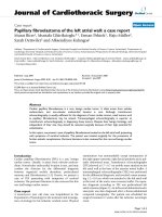
Hypercellular-hypocellular regions, hyalinised blood vessels and hemosiderin pigment (HsP) collections are observed (LM, H&E, ×100)Figure 2
Hypercellular-hypocellular regions, hyalinised blood vessels
and hemosiderin pigment (HsP) collections are observed
(LM, H&E, ×100).
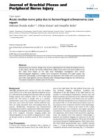
Median nerve (MN) and Schwannoma mass (Sc) are seen intraoperativelyFigure 1
Median nerve (MN) and Schwannoma mass (Sc) are seen
intraoperatively.
Publish with BioMed Central and every
scientist can read your work free of charge
"BioMed Central will be the most significant development for
disseminating the results of biomedical research in our lifetime."
Sir Paul Nurse, Cancer Research UK
Your research papers will be:
available free of charge to the entire biomedical community
peer reviewed and published immediately upon acceptance
cited in PubMed and archived on PubMed Central
yours — you keep the copyright
Submit your manuscript here:
/>BioMedcentral
Journal of Brachial Plexus and Peripheral Nerve Injury 2007, 2:19 />Page 3 of 3
(page number not for citation purposes)
Authors' contributions
MDA performed surgery, played role in clinical evalua-
tionn and treatment protocol. DK conducted electromyo-
grahy and interpreted results. MK evaluated
histopathology. All authors read and approvaed the final
manuscript.
References
1. Lee SH, Jung HG, Park YC, Kim HS: Results of neurilemoma
treatment: a review of 78 cases. Orthopedics 2001, 24:977-80.
2. Harkin JC, Reed RS: Tumors of the peripheral nervous system.
Washington DC, Armed Forces Institute of Pathology 1968:110-6.
3. Phalen G: Neurilemmomas of the forearm and hand. Clin Ortop
1976, 114:212-22.
4. Louis D: Peripheral nerve tumors of the upper etremity. Hand
Clin 1987, 3:311-8.
5. MacDonell RAL, Schwartz MS, Swash M: Carpal Tunel Syndrome:
Which finger should be tested? An analysis sensory conduc-
tion in digital branches of the median nerve. Musle Nerve 1990,
13:601-6.
6. Hems TE, Burge PD, Wilson DJ: The role of magnetic resonance
imaging in the management of peripheral nerve tumours. J
Hand Surg 1997, 22:57-60.
7. Kuo YL, Yao WJ, Chiu HY: Role of sonography in the preopera-
tive assessment of neurilemmoma. J Clin Ultrasound 2005,
33:87-9.
8. Mintz A, Carneiro R: Schwannoma associated with anomalous
division of the median nerve. Case report. Neurosurgery 1989,
25:965-8.
9. Charitides J, Charisopoulos G, Tsakonas A: Neurilemmona of the
median nerve in a child. Case report. Chir Organi Mov 2000,
85:281-3.
10. Josty IC, Sykes PJ: An unusual Schwannoma of the median
nerve: effects on the motor branch. Br J Plast Surg 2001, 54:71-3.
11. Haussmann P: Malignant Schwannoma of the median nerve.
Handchir Mikrochir Plast Chir 1988, 20:147-9.
12. Zingale A, Consoli V, Tigano G, Pero G, Albanese V: CT morphol-
ogy of a median nerve neurilemmoma at the arm. Case
report and review. J Neurosurg Sci 1993, 37:57-9.
13. Williams GD, Hoffman S, Schwartz IS: Malignant transformation
in a plexiform neurofibroma of the median nerve. J Hand Surg
1984,
9:583-7.