Báo cáo y học: "Pain as a symptom of peripheral nerve sheath tumors: clinical significance and future therapeutic directions" potx
Bạn đang xem bản rút gọn của tài liệu. Xem và tải ngay bản đầy đủ của tài liệu tại đây (262.6 KB, 5 trang )
BioMed Central
Page 1 of 5
(page number not for citation purposes)
Journal of Brachial Plexus and
Peripheral Nerve Injury
Open Access
Review
Pain as a symptom of peripheral nerve sheath tumors: clinical
significance and future therapeutic directions
Michael E Sughrue*
1
, Jon Levine
2
and Nicholas M Barbaro
1
Address:
1
Department of Neurological Surgery, University of California at San Francisco, 505 Parnassus Ave, San Francisco, California, USA and
2
Department of Medicine, University of California at San Francisco, 505 Parnassus Ave, San Francisco, California, USA
Email: Michael E Sughrue* - ; Jon Levine - ;
Nicholas M Barbaro -
* Corresponding author
Abstract
Tumors arising from the supporting cells of peripheral nerve sheaths are relatively uncommon
neoplasms, and as such many clinicians are unfamiliar with the details of their presentation,
diagnosis and management. Further, little is known regarding the pathogenesis of these tumors,
how they cause symptoms, and how to treat these symptoms. One classic symptom of peripheral
nerve tumors is pain, however there has been little formal discussion regarding the significance of
pain in this setting. Here we present a brief review of the clinical significance of pain, its relevance
in pre-operative planning for the treatment of these tumors, and what is known regarding the
molecular mechanisms of pain generation by these tumors.
Epidemiology and clinical presentation of
peripheral nerve tumors
Tumors arising from the supporting cells of peripheral
nerve sheaths are relatively uncommon neoplasms, and as
such many clinicians are unfamiliar with the details of
their presentation, diagnosis and management. Further,
little is known regarding the pathogenesis of these
tumors, how they cause symptoms, and how to treat these
symptoms.
Tumors of peripheral nerve are benign in at least 85–90%
of clinically symptomatic cases, and likely a larger per-
centage of subclinical cases [1]. In normal patients, the
majority of these tumors are histologically schwannomas,
with lesser percentages made up of other benign lesions
such as hemangiomas, ganglion cysts, desmoids, malig-
nant peripheral nerve sheath tumors (MPNST's), and
other malignant lesions, such as lymphoma and metas-
tases [2]. For patients, with neurofibromatosis type 1 (NF-
1), the incidence of malignancy is significantly greater:
8–10% of NF-1 patients will develop an MPNST during
their lifetimes, and nearly 50% of patients with MPNST
have NF-1 [3].
The typical presenting signs and symptoms of a peripheral
nerve sheath tumor (PNST) involves some combination
of a palpable (or radiographically visible) mass involving
a peripheral nerve, loss of nerve function, and/or pain
[1,3]. The etiology and significance of the first two symp-
toms should be relatively intuitive, for instance, the pres-
ence of a significant nerve palsy is likely due to nerve
invasion and destruction by the tumor, and is highly sug-
gestive of malignancy. However, the significance of pain
in the setting of PNST is significantly less well defined, the
mechanisms that cause it are more complex and poorly
understood, and the proper tools to specifically or effec-
Published: 29 February 2008
Journal of Brachial Plexus and Peripheral Nerve Injury 2008, 3:6 doi:10.1186/1749-7221-3-
6
Received: 24 October 2007
Accepted: 29 February 2008
This article is available from: />© 2008 Sughrue et al; licensee BioMed Central Ltd.
This is an Open Access article distributed under the terms of the Creative Commons Attribution License ( />),
which permits unrestricted use, distribution, and reproduction in any medium, provided the original work is properly cited.
Journal of Brachial Plexus and Peripheral Nerve Injury 2008, 3:6 />Page 2 of 5
(page number not for citation purposes)
tive treat it are currently not available. A brief summary of
what is currently known is the topic of the present review.
Pain as a presenting symptom of peripheral
nerve tumors
Of clear importance is the ability to differentiate between
benign and malignant lesions as early as possible in the
clinical work-up and management of these lesions, as they
are treated very differently, and exhibit very different clin-
ical and intraoperative behaviors. Ideally, the probable
diagnosis should be known prior to surgery, as malignant
tumors are more likely to require aggressive resection and
possibly amputation in order to achieve any degree of
oncologic control of these aggressive tumors [2,4,5].
Despite even aggressive management, the prognosis for
these tumors remains poor [5-7]. Benign lesions, in con-
trast, are often able to be easily resected away from nerve
fibers with minimum morbidity [2,4,5]. Further, resective
surgery is likely to resolve or significantly improve pain in
75–85% of patients with benign tumors and pre-operative
pain [2].
Current evidence suggests that in most cases, benign and
malignant lesions can be differentiated pre-operatively
based on clinical and radiographic characteristics. Most
importantly, while either a palpable/visible mass, nerve
palsy, or pain can occur in either benign or malignant
tumors, all three are more common and more notable in
malignant tumors. For example, rapid enlargement of a
nerve mass was found in one study to predict malignant
histology have a positive predictive value (PPV) of
approximately 95% [3]. Also, the presence of any neuro-
logic deficit predicts malignancy with a PPV of 73% which
was identical in results published by 2 different groups
[1,3]. Greater degrees of neurologic deficit (i.e. motor
strength less than 3/5), appears to be exclusively a symp-
tom of malignant tumors (PPV = 100%) [1].
Less clear is what to make of pain in the setting of a PNST,
as approximately 75% of all patients with PNST (benign
or malignant) have pain in some setting, and the positive
predictive value of the symptom "pain" to predict malig-
nancy is about 20–30% [1,3]. Far more important is the
distinction of pain at rest versus positional pain or pain
induced by pressure (i.e. the Tinel's sign). For example,
one group reported that pain at rest occurred in nearly all
(15/16) patients with MPNST, however only 5/99 (5%)
patients with benign schwannomas or neurofibromas [1].
In contrast, 94/99 of patients with benign tumors had a
positive Tinel's test [1]. Thus, further clarification of the
character and timing of the pain increases the PPV of the
symptom "pain" to 75%, making it a much more useful
piece of information in surgical planning.
Potiential mechanisms of pain generation in
PNST and future therapeutic directions
The dichotomy seen clinically between pressure induced
pain (which occurs in both benign and malignant
tumors) and rest pain (largely a symptom of malignancy)
suggests that these types of pain might result from distinct
pathophysiologic mechanisms. It follows from this
hypothesis that development of optimal therapies for
each of these types of pain probably should be directed at
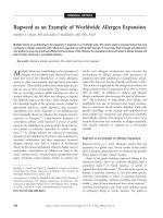
different molecular targets. Figure 1 briefly summarizes
some of the possible mechanisms involved in neuro-
pathic pain caused by nerve tumors.
Ectopic mechanosensitivity
The cause of pressure induced nerve pain in the setting of
nerve sheath tumors is unknown. The best hypotheses for-
mulated about the cause are extrapolations from work
regarding the mechanisms of mechanosensitivity-type
pain in Ad- and C-fibers seen in non-neoplastic, mech-
anosensitive lesions such as neuromas. Mechanosensitiv-
ity in these lesions is thought to result from progressive
incorporation and buildup of a number of proteins,
including mechanosensitive receptors intended for the
receptor terminal of a pain receptor, into an ectopic site
along an axon [8-11]. To date, the exact molecular trans-
ducer of these mechanical pain impulses is unknown.
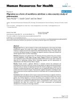
Schematic representation of possible mechanisms of algogen-esis in the setting of nerve sheath tumorsFigure 1
Schematic representation of possible mechanisms of algogen-
esis in the setting of nerve sheath tumors. Mechanisms
depicted include: (A) Ectopic mechanosensitivity possibly due
to increase in local concentrations of the Nav 1.4 sodium
channel leading to increased axonal transmission in response
to mechanical stimulation, (B) Continuous secretion of
chemical algogens leading to rest pain in the absence of stim-
ulus, (C) Aberrant axonal sprouting which fire pain stimuli
constitutively.
Journal of Brachial Plexus and Peripheral Nerve Injury 2008, 3:6 />Page 3 of 5
(page number not for citation purposes)
One protein known to accumulate near areas of axonal
compression, which may augment signal transduction,
and thus promote the development of a mechanosensitive
state, is the tetrodotoxin resistant sodium channel Nav1.8.
Clinical studies have demonstrated that Nav1.8 is densely
immunolocalized in the region immediately surrounding
focal sites of axonal injury, such as neuromas [12-14].
Roza et al. subsequently demonstrated in vitro that Ad-
and C-fibers taken from Nav1.8 null mice were markedly
less prone to the development of ectopic mechanosensi-
tivity than nerves taken from wild type mice in an experi-
mental model of neuroma formations, and that these
fibers were less likely to develop delayed spontaneous dis-
charges in that same model [15]. The Nav1.8 is a particu-
larly appealing therapeutic target as its expression appears
to be largely restricted to peripheral nerves [15], however
to date the development of a specific inhibitor has been
elusive.
Malignancy-induced nerve pain
Nerve pain caused by malignant tumors is likely chemical
in nature, and results from the release of substances by
malignant cells that stimulate chemoreceptive pain fibers,
such as H
+
, proteolytic enzymes, cytokines and growth
factors [1,16,17]. The later two classes of molecules have
the particularly appealing characteristic of potentially spe-
cific inhibition as a means of alleviating cancer pain. In
many cancers, invasion of the perineurium is necessary for
the development of rest pain, suggesting that high periax-
onal concentration of the offending algesic substance is
required [18]. Given the close proximity of Schwann cells
to the axon (originating inside the perineurium), it is
likely that any secreted algogen is present in biologically
relevant concentrations in the periaxonal space.
One potential algogen, the vasoconstrictive molecule
endothelin-1 (ET-1), has been found to be released in
high local concentrations in murine fibrosarcoma models
of hyperalgesia [19], and was not seen in a non-sarcoma
model (melanoma) [19]. Interestingly, local administra-
tion of an endothelin-A receptor antagonist significantly
reduced the morphine requirement in this model [19].
Similar to fibrosarcomas, MPNST's were also found to
have nearly 3-fold increased expression of the ET-1 gene
compared to normal schwann cells [20], suggesting that
ET-1 upregulation may be a consistent feature of sarcomas
in general, and raising the possibility that ET-1 antago-
nism might be useful in treating MPNST rest pain, though
formal in vivo evidence is presently lacking.
Nerve growth factor (NGF) is another algogen that has
been commonly implicated as an important cause of both
neuropathic and malignant cancer pain. For example, Zhu
et al found that human pancreatic cancers with high levels
of NGF expression demonstrated more extensive perineu-
real invasion, and more severe and refractory pain [18].
Other groups have demonstrated significant reduction of
cancer pain with systemic NGF antagonism in murine
models [16,21] While NGF may mediate these effects, in
part, by inducing cancer cells to invade the perineurium,
a great deal of evidence suggests that NGF may directly
induce hypersensitivity in sensory neurons in both in vitro
and in vivo models of neuropathic pain [22,23].
Sarcoma cells have been known for almost 50 years to
produce and secrete NGF [24], and in fact, the molecule
was originally discovered in experiments with sarcoma
cells [25,26]. The experience with NGF expression in
nerve sheath tumors is much more limited, but it seems
reasonable to hypothesize that NGF might be secreted by
MPNST's. More investigation is needed to further investi-
gate this issue.
Significance of the Schwann cell lineage to tumor-
associated neuropathic pain
A large body of published work supports the notion that
Schwann cells are involved in a number of dynamic inter-
actions with their associated axons, many of which can
promote (or in some cases inhibit) the development of
neuropathic pain in the setting of neuronal injury. This is
of special significance to the present discussion given that
a significant number of peripheral nerve tumors are of
Schwann cell lineage, and in theory, the disinhibition or
loss of Schwann cell functions could also play a role in the
production of neuropathic pain.
Schwann cells produce a number of cytokines in response
to injury, many of which have been implicated in the
development of neuropathic pain in the injured periph-
eral nerve. For example, normal Schwann cells have been
found to increase expression of Tumor necrosis factor-
alpha (TNF-a) in response to ex vivo compressive injury
[27], and sub-endoneureal TNF-a injection in vivo
induces neuropathic pain in rats [28]. Similar lines of evi-
dence implicate Schwann cell production of matrix metal-
loproteinase-9 (MMP9) [29], cyclooxgenase-2 [30], and
cytokines [31,32] in development of neuropathic pain.
Conversely, Schwann cells play a critical role in guidance
of sprouting axons during neuronal regeneration. Regen-
erating neurons which lack functional Schwann cell guid-
ance, often sprout in random directions and fail to form
functional connections, which some investigators
hypothesize may spontaneously fire causing neuropathic
pain [33,34]. It is unclear whether this dysfunctional
sprouting occurs in the setting of Schwann cell neoplasia,
however it is reasonable to hypothesize that these cells
likely are atleast less than ideal cellular guideposts for
regenerating neurons.
Journal of Brachial Plexus and Peripheral Nerve Injury 2008, 3:6 />Page 4 of 5
(page number not for citation purposes)
While little published work has focused on the occurrence
of either phenomenon in peripheral nerve tumor, either
seems like a reasonable starting point for further investi-
gation in this area.
Conclusion
Some form of pain is seen in most patients with periph-
eral nerve tumor, regardless of their histopathology. How-
ever, careful delineation of the nature and character of the
pain seems to provide valuable information for planning
the surgical approach to these tumors. Lesions with a sig-
nificant degree of rest pain should be considered as poten-
tially malignant in terms of pre-surgical planning.
Additionally, a better understanding of the chemical and
molecular causes of pain in these lesions will likely lead to
increased therapeutic options for palliating pain from
tumors involving and invading peripheral nerves.
Authors' contributions
MS wrote substantial portion of manuscript. JL contibuted
significant portion of ideas, especially section on mecha-
nisms of neuropathic pain. NMB concept conception,
wrote significant portion of manuscript.
References
1. Ogose A, Hotta T, Morita T, Yamamura S, Hosaka N, Kobayashi H,
Hirata Y: Tumors of peripheral nerves: correlation of symp-
toms, clinical signs, imaging features, and histologic diagno-
sis. Skeletal radiology 1999, 28(4):183-188.
2. Kim DH, Murovic JA, Tiel RL, Kline DG: Operative outcomes of
546 Louisiana State University Health Sciences Center
peripheral nerve tumors. Neurosurgery clinics of North America
2004, 15(2):177-192.
3. Valeyrie-Allanore L, Ismaili N, Bastuji-Garin S, Zeller J, Wechsler J,
Revuz J, Wolkenstein P: Symptoms associated with malignancy
of peripheral nerve sheath tumours: a retrospective study of
69 patients with neurofibromatosis 1. The British journal of der-
matology 2005, 153(1):79-82.
4. Kim DH, Murovic JA, Tiel RL, Kline DG: Management and out-
comes in 318 operative common peroneal nerve lesions at
the Louisiana State University Health Sciences Center. Neu-
rosurgery 2004, 54(6):1421-8; discussion 1428-9.
5. Fein DA, Lee WR, Lanciano RM, Corn BW, Herbert SH, Hanlon AL,
Hoffman JP, Eisenberg BL, Coia LR: Management of extremity
soft tissue sarcomas with limb-sparing surgery and postoper-
ative irradiation: do total dose, overall treatment time, and
the surgery-radiotherapy interval impact on local control?
International journal of radiation oncology, biology, physics 1995,
32(4):969-976.
6. Leroy K, Dumas V, Martin-Garcia N, Falzone MC, Voisin MC,
Wechsler J, Revuz J, Creange A, Levy E, Lantieri L, Zeller J, Wolken-
stein P: Malignant peripheral nerve sheath tumors associated
with neurofibromatosis type 1: a clinicopathologic and
molecular study of 17 patients. Archives of dermatology 2001,
137(7):908-913.
7. Mundt AJ, Awan A, Sibley GS, Simon M, Rubin SJ, Samuels B, Wong
W, Beckett M, Vijayakumar S, Weichselbaum RR: Conservative
surgery and adjuvant radiation therapy in the management
of adult soft tissue sarcoma of the extremities: clinical and
radiobiological results. International journal of radiation oncology,
biology, physics 1995, 32(4):977-985.
8. Devor M, Govrin-Lippmann R, Angelides K: Na+ channel immu-
nolocalization in peripheral mammalian axons and changes
following nerve injury and neuroma formation. J Neurosci
1993, 13(5):1976-1992.
9. Gold MS, Weinreich D, Kim CS, Wang R, Treanor J, Porreca F, Lai J:
Redistribution of Na(V)1.8 in uninjured axons enables neuro-
pathic pain. J Neurosci 2003, 23(1):158-166.
10. Koschorke GM, Meyer RA, Tillman DB, Campbell JN: Ectopic excit-
ability of injured nerves in monkey: entrained responses to
vibratory stimuli. Journal of neurophysiology 1991, 65(3):693-701.
11. Michaelis M, Blenk KH, Vogel C, Janig W: Distribution of sensory
properties among axotomized cutaneous C-fibres in adult
rats. Neuroscience 1999, 94(1):7-10.
12. Coward K, Plumpton C, Facer P, Birch R, Carlstedt T, Tate S, Bountra
C, Anand P: Immunolocalization of SNS/PN3 and NaN/SNS2
sodium channels in human pain states. Pain 2000, 85(1-
2):41-50.
13. Yiangou Y, Birch R, Sangameswaran L, Eglen R, Anand P: SNS/PN3
and SNS2/NaN sodium channel-like immunoreactivity in
human adult and neonate injured sensory nerves. FEBS letters
2000, 467(2-3):249-252.
14. Yiangou Y, Facer P, Birch R, Sangameswaran L, Eglen R, Anand P:
P2X3 receptor in injured human sensory neurons. Neurore-
port 2000, 11(5):993-996.
15. Roza C, Laird JM, Souslova V, Wood JN, Cervero F: The tetrodo-
toxin-resistant Na+ channel Nav1.8 is essential for the
expression of spontaneous activity in damaged sensory
axons of mice. The Journal of physiology 2003, 550(Pt 3):921-926.
16. Sevcik MA, Ghilardi JR, Peters CM, Lindsay TH, Halvorson KG, Jonas
BM, Kubota K, Kuskowski MA, Boustany L, Shelton DL, Mantyh PW:
Anti-NGF therapy profoundly reduces bone cancer pain and
the accompanying increase in markers of peripheral and
central sensitization. Pain 2005, 115(1-2):128-141.
17. Wacnik PW, Baker CM, Herron MJ, Kren BT, Blazar BR, Wilcox GL,
Hordinsky MK, Beitz AJ, Ericson ME: Tumor-induced mechanical
hyperalgesia involves CGRP receptors and altered innerva-
tion and vascularization of DsRed2 fluorescent hindpaw
tumors. Pain 2005, 115(1-2):95-106.
18. Zhu Z, Friess H, diMola FF, Zimmermann A, Graber HU, Korc M,
Buchler MW: Nerve growth factor expression correlates with
perineural invasion and pain in human pancreatic cancer. J
Clin Oncol 1999, 17(8):2419-2428.
19. Wacnik PW, Eikmeier LJ, Ruggles TR, Ramnaraine ML, Walcheck BK,
Beitz AJ, Wilcox GL: Functional interactions between tumor
and peripheral nerve: morphology, algogen identification,
and behavioral characterization of a new murine model of
cancer pain. J Neurosci 2001, 21(23):9355-9366.
20. Lee PR, Cohen JE, Tendi EA, Farrer R, GH DEV, Becker KG, Fields
RD: Transcriptional profiling in an MPNST-derived cell line
and normal human Schwann cells. Neuron Glia Biol 2004,
1(2):135-147.
21. Halvorson KG, Kubota K, Sevcik MA, Lindsay TH, Sotillo JE, Ghilardi
JR, Rosol TJ, Boustany L, Shelton DL, Mantyh PW: A blocking anti-
body to nerve growth factor attenuates skeletal pain induced
by prostate tumor cells growing in bone. Cancer research 2005,
65(20):9426-9435.
22. Anand P: Neurotrophic factors and their receptors in human
sensory neuropathies. Progress in brain research 2004,
146:477-492.
23. Kitamura N, Konno A, Kuwahara T, Komagiri Y: Nerve growth fac-
tor-induced hyperexcitability of rat sensory neuron in cul-
ture. Biomedical research (Tokyo, Japan) 2005, 26(3):123-130.
24. Werrbach-Perez K, Perez-Polo JR: De novo synthesis of NGF
subunits in S-180 mouse sarcoma cell line. Neurochemical
research 1987, 12(10):875-883.
25. Cohen S, Levi-Montalcini R, Hamburger V: A Nerve Growth-Stim-
ulating Factor Isolated from Sarcom as 37 and 180. Proceed-
ings of the National Academy of Sciences of the United States of America
1954, 40(10):1014-1018.
26. Levi-Montalcini R, Meyer H, Hamburger V: In vitro experiments
on the effects of mouse sarcomas 180 and 37 on the spinal
and sympathetic ganglia of the chick embryo. Cancer research
1954, 14(1):49-57.
27. Wagner R, Myers RR: Schwann cells produce tumor necrosis
factor alpha: expression in injured and non-injured nerves.
Neuroscience 1996, 73(3):625-629.
28. Wagner R, Myers RR: Endoneurial injection of TNF-alpha pro-
duces neuropathic pain behaviors. Neuroreport 1996,
7(18):2897-2901.
Publish with BioMed Central and every
scientist can read your work free of charge
"BioMed Central will be the most significant development for
disseminating the results of biomedical researc h in our lifetime."
Sir Paul Nurse, Cancer Research UK
Your research papers will be:
available free of charge to the entire biomedical community
peer reviewed and published immediately upon acceptance
cited in PubMed and archived on PubMed Central
yours — you keep the copyright
Submit your manuscript here:
/>BioMedcentral
Journal of Brachial Plexus and Peripheral Nerve Injury 2008, 3:6 />Page 5 of 5
(page number not for citation purposes)
29. Chattopadhyay S, Myers RR, Janes J, Shubayev V: Cytokine regula-
tion of MMP-9 in peripheral glia: Implications for pathologi-
cal processes and pain in injured nerve. Brain Behav Immun
2006.
30. Takahashi M, Kawaguchi M, Shimada K, Konishi N, Furuya H,
Nakashima T: Cyclooxygenase-2 expression in Schwann cells
and macrophages in the sciatic nerve after single spinal
nerve injury in rats. Neuroscience letters 2004, 363(3):203-206.
31. Bhangoo SK, Ren D, Miller RJ, Chan DM, Ripsch MS, Weiss C, McGin-
nis C, White FA: CXCR4 chemokine receptor signaling medi-
ates pain hypersensitivity in association with antiretroviral
toxic neuropathy. Brain Behav Immun 2007.
32. Abbadie C, Lindia JA, Cumiskey AM, Peterson LB, Mudgett JS, Bayne
EK, DeMartino JA, MacIntyre DE, Forrest MJ: Impaired neuro-
pathic pain responses in mice lacking the chemokine recep-
tor CCR2. Proceedings of the National Academy of Sciences of the
United States of America 2003, 100(13):7947-7952.
33. Koltzenburg M, Scadding J: Neuropathic pain. Current opinion in
neurology 2001, 14(5):641-647.
34. Tyner TR, Parks N, Faria S, Simons M, Stapp B, Curtis B, Sian K,
Yamaguchi KT: Effects of collagen nerve guide on neuroma for-
mation and neuropathic pain in a rat model. American journal
of surgery 2007, 193(1):e1-6.