Báo cáo y học: "Capnography as an aid in localizing the phrenic nerve in brachial plexus surgery. Technical note" potx
Bạn đang xem bản rút gọn của tài liệu. Xem và tải ngay bản đầy đủ của tài liệu tại đây (826.01 KB, 4 trang )
BioMed Central
Page 1 of 4
(page number not for citation purposes)
Journal of Brachial Plexus and
Peripheral Nerve Injury
Open Access
Research article
Capnography as an aid in localizing the phrenic nerve in brachial
plexus surgery. Technical note
Hemant Bhagat
†1
, Anil Agarwal
†1
and Manish S Sharma*
2
Address:
1
Department of Neuroanesthesia, All India Institute of Medical Sciences, New Delhi-110029, India and
2
Department of Neurosurgery,
All India Institute of Medical Sciences, New Delhi-110029, India
Email: Hemant Bhagat - ; Anil Agarwal - ;
Manish S Sharma* -
* Corresponding author †Equal contributors
Abstract
Background: To determine whether monitoring end- tidal Carbon Dioxide (capnography) can be
used to reliably identify the phrenic nerve during the supraclavicular exploration for brachial plexus
injury.
Methods: Three consecutive patients with traction pan-brachial plexus injuries scheduled for
neurotization were evaluated under an anesthetic protocol to allow intraoperative
electrophysiology. Muscle relaxants were avoided, anaesthesia was induced with propofol and
fentanyl and the airway was secured with an appropriate sized laryngeal mask airway. Routine
monitoring included heart rate, noninvasive blood pressure, pulse oximetry and time capnography.
The phrenic nerve was identified after blind bipolar electrical stimulation using a handheld bipolar
nerve stimulator set at 2–4 mA. The capnographic wave form was observed by the
neuroanesthetist and simultaneous diaphragmatic contraction was assessed by the surgical
assistant. Both observers were blinded as to when the bipolar stimulating electrode was actually in
use.
Results: In all patients, the capnographic wave form revealed a notch at a stimulating amplitude of
about 2–4 mA. This became progressively jagged with increasing current till diaphragmatic
contraction could be palpated by the blinded surgical assistant at about 6–7 mA.
Conclusion: Capnography is a sensitive intraoperative test for localizing the phrenic nerve during
the supraclavicular approach to the brachial plexus.
Background
Early surgical intervention after brachial plexus injury is
the best predictor of a favourable functional outcome
after a trial of conservative management. Electrodiagnos-
tic studies like sensory evoked potentials (SEP), electro-
myography (EMG) and nerve compound action
potentials (NCAPs) are performed intraoperatively to aid
in monitoring, guiding, identifying and localizing nerve
function.[1] Though these diagnostic modalities have
contributed immensely to the improved surgical out-
comes following brachial plexus repair, their use may
prove cumbersome and prone to errors of interpretation.
Direct observation of muscle belly contraction after nerve
Published: 22 May 2008
Journal of Brachial Plexus and Peripheral Nerve Injury 2008, 3:14 doi:10.1186/1749-7221-3-
14
Received: 22 March 2008
Accepted: 22 May 2008
This article is available from: />© 2008 Bhagat et al; licensee BioMed Central Ltd.
This is an Open Access article distributed under the terms of the Creative Commons Attribution License ( />),
which permits unrestricted use, distribution, and reproduction in any medium, provided the original work is properly cited.
Journal of Brachial Plexus and Peripheral Nerve Injury 2008, 3:14 />Page 2 of 4
(page number not for citation purposes)
stimulation remains the gold standard to detect intact
neuronal function.
Phrenic nerve identification is a key step during the supr-
aclavicular approach for brachial plexus surgery. Capnog-
raphy is a technique to record end-tidal carbon dioxide
(ETCO
2
) and is one of the standards of monitoring in
anesthetic care. The authors describe the use of capnogra-
phy as an aid in the intraoperative localization of the
phrenic nerve.
Methods
Three adult patients with diagnosed traction panbrachial
plexus lesions were scheduled for supra and infraclavicu-
lar exploration and neurotization of the suprascapular,
axillary and musculocutaneous nerves.
The general anaesthetic technique was tailored to allow
intraoperative electrophysiological techniques to guide
the localization and repair of the injured nerves. Conse-
quently, muscle relaxants were avoided. Anaesthesia was
induced with propofol and fentanyl and the airway was
secured with a laryngeal mask. Anaesthesia was main-
tained with a propofol infusion and intermittent boluses
of fentanyl. Routine monitoring included heart rate, non-
invasive blood pressure, pulse oximetry and time capnog-
raphy.
Supraclavicular exploration was commenced in the
supine position with the head extended and turned to the
opposite side and the injured arm in an adducted posi-
tion. The skin incision was extended inferiorly over the
lower 1/3
rd
of the posterior border of the clavicular head
of the sternocleidomastoid and then curved laterally over
the medial 2/3
rd
of the superior surface of the clavicle. The
platysma was incised and the supraclavicular pad of fat
was dissected sharply under the microscope away from
the carotid sheath and the subclavian vein and retracted
posterolaterally. The omohyoid bellies were then identi-
fied and their common tendinous insertion was divided
between ligatures. The Scalenus anticus was then sought
as the musculofascial structure behind the phrenic nerve.
In view of the extensive scarring, the visual identification
of the phrenic nerve was not possible at first. Hence, blind
bipolar electrical stimulation using a handheld bipolar
nerve stimulator was used to localize the same by eliciting
diaphragmatic contraction. The nerve stimulator was ini-
tially used at low amplitude (1 mA) and the capnographic
wave form was observed. The changes in waveform were
monitored by the neuroanesthetist as the stimulating cur-
rent was gradually increased. Simultaneously, the pres-
ence of diaphragmatic contraction was judged by the
surgical assistant with his hand placed over the patient's
draped epigastrium. Both the neuroanesthetist and the
surgical assistant were blinded as to when the bipolar
stimulating electrode was actually in use. Once the
phrenic nerve was approximately localized, sharp dissec-
tion was commenced to identify the same.
Results
In all patients, the capnographic wave form revealed a
notch at a low electrical stimulating current of about 2–4
mA. This became progressively jagged with increasing cur-
rent strengths till diaphragmatic contraction could be pal-
pated by the blinded surgical assistant at about twice the
amplitude (6–7 mA). (Fig 1)
Discussion
Brachial plexus lesions most frequently affect the supra-
clavicular region rather than the retroclavicular or infra-
clavicular levels.[2] Hence, the supraclavicular approach
is the most commonly performed for traumatic brachial
plexus repair. Intraplexal and extraplexal nerve-transfers
are increasingly being utilized for brachial plexus recon-
struction aimed at restoring elbow flexion and shoulder
abduction.[3] Commonly used donor nerves are the tho-
racic intercostals, the medial pectoral, the phrenic and the
spinal accessory nerves.
Intraoperative monitoring of nerve repair using electrodi-
agnostic techniques aids the surgeon in the dissection,
identification and localization of nerves and also helps in
assessing nerve function. Electrodiagnosis proves valua-
ble, more so, in a setting of extensive fibrosis in the supr-
aclavicular compartment frequently encountered after
traction brachial plexus injuries. This makes identification
of the Scalenus anticus, behind which the C5 and C6
nerve roots lie, very difficult especially when this key mus-
cle is fibrosed and merges with the surrounding neuroma.
The muscle is then indirectly identified as the tissue lying
behind the phrenic nerve. The phrenic nerve is the only
structure in the medial supraclavicular area which passes
from lateral to medial. Thus, phrenic nerve identification
is the crucial initial step in the supraclavicular approach
for brachial plexus repair. Direct visualization may not be
possible even under high magnification as the phrenic
nerve too is often encased by scar tissue. Hence, blind
stimulation using a hand held bipolar electrical stimula-
tor and judging the contractile response of the diaphragm
manually is a useful aid in initial localization before
attempting scar tissue release with sharp dissection.
Other surgical techniques to identify the phrenic nerve
include following the supraclavicular nerve proximally till
the C4 root in order to identify the Phrenic nerve.[4]
However, most brachial plexus surgeons prefer to use
intraoperative electrical stimulation.
Monitoring phrenic nerve stimulation using lower chest
wall electrodes may produce false-positive results due to
Journal of Brachial Plexus and Peripheral Nerve Injury 2008, 3:14 />Page 3 of 4
(page number not for citation purposes)
co-activation of the brachial plexus.[5,6] Other possible
technical problems include overstimulation, stimulus
artifacts, electrical noise, and high recording electrode
impedance which may diminish reliability and increase
the duration of the procedure.[7]
On the other hand, capnography is a routine and manda-
tory anaesthetic monitoring device. In this study, a notch
in the capnograph could be obtained at a lower stimulus
intensity than palpable diaphragmatic contraction.
Phrenic nerve stimulation in an anesthetized non-para-
lyzed patient produces sub-clinical diaphragmatic con-
traction which mimics inspiration. This produces a drop
in ETC0
2
which is reflected as a notch on the time capno-
graph. These observations were similar and reproducible
in all the three patients. The authors could not come
across any report in medical literature utilizing this
attribute of capnography as an indicator of phrenic nerve
stimulation in brachial plexus surgery in a non-paralysed
patient. Electromyographic electrode placement to detect
phrenic nerve activity may also be affected by concurrent
stimulation of the other intraplexal nerves such as the tho-
racodorsal.[8] A notch in the capnogram, however, can-
not be produced upon stimulation of the brachial plexus,
thereby rendering this technique not only highly sensitive
but also highly specific. The phrenic nerve also has a large
number of motor axons and thus serves as an excellent
donor nerve.[9] Capnography thus may help prevent
inadvertent damage to the same by alerting the surgeon to
its presence in difficult cases with extensive scarring.
Conclusion
Capnography is a sensitive intraoperative test for localiz-
ing the phrenic nerve during the supraclavicular approach
to the brachial plexus.
Competing interests
Financial competing interests
In the past five years have you received reimbursements,
fees, funding, or salary from an organization that may in
any way gain or lose financially from the publication of
this manuscript, either now or in the future? Is such an
organization financing this manuscript? If so, please spec-
ify.
No.
Do you hold any stocks or shares in an organization that
may in any way gain or lose financially from the publica-
tion of this manuscript, either now or in the future? If so,
please specify.
No.
Do you hold or are you currently applying for any patents
relating to the content of the manuscript? Have you
received reimbursements, fees, funding, or salary from an
organization that holds or has applied for patents relating
to the content of the manuscript? If so, please specify.
No.
Do you have any other financial competing interests? If
so, please specify.
No.
Non-financial competing interests
Are there any non-financial competing interests (political,
personal, religious, ideological, academic, intellectual,
commercial or any other) to declare in relation to this
manuscript? If so, please specify. No.
Authors' contributions
HB made the original observation on the capnograph, was
the blinded anesthetist and helped edit the manuscript.
AA wrote the manuscript's first draft, carried out the liter-
ature search and was a blinded anesthetist. MSS conceived
the concept, elucidated the methodology and edited the
manuscript.
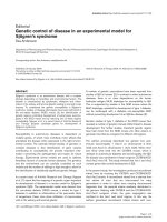
Fused capnograms as seen on the patient monitorFigure 1
Fused capnograms as seen on the patient monitor.
The top row is normal. After electrical stimulation, the mid-
dle row reveals progressive notching of the wave form (sub-
clinical diaphragmatic contraction) which degenerates into
frank spikes with increasing current corresponding to palpa-
ble diaphragmatic contractions. The progressive drop in end
tidal CO
2
from a baseline of 39 mm Hg to 31 mm Hg is note-
worthy.
Publish with BioMed Central and every
scientist can read your work free of charge
"BioMed Central will be the most significant development for
disseminating the results of biomedical research in our lifetime."
Sir Paul Nurse, Cancer Research UK
Your research papers will be:
available free of charge to the entire biomedical community
peer reviewed and published immediately upon acceptance
cited in PubMed and archived on PubMed Central
yours — you keep the copyright
Submit your manuscript here:
/>BioMedcentral
Journal of Brachial Plexus and Peripheral Nerve Injury 2008, 3:14 />Page 4 of 4
(page number not for citation purposes)
Acknowledgements
No financial support was provided in any form to the authors of this man-
uscript.
References
1. Slimp J: Intraoperative monitoring of nerve repairs. Hand Clin
2000, 16:25-36.
2. Moran SL, Steinmann SP, Shin AY: Adult brachial plexus injuries:
mechanism, patterns of injury and physical diagnosis. Hand
Clin 2005, 21:13-24.
3. Spinner RJ, Kline DG: Surgery for peripheral nerve and brachial
plexus injuries or other nerve lesions. Muscle Nerve 2000,
23:680-695.
4. Al-Qattan MM: Identification of the phrenic nerve in surgical
exploration of the brachial plexus in obstetrical palsy. J Hand
Surg [Am] 2004, 29:391-392.
5. Luo YM, Polkey MI, Lyall RA, Moxham J: Effect of brachial plexus
co-activation on phrenic nerve conduction time. Thorax 1999,
54:765-770.
6. American Thoracic Society; European Respiratory Society: ATS/ERS
statement on respiratory muscle testing. Am J Respir Crit Care
Med 2002, 166:528-547.
7. Harper CM: Preoperative and intraoperative electrophysio-
logic assessment of brachial plexus injuries. Hand Clin 2005,
21:39-46.
8. Hemmerling TM, Schmidt J, Hanusa C, Wolf T, Jacobi KE: The lum-
bar paravertebral region provides a novel site to assess neu-
romuscular block at the diaphragm. Can J Anaesth 2001,
48:356-360.
9. Luedemann W, Hamm M, Blömer U, Samii M, Tatagiba M: Brachial
plexus neurotization with donor phrenic nerves and its effect
on pulmonary function. J Neurosurg 2002, 96:523-526.