Báo cáo y học: "Rho kinase inhibitors Y27632 and H1152 augment neurite extension in the presence of cultured Schwann cells" pdf
Bạn đang xem bản rút gọn của tài liệu. Xem và tải ngay bản đầy đủ của tài liệu tại đây (1.69 MB, 11 trang )
BioMed Central
Page 1 of 11
(page number not for citation purposes)
Journal of Brachial Plexus and
Peripheral Nerve Injury
Open Access
Research article
Rho kinase inhibitors Y27632 and H1152 augment neurite extension
in the presence of cultured Schwann cells
Erick O Fuentes
1
, Jost Leemhuis
2
, G Björn Stark
1
and Eva M Lang*
1
Address:
1
Department of Plastic and Hand Surgery, University of Freiburg Medical Centre, Freiburg, Germany and
2
Institute of Experimental and
Clinical Pharmacology and Toxicology, Centre for Neuroscience, Freiburg, Germany
Email: Erick O Fuentes - ; Jost Leemhuis - ; G
Björn Stark - ; Eva M Lang* -
* Corresponding author
Abstract
Background: RhoA and Rho kinase inhibitors overcome the inhibition of axonal regeneration
posed by central nervous system (CNS) substrates.
Methods: To investigate if inhibition of the Rho pathway augments the neurite extension that
naturally occurs in the peripheral nervous system (PNS) following nerve damage, dorsal root
ganglion neurons and Schwann cell co-cultures were incubated with culture medium, C3 fusion
toxin, and the Rho kinase (ROCK) inhibitors Y27632 and H1152. The longest neurite per neuron
were measured and compared. Incubation with Y27632 and H1152 resulted in significantly longer
neurites than controls when the neurons were in contact with Schwann cells. When separated by
a porous P.E.T. membrane, only the group incubated with H1152 developed significantly longer
neurites. This work demonstrates that Rho kinase inhibition augments neurite elongation in the
presence of contact with a PNS-like substrate.
Background
The CNS is an environment normally hostile to nerve
regeneration due to the presence of axonal inhibitory sub-
strates like chondroitin sulphate proteoglycans (CSPGs) –
present in both the glial scar and in myelin – NOGO and
myelin associated glycopeptide (MAG) [1]. These sub-
stances inhibit axonal regeneration by activating on RhoA,
a member of the Rho GTPase family. Active RhoA causes
the retraction of growth cones by increasing the net phos-
phorylation of the myosin regulatory light chain. It also
activates Rho kinase (ROCK) which directly phosphor-
ylates the regulatory light chain of the major cytoplasmic
myosin, myosin II, increasing its actin-activated ATPase
and thus contractility [2] resulting in growth cone col-
lapse and retraction [3,4]. RhoA activity is increased fol-
lowing CNS injury [5] further augmenting the inhibition
of axonal regeneration that is already present. It is known
that this effect is overcome by the RhoA specific inhibitor
C3 transferase and the ROCK-specific inhibitor Y27632
[6-8,1]. The p75 nerve growth factor (p75NTR) plays an
important role in the axon and neurite extension through
modulation of the RhoA pathway. In the unbound state,
the p75NTR constitutively activates RhoA. When neuro-
trophin binds to the p75NTR, RhoA activation is switched
off [9-11]. The CNS inhibitory substrates such as NOGO
mediate their effect by binding to the p75NTR however,
this binding causes the activation of RhoA and hence the
inhibition of axonal regeneration [12,7].
Published: 25 September 2008
Journal of Brachial Plexus and Peripheral Nerve Injury 2008, 3:19 doi:10.1186/1749-7221-3-19
Received: 8 April 2008
Accepted: 25 September 2008
This article is available from: />© 2008 Fuentes et al; licensee BioMed Central Ltd.
This is an Open Access article distributed under the terms of the Creative Commons Attribution License ( />),
which permits unrestricted use, distribution, and reproduction in any medium, provided the original work is properly cited.
Journal of Brachial Plexus and Peripheral Nerve Injury 2008, 3:19 />Page 2 of 11
(page number not for citation purposes)
In contrast to the CNS, the peripheral nervous system
(PNS) allows nerve regeneration to occur following nerve
injury such as axotomy or crush injury. This is assisted by
Schwann cells (SC), which provide neurons with adhe-
sion molecules and a myriad of neutrophins to support
neurite and axonal growth. Little is known of the role that
Rho GTPases play in peripheral nerve regeneration. Whilst
RhoA is present and expressed in peripheral nerve axons
and SC [13], recent work suggests that RhoA activity is not
increased in SC following PNS injury [14]. Rho has also
been shown to play a role in the process of PNS myelina-
tion [15,16]. and SC migration [17]. There is however,
sparse evidence showing that axonal regeneration or neu-
rite elongation are enhanced by the inhibition of RhoA or
ROCK in the PNS hence, this work aimed to measure the
effect of Rho and ROCK inhibition on neurite extension
of neurons on a PNS like environment.
Materials and methods
RhoA and ROCK inhibitors
The C3 fusion toxin (C3 FT), a chimeric protein consisting
of the Clostridium limosum toxin C3 and the N-terminal
adaptor domain of Clostridium botulinum C2I, which
interacts with the binding/transport component C2II of
the C2 toxin. In this construct, the C2II protein acts as a
pore forming protein which allows the efficient delivery
of the C3 protein into target cells [18]. C3 FT/C2II toxin
was used at 10 ng/ml:20 ng/ml concentration. (C3 FT and
C2II proteins were kindly donated by Dr. K Aktories, Insti-
tute of Experimental and Clinical Pharmacology and Tox-
icology, Freiburg, Germany).
Y27632, is a well established inhibitor of ROCK in a vari-
ety of systems. This pyridine derivative is the oldest syn-
thesised and reported specific inhibitor of Rho-kinase
family enzymes. Y27632 inhibits ROCK activity by com-
petitive binding with ATP to the catalytic domain. Y27632
is reported to have a specificity 100 times greater for
ROCK than for protein kinase A, protein kinase C, or
myosin light chain kinase, as well as over 20 times greater
than that for two other downstream Rho effectors, citron
kinase and protein kinase N [19,20]. Y27632 (Calbio-
chem, USA) was dissolved in 1 ml of distilled water,
smaller aliquots using culture medium were made and a
final concentration of 10 μM was used.
The newer H1152 is a more specific, stronger and mem-
brane-permeable inhibitor of ROCK with a Ki value of 1.6
nM. It is a poor inhibitor of the serine/threonine kinases,
PKA, PKC and MLCK. The Ki values of H1152 for these
kinases are about 390, 5800 and 6300 times higher than
for Rho-kinases, respectively [21-23]. H1152 (Calbio-
chem, USA) was dissolved in distilled water and used in a
concentration of 100 nM.
Cell Cultures
Schwann cell cultures were prepared from sciatic nerves of
2 to 3-day-old Wistar rats. These were surplus animals
from the animal breeding program belonging to the Fac-
ulty of Veterinary Science of the University of Freiburg.
The animals were housed and handled in accordance to
the local animal ethics committee rules. The rats were
given a lethal dose of CO
2
, the sciatic nerves excised and
placed into ice cooled DMEM (GibcoBRL Life Technolo-
gies, Germany). The epineurium was removed, the nerves
then cut into small blocks and digested in 2 ml of DMEM
with 0.25% trypsin (Sigma, Germany) and 0.1% colla-
genase A (Roche Diagnostics, Germany) for 45 min at
37°C. The nerve pieces were mechanically dissociated
using a fire polished Pasteur pipette and the cells collected
by centrifugation and resuspended in DMEM with 1000
IU/ml penicillin, 1000 mg/ml streptomycin, and 10%
FCS. The SC were plated on 25 ml culture bottles, the
medium was changed after 24 h and cytosine-arabinoside
1 μM/ml (Sigma Aldrich, Germany) added. After 48 h, the
cells were passaged under microscope control using 1 ml
of trypsin/EDTA (PAA Laboratories, Austria). The trypsin
activity was stopped with serum containing medium and
the cells gathered by centrifugation at 1000 rpm for 5 min
at 4°C. The cells were resuspended in 5 ml of medium,
plated onto a new 25 ml culture bottle and the mitogen
activator forskolin 1 μM/ml (Calbiochem, USA) added to
the medium. Primary SC cultures became 80% confluent
after 14 to 20 days. The medium was changed every 7
days.
Dorsal Root Ganglion Neurons
Cultures of dorsal root ganglions were harvested from the
same rats and DRG neuron cultures established using the
method described by Seilheimer [24]. In short, the spinal
columns from 10 rats were excised and washed in ice
cooled PBS (Biochrom, Germany). Under a dissecting
microscope the vertebral lamini were removed and 20 to
25 DRG were dissected from each animal and placed in
ice cooled DMEM. The neurons were dissociated in 0.25%
trypsin and 0.1% collagenase in 2 ml of DMEM for 45 min
at 37°C followed by mechanical dissociation and centrif-
ugation. The cells were resuspended and passed through a
50 μm pore size cell strainer. After 5 min of gentle centrif-
ugation they were transferred onto a cushion of 32% Per-
coll (Sigma Aldrich, Germany) and centrifuged for 10 min
at 4°C and 600 rpm. Using a cell counting chamber the
concentration of neurons was estimated and an appropri-
ate dilution and aliquots of the suspension set aside for
plating onto the coverslips.
Neuron enriched cultures
150–200 neurons were plated onto rat-tail collagen
coated coverslips. The cultures were kept overnight in
DMEM medium with 1% penicillin and 10% fetal calf
Journal of Brachial Plexus and Peripheral Nerve Injury 2008, 3:19 />Page 3 of 11
(page number not for citation purposes)
serum (FCS) and supplemented 10 ng/ml of nerve growth
factor (NGF) (Sigma, Germany). The medium was subse-
quently replaced, NGF omitted, and C3 FT, Y27632 and
H1152 (Merk/Calbiochem, USA) at concentrations of 10
ng/ml, 10 μM and 100 nM respectively, added to the
medium and the culture incubated for 8 h. The control
group was incubated only in medium without NGF. At the
end of the incubation, the cells were fixed with 4% para-
formaldehyde in cytosol stabilization buffer (CSB).
Co-culture experiments
DRG neurons and Schwann cells were put in co-culture in
two distinct ways. For direct neuron-SC contact, 30,000
Schwann Cells were plated onto 10 mm diameter poly-L-
lysine (Sigma-Aldrich, Germany) coated glass coverslips
and kept for 48 h in DMEM with 10% FCS, 150–200 dis-
sociated DRG neurons were seeded directly onto this con-
fluent SC culture. Alternatively, the same number of SC
were plated onto a chamber insert containing a P.E.T.
membrane with 1 μm pores (Falcon, Denmark). 48 h
later, 150–200 neurons were seeded onto coverslips
coated with rat tail collagen and the aforementioned
chambers inserts were placed in the same culture well
sharing the same medium.
The co-cultures were allowed to stabilize for 4 h after
which C3 Fusion Toxin, Y27632 and H1152 were added
to the medium at a concentration of 10 ng/ml, 10 uM and
100 nM respectively. They were then incubated for a fur-
ther 8 hours. The length of the longest or more dominant
neurite seen to arise from a neuronal cell body was meas-
ured. Neurons were excluded from the analysis if; a) the
neurites were in contact with neurites or the cell body of
other neurons, b) had highly branched neurites that made
it impossible to determine the presence of a dominant
neurite. The neurons plated onto coverslips not contain-
ing SC invariably had some contaminating SC. In this
case, only the neurites of neurons not in contact with SC
were included in the analysis. Two coverslips were used in
each experiment for each substance. Each experiment was
repeated 4 times.
Immunohistochemistry
The method used for immunohistochemistry was adapted
from that described by Henle [25]. In short, coverslips
containing co-cultures were fixed for 12 min in 4% para-
formaldehyde (PFA) in cytosol stabilization buffer (CSB),
followed by 2 min in 0.1% triton-x in CSB. The S100 rab-
bit anti-rat primary antibody (DAKO, Denmark) and pri-
mary mouse anti-βtubulin III (Sigma Aldrich, Germany)
were added at a concentration of 1:200 and 1:650 v/v
respectively in 1% goat serum in PBS and incubated for 1
h. After rinsing with PBS the coverslips were incubated
with the Cy3-conjugated goat anti-rabbit (Dinova, Ger-
many) and Alexa-488 coupled goat anti-mouse secondary
antibodies for 1 h in the dark at room temperature. The
coverslips were mounted onto glass slides using Prolong
Gold anti-fade (Molecular Probes, Neatherlands) mount-
ing medium (Figure. 1).
Microscopy and image and data analysis
Fixed co-culture coverslips were loaded onto glass slides
and looked at using the ×40 magnification under an Axi-
oplan 2 microscope with epifluorescence mounted with a
digital Axiocam camera (Carl Zeiss, Germany). Cells of
interest were photographed using the Axiovision 3.01
program (Carl Zeiss Vision, Germany). The images were
saved and later analysed using the built-in measuring
function "length", available in the Metamorph Version
6.1r4 image analysis software (Universal Imaging Corp.
USA). This function automatically measured the overall
length after drawing a box around the selected neurite
[26].
The data obtained from the neurite measurements were
analysed with Microsoft Excel 2002 (Microsoft Corpora-
tion) and SPSS 12.0.1 for Windows (SPSS Inc.). The two-
tailed T-test was used to compare of the proportion of
neurons that responded to the different stimuli. Neurite
lengths were compared using one-way ANOVA with Tam-
hane's post hoc test.
Results
C3 fusion toxin and the Rho kinase inhibitors Y27632 and
H1152 increase neurite length in neuron-enriched cultures
Dissociated DRG neuron cultures incubated overnight
with 10 ng/ml of NGF followed by an 8 h incubation with
C3 fusion toxin and the ROCK inhibitors Y27632 and
H1152 showed an increase in the average length of their
longest neurites when compared to the control group
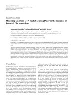
(medium only). The average maximal neurite lengths
were in the controls 110.1 μm (SE ± 7.8) in the C3FT
group 162.4 μm (SE ± 10.4), in the Y27632 group 167.8
μm (SE ± 12.3), and 185.4 μm (SE ± 16.6) in the H1152
group. All groups showed significantly longer neurites
than the control group (p < 0.001). There was no signifi-
cant difference between the three treatment groups (Fig-
ure. 2).
Effects of Rho and ROCK inhibition on SC morphology
The effect of Rho kinase inhibition on Schwann cell mor-
phology was previously described [15]. The most notable
change in SC morphology observed in this study was
caused by Y27632 and to a lesser extent by H1152. These
SC showed narrower and longer spindles with a more tri-
angular cell body. There was no obvious morphologic
effect of C3 FT on Schwann cells.
Journal of Brachial Plexus and Peripheral Nerve Injury 2008, 3:19 />Page 4 of 11
(page number not for citation purposes)
Double immuno-fluoresence of Schwann cell and DRG neuron co-cultures seen under ×40 objectiveFigure 1
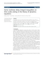
Double immuno-fluoresence of Schwann cell and DRG neuron co-cultures seen under ×40 objective. Images A
(control), C (C3 Toxin-treated), E (Y27632-treated), and G (H1152-treated) show the Schwann cell-specific S100 stain. Images
B, D, F, and H are the corresponding Beta tubulin stains of A, C, E and G respectively.
Journal of Brachial Plexus and Peripheral Nerve Injury 2008, 3:19 />Page 5 of 11
(page number not for citation purposes)
Inhibition of RhoA and ROCK has no effects on number of
neurons that extend neurites
The proportion of neurite-bearing neurons (i.e. DRG neu-
rons with neurite sprouts) was estimated by counting all
neurons with neurites present in one half of the coverslips
and dividing by the total number of neurons present in
that section of the coverslip. The proportion of neurite-
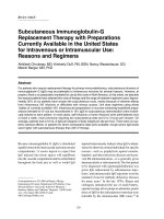
bearing DRG neurons in direct contact with SC was 44.5%
in the control group, 38.6% in the C3 FT group, 52.4% in
the Y27632 group and 52.0% in the H1152 group. The
proportion of neurite-bearing neurons separated form SC
by the presence of the separation membrane were 10.3%,
7.2%, 10.3%, 13.1% respectively. In both experimental
settings, there were no statistically significant differences
between stimuli and controls.
The proportion of neurite-bearing neurons in contact with
Schwann cells were clearly higher in all groups when com-
pared to neurons separated from SC by the 1 μm pore-size
P.E.T. membrane with permeable membrane (p < 0.001,
Figure. 3).
Y27632 and H1152 promote neurite extension in neurons
which are in contact with Schwann cells
Next the length of the longest neurite of the neurons,
which were in direct contact with SC were studied. To this
end 144 control, 141 C3 FT, 138 Y27632 and 137 H1152-
treated neurons were analysed. Interestingly, neurons in
the C3 FT, Y27632 and H1152 groups had on average
longer neurites than in the control group (Figure. 4). The
average neurite lengths obtained were 190.4 μm (SE ±
8.3) in the controls, 213.8 μm (SE ± 9.5) in the C3 FT
group, 259.7 μm (SE ± 9.1) in the Y27632 group and
244.4 μm (SE ± 11.4) in the H1152 group. However, only
the neurite lengths in the Y27632 and H1152 groups were
significantly longer than controls (p < 0.001 and p = 0.001
respectively). There was no significant difference between
the C3 FT, Y27632 and H1152 groups (Figure. 4).
When using the separation chamber to prevent SC-neuron
contact, the longest neurites from 60 control, 45 C3 FT, 58
Y27632 and 38 H1152-treated neurons were included in
the analysis. The average neurite lengths for the separated
neurons were for 92.2 μm (SE ± 8.0) in the Control group,
98.8 μm (SE ± 10.1) in the C3 FT group, 118.0 μm (SE ±
Neurite length of purified DRG neurons after an overnight incubation with 10 ng/ml NGF followed by 8 h incubation with C3 FT, Y27632 and H1152Figure 2
Neurite length of purified DRG neurons after an overnight incubation with 10 ng/ml NGF followed by 8 h incu-
bation with C3 FT, Y27632 and H1152.
Journal of Brachial Plexus and Peripheral Nerve Injury 2008, 3:19 />Page 6 of 11
(page number not for citation purposes)
Histogram comparing the proportion of neurite-bearing neurons under the two co-culture conditionsFigure 3
Histogram comparing the proportion of neurite-bearing neurons under the two co-culture conditions. As
expected, there was a highly significant difference between the neurons in contact vs separated from Schwann cells for the
same test stimuli (p ≤ 0.005) however, there were no significant differences between the different stimuli under the same cul-
ture conditions.
Histogram comparing neurite lengths of neurons in contact with Schwann cells after incubation with C3 FT, Y27632 and H1152Figure 4
Histogram comparing neurite lengths of neurons in contact with Schwann cells after incubation with C3 FT,
Y27632 and H1152. *** p ≤ 0.001.
Journal of Brachial Plexus and Peripheral Nerve Injury 2008, 3:19 />Page 7 of 11
(page number not for citation purposes)
12.6) in the Y27632 group and 123.5 μm (SE ± 10.9) in
the H1152 group. Although the average neurite lengths
were longer in the C3 FT, Y27632 and H1152 groups, only
the H1152 treated group was significantly longer than the
control group (p = 0.02, see Figure. 5).
The neurite lengths of the neurons in contact with SC were
significantly longer than the separated neurons (see Fig-
ure. 6). The p values were all < 0.001.
Discussion
Axonal or neurite elongation following the inhibition of
RhoA and Rho Kinase is well described. For instance,
recombinant constitutively active RhoA transfected into
PC12 cells causes suppression of neurite extension, which
is reversed by C3 whilst transfection of dominant negative
Rho results in increased axonal and neurite length [27].
The use of both C3 transferase and the Rho kinase inhibi-
tors Y27632 to promote neurite extension on foetal neu-
rons has been demonstrated by various authors [27-29].
Other authors have reported increases in axonal length
form DRG explants following incubation with C3 and
Y27632 [1]. We observed a similar response in neonatal
DRG neuron enriched culture experiments, where neurite
length was increased by C3 FT, H26732 and the newer
and more specific Rho kinase inhibitor H1152.
Rho and ROCK inhibitors overcome the inhibitory effect
that CNS substrates have on axonal regeneration [30]. It
has been shown that substances like chondroitin sulphate
proteoglycans (CSPGs), NOGO and myelin associated
glycopeptide (MAG) [31,32]. mediate their inhibitory
effect on axonal regeneration by the activation of the
RhoA pathway [1] by binding to the NOGO receptor
which interacts with the p75NTR receptor which, in turn
constitutively activates Rho [33,7,34,28]. This work aimed
to ascertain if the regenerative properties of Rho and
ROCK inhibitors also apply in the peripheral nervous sys-
tem model when the aforementioned inhibitory sub-
strates are not present i.e. in the in vitro non-myelinating
SC culture.
Cultured SC have properties similar to the SC in injured
nerves following Wallerian regeneration [35,36]. Impor-
tantly, SC synthesise and secrete various neurotrophins
including nerve growth factor (NGF), neurotrophin 3
(NT-3), brain derived neurotrophic factor (BDNF) and cil-
iary neurotrophic factor (CNTF) [37,38]. Since the
p75NTR binds all neurotrophins with a similar affinity
[39,40], it can be expect that all these neurotrophins will
cause the inactivation of RhoA by their binding to the
p75NGFR.
As a result, the null hypothesis ie. that additional extrinsic
inhibition of the Rho/ROCK pathway would have no
added effect on axonal/neurite elongation in our PNS
model was formulated. This hypothesis is supported by
the work of Geheler et al who demonstrated that neurons
treated with C3 showed no further increase in filopodial
length when co-incubated with BNDF. This effect was
shown to be independent of the Trk pathway since block-
ade with K252a at the same time that RhoA inhibition was
induced by neurotropin binding to the p75 receptor,
resulted in filopodial extension of the same magnitude as
when the p75 receptor was stimulated with an intact Trk
pathway [11]. In contrast to previous studies where DRG
explants were incubated in medium containing NGF, NT-
3 or BDNF [30,11]., the co-culture experiments in this
study did not use extrinsic growth factors. Rather, they
relied on the trophic substances secreted by the SC to
ensure the survival of the cultured neurons. This makes it
possible to observe if these Rho kinase inhibitors also
influence the number of neurons that extend neurites in
the co-culture setting.
The proportion of neurons that sprouted neurites follow-
ing a challenge with the experimental substances were not
significantly different to the control group or to each other
in the two co-culture conditions tested in this study. If we
consider neurite sprouting/bearing as a surrogate measure
of neuronal survival or viability, then it is clear that inhi-
bition of the RhoA pathway does not improve nor worsen
neuronal survival. This agrees with previous findings by
other authors where RhoA inhibition resulted in motor
neuron apoptosis during development but it did not seem
to affect sympathetic or DRG neuron survival [41] with
the essential difference that this study used neonatal
rather than embryonic cells. Our experiments showed sig-
nificant differences between the number of neurons-bear-
ing neurites in the two co-culture conditions. Although
equal numbers of SC were seeded onto the coverslips
Histogram comparing neurite lengths of neurons separated from Schwann cells by a porous P.E.T. membrane after incu-bation with C3 FT, Y27632 and H1152Figure 5
Histogram comparing neurite lengths of neurons
separated from Schwann cells by a porous P.E.T.
membrane after incubation with C3 FT, Y27632 and
H1152.
Journal of Brachial Plexus and Peripheral Nerve Injury 2008, 3:19 />Page 8 of 11
(page number not for citation purposes)
(neurons in contact with SC) and separation chambers
(neurons separated from SC), a significantly greater pro-
portion of neurons sprouted neurites when in contact
with SC. Similarly, the neurite lengths of neurons in con-
tact with SC were significantly greater in comparison to
their counterparts separated from SC by the permeable
membrane. These results would suggest that the number
of SC used did not provide enough trophic support via
diffusible factors to adequately maintain the viability of
the seeded neurons in the separation chamber experi-
ment. This result was anticipated and is consistent with
the literature where it is known that neuron-SC contact
improves neuron survival and axonal regeneration
[42,43,24,37]. Adhesion molecules present and SC such
as L1 have been shown to mediate a trophic effect that
ensures axonal survival [44]. Similarly, laminin which is a
constituent of the Schwann cell basal lamina, has also
been demonstrated to override the inhibitory effects of
PNS and CNS derived inhibitors of neurite growth [45].
Hence it can be concluded that the discrepancy in results
between the two co-culture conditions is due to a funda-
mental difference in the strength of the trophic support
provided by the Schwann cells rather than a lack of effect
from the RhoA and ROCK inhibitors used. It may be
argued that the results observed in the neurons in contact
with SC more closely resemble peripheral nerve injury in
vivo.
Our null hypothesis was disproved. The co-culture exper-
iments showed that both Y27632 and H1152 highly sig-
nificantly increased the lengths of neurites when the
neurons were in contact with SC. Neurons separated from
SC also showed an increase in neurite length when incu-
bated with C3 FT, Y26732 and H1152 however, this
increase was only significant in the H1152 group. Unfor-
tunately, technical difficulties with this experiment,
namely the extremely low number of neurites extended by
the neurons (see above), did not allow the collection of a
greater number of observations for each group. It may
well be that with a greater number of observations, the
effects of Y27632 and C3 FT would become significant.
Another strange observation was that, although C3 trans-
ferase showed a small increase in neurite length, this was
not significant in any of the co-culture experiments. It is
difficult to explain why C3 transferase had a significant
effect in the neuron enriched culture and yet no effect in
the co-culture experiments. One possibility is that either
the C3 or the C2II proteins were denatured or had expired
and thus no longer effective at the time of the co-culture
experiments however, a fresh batch of these proteins was
Histogram comparing the neurite lengths for neurons in contact with vs separated from Schwannn cells after treatment with C3 FT, Y27632 and H1152Figure 6
Histogram comparing the neurite lengths for neurons in contact with vs separated from Schwannn cells after
treatment with C3 FT, Y27632 and H1152. *** p ≤ 0.001.
Journal of Brachial Plexus and Peripheral Nerve Injury 2008, 3:19 />Page 9 of 11
(page number not for citation purposes)
used in each experiment. Another possible explanation,
although less likely, is that the toxin complex was prefer-
entially taken up by the SC thus decreasing the effective
concentration delivered to the neurons. If this were the
case, one would expect the SC to undergo a morphologi-
cal change. However, in this study, the SC did not show a
significant morphological change with C3. Nevertheless,
given the increases in neurite lengths observed in these
experiments, it can be expected that ROCK inhibitors
would increase axonal extension in vivo.
Whilst a recent study using the sciatic nerve crush injury
model showed that peripheral nerve regeneration is
enhanced by ROCK inhibition, it did not describe a direct
measure of increased axonal or neurite length [14], rather
they used surrogate measures such as compound muscle
action potential and motor nerve conduction studies. For
the first time, we show that incubation of neurons with
ROCK inhibitors increase neurite length while neurons
are in the presence of an environment similar to that of
the PNS following Wallerian degeneration. In this state,
Schwann cells shed their myelin and the myelin is then
phagocytosed [46,47]. This leaves connective tissue tubes
lined with proliferating Schwann cells that form linear
bands within the endoneurial sheath, known as Bands of
Bungner, which are important in guiding regenerating
axons across the injury site and into the distal nerve stump
[36]. However, given that neonatal neurons have a higher
intrinsic growth capacity [48] it is uncertain if these obser-
vations would hold true when using an adult animal
model.
Taken together, these results suggest that inhibition of
RhoA and ROCK in peripheral sensory neurons in the
presence of cultured Schwann cells, will result in axonal or
neurite extension. A reasonable explanation is that given
that RhoA and ROCK inhibition favour growth cone
extension by limiting growth cone collapse and retraction
[2,1], contact with Schwann cells (in their unmyelinating
state) favours further extension because of the presence of
adhesions molecules on their surface, which provide
greater stabilisation of the growth cone and in turn allows
further elongation. However, the short duration of these
experiments pose further questions. For instance, it was
observed that the ROCK inhibitors Y27632 and H1152
altered the morphology of the SC. These changes were
similar to those observed by Melendez et al [15]. They also
described a shortening of the myelin segments provided
by myelinating SC and detachment of SC for the culture
substrate after 48 hours or incubation with C3 and
Y27632. The latter is in keeping with the fact that Rho
GTPases are involved in the regulation of cell adhesion to
substratum and in cell to cell adhesion [49]. How these
observations would translate to in vivo peripheral nerve
regeneration in the presence of Rho and Rho kinase inhi-
bition remain undescribed.
Conclusion
In conclusion, we propose that inhibition of the RhoA
pathway in the peripheral nerve model, results in
increased neurite or axonal length. This may be due to
Schwann cells promoting the stabilisation of the growth
cone and thus, shifting the balance in the dynamics of
growth cones in favour of axonal elongation. Further
work using an in vivo model is warranted to gain further
knowledge of the effects of RhoA and ROCK inhibitors on
the functional recovery afterperipheral nerve injury.
List of abbreviations used
BDNF: brain derived neurotrophic factor; CNTF: ciliary
neurotrophic factor; CNS: central nervous system; C3 FT:
C3 fusion toxin; CSPGs: chondroitin sulphate proteogly-
cans; CSB: cystosol stabilization buffer; DRG: dorsal root
ganglion; FCS: fetal calf serum; MAG: myelin associated
glycopeptide; NGF: Nerve growth factor; NT-3: neuro-
trophin 3; PBS: phosphate buffered saline; PFA: parafor-
maldehyde; PNS: peripheral nervous system; ROCK: Rho
kinase.
Competing interests
The authors declare that they have no competing interests.
Authors' contributions
EF designed the experimental protocols, carried out the
experimental work, microscopy, data analysis, and pre-
pared the manuscript. EL helped to design the experimen-
tal protocols, assisted in data analysis and interpretation
and preparation of the manuscript. JL intellectually con-
tributed to the experimental design. GBS helped in correc-
tion the manuscript. All Authors have read and approved
the final manuscript.
Acknowledgements
Prof. K Actories and Prof D Meyer of the Institute of Experimental and
Clinical Pharmacology and Toxicology, and Centre for Neuroscience,
Freiburg, Germany for supplying experimental materials, in particular the
C3 FT, Rho kinase inhibitors, immunohistochemistry reagents and labora-
tory space. Dr F Henle instructed EF in the use of the image analysis soft-
ware. Prof B Stark of the Department of Plastic and Hand Surgery,
University of Freiburg Medical Centre, Freiburg, Germany, who's depart-
ment supplied the tissue culture equipment and laboratory. Dr Vincenzo
Penna aided the EF in the establishment of the Schwann cell cultures.
References
1. Giniger E: How do Rho family GTPases direct axon growth
and guidance? A proposal relating signaling pathways to
growth cone mechanics. Differentiation 2002, 70:385-396.
2. Amano M, Ito M, Kimura K, Fukata Y, Chihara K, Nakano T, Matsuura
Y, Kaibuchi K: Phosphorylation and activation of myosin by
Rho-associated kinase (Rho-kinase). J Biol Chem 1996,
271:20246-20249.
3. Nobes CD, Hall A: Rho, rac, and cdc42 GTPases regulate the
assembly of multimolecular focal complexes associated with
Journal of Brachial Plexus and Peripheral Nerve Injury 2008, 3:19 />Page 10 of 11
(page number not for citation purposes)
actin stress fibers, lamellipodia, and filopodia. Cell 1995,
81:53-62.
4. Mackay DJ, Nobes CD, Hall A: The Rho's progress: a potential
role during neuritogenesis for the Rho family of GTPases.
Trends Neurosci 1995, 18:496-501.
5. Sung JK, Miao L, Calvert JW, Huang L, Louis HH, Zhang JH: A possi-
ble role of RhoA/Rho-kinase in experimental spinal cord
injury in rat. Brain Research 2003, 959:29-38.
6. Monnier PP, Sierra A, Schwab JM, Henke-Fahle S, Mueller BK: The
Rho/ROCK pathway mediates neurite growth-inhibitory
activity associated with the chondroitin sulfate proteogly-
cans of the CNS glial scar. Mol Cell Neurosci 2003, 22(3):319-330.
7. Niederost B, Oertle T, Fritsche J, McKinney RA, Bandtlow CE:
Nogo-A and myelin-associated glycoprotein mediate neurite
growth inhibition by antagonistic regulation of RhoA and
Rac1. J Neurosci 2002, 22:10368-10376.
8. Bandtlow CE: Regeneration in the central nervous system.
Experimental Gerontology 2003, 38:79-86.
9. Yamashita T, Tucker KL, Barde YA: Neurotrophin binding to the
p75 receptor modulates Rho activity and axonal outgrowth.
Neuron 1999, 24:585-593.
10. Yamashita T, Tohyama M: The p75 receptor acts as a displace-
ment factor that releases Rho from Rho-GDI. Nat Neurosci
2003, 6:461-467.
11. Gehler S, Gallo G, Veien E, Letourneau PC: p75 neurotrophin
receptor signaling regulates growth cone filopodial dynam-
ics through modulating RhoA activity. J Neurosci 2004,
24:4363-4372.
12. Mi S, Lee X, Shao Z, Thill G, Ji B, Relton J, Levesque M, Allaire N, Per-
rin S, Sands B, Crowell T, Cate RL, McCoy JM, Pepinsky RB: LINGO-
1 is a component of the Nogo-66 receptor/p75 signaling
complex. Nat Neurosci 2004, 7:221-228.
13. Terashima T, Yasuda H, Terada M, Kogawa S, Maeda K, Haneda M,
Kashiwagi A, Kikkawa R: Expression of Rho-family GTPases
(Rac, cdc42, RhoA) and their association with p-21 activated
kinase in adult rat peripheral nerve. J Neurochem
2001,
77:986-993.
14. Hiraga A, Kuwabara S, Doya H, Kanai K, Fujitani M, Taniguchi J, Arai
K, Mori M, Hattori T, Yamashita T: Rho-kinase inhibition
enhances axonal regeneration after peripheral nerve injury.
J Peripher Nerv Syst 2006, 11:217-224.
15. Melendez-Vasquez CV, Einheber S, Salzer JL: Rho kinase regulates
schwann cell myelination and formation of associated axonal
domains. J Neurosci 2004, 24:3953-3963.
16. Fernandez-Valle C, Wood PM, Bunge MB: Localization of focal
adhesion kinase in differentiating Schwann cell/neuron cul-
tures. Microsc Res Tech 1998, 41(5):416-430.
17. Yamauchi J, Chan JR, Shooter EM: Neurotrophins regulate
Schwann cell migration by activating divergent signaling
pathways dependent on Rho GTPases. Proc Natl Acad Sci USA
2004, 101:8774-8779.
18. Barth H, Blocker D, Aktories K: The uptake machinery of
clostridial actin ADP-ribosylating toxins – a cell delivery sys-
tem for fusion proteins and polypeptide drugs. Naunyn
Schmiedebergs Arch Pharmacol 2002, 366:501-512.
19. Ishizaki T, Uehata M, Tamechika I, Keel J, Nonomura K, Maekawa M,
Narumiya S: Pharmacological properties of Y-2 a specific
inhibitor of rho-associated kinases. Mol Pharmacol 7632,
57:976-983.
20. Uehata M, Ishizaki T, Satoh H, Ono T, Kawahara T, Morishita T, Tam-
akawa H, Yamagami K, Inui J, Maekawa M, Narumiya S: Calcium sen-
sitization of smooth muscle mediated by a Rho-associated
protein kinase in hypertension. Nature 1997, 389:990-994.
21. Ikenoya M, Hidaka H, Hosoya T, Suzuki M, Yamamoto N, Sasaki Y:
Inhibition of rho-kinase-induced myristoylated alanine-rich
C kinase substrate (MARCKS) phosphorylation in human
neuronal cells by H- a novel and specific Rho-kinase inhibitor.
J Neurochem 1152, 81:9-16.
22. Sasaki Y, Suzuki M, Hidaka H: The novel and specific Rho-kinase
inhibitor (S)-(+)-2-methyl-1-[(4-methyl-5-isoquinoline)sulfo-
nyl]-homopiperazine as a probing molecule for Rho-kinase-
involved pathway. Pharmacol Ther 2002, 93:225-232.
23. Breitenlechner C, Gassel M, Hidaka H, Kinzel V, Huber R, Engh RA,
Bossemeyer D: Protein kinase A in complex with Rho-kinase
inhibitors Y-27632 Fasudil, and H-1152P: structural basis of
selectivity. Structure 2003, 11(12):
1595-1607.
24. Seilheimer B, Persohn E, Schachner M: Neural cell adhesion mol-
ecule expression is regulated by Schwann cell-neuron inter-
actions in culture. Journal of Cell Biology 1989, 108:1909-1915.
25. Henle F, Leemhuis J, Fischer C, Bock HH, Lindemeyer K, Feuerstein
TJ, Meyer DK: Gabapentin-lactam induces dendritic filopodia
and motility in cultured hippocampal neurons. J Pharmacol Exp
Ther 2006, 319:181-191.
26. Henle F, Fischer C, Meyer DK, Leemhuis J: Vasoactive intestinal
peptide and PACAP38 control N-methyl-D-aspartic acid-
induced dendrite motility by modifying the activities of Rho
GTPases and phosphatidylinositol 3-kinases. J Biol Chem 2006,
281:24955-24969.
27. Lehmann M, Fournier A, Selles-Navarro I, Dergham P, Sebok A,
Leclerc N, Tigyi G, McKerracher L: Inactivation of Rho signaling
pathway promotes CNS axon regeneration. J Neurosci 1999,
19:7537-7547.
28. Thies E, Davenport RW: Independent roles of Rho-GTPases in
growth cone and axonal behavior. Journal of Neurobiology 2003,
54:358-369.
29. Ellezam B, Dubreuil C, Winton M, Loy L, Dergham P: Inactivation
of intracellular Rho to stimulate axon growth and regenera-
tion. Progress in Brain Research 2002, 137:371-380.
30. Borisoff JF, Chan CC, Hiebert GW, Oschipok L, Robertson GS: Sup-
pression of Rho-kinase activity promotes axonal growth on
inhibitory CNS substrates. Mol Cell Neurosci 2003,
22(3):405-416.
31. Goldberg JL, Barres BA: Nogo in nerve regeneration. Nature
2000, 403:369-370.
32. Sandvig A, Berry M, Barrett LB, Butt A, Logan A: Myelin-, reactive
glia-, and scar-derived CNS axon growth inhibitors: expres-
sion, receptor signaling, and correlation with axon regener-
ation. Glia 2004, 46:225-251.
33. McGee AW, Strittmatter SM: The Nogo-66 receptor: focusing
myelin inhibition of axon regeneration. Trends in Neurosciences
2003, 26:
193-198.
34. Schwab ME: Nogo and axon regeneration. Current Opinion in Neu-
robiology 2004, 14:118-124.
35. Fu SY, Gordon T: The cellular and molecular basis of periph-
eral nerve regeneration. Mol Neurobiol 1997, 14:67-116.
36. Bunge MB, Bunge RP, Kleitman N, Dean AC: Role of peripheral
nerve extracellular matrix in Schwann cell function and in
neurite regeneration. Dev Neurosci 1989, 11:348-360.
37. Bunge RP: The role of the Schwann cell in trophic support and
regeneration. J Neurol 1994, 242:S19-S21.
38. Meyer M, Matsuoka I, Wetmore C, Olson L, Thoenen H: Enhanced
synthesis of brain-derived neurotrophic factor in the
lesioned peripheral nerve: different mechanisms are respon-
sible for the regulation of BDNF and NGF mRNA. J Cell Biol
1992, 119:45-54.
39. Hempstead BL: The many faces of p75NTR. Curr Opin Neurobiol
2002, 12:260-267.
40. Chao MV: Neurotrophins and their receptors: a convergence
point for many signalling pathways. Nat Rev Neurosci 2003,
4:299-309.
41. Kobayashi K, Takahashi M, Matsushita N, Miyazaki J, Koike M: Sur-
vival of developing motor neurons mediated by Rho GTPase
signaling pathway through Rho-kinase. J Neurosci 2004,
24:3480-3488.
42. Seilheimer B, Schachner M: Regulation of neural cell adhesion
molecule expression on cultured mouse Schwann cells by
nerve growth factor. EMBO J 1987, 6:1611-1616.
43. Seilheimer B, Schachner M: Studies of adhesion molecules medi-
ating interactions between cells of peripheral nervous sys-
tem indicate a major role for L1 in mediating sensory neuron
growth on Schwann cells in culture. Journal of Cell Biology 1988,
107:341-351.
44. Haney CA, Sahenk Z, Li C, Lemmon VP, Roder J, Trapp BD: Heter-
ophilic binding of L1 on unmyelinated sensory axons medi-
ates Schwann cell adhesion and is required for axonal
survival.
Journal of Cell Biology 1999, 146:1173-1184.
45. David S, Braun PE, Jackson DL, Kottis V, McKerracher L, David S,
Braun PE, Jackson DL, Kottis V, McKerracher L: Laminin overrides
the inhibitory effects of peripheral nervous system and cen-
tral nervous system myelin-derived inhibitors of neurite
growth. Journal of Neuroscience Research 1995, 42:594-602.
Publish with BioMed Central and every
scientist can read your work free of charge
"BioMed Central will be the most significant development for
disseminating the results of biomedical researc h in our lifetime."
Sir Paul Nurse, Cancer Research UK
Your research papers will be:
available free of charge to the entire biomedical community
peer reviewed and published immediately upon acceptance
cited in PubMed and archived on PubMed Central
yours — you keep the copyright
Submit your manuscript here:
/>BioMedcentral
Journal of Brachial Plexus and Peripheral Nerve Injury 2008, 3:19 />Page 11 of 11
(page number not for citation purposes)
46. Bruck W, Bruck Y, Maruschak B, Friede RL: Mechanisms of mac-
rophage recruitment in Wallerian degeneration. Acta Neu-
ropathol 1995, 89:363-367.
47. Stoll G, Griffin JW, Li CY, Trapp BD: Wallerian degeneration in
the peripheral nervous system: participation of both
Schwann cells and macrophages in myelin degradation. J
Neurocytol 1989, 18:671-683.
48. Cai D, Qiu J, Cao Z, McAtee M, Bregman BS, Filbin MT: Neuronal
cyclic AMP controls the developmental loss in ability of
axons to regenerate. J Neurosci 2001, 21:4731-4739.
49. Fukata M, Nakagawa M, Kuroda S, Kaibuchi K: Cell adhesion and
Rho small GTPases. J Cell Sci 1999, 112(Pt 24):4491-4500.