Báo cáo Y học: Thiol-modifying inhibitors for understanding squalene cyclase function pdf
Bạn đang xem bản rút gọn của tài liệu. Xem và tải ngay bản đầy đủ của tài liệu tại đây (313.16 KB, 9 trang )
Thiol-modifying inhibitors for understanding squalene cyclase function
Paola Milla
1
, Alexander Lenhart
2
, Giorgio Grosa
3
, Franca Viola
1
, Wilhelm A. Weihofen
2
, Georg E. Schulz
2
and Gianni Balliano
1
1
Universita
`
degli Studi di Torino, Dipartimento di Scienza e Tecnologia del Farmaco, Torino, Italy;
2
Universita
¨
t Freiburg,
Institut fu
¨
r Organische Chemie und Biochemie, Freiburg, Germany;
3
Universita
`
degli Studi del Piemonte Orientale ÔA. Avogadro,
Dipartimento di Scienze Chimiche, Alimentari, Farmaceutiche e Farmacologiche, Novara, Italy
The function of squalene-hopene cyclase from Alicycloba-
cillus acidocaldarius was studied by labelling critical cysteine
residues of the enzyme, either native or inserted by site-
directed mutagenesis, with different thiol-reacting molecules.
The access of the substrate to the active centre cavity through
a nonpolar channel that contains a narrow constriction
harbouring a cysteine residue (C435) was probed by labelling
experiments on both a C435S mutant, lacking C435 of the
channel constriction, and a C25S/C50S/C455S/C537S mu-
tant, bearing C435 as the only cysteine residue. Labelling
experiments with tritiated 3-carboxy-4-nitrophenyl-dithio-
1,1¢,2-trisnorsqualene (CNDT-squalene) showed that the
cysteine residue at the channel constriction was covalently
modified by the squalene-like inhibitor. Time-dependent
inactivation of the C25S/C50S/C455S/C537S mutant by a
number of squalene analogues and other agents with thiol-
modifying activity suggested that modifying C435 caused the
obstruction of the channel constriction thus blocking access
of the substrate to the active site. The tryptic fragment
comprising C435 of the quadruple mutant labelled with the
most effective inhibitor had the expected altered molecular
mass, as determined by LC-ESI-MS measurements. The
arrangement of the substrate in the active site cavity was
studied by using thiol reagents as probes in labelling
experiments with the double mutant D376C/C435S in which
D376, supposedly the substrate-protonating residue, was
substituted by cysteine. The inhibitory effect was evaluated
in terms of the reduced ability to cyclize oxidosqualene, as
the mutant is unable to catalyse the reaction of squalene to
hopene. Among the inhibitors tested, the substrate analogue
squalene-maleimide proved to be a very effective time-
dependent inhibitor.
Keywords: Alicyclobacillus acidocaldarius;membrane
protein; site-directed mutagenesis; squalene cyclase; thiol
reagents.
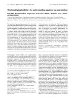
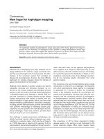
Oxidosqualene cyclases (OSCs) and squalene-hopene
cyclases (SHCs) are key enzymes in triterpenoid biosynthesis:
they transform acyclic isoprenoid precursors into tetra- and
pentacyclic compounds [1]. OSCs can be considered
taxonomic markers, as they catalyse the conversion of 2,3-
oxidosqualene into lanosterol in nonphotosynthetic organ-
isms (fungi and mammals), and into cycloartenol and other
tetra- and pentacyclic triterpenes in plants [2]. In prokary-
otes, SHCs convert squalene into hopene or diplopterol
(Fig. 1), pentacyclic triterpene precursors of hopanoids.
These compounds are thought to have functions similar to
those of sterols in eukaryotic membranes [3].
An important contribution to the understanding of the
catalytic mechanisms controlled by OSCs and SHCs came
from the crystal structure of SHC from Alicyclobacillus
acidocaldarious [4,5]. X-ray analysis revealed a membrane
protein with membrane-binding characteristics similar to
those of two prostaglandin-H
2
synthase isoenzymes [6,7].
These membrane proteins are called monotopic as they are
shaped so as to submerge from one side of the membrane
into the nonpolar part of the phospholipid bilayer without
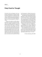
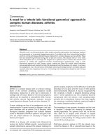
protruding through it [8]. The enzyme has a hydrophobic
plateau plunging into the lipophilic centre of the membrane.
A nonpolar channel connects the plateau and the active
centre through a narrow constriction formed by four
amino-acid residues, which appear to act as a gate that
permits substrate passage (Fig. 2). The cavity hosting the
active site is lined by nonpolar residues, but has a highly
polar patch at the top. It seems to be shaped so as to bind
the substrate in a specific product-like conformation, to
trigger cyclization by protonating a terminal double bond,
to assist ring-closures by stabilizing the cationic intermedi-
ates and, finally, to deprotonate the hopanyl cation to form
hopene or, in a side reaction, to hydroxylate the cation to
form hopan-22-ol (diplopterol).
We report here a new approach for studying the access of
the substrate to the active site cavity of A. acidocaldarius
SHC (E.C. 5.4.99.x) and its arrangement in it. To this aim,
Correspondence to G. Balliano, Dipartimento di Scienza e Tecnologia
del Farmaco, Via P. Giuria 9, I-10125 Torino, Italy.
Fax: +39 011 6707695, Tel.: +39 011 6707698,
E-mail:
Abbreviations: OSC, oxidosqualene cyclase; SHC, squalene-hopene
cyclase; CNDT-squalene, 3-carboxy-4-nitrophenyl-dithio-1,1¢,
2-trisnorsqualene; U14266A, (U14), 3b-(2-dimethylaminoethoxy)-
androst-5-en-17-one; CPTO, 2-(4-chlorophenyl)-D
2
-thiazoline-1-
oxide; DTS, (dimeric thiolsulfinate), 2(4-chlorobenzamido) ethane-
thiosulfinic acid S-2(4-chlorobenzamido) ethyl ester; NaB
3
H
4
,sodium
borotritiure (Ph)
3
P, triphenylphosphine,
Enzymes: oxidosqualene cyclase (EC 5.4.99.7); squalene-hopene
cyclase (EC 5.4.99.x).
Note: P. Milla and A. Lenhart contributed equally to this work.
Note: a web site is available at
/>(Received 1 November 2001, revised 18 February 2002, accepted 25
February 2002)
Eur. J. Biochem. 269, 2108–2116 (2002) Ó FEBS 2002 doi:10.1046/j.1432-1033.2002.02861.x
critically located Cys residues, either present in native
protein or inserted by site-directed mutagenesis, were
labelled with different thiol-reacting molecules, designed
and synthesized in our laboratories.
MATERIALS AND METHODS
NMR and MS of chemical products
1
H-NMR spectra were recorded on a Jeol EX-400 or Jeol
GX-270, with SiMe
4
as internal standard. Mass spectra
were obtained on a VG Analytical 7070 EQ-HF spectro-
meter by electron impact ionization. IR and UV spectra
were recorded, respectively, on Perkin-Elmer 781 and
Beckman DU 70 spectrophotometers.
Chemicals
Light petroleum refers to the fractions of bp 40–60 °C.
Tetrahydrofuran was distilled under sodium benzophenone
ketyl. Silica gel was 70–230 mesh. Squalene was from
Merck, polyoxyethylene 9 lauryl ether (polidocanol) was
from Sigma-Aldrich, Italy), 2,3-oxidosqualene was prepared
as described in [10]. 3-Carboxy-4-nitrophenyl-dithio-1,1¢,2-
trisnorsqualene (CNDT-squalene) (1), dodecyl- (3)and
squalene-maleimide (2) were synthetized as described else-
where [11]. U14266A (U14; 3b-(2-dimethylaminoethoxy)-
androst-5-en-17-one) [12] was provided by Upjohn
Company. 2-(4-chlorophenyl)-D
2
-thiazoline was synthetized
as reported previously [13]. m-chloroperbenzoic acid,
cystamine dihydrochloride, 4-chlorobenzoyl chloride and
triethylamine were purchased from Sigma-Aldrich.
2-(4-Chlorophenyl)-D
2
-thiazoline-1-oxide (CPTO) (4).
To an ice-cold and well stirred solution of 2-(4-chlorophe-
nyl)-D
2
-thiazoline (2 g, 0.0101 mol) in CH
2
Cl
2
(20 mL),
m-chloroperbenzoic acid (2.05 g, 0.0101 mol) dissolved in
CH
2
Cl
2
(40 mL) was slowly added. The mixture was stirred
for 3 h on ice while a precipitate appeared. CHCl
3
(60 mL)
was then added and the solution was washed with 5%
NaHCO
3
(2 · 120 mL) and saturated brine (40 ng NaCl in
100 mL H
2
O; 1 · 100 mL). The organic phase was dried
over anhydrous sodium sulfate and evaporated in vacuo.
The crude product was purified by flash-chromatography
using CHCl
3
as eluant to give 1.36 g CPTO (63% yield).
ESI-MS m/z:213(M
+
, 11), 195 (100), 185 (8), 137 (43);
1
H-
NMR (CDCl
3
) d:3.14(m,1H,5-H
a
), 3.36 (m, 1H, 5-H
b
),
4.68 (m, 1H, 4-H
a
), 4.91 (m, 1H, 4-H
b
), 7.49 (d, 2H,
aromatic protons), 8.04 (d, 2H, aromatic protons); IR (KBr)
m
max
: 3375, 3040, 2980, 2920, 1612, 1590, 1485, 1400Æcm
)1
;
UV (CH
3
OH) k
max
: 203, 260.
N,N¢-(Dithiodi-2,1-ethanediyl)bis 4-chlorobenzamide. To
an ice-cold and well stirred suspension of cystamine
dihydrochloride (0.77 g, 0.00343 mol) and triethylamine
(12 mL, great excess) in CHCl
3
(10 mL), 4-chlorobenzoyl
chloride (1.5 g, 0.00857 mol) dissolved in chloroform
(4 mL) was slowly added under argon atmosphere. The
mixture was stirred overnight at room temperature. CHCl
3
(10 mL) was then added and the mixture was extracted with
chloroform (2 · 50 mL) and chloroform/ethyl acetate 50/
50 (1 · 50 mL). The pooled organic phases were washed
with 5% NaHCO
3
(2 · 50 mL) and saturated brine
(1 · 50 mL). After anhydrification with anhydrous sodium
sulfate the solvent was evaporated under reduced pressure.
The crude product was purified by column chromatography
on silica with chloroform as eluant to give 1.05 g N,N¢-
(dithiodi-2,1-ethanediyl)bis 4-chlorobenzamide (68% yield).
ESI-MS m/z: 428 (M
+
, < 1), 215 (33), 182 (60), 139 (100),
111 (39);
1
H-NMR (CDCl
3
/CD
3
OD) d:2.81(t,4H,-CH
2
-
S), 3.57 (t, 4H, -CH
2
-N), 7.23 (d, 4H, aromatic protons),
7.61 (d, 4H, aromatic protons); IR (KBr) m
max
: 3302, 3236,
1638, 1628, 1597, 1541, 1489Æcm
)1
;UV(CH
3
OH) k
max
: 204,
235.
2(4-Chlorobenzamido) ethanethiosulfinic acid S-2(4-
chlorobenzamido) ethyl ester (DTS ¼ dimeric thiolsulf-
inate) (5). A solution of N,N¢-(dithiodi-2,1-ethanediyl)bis
4-chlorobenzamide (400 mg, 0.932 mmol) dissolved in
CH
2
Cl
2
(25 mL) was stirred while m-chloroperbenzoic acid
(85% purity, 147 mg, 0.932 mmol) was slowly added; it was
then allowed to react for a further 3 h with continuous
stirring. CH
2
Cl
2
(20 mL) was then added and the mixture
was washed with 5% NaHCO
3
(2 · 30 mL) and saturated
brine (1 · 50 mL). After anhydrification with anhydrous
Fig. 1. Reactions catalysed by (A) prokaryotic SHC and (B) eukaryotic
OSC.
Fig. 2. Surface representation of SHC sliced in the middle of the
molecule with nonpolar (yellow), positively charged (blue) and negatively
charged (red) areas. The large internal cavity is connected with the
hydrophobic plateau on the right by a nonpolar channel. A detergent
molecule LDAO that has been found in a crystal structure of SHC [4]
is shown as ball-and-stick model. The catalytic acid D376 and the
residues forming the channel constriction are indicated (V174 lying in
front of C435 was omitted for clarity). The figure was produced with
GRASP
[9].
Ó FEBS 2002 Effects of SH-modifying agents on squalene cyclase (Eur. J. Biochem. 269) 2109
sodium sulfate the solvent was evaporated under reduced
pressure. The crude product was purified by column
chromatography on silica with CHCl
3
andthenCHCl
3
/
CH
3
OH 98 : 2 to give DTS (5) (220 mg, 53% yield). ESI-
MS m/z: 429 (M ± 16, 54), 214 (29), 182 (44), 156 (35), 139
(100), 111 (54);
1
H-NMR (CD
3
OD) d:3.57(m,4H,-CH
2
-
SO and -CH
2
-S), 3.82–3.93 (m, 4H, -CH
2
-N), 7.53 (m, 4H,
aromatic protons), 7.88 (m, 4H, aromatic protons); IR
(KBr) m
max
: 3680, 3412, 2920, 1664, 1597, 1541, 1480Æcm
)1
;
UV (CH
3
OH) k
max
: 203, 236.
Radiochemicals
[2-
14
C]-mevalonate (50 mCiÆmmol
)1
) was from NEN. [
14
C]-
squalene and [
14
C]-3S-2,3-oxidosqualene were prepared by
incubating an S
10
supernatant (25 mg proteins) of a pig liver
homogenate with 1 lCi [
14
C]-mevalonate in the presence of
the OSC inhibitor U14266A (U14) [12], essentially as
described by Popjak [14]. The nonsaponifiable lipids were
separated by two-step TLC on silica gel plates (Merck)
(20 · 20 cm, 0.5 mm layer). The plates were first developed
in light petroleum to a height of about 10 cm above the
origin. After drying, the plates were developed to 15 cm
above the origin with n-hexane/ethyl acetate (90 : 10; v/v).
Radioactive areas corresponding to squalene and 2,3-oxido-
squalene were scraped off and eluted with dichloromethane.
The solvent was dried under N
2
and [
14
C]-squalene and
[
14
C]-3S-2,3-oxidosqualene were dissolved in benzene. The
radiochemical purity of products was evaluated by scanning
TLC plates with a System 2000 Imaging Scanner (Packard).
Radioactivity was measured by Liquid Scintillation Count-
ing (Beckman).
All of the radiolabelled compounds were compared
chromatographically with authentic radio-inert samples.
Determination of the radioactive substances and isotope
counting were carried out as already described [15,16].
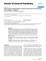
Radiolabelled CNDT-squalene (1) was synthesized via
the following steps (Fig. 3): (i) synthesis of [1-
3
H]trisnor-
squalene alcohol; (ii) synthesis of [1-
3
H]trisnorsqualene
thioacetate; (iii) synthesis of [1-
3
H]trisnorsqualene thiol;
(iv) transformation of [1-
3
H]trisnorsqualene thiol into [
3
H]-
CNDT-squalene (1).
(i) [1-
3
H]Trisnorsqualene alcohol: [1-
3
H]-(4E,8E,12E,
16E)-4,8,13,17,21-pentamethyl-4,8,12,16,20-docosapentaen-
1-ol. Pure trisnorsqualene aldehyde, obtained as described
by Ceruti et al. [17] (20.5 mg, 0.053 mmol) was dissolved in
methanol (0.5 mL) and added to the NaB
3
H
4
-containing
phial (total activity 25.0 mCi, specific activity 500 mCiÆ
mmol
)1
, total amount 0.05 mmol). After 3 h, NaBH
4
(excess, 6.0 mg, 0.159 mmol) was added to complete the
reaction. After an additional hour, the methanol was
evaporated under nitrogen and the reaction mixture was
dissolved in dichloromethane, transferred to a single necked
flask, and evaporated to dryness under reduced pressure to
give crude [1-
3
H]trisnorsqualene alcohol. The product was
purified, after dissolution with light petroleum, by column
chromatography on silica gel with 100% light petroleum to
remove impurities, then light petroleum/diethylether 90 : 10
to give 18 mg (0.046 mmol) of pure [1-
3
H]trisnorsqualene
alcohol. The radiochemical purity of the alcohol was
determined by radiochromatogram with light petroleum/
diethylether 80 : 20 and then revealed with iodine vapour.
Total activity: 4.2 mCi; specific activity: 92 mCiÆmmol
)1
;
chemical yield: 88%.
(ii) [1-
3
H]Trisnorsqualene thioacetate: [1-
3
H]-(4E,8E,
12E,16E) S-[4,8,13,17,21-pentamethyl-4,8,12,16,20-docos-
apentaenyl] thioacetate. A solution of diisopropyl azodi-
carboxylate (71.6 mg, 0.354 mmol) in 0.5 mL anhydrous
tetrahydrofuran was added to a well-stirred solution of
triphenylphosphine (92.5 mg, 0.350 mmol) in 3 mL anhy-
drous tetrahydrofuran at 0 °C. The mixture was stirred at
0 °C for 30 min and produced a white precipitate. A solu-
tion of [1-
3
H] trisnorsqualene alcohol (18 mg, 0.046 mmol)
and thiolacetic acid (37.2 mg, 0.488 mmol) in 0.5 mL
anhydrous tetrahydrofuran was added dropwise under
nitrogen and the mixture stirred for 1 h at 0 °Cand1h
at room temperature. The mixture was evaporated under
nitrogen and the crude product triturated in light petroleum;
the suspension was purified by column chromatography on
silica gel with 100% light petroleum to remove impurities,
then light petroleum/diethylether 99.5 : 0.5 to give pure
[1-
3
H] trisnorsqualene thioacetate (16.5 mg, 0.037 mmol).
The radiochemical purity of the thioacetate was determined
by radiochromatogram with light petroleum/diethylether
98 : 2 and then revealed with iodine vapour. Total activity:
3.6 mCi; specific activity: 92 mCi mmol
)1
; chemical yield:
80%.
(iii) [1-
3
H]Trisnorsqualene thiol: [1-
3
H]-(4E,8E,12E,16E)
4,8,13,17,21-pentamethyl-4,8,12,16,20-docosapentaenyl
1-thiol. [1-
3
H]trisnorsqualene thioacetate (16.5 mg,
0.037 mmol) was dissolved in anhydrous diethylether
(2 mL) and added dropwise to a suspension of LiAlH
4
(11.6 mg, 0.31 mmol) in anhydrous diethylether (5 mL)
under nitrogen. The mixture was stirred 25 min at room
temperature then 25 min under reflux. LiAlH
4
excess was
destroyed by the careful addition of 7 mL 1
M
HCl solution.
The ether layer was separated and the aqueous phase
extracted with dichloromethane. The combined organic
phases were dried over sodium sulfate and the solvent
evaporated under reduced pressure. The crude product was
used without purification for the following step.
(iv) [1-
3
H]Trisnorsqualene nitrobenzoic acid (1): [1-
3
H]-
6-nitro-3-[(4E,8E,12E,16E)-4,8,13,17,21-pentamethyl-4,8,12,
16,20-docosapentaenyldisulfamyl] benzoic acid; 5,5¢-dithio-
bis(2-nitrobenzoic acid) (41 mg, 0.103 mmol) was dissolved
Fig. 3. Scheme for the synthesis of [1-
3
H] CNDT-squalene. The asterisk
indicates position of
3
H-label: (i) NaB
3
H
4
(ii) (Ph)
3
P, CH
3
COSH,
diisopropyl azodicarboxylate; (iii) LiAlH
4
(iv) 5,5¢-dithiobis(2-nitro-
benzoic acid).
2110 P. Milla et al. (Eur. J. Biochem. 269) Ó FEBS 2002
in ethanol (9 mL). To this solution [1-
3
H] trisnorsqualene
thiol dissolved in ethanol (10 mL) was added under
nitrogen. The mixture was stirred overnight at room
temperature and then the solvent evaporated under reduced
pressure. The resulting yellow oil was purified by column
chromatography on silica gel with dichloromethane/meth-
anol 92 : 8 as eluent to give [1-
3
H] trisnorsqualene nitro-
benzoic acid (1) (11.1 mg, 0.0185 mmol). The radiochemical
purity of the disulfide was determined by radiochromato-
gram with dichloromethane/methanol 85 : 15 and then
revealed with iodine vapour. Total activity: 0.91 mCi;
specific activity: 92 mCiÆmmol
)1
;chemicalyield:50%.
Expression and purification of recombinant SHC
Wild-type SHC was kindly provided by Prof. K. Poralla
(Universita
¨
tTu
¨
bingen) [18].
For production of recombinant SHC, the overexpression
system described elsewhere [19] was used. Mutants D376C/
C435S, C455S and C50S had been generated for structure
analysis [5] utilizing the phosphorothioate method (Sculp-
tor
TM
, Amersham).
To generate the quadruple mutant, gene fragments
containing mutations C455S and C50S were introduced
into expression plasmid pKSHC1 using restriction sites
SacI/HindIII and EcoRI/ApaI, respectively. Mutations
C25S and C537S were created with the megaprimer method
[20,21]. The first round of PCR amplification was per-
formed with Pwo polymerase (Peqlab, Erlangen, Germany)
and primers MP-C25S and MP-ApaI or MP-C537S and
MP-HindIII, respectively. The PCR products were purified
by agarose gel electrophoresis and gel extraction (QIAquick
`
Gelextraction-Kit, Qiagen) and were used as ÔmegaprimersÕ
in the second round of PCR with the additional primers
MP-EcoRI or MP-SacI. Sequences of the synthetic
oligonucleotides (MWG, Ebersberg, Germany) were as
follows (with changes from the wild-type sequence under-
lined): Mp-C25S, 5¢-CTCCTCTCC
AGCCAAAAGG-3¢;
MP-C537S, 5¢-GGCGAGGAC
AGCCGCTCGTAC-3¢;
MP-ApaI, 5¢-GTACAGGGCCCACGTGCCG-3¢;MP-
EcoRI, 5¢-AACAGAATTCATGGCTGAGCAGTTGG
TG-3¢; MP-HindIII, 5¢-CAGCCAAGCTTGCATGCCTG
-3¢;MP-SacI,5¢-CATGCAGAGCTCGAAC GGCG-3¢.
The purified PCR products were digested with EcoRI/
ApaIandSacI/HindIII, respectively, purified by agarose gel
electrophoresis and gel extraction, and were ligated into the
vector fragments obtained in an analogous manner. Ligation
products were transformed into Escherichia coli JM105 cells
according to standard protocols [22]. Mutagenesis results
were validated by DNA sequencing (SeqLab, Go
¨
ttingen,
Germany). Expression and purification of the mutant SHC
was performed as described by Wendt et al.[19].
Enzyme assay
Cyclization of squalene. Purified SHC (3–5 lg) was
incubated at 55 °C for 30 min in 1 mL 0.1
M
Na citrate
buffer (pH 6.0) containing 1.5 mgÆmL
)1
polidocanol and
10 l
M
[
14
C]squalene (3000 c.p.m.). The reaction was
stopped by adding 1 mL 10% KOH in MeOH, and the
nonsaponifiable lipids were extracted twice with 1 mL
petroleum ether. The extract was chromatographed on silica
gel plates developed in petroleum ether. The radioactivities
of squalene, hopene and diplopterol ( 15% of products
formed) were evaluated by a System 2000 Imaging Scanner
(Packard).
Cyclization of oxidosqualene. Purified SHC (3–80 lg) was
incubated at 55 °Cfor30minin1mL0.1
M
Na citrate
buffer (pH 6.0) containing 1.5 mgÆmL
)1
polidocanol and
10 l
M
[
14
C]oxidosqualene (3000 c.p.m.). The reaction was
stopped with KOH and the nonsaponifiable lipids were
extracted as described for squalene. The extract was
chromatographed on silica gel plates developed in CH
2
Cl
2
.
The radioactivities of chromatographic bands (oxidosqua-
lene and hop-22(29)-en-3-ol) [23] were evaluated as des-
cribed for squalene.
Time-dependent inactivation
SHC (0.12–3.2 mgÆmL
)1
) was preincubated at 55 °Cin0.1
M
Na citrate buffer (pH 6.0) containing 1.5 mgÆmL
)1
polido-
canol and different concentrations of reagents. After the
preincubation period, the inhibitor concentration was de-
creased by 40-fold dilution, and the squalene or oxidosqua-
lene cyclizing activity was determined as described above.
Labelling with radioactive CNDT-squalene (1)
SHC wild-type and mutants (40 lg) were incubated at 55 °C
for1hin0.1
M
Na citrate buffer (pH 6.0) containing
1.5 mgÆmL
)1
polidocanol and 2 m
M
[
3
H]-CNDT-squalene
(1)(10 · 10
6
d.p.m., 5.26 · 10
10
d.p.m./mmol). The enzyme
was precipitated with 5 vol. cold acetone. The precipitate was
washed twice with cold acetone and resuspended in sample
buffer for SDS/PAGE with or without 2-mercaptoethanol.
The samples were then analysed by 10% SDS/PAGE, the gel
was stained, enhanced with 20% 2,5-diphenyloxazole in
acetic acid, washed in water, dried and exposed to a Kodak
X-OMAT XAR-5 film at )80 °C for 7–10 days.
Digestion and MS analysis of labelled protein
For tryptic proteolysis, labelled protein (1 mg) dissolved in
buffer-B (20 lL, 20 m
M
Tris/HCl, pH 8.0; 0.6% C
8
E
4
;
200 m
M
NaCl) was precipitated with 80 lLEtOHat0°C
to eliminate any detergent present. Precipitated protein was
resuspended in 50 lL buffer-D (10 m
M
N-ethyl-morpho-
line-HOAc, pH 7.8, 2 m
M
CaCl
2
) and digested with 10 lg
trypsin (Promega) at 37 °C and occasional agitation for 2 h.
Trypsin addition and incubation were repeated, the protease
was heat inactivated (5 min at 98 °C) and fragments were
analysed by LC-ESI-MS. Ten lLdigestedSHCwere
applied onto a Phenomenex RP column (Jupiter 5 l
C4300 A, 150 mm · 2.0 mm), the fragments were eluted in
a gradient 0–50% CH
3
CN in 0.1% formic acid and mapped
by ESI-MS (Finnigan TSQ7000, scanzone 200–2300 amu/z).
Deconvolution and data analysis were performed with the
program
BIOWORKS
8.2 (Finnigan).
RESULTS
Blocking substrate access to the active centre
The idea of inhibiting the enzyme by blocking substrate
access to the active centre stemmed from the observation
Ó FEBS 2002 Effects of SH-modifying agents on squalene cyclase (Eur. J. Biochem. 269) 2111
that one of the four amino-acid residues present at the
constriction of the hypothetical access channel is a cysteine
(C435), an amino-acid residue that is easy to modify
(Fig. 2). The possible inhibitory effect of labelling the C435
residue with a series of substrate analogues or other thiol
modifyingagents(Fig.4)wasthenstudied.AsSHC
possesses five cysteine residues and no disulfide bridges,
labelling experiments were performed not only on the native
protein but also on two mutants obtained by site-directed
mutagenesis: C435S, lacking C435 at the channel constric-
tion, and the C25S/C50S/C455S/C537S-mutant (Ôquadruple
mutantÕ), bearing C435 as its only cysteine residue. The
mutants were first characterized for their optimal tempera-
ture and kinetic parameters (Table 1). All of the mutants
showed the optimal temperature at 60 °C like wild-type
SHC (data not shown). The quadruple mutant proved to be
less active than either the wild-type or the single mutant, as
indicated by the values of k
cat
and k
cat
/K
M
.
A radioactive thiol modifying squalene-analogue
[(CNDT-squalene (1)] was first used to test the ability of a
squalenoid molecule to reach the channel constriction.
Native SHC and quadruple mutant (40 lgprotein)were
incubated separately in 0.1
M
Na citrate buffer (pH 6)
containing 1.5 mgÆmL
)1
polidocanol, for 60 min at 55 °C
with 0.2 m
M
[1-
3
H]-CNDT-squalene (1)(10· 10
6
d.p.m.,
5.26 · 10
10
d.p.m.Æmmol
)1
). SDS/PAGE of modified
enzyme followed by fluorography were then used to verify
labelling of the protein by the inhibitor. An aliquot of
incubated protein was treated with 2-mercaptoethanol
followed by electrophoretic analysis to verify formation of
disulfide bridges between thiol residues of the protein and
the squalenoid radioactive moiety of the inhibitor.
As shown by the results reported in Fig. 5, both wild-type
protein and mutant were covalently labelled by the radio-
active inhibitor. The unambiguous labelling of the quad-
ruple mutant, bearing only C435, indicates that inhibitor
has reached and modified the cysteine residue at the channel
constriction. Inactivation experiments carried out at con-
centrations of inhibitors up to 0.5 m
M
showed that CNDT-
squalene (1) behaves as a poorly irreversible inhibitor of the
quadruple mutant.
Fig. 4. Structures of synthesized inhibitors.
Table 1. Kinetic parameters for the wild-type and mutant SHCs with squalene or oxidosqualene as substrates. The initial rates were measured at 55 °C
as described in Material and methods. The kinetic values were determined from double-reciprocal plots. NA, not applicable.
Protein
Substrate: squalene Substrate: oxidosqualene
K
M
(l
M
)
k
cat
(min
)1
)
k
cat
/K
M
(min
)1
l
M
)1
)
K
M
(l
M
)
k
cat
(min
)1
)
k
cat
/K
M
(min
)1
l
M
)1
)
Wild-type 13.2 ± 2.5 2.08 ± 0.28 0.16 1.5 ± 0.6 0.547 ± 0.07 0.304
C435S 9.5 ± 2.9 1.43 ± 0.19 0.15 1.4 ± 0.5 0.232 ± 0.02 0.166
C25S/C50S/C455S/C537S 13.6 ± 5.4 0.39 ± 0.16 0.03 1.0 ± 0.5 0.042 ± 0.01 0.042
D376C/C435S NA NA NA 1.3 ± 0.2 0.020 ± 0.002 0.015
Fig. 5. SDS/PAGE and fluorography of SHC wild-type and quadruple
mutant incubated with [1-
3
H] CNDT-squalene (1). Coomassie stained
SDS/PAGE (lanes 1 and 2) and fluorography (lanes 3–6). Samples
without 2-mercaptoethanol treatment (lanes 3 and 5) and with
2-mercaptoethanol treatment (lanes 4 and 6). M, markers.
2112 P. Milla et al. (Eur. J. Biochem. 269) Ó FEBS 2002
The effect of obstructing the channel constriction bearing
C435 was then evaluated by treating the quadruple mutant,
having C435 as the only thiol residue, with other thiol-
modifying agents of different shapes and sizes (Fig. 4).
Some of them [squalene-maleimide (2) and dodecyl-malei-
mide (3)] were designed to link a cysteine residue with a
maleimide ring bearing a flexible lipophilic chain, others
[CPTO (4)andDTS(5)] to modify the thiol residue with an
arm bearing the bulky and rigid chlorophenylketone group
at its ends. The effect of the inhibitors on the quadruple
mutant was compared with that observed on the C435S
mutant, lacking only the cysteine residue of the channel
constriction (Table 2).
Inhibitors proved to have little effect on the mutant
lacking the cysteine at the channel constriction, whereas
they were all good inactivating agents for the mutant
bearing only the cysteine residue of the channel constriction.
DTS (5) was the most effective inactivating agent of the
quadruple mutant, as indicated by t
½
inactivation values
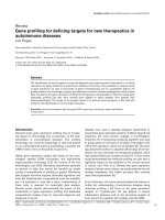
(Table 2 and Fig. 6). Covalent modification of C435 by
DTS (5) was confirmed by tryptic digestion and MS analysis
of the quadruple mutant treated with the inhibitor. The
DTS-labelled tryptic peptide comprising C435 has a theor-
etical molecular mass of 6895.8 Da. Deconvoluted mass
spectra (Fig. 7B) indicated a molecular mass of 6893.0 Da
and refined data analysis a molecular mass of 6894.2 Da for
the expected peptide recorded by scans nos 1481–1486. The
corresponding peak with elution time 47.85 min was the
most prominent peak of the chromatogram (Fig. 7A). A
difference between the theoretical and the experimental
values of 1.6 Da can be tolerated due to the systematic error
of the mass spectrometer (0.01–0.05%). Even if an incom-
plete tryptic digest of SHC with up to two missed cleavages
is taken into account, it can be ruled out that other peptides
caused the observed MS signals, because potential tryptic
peptides are residues 403–466 (one missed cleavage,
7042.6 Da instead of the observed value 6894.2 Da), the
unlabelled peptide containing C435 (6682.2 Da) and resi-
dues 274–332 (two missed cleavages, 6500.4 Da). Further-
more, due to the high intensity of the chromatogram peak
assigned to the labelled peptide it is unlikely that peptides
originating from unspecific cleavage of the labelled SHC
and having a molecular mass close to that of the labelled
peptide contributed to the deconvolution peak. Therefore
the identification of the labelled peptide could be interpreted
as unambiguous.
Binding of a squalene analogue inside the active centre
Labelling a specific amino-acid residue at the active centre
with a substrate analogue is a crucial step in studying the
Table 2. Inactivating effect t
½
of thiol-modifying inhibitors on enzy-
matic activity of mutant C435S and quadruple mutant C25S/C50S/
C455S/C537S with squalene as substrate. Experimental conditions
were as described in Materials and methods. Data are mean values
from three independent experiments with a mean deviation of ± 10%.
Inhibitor
Inhibitor
concentration
(l
M
)
t
½
(min)
C435S
C25S/C50S/
C455S/C537S
Dodecyl-maleimide (3) 500 > 60 30
Squalene-maleimide (2) 200 > 60 45–60
CPTO (4) 200 > 60 23
DTS (5) 100 45 4.5
DTS (5) 20 > 60 22
Fig. 6. Half-logarithmic plot of time-dependent inactivation of mutant
C25S/C50S/C455S/C537S by DTS (5). Data are shown for DTS (5)
concentrations of 100 l
M
(r)and20l
M
(j).
Fig. 7. LC-ESI-MS results: RP-HPLC separation of tryptic digested
mutant C25S/C50S/C455S/C537S labelled with DTS (5). Elution time
[min] is denoted above the peaks (A). Deconvoluted spectra of ESI-MS
scans nos 1481–1486 corresponding to the elution time 47.85 min (B).
Ó FEBS 2002 Effects of SH-modifying agents on squalene cyclase (Eur. J. Biochem. 269) 2113
complex interaction between substrate and enzyme. This
approach was successfully adopted with yeast oxidosqua-
lene cyclase, which was irreversibly inhibited by CNDT-
squalene (1), a thiol-reacting squalene-like molecule [24].
SHCs, unlike OSCs, bear no cysteine residues at the active
centre, as they lack the cysteine residue present in the
conserved active site motif DCTA of eukaryotic OSCs (in
prokaryotic cyclases the motif is DDTA). No other cysteine
residues appear at the active centre cavity of SHC. The goal
of binding a squalene analogue inside the active site of SHC
covalently may thus be pursued by introducing a critical
cysteine residue into the active centre cavity by site-directed
mutagenesis, able to serve as a Ôsticky pointÕ for squalene
analogues with thiol-modifying activity. The D376C/C435S
mutant, already used in crystallization experiments of SHC
[5], seemed to fit the above purpose. In this mutant, the
D376 at the reaction initiation site of the central cavity has
been replaced by a cysteine residue, and C435 at the channel
constriction has been replaced by serine. The latter substi-
tution was required to allow thiol-reacting molecules to
move to their target located in the active site cavity, without
interacting with the cysteine residue at the channel constric-
tion.
A series of thiol-reacting agents were tested as time-
dependent inhibitors of the D376C/C435S mutant. The
SHC mutant C435S was used as control. Mutant D376C/
C435S lost the ability to cyclize squalene due to the absence
of one of the two aspartate residues in the crucial DDTA
motif [25], but it still shared the ability to cyclize oxido-
squalene with wild-type SHC [23] and with the control
C435S mutant. Therefore, the inhibitory properties of thiol
reagents were assayed towards the oxidosqualene cyclizing
activity. First, enzymatic activity of the mutants with
oxidosqualene as a substrate, was characterized and
compared with wild-type enzyme. Both the D376C/C435S
and the C435S mutants showed the same temperature
profile (data not shown) for oxidosqualene cyclizing activ-
ity, with maximum activity at 60 °C, coincident with the
optimal temperature for squalene cyclization by wild-type
SHC [3]. Comparison of kinetic parameters showed that the
double mutant was less efficient in cyclizing oxidosqualene
than either the wild-type or the mutants bearing the native
DDTA motif (Table 1).
All thiol-reacting agents tested had a stronger inhibitory
effect on the D376C/C435S mutant, which was time-
dependent, than they did on the C435S mutant with intact
initiation site motif DDTA (Table 3). The reactive sub-
strate-analogue squalene-maleimide appeared to be the
most effective inhibitor of the D376C/C435S mutant
bearing a cysteine residue at the active site.
DISCUSSION
A series of thiol modifying agents were used as tools to
elucidate some structural/functional features of squalene-
hopene cyclase. The study started from the observation that
C435 of the enzyme, is located on the putative path of the
substrate from the membrane interior to the active centre.
C435 is in fact located at a constriction formed by four
amino-acid residues, which separates the large central cavity
containing residues critical for catalysis from a lipophilic
channel open towards the inner surface of the protein
(Fig. 2). This constriction appears to be sufficiently mobile
to act as a gate for substrate passage, due to the flexibility of
a loop bearing two of the four amino-acid residues as
indicated by the higher crystallographic B-factors [5]. The
ability of the inhibitor CNDT-squalene (1)tomodifyC435
covalently provided the first evidence that a substrate
analogue can move along the lipophilic channel and reach
the enzyme’s putative gate-constriction, establishing that
this is in fact the entrance to the active centre.
Even stronger support for the position of the entrance
gate came from the inactivating experiments with the C25S/
C50S/C455S/C537S mutant, which bears C435 as the only
cysteine of the protein. When this SHC-mutant was exposed
to thiol-modifying agents, especially to the inhibitor DTS
(5), rapid time-dependent inhibition was observed (Fig. 6).
Such inhibition can be explained as a consequence of an
obstruction of the channel constriction. Other explanations,
such as a direct influence of the inhibitor on the catalytic
process, may be ruled out since C435 is not involved in the
reaction mechanism as shown by the essentially unchanged
activity of the C435S mutant compared to wild-type
(Table 1). Moreover, when the inhibitors could in principle
overcome the barrier of the channel constriction and act
directly in the active site cavity, as in the case of the C435S
mutant, poor inhibition effect was observed (Table 2).
Interestingly, the inhibitors squalene- and dodecyl-malei-
mide, which modify thiol residues with a group bearing a
mobile and flexible chain, proved to be less active than
CPTO (4)andDTS(5).Thereasonforthedifferent
effectiveness of the inhibitors could, of course, simply result
from their different reactivity or, as in the case of DTS (5),
from some enhancement effect due to the formation of
reactive intermediates during the reaction between the intact
inhibitor and thiol groups [26,27]. Moreover, a difference
depending on size and rigidness of groups modifying C435
may occur.
The series of thiol-reacting agents used to study access of
the substrate to the active site of SHC was also found to be a
useful tool for studying the possibility of forming an
enzyme–inhibitor complex with the inhibitor covalently
bound inside the active centre cavity. The mutant used for
the study was the D376C/C435S mutant bearing a cysteine
residue at the reaction initiation site in the central cavity and
a serine substituting the cysteine at the channel constriction.
This mutant is unable to cyclize squalene [25], but recognizes
2,3-oxidosqualene as a substrate, an ability shared with
wild-type SHC and with mutants bearing at least one of the
two aspartate residues of the conserved sequence DDTA
Table 3. Inactivating effect t
½
of thiol-modifying inhibitors on enzy-
matic activity of mutant C435S and mutant D376C/C435S with
oxidosqualene as substrate. Experimental conditions were as described
in Materials and methods. Data are mean values from three inde-
pendent experiments with a mean deviation of ± 10%.
Inhibitor
Inhibitor
concentration
(l
M
)
t
½
(min)
C435S D376C/C435S
Dodecyl-maleimide (3) 200 > 60 12
Squalene-maleimide (2) 200 > 60 1.5
CPTO (4) 200 > 60 36
DTS (5) 200 45 7.5
2114 P. Milla et al. (Eur. J. Biochem. 269) Ó FEBS 2002
[28]. Recently, the preference for oxidosqualene of such
mutants was confirmed with a SHC mutant bearing the
eukaryotic DCTAEA OSC-motif instead of the DDTAVV
SHC-motif [28]. The exchange of the prokaryotic by the
eukaryotic motif was carried out in [23].
The strong time-dependent inactivation of inhibitors on
the D376C/C435S mutant, and the poor inhibition of the
C435S mutant, indicate that inactivation occurs at C376.
Interestingly, squalene-maleimide (2) [11] which should have
a higher ability to deliver a reactive group to the squalene-
hosting cavity of the enzyme, proved to be the most
powerful inactivating agent. It appears possible that, for the
first time, a covalent SHC–inhibitor complex has been
formed, in which a squalene analogue in a noncyclized form
is bound to the squalene-hosting cavity covalently. These
results open the fascinating prospect of solving the structure
of a SHC with a complexed squalenoid molecule, and
determining in detail the interactions between residues at the
active site and the squalene skeleton in its not-yet cyclized
conformation.
In summary, rationally designed mutants of SHC, C25S/
C50S/C455S/C537S (Ôquadruple mutantÕ)andD376C/
C435S, obtained by site-directed mutagenesis, have been
effectively inhibited by thiol-reacting molecules DTS (5)and
squalene-maleimide (2) in a time-dependent manner. Ex-
perimental evidence suggests that DTS (5) inhibits the
quadruple mutant by obstructing the substrate access to the
active site, while squalene-maleimide (2) inactivates the
D376C/C435S mutant by irreversible occupation of the
active centre cavity. These results, in particular those
relating to the D376C/C435S mutant, may be regarded as
the first step towards the goal of preparing stable covalent
complexes for structural analysis.
ACKNOWLEDGEMENTS
This work was supported by the Ministero dell’Universita
`
edella
Ricerca Scientifica e Tecnologica (MURST), Italy (ex 60%), and by the
Deutsche Forschungsgemeinschaft under SFB-388. The authors thank
Prof. K. Poralla (Universita
¨
tTu
¨
bingen) for providing wild-type SHC,
D. Kessler and B. Fu
¨
llgrabe for help with mutagenesis and C. Warth
for ESI-MS measurements.
REFERENCES
1. Abe, I., Rohmer, M. & Prestwich, G.D. (1993) Enzymatic cycli-
zation of squalene and oxidosqualene to sterols and triterpenes.
Chem. Rev. 93, 2189–2206.
2. Nes, W.D. (1990) Control of sterol biosynthesis and its import-
ance to developmental regulation and evolution. Recent Adv.
Phytochem. 242, 283–327.
3. Ourisson, G., Rohmer, M. & Poralla, K. (1987) Characterization
and partial purification of squalene-hopene cyclase from Bacillus
acidocaldarius. Biochim. Biophys. Acta 881, 356–363.
4. Wendt, K.U., Poralla, K. & Schulz, G.E. (1997) Structure and
function of squalene cyclase. Science 277, 1811–1815.
5. Wendt, K.U., Lenhart, A. & Schulz, G.E. (1999) The structure
of the membrane protein squalene-hopene cyclase at 2 A
˚
resolu-
tion. J. Mol. Biol. 286, 175–187.
6. Picot, D., Loll, P.J. & Garavito, R.M. (1994) The X-ray crystal
structure of the membrane protein prostaglandin H
2
synthase-1.
Nature 367, 243–249.
7. Luong, C., Miller, A., Barnett, J., Chow, J., Ramesha, C. &
Browner, M.F. (1996) Flexibility of the NSAID binding site in the
structure of human cyclooxygenase-2. Nature Struct. Biol. 3, 927–
933.
8. Blobel, G. (1980) Intracellular protein topogenesis. Proc. Natl
Acad.Sci.USA77, 1496–1500.
9. Nicholls, A., Sharp, K.A. & Honig, B. (1991) Protein folding and
association: insight from the interfacial and thermodynamic
properties of hydrocarbons. Proteins: Struct. Funct. Genet. 11,
281–296.
10. Duriatti, A., Bouvier-Nave
´
, P., Benveniste, P., Schuber, F.,
Delprino, L., Balliano, G. & Cattel, L. (1985) In vitro inhibition
of animal and higher plants, 2,3-oxidosqualene-sterol cyclases
by 2-aza-2,3-dihydrosqualene and derivatives and by other
ammonium-containing molecules. Biochem. Pharmacol. 34, 2765–
2777.
11. Grosa, G., Viola, F., Ceruti, M., Brusa, P., Delprino, L., Dosio, F.
& Cattel, L. (1994) Synthesis and biological activity of a squale-
noid maleimide and other classes of squalene derivatives as irre-
versible inhibitors of 2,3-oxidosqualene cyclase. Eur. J. Med.
Chem. 29, 17–23.
12. Field, R.B., Holmlund, C.E. & Whittaker, N.F. (1979) The effects
of the hypocholesteremic compound 3 b-(b-dimethylaminoeth-
oxy) -androst-5-en-17-one on the sterol and steryl ester composi-
tion of Saccharomyces cerevisiae. Lipids 14, 741–747.
13. Caujolle, R., Favrot, J.D., Loiseau, P.R., Payard, M., Amarouch,
H., Lazrek, H., Seguela, J.P., Loiseau, P.M., Bories, C. & Gayral,
P. (1991) Synthe
`
se, activite
´
antiparasitaire et antifongique d’aryl-2-
thiazolines. Pharm. Acta Helv. 66, 237–240.
14. Popjak, G. (1969) Enzymes of sterol biosynthesis in liver and
intermediates of sterol biosynthesis. Methods Enzymol. XV, 393–
454.
15. Ceruti, M., Grosa, G., Rocco, F., Dosio, F. & Cattel, L. (1994) A
convenient synthesis of [3-
3
H]-squalene and [3-
3
H]-oxidosqualene.
J. Labelled Compounds Radiopharmaceuticals 34, 577–585.
16. Balliano, G., Viola, F., Ceruti, M. & Cattel, L. (1992) Char-
acterization and partial purification of squalene-2,3-oxide cyclase
from Saccharomyces cerevisiae. Arch. Biochem. Biophys. 293, 122–
129.
17.Ceruti,M.,Balliano,G.,Viola,F.,Cattel,L.,Gerst,N.&
Schuber, F. (1987) Synthesis and biological activity of azasqua-
lenes, bis-azasqualenes and derivatives. Eur. J. Med. Chem. 22,
199–208.
18. Ochs,D.,Kaletta,C.,Entian,K.D.,Beck-Sickinger,A.&Poralla,
K. (1992) Cloning, expression, and sequencing of squalene-hopene
cyclase, a key enzyme in triterpenoid metabolism. J. Bacteriol. 174,
298–302.
19. Wendt, K.U., Feil, C., Lenhart, A., Poralla, K. & Schulz, G.E.
(1997) Crystallization and preliminary X-ray crystallographic
analysis of squalene-hopene cyclase from Alicyclobacillus acid-
ocaldarius. Protein Sci. 6, 722–724.
20. Barik, S. (1993) Site-directed mutagenesis by double polymerase
chain reaction. In PCR Protocols; Current Methods and Applica-
tions (White, B.A., ed.), pp. 277–286. Humana Press, Totowa,
New Jersey.
21. Sakar, G. & Sommer, S.S. (1990) The ÔmegaprimerÕ method of site-
directed mutagenesis. Biotechniques 8, 404–407.
22. Sambrook, J., Fritsch, E.F. & Maniatis, T. (1989) Molecular
Cloning: a Laboratory Manual, 2nd edn. Cold Spring. Harbor
Laboratory Press, Cold Spring Harbor, New York.
23. Dang, T. & Prestwich, G.D. (2000) Site-directed mutagenesis of
squalene-hopene cyclase: altered substrate specificity and product
distribution. Chem. Biol. 7, 463–469.
24. Balliano, G., Grosa, G., Milla, P., Viola, F. & Cattel. L. (1993)
3-Carboxy-4-nitrophenyl-dithio-1,1¢,2-trisnorsqualene: a site-direc-
ted inactivator of yeast oxidosqualene cyclase. Lipids 28, 903–906.
25. Feil, C., Su
¨
ssmuth, R., Jung, G. & Poralla, K. (1996) Site-directed
mutagenesis of the putative active-site residues in squalene-hopene
cyclase. Eur. J. Biochem. 242, 51–55.
Ó FEBS 2002 Effects of SH-modifying agents on squalene cyclase (Eur. J. Biochem. 269) 2115
26. Kice, J.L. & Cleveland, J.P. (1973) Nucleophilic substitution
reaction involving sulfenic acids and sulfenyl derivatives. The
nucleophile- and acid-catalyzed oxygen-18 exchange of phenyl
benzenthiosulfinate. J. Am. Chem. Soc. 95, 104–109.
27. Oae, S. & Okuyama, T. (1992) Organic Sulfur Chemistry: Bio-
chemical Aspects. CRC Press Inc. Boca Raton, Florida.
28. Sato, T. & Hoshino, T. (1999) Functional analysis of the
DXDDTA motif in squalene-hopene cyclase by site-directed
mutagenesis experiments: initiation site of the polycyclization
reaction and stabilization site of the carbocation intermediate of
the initially cyclized A-ring. Biosci.Biotechnol.Biochem.63, 2189–
2198.
2116 P. Milla et al. (Eur. J. Biochem. 269) Ó FEBS 2002