Báo cáo y học: "Clinical and neuropathological study about the neurotization of the suprascapular nerve in obstetric brachial plexus lesions" potx
Bạn đang xem bản rút gọn của tài liệu. Xem và tải ngay bản đầy đủ của tài liệu tại đây (481.78 KB, 11 trang )
BioMed Central
Page 1 of 11
(page number not for citation purposes)
Journal of Brachial Plexus and
Peripheral Nerve Injury
Open Access
Research article
Clinical and neuropathological study about the neurotization of the
suprascapular nerve in obstetric brachial plexus lesions
Dominique Schaakxs*
1
, Jörg Bahm
2
, Bernd Sellhaus
1
and Joachim Weis
1
Address:
1
Institute for Neuropathology, Klinikum RWTH Aachen, Germany and
2
Euregio Reconstructive Microsurgery Unit, Franziskushospital
Aachen, Germany
Email: Dominique Schaakxs* - ; Jörg Bahm - ;
Bernd Sellhaus - ; Joachim Weis -
* Corresponding author
Abstract
Background: The lack of recovery of active external rotation of the shoulder is an important
problem in children suffering from brachial plexus lesions involving the suprascapular nerve. The
accessory nerve neurotization to the suprascapular nerve is a standard procedure, performed to
improve shoulder motion in patients with brachial plexus palsy.
Methods: We operated on 65 patients with obstetric brachial plexus palsy (OBPP), aged 5-35
months (average: 19 months). We assessed the recovery of passive and active external rotation
with the arm in abduction and in adduction. We also looked at the influence of the restoration of
the muscular balance between the internal and the external rotators on the development of a
gleno-humeral joint dysplasia. Intraoperatively, suprascapular nerve samples were taken from 13
patients and were analyzed histologically.
Results: Most patients (71.5%) showed good recovery of the active external rotation in abduction
(60°-90°). Better results were obtained for the external rotation with the arm in abduction
compared to adduction, and for patients having only undergone the neurotization procedure
compared to patients having had complete plexus reconstruction. The neurotization operation has
a positive influence on the glenohumeral joint: 7 patients with clinical signs of dysplasia before the
reconstructive operation did not show any sign of dysplasia in the postoperative follow-up.
Conclusion: The neurotization procedure helps to recover the active external rotation in the
shoulder joint and has a good prevention influence on the dysplasia in our sample. The nerve quality
measured using histopathology also seems to have a positive impact on the clinical results.
Background
Brachial plexus lesions during birth affect one in 2000
newborns [1]. Ten percent of them need early or second-
ary surgical reconstruction [1]. In the treatment of obstet-
ric brachial plexus lesions, one of the main problems is
the poor recovery of abduction and external rotation in
the shoulder joint [2].
In children with upper and total brachial plexus lesions,
the suprascapular nerve, the first motor branch of the
upper trunk located in the center of the obstetric brachial
plexus lesion, is usually affected. The clinical manifesta-
tion is the lack of active external rotation in the gleno-
humeral joint. The child adopts an internal rotation
position and might be restricted in many activities such
Published: 11 September 2009
Journal of Brachial Plexus and Peripheral Nerve Injury 2009, 4:15 doi:10.1186/1749-7221-4-15
Received: 24 May 2009
Accepted: 11 September 2009
This article is available from: />© 2009 Schaakxs et al; licensee BioMed Central Ltd.
This is an Open Access article distributed under the terms of the Creative Commons Attribution License ( />),
which permits unrestricted use, distribution, and reproduction in any medium, provided the original work is properly cited.
Journal of Brachial Plexus and Peripheral Nerve Injury 2009, 4:15 />Page 2 of 11
(page number not for citation purposes)
as: eating, writing, dressing or combing their hair. Some of
them develop a "trumpet sign" posture, indicating that
elbow flexion is executed with an abducted arm and a pro-
nated forearm, the supination of the forearm being lim-
ited. The lack of external rotation can lead to secondary
soft tissue contractures, deformities in the shoulder joint
such as a posterior subluxation together with an enhanced
retroversion of the humeral head and various glenoid
deformities. An important point is to restore the external
rotation in order to prevent these deformities [1,3].
The main goal of the plexus reconstruction is the recovery
of the motor and sensory functions of the hand, as well as
elbow flexion, shoulder stability and motion. Many mus-
cles are involved in the shoulder motion, mainly control-
led by four nerves (axillary nerve, deltoid muscle:
abduction; suprascapular nerve, supraspinatus and infra-
spinatus muscles: external rotation of the humerus and
abduction in the supraspinatus muscle; dorsal scapular
nerve, rhomboidei muscles and long thoracic nerve, sera-
tus anterior muscle: scapular stabilization). The natural
balance between the lateral (infraspinatus and suprasp-
inatus muscle) and the medial rotators (latissimus dorsi,
teres major, subscapularis, pectoralis major muscles)
favors the internal rotation [1].
In our study, we wanted to answer the following ques-
tions:
1. Can we get a good recovery of the active external
rotation after the spinal accessory nerve neurotization
to the suprascapular nerve? What could be the reasons
for insufficient results?
2. Does neurotization of the suprascapular nerve
reduce the amount of shoulder dysplasia seen by
allowing the recovery of muscle balance between the
internal and external rotators? Could an existing dys-
plasia be treated? Is it possible through this procedure
to prevent the development of a shoulder dysplasia?
3. Is there a correlation between the quality criteria of
the nerves involved in the reconstruction measured by
the histopathology (morphometry and microscopic
qualitative analysis) and the clinical results? Is it pos-
sible to identify clinical prognostic factors with the
analysis of these parameters?
Methods
We examined 65 patients (37 girls and 28 boys) who
required brachial plexus reconstruction between 2001
and 2007. We operated on all 65 patients at ages ranging
between 5 and 35 months (average: 19 months) and
assessed their recovery for a mean postoperative observa-
tion period of 2.5 years.
Surgical techniques
Our 65 patients presented varying grades of severity of
obstetric brachial plexus lesions involving the suprascapu-
lar nerve. Depending on lesion severity, 3 groups of
patients were operated on using different surgical proce-
dures:
1. Accessory nerve neurotization to the suprascapular
nerve using the dorsal approach (N = 38). All patients
in this group presented an upper brachial plexus palsy.
2. Accessory nerve neurotization to the suprascapular
nerve and neurolysis of the other cervical nerve roots:
ventral approach (N = 6). All patients in this group
presented an upper brachial plexus palsy.
3. Plexus reconstruction on patients with complete
brachial plexus lesion and accessory nerve neurotiza-
tion to the suprascapular nerve: ventral approach (N =
21). Out of these patients, 10 presented a lesion of C5-
C7 and 11 had a total brachial plexus palsy.
The dorsal approach has been described previously [1].
For a ventral approach, the patient was placed in a supine
position under general anesthesia and orotracheal intuba-
tion. A 4 cm horizontal incision was made laterally begin-
ning at the border of the sternocleidomastoideus muscle.
The subcutaneous tissue and the platysma were divided
and then the adipolymphatic tissue was dissected. The
jugularis vein, the carotis and the phrenic nerve were iden-
tified. Then, on the scalenus anterior muscle, the phrenic
nerve was stimulated. The dissection was carried out far
enough proximally (root C4) and distally (under the clav-
icle) according to the extent of the lesion to expose the
brachial plexus. The trunks and the roots of the brachial
plexus down to their foramen were progressively identi-
fied and individualized by rubber loops [4,5]. In case of
neuroma in continuity, a neurolysis can be performed to
release the intraneural pressure caused by the scar tissue
and favor a good recovery in patients with Erb's palsy
[6,7]. Functional recovery was assessed using electrical
stimulation. After that, the neurotization of the supras-
capular nerve was performed. The suprascapular nerve
was followed close to its emergence from the upper trunk
and cut. The accessory nerve was followed as distal as pos-
sible, cut and the proximal collaterals were spared to pro-
tect the horizontal trapezius function. Neuropathology
samples were taken and a classic epineural repair by 10-0
sutures or by fibrin glue was performed as distal as possi-
ble to reduce the reinnervation time [1]. In case of a sim-
ple neurotization, using a dorsal or ventral approach, no
cast was needed but only 10 days of immobilization with
the elbow against the body.
Journal of Brachial Plexus and Peripheral Nerve Injury 2009, 4:15 />Page 3 of 11
(page number not for citation purposes)
In the complete plexus reconstruction including an acces-
sory nerve neurotization (ventral approach), the patient
was in a supine position, under general anesthesia and
orotracheal intubation. The same operative procedure as
described above was used to expose the brachial plexus.
The topographic anatomy of the different brachial plexus
branches was exposed by using electrostimulation and
assessing the muscular motor response. When no motor
response was obtained, it is a sign that the nerve or its
roots are non conducting [7].
In case of severe upper brachial plexus lesions, reconstruc-
tive priorities must be defined. The main goal is the recov-
ery of elbow flexion and shoulder stability. In the
exploration of the damaged brachial plexus, several plexus
reconstruction options are available: the neurolysis, the
intraplexal nerve suture (with or without nerve graft) and
the nerve transfer. The choice of the reconstruction tech-
niques is individual and depends on the intraoperative
findings [7]. In general, better results are obtained with
neuroma resection and nerve transplantation than with
the neurolysis [6,8]. When the nerve is ruptured, an autol-
ogous nerve graft is used. The sural nerve is most often
used as donor nerve [7]. The neuroma parts were removed
and nerve samples were taken for the neurohistopathol-
ogy. The sural nerve parts were taken and an interfascicu-
lar transplantation was performed. The coaptation is
performed under microscope by 10/0 sutures or fibrin
glue. Then the accessory nerve neurotization to the supras-
capular nerve was performed as described above. The skin
was closed, and a handmade well-padded head and neck
plaster was used, which was worn for a period of 3 weeks.
Clinical examination methods
We studied the recovery of the active external rotation and
the issue of the shoulder dysplasia.
We assessed the recovery of external rotation using the
range of motion method. Only clinical examination was
used, without any device-assisted diagnostic procedure.
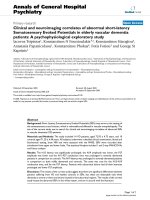
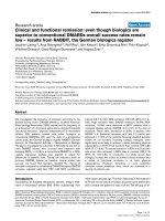
Different parameters were measured: active and passive
external rotation in adduction and in abduction as well as
a part of the Mallet score (hand to mouth and hand to
head, see Table 1) to assess the functional recovery. For
the external rotation in adduction, the neutral position
(0°) is with the arm along the lateral chest and the fore-
arm forming a 90° angle with the arm and pointing for-
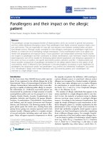
wards (see Figure 1). For the external rotation in
abduction, the neutral position (0°) is with the arm in
90° abduction and the forearm at a 90° angle with the
arm and pointing forwards (see Figure 2).
The glenohumeral joint was also assessed to observe the
presence or absence of dysplasia and the impact on the
dysplasia of the restoration of the muscle balance (lateral
and medial rotators) by means of the neurotization pro-
cedure. The mean follow-up period of 2.5 years provides
a good indication of the impact of reconstruction on the
proper development of the glenohumeral joint, although
an additional follow-up after 5 years would be desirable
in order to confirm the results. We analyzed the gleno-
humeral joint clinically without using magnetic reso-
nance imaging (MRI). Although MRI would have been
useful from a radiological point of view, it was not possi-
ble to carry out this test consistently on a wide sample of
young children as it requires general anesthesia, which
parents would not have accepted without therapeutical
justification. For this reason, we focused on the clinical
examination of the joint, checking that there was no
major deformity. We assessed the shoulder joint by meas-
uring the range of motion, assessing the presence of con-
tractures and the articular mobility. We stabilized the
scapulothoracic joint with one hand and used the other
hand to assess the glenohumeral joint external rotation
[9]. In case of severe dysplasia, there is an audible "click"
during the examination of the passive external rotation
and a reduction of the mobility. The literature shows a
strong correlation between clinical measures and the pres-
ence of dysplasia, detected by MRI [9]. Strongly reduced
passive glenohumeral external rotation motion and the
presence of internal rotation contracture are indicators of
underlying joint deformity.
Table 1: Mallet scoring
01 2
Hand to nape of neck impossible difficult Easy
Hand to mouth impossible difficult (trumpet sign) Easy
Active external rotation in adductionFigure 1
Active external rotation in adduction.
Journal of Brachial Plexus and Peripheral Nerve Injury 2009, 4:15 />Page 4 of 11
(page number not for citation purposes)
Neurohistopathology and morphometry
During the surgical procedures, we took nerve samples of
the suprascapular nerve from 13 patients for neurohis-
topathology. Analyses by microscope and morphometry
were carried out at Institute for Neuropathology (Head:
Univ. Professor Dr J.Weis), Klinikum RWTH Aachen.
Unfortunately, only 11 patients could be compared for
clinical and neurohistopathology results due to sample
attrition.
Coloration
Semithin nerve sample sections were obtained by using
paraphenylenediamin and toluidine-blue staining to vali-
date the structural details.
Morphometry
1 μm semithin sections of the suprascapular nerve from
13 patients were observed under microscope (100×) in oil
immersion. We used a KS 300 automatic, optical-elec-
tronic digital evaluation system to measure 2 fields per
section. All nerve fibers were marked manually, excluding
the fibers which were located on the edges of the sample
as well as those which were incomplete or had artifacts.
For all the fibers that had been marked, various parame-
ters per field were measured: myelin surface (μm
2
), total
nerve fiber surface (μm
2
), axon surface (μm
2
), myelin
diameter (μm), axon diameter (μm), total nerve fiber
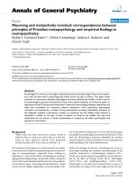
diameter (μm) and number of fibers. Figure 3 shows a
part of this marking process.
Quality criteria of the suprascapular nerve
We measured the following parameters from the endone-
urium using the morphometry, and calculated different
ratios to assess the nerve quality:
- The M/A ratio (surface of myelin (μm
2
)/surface of
axon (μm
2
)) gives an indication of the thickness of the
myelin in the axon.
- The G-ratio (axon diameter (μm)/total nerve fiber
diameter (μm)) is often used in the literature indicat-
ing the degree of myelinization of the axon. Normal
values are comprised between 0.5 and 0.7 [10].
- The ratio between the surface of the axon (μm
2
) and
the total surface of nerve fiber (μm) corresponds to the
proportion of axon material in the nerve fascicle. This
ratio is comparable to the G-ratio but is more precise,
because the shape of the nerve fiber is not exactly
round. For this reason, the diameter only gives an
approximation of the relative surface in the nerve fas-
cicle.
- The ratio between the surface of myelin (μm
2
) and
the total surface of the observed nerve sample (μm
2
)
indicates the proportion of myelin in the total
observed nerve sample.
- The ratio between the total surface of nerve fiber
(μm
2
) and the total surface of the observed nerve sam-
ple (μm
2
) indicates the proportion of nerve fiber in
the total nerve sample.
Under the microscope, we observed the following qualita-
tive criteria of the suprascapular nerve [5]:
Active external rotation in abductionFigure 2
Active external rotation in abduction.
Morphometry marking processFigure 3
Morphometry marking process. Nerve fibers are
marked in color. Fibers colored in yellow were selected
manually, excluding incomplete fibers located on the edges of
the sample or fibers that presented artifacts. Fibers marked
in other colors were selected automatically by the KS 300
optical-electronic digital evaluation system.
Journal of Brachial Plexus and Peripheral Nerve Injury 2009, 4:15 />Page 5 of 11
(page number not for citation purposes)
- number and orientation of nerve fascicles
- presence of peri- or endoneural fibrosis
- remnants of nerve degeneration (clusters of Schwann
cells called "Büngner" bands)
- indirect signs of reinnervation: presence of minifasci-
cles
- presence or absence of minifascicles in the perineu-
rium or epineurium, which are a sign of neuroma.
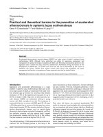
Figure 4 shows a suprascapular nerve with good endone-
ural regeneration and Figure 5 shows another suprascapu-
lar nerve with minifascicles in the perineurium, which
indicates the presence of a neuroma. The higher the pres-
ence of minifascicles in the perineurium and epineurium,
the more important is the neuroma. Therefore, the pres-
ence of minifascicles is an indicator of the lower quality of
the nerve involved in the neurorrhaphy, potentially com-
promising the clinical results.
Statistical evaluation
The clinical values for the external rotation are measured
in degrees, the minimum being 0° (no result) and the
maximum 90° (goal value). We assessed the clinical
parameters at different times and used a one-sample T-test
to show the postoperative improvement of the clinical
parameters.
We distinguished between the 3 procedure groups and
tried to show the influence of the primary reconstructive
procedure on the postoperative results by using a one-way
analysis of variance (ANOVA) procedure. Given the rela-
tively small sample and potentially non-normal distribu-
tion, we further confirmed this statistic using the non-
parametric rank-based Kruskal-Wallis test. In our clinical
examinations, we looked also for the presence of dyspla-
sia in the glenohumeral joint (before and after the proce-
dure). We used descriptive statistics and variance analysis
to show the influence of the dysplasia on the postopera-
tive results and frequency tables to assess the influence of
the neurotization (restoration of the muscle balance) on
the dysplasia.
The morphometry criteria were assessed using descriptive
statistics.
Results
Clinical results
We examined the children (N = 65) postoperatively at dif-
ferent times, which were statistically distributed in differ-
ent groups:
- 0-6 months after the procedure
- after 7-12 months
- after 13-18 months
- after 19-24 months
- after 25-36 months
- after 36 months or more
Suprascapular nerve showing good endoneural regenerationFigure 4
Suprascapular nerve showing good endoneural
regeneration.
Suprascapular nerve with minifascicles in the perineurium (*)Figure 5
Suprascapular nerve with minifascicles in the
perineurium (*).
Journal of Brachial Plexus and Peripheral Nerve Injury 2009, 4:15 />Page 6 of 11
(page number not for citation purposes)
Before the operation, all the patients presented an active
external rotation (in abduction and in adduction) close to
0°. In general, we observed an improvement of the clini-
cal values in the 3 years following the procedure, as shown
in Table 2.
A one-sample T-test confirmed the improvement of the
clinical parameters with all postoperative values being sig-
nificantly different from the initial value of 0° (p = 0.00).
We calculated the mean of the maximal values reached by
all the patients. The maximum goal value for the different
clinical parameters is 90° of rotation. For the active exter-
nal rotation in abduction parameter, 71.5% of patients
reached a value comprised between 60°-90°. For passive
external rotation in abduction, 96.6% of patients reached
70°-90°. For passive external rotation in adduction, 50%
of patients obtained a value between 60° and 90°. The
results for active external rotation in adduction were more
disappointing with only 18% of patients obtaining 60°-
90°, while 66% reported 0°-30°. The detailed frequencies
are shown in Table 3.
In general, we observe better results for the external rota-
tion in abduction than in adduction. Active external rota-
tion improves gradually up until 3 years following the
procedure, then stabilizes. Passive external rotation
decreases at first, maybe due to the immobilization fol-
lowing the procedure. Evolutions from one period to the
next should be considered with some caution as the
patients observed are not always the same for practical
reasons. We observe better results for passive than for
active values (see Table 4).
We sought an explanation for the unsatisfactory results
achieved by some patients in their individual follow-up
(active external rotation in abduction between 0° and
20°). Two patients could not obtain any active external
rotation in abduction. One of them had no active external
rotation in abduction 5.5 months after the procedure and
did not show up for the next examination. The other one,
who underwent a ventral approach with a neurolysis from
the trunci superior and medius and a neurotization of the
suprascapular nerve developed a shoulder contracture.
Three patients reached an active external rotation in
abduction comprised only between 0°-20° because they
developed a shoulder contracture. Another patient with a
heavy subtotal plexus lesion reached an active external
rotation in abduction of 10° eight months after the oper-
ation and did not come to the examination afterwards.
The Mallet score results also show marked improvement
over the initial values. For hand-to-mouth, the value
reached was an average of 1.90, with 10.2% of patients
reaching the value of 1 and 89.8% reaching the maximum
value of 2. For hand-to-head movement, 84.7% of
patients registered an improvement with an average max-
imum value of 1.39.
We also examined the influence of the type of the primary
reconstructive operation on the clinical results. We used a
one-way ANOVA procedure as well as a Kruskal-Wallis
test to test for differences in the clinical results in our 3
operation groups. We obtained significant differences
between the 3 operation groups for active external rota-
tion in abduction (ANOVA p = 0.026, Kruskal-Wallis p =
0.036), passive external rotation in abduction (ANOVA p
= 0.017, Kruskal-Wallis p = 0.048) and Mallet Score
parameters (ANOVA p = 0.000, Kruskal-Wallis p = 0.000
for both Hand-to-Mouth and Hand-to-Head). The
patients who underwent only the neurotization operation
obtained better results than the patients who had com-
plete plexus reconstruction. The descriptive statistics for
the maximum values reached for each clinical parameter
in each operation group are shown in Table 5. We did not
find any significant difference between the 3 groups for
parameters passive external rotation in adduction
(ANOVA p = 0.198, Kruskal-Wallis p = 0.471) and active
external rotation in adduction (ANOVA p = 0.447,
Table 2: Clinical result evolution (degrees of rotation achieved)
Average 0-6 months 7-12 months 13-18 months 19-24 months 25-36 months 36+ months Max. reached
active external
rotation (abduction)
36.79 47 58.61 64.67 70.37 70.27 67.54
passive external
rotation (abduction)
68.75 57.92 77.73 79.58 76.43 85.81 86.1
passive external
rotation (adduction)
77.5 56.67 48.33 47.08 48.93 53.57 56.61
active external
rotation (adduction)
40 16.25 47.5 25 24.29 28.75 28.1
Journal of Brachial Plexus and Peripheral Nerve Injury 2009, 4:15 />Page 7 of 11
(page number not for citation purposes)
Kruskal-Wallis p = 0.568). It should be cautioned that this
result is in need of verification as one of the three groups
(patients having undergone neurotization and neurolysis)
was much smaller (N = 6) than the other groups (N = 38
and 21, respectively).
In our clinical examinations, we also looked for the pres-
ence of dysplasia in the glenohumeral joint (pre and post-
operatively). The patients showed a muscular imbalance
between the external rotators and the internal rotators in
favor of the internal rotators. The neurotization operation
contributes to the restoration of the muscular balance in
the glenohumeral joint and should have a positive influ-
ence on the shoulder joint and therefore prevent the
development of a shoulder dysplasia. In our sample, 7
patients affected by dysplasia before the operation did not
show any sign of dysplasia in the postoperative follow-up.
Although this is a small sample, we observe a positive
influence from the reconstructive operation on the gleno-
humeral joint.
Histopathology results
We looked for tendencies in the relation between the his-
topathology and clinical results. The histopathology
results in our 13 samples were normal on average, as
shown in Table 6. In particular, the G Ratio (axon diame-
ter (μm)/total nerve fiber diameter (μm)) is often used in
the literature to indicate the degree of myelinization of the
axon. Normal values are comprised between 0.5 and 0.7
[10]. In our results, all the G Ratio values were contained
within this interval, with the exception of one value which
was very close to the normal range (0.48) and one patient
with a G Ratio value of 1.
We also assessed the number and orientation of the nerve
fascicles, the presence of perineural or endoneural fibro-
sis, signs of regeneration and the presence of minifascicles
in the perineurium or in the epineurium to check if these
criteria could influence the clinical results. Most patients
did not have minifascicles in the perineurium or in the
epineurium and showed signs of good endoneural regen-
eration and no sign of degeneration. Two of our patients
showed a very small presence of minifascicles in the
perineurium with signs of endoneural regeneration. Both
patients achieved good clinical results. One patient had a
very small presence of minifascicles in the perineurium
and in the epineurium, good endoneural regeneration
and achieved good clinical results as well. Only one
Table 3: Clinical result frequencies (maximum degrees of rotation achieved)
Maximum degrees
achieved
(Percent of patients)
Active external
rotation (Abduction)
Passive external
rotation (Abduction)
Active external
rotation (Adduction)
Passive external
rotation (Adduction)
0-10 6.3 0.0 36.0 3.6
11-20 3.2 0.0 8.0 3.6
21-30 6.3 0.0 22.0 16.1
31-40 4.8 1.7 8.0 7.1
41-50 6.3 1.7 8.0 17.9
51-60 6.3 0.0 8.0 10.7
61-70 12.7 6.8 0.0 12.5
71-80 9.5 6.8 6.0 10.7
81-90 44.4 83.1 4.0 17.9
Table 4: Comparison of maximum results achieved between exercise types
Patients achieving 90° rotation after 36 months Abduction Adduction
Active 37.8% 3.1%
Passive 81.1% 11.4%
Journal of Brachial Plexus and Peripheral Nerve Injury 2009, 4:15 />Page 8 of 11
(page number not for citation purposes)
patient, who presented an important lesion treated by
complete plexus reconstruction and the neurotization of
the suprascapular nerve showed an important neuroma
(high number of minifascicles in the perineurium and in
the epineurium) and a G Ratio value of 1. The clinical
results for this patient were insufficient in the follow-up,
leading us to suspect a problem of quality of the supras-
capular nerve involved in the neurorrhaphy with the
accessory nerve.
Discussion
The lack of recovery of active external rotation is an
important problem in children suffering from brachial
plexus lesions involving the suprascapular nerve. The res-
Table 5: Descriptive statistics for clinical parameters of different operation groups (ANOVA analysis)
N Mean Std. Deviation 95% Confidence Interval for Mean Min Max
Low High
Active external rotation
(abduction) - max. reached
Neurotisation N. XI/SSC 38 74.74 22.480 67.35 82.13 0 90
Neurotisation+
Neurolysis
6 61.67 33.116 26.91 96.42 0 90
Complete Plexus
reconstruction
19 55.00 29.627 40.72 69.28 10 90
Total 63 67.54 26.984 60.74 74.34 0 90
Passive external rotation
(abduction) max.
reached
Neurotisation N. XI/SSC 35 88.71 3.900 87.37 90.05 70 90
Neurotisation+
Neurolysis
6 77.50 17.819 58.80 96.20 45 90
Complete Plexus
reconstruction
18 83.89 12.897 77.48 90.30 40 90
Total 59 86.10 9.916 83.52 88.69 40 90
Passive external rotation
(adduction) max.
reached
Neurotisation N. XI/SSC 33 59.70 21.612 52.03 67.36 5 90
Neurotisation+
Neurolysis
6 40.83 28.358 11.07 70.59 10 80
Complete Plexus
reconstruction
17 56.18 24.656 43.50 68.85 20 90
Total 56 56.61 23.551 50.30 62.91 5 90
Active external rotation
(adduction) max.
reached
Neurotisation N. XI/SSC 30 31.17 25.281 21.73 40.61 0 90
Neurotisation+
Neurolysis
4 13.75 24.281 -24.89 52.39 0 50
Complete Plexus
reconstruction
16 25.94 30.068 9.92 41.96 0 90
Total 50 28.10 26.743 20.50 35.70 0 90
Hand-Head max.
reached
Neurotisation N. XI/SSC 36 1.67 .586 1.47 1.86 0 2
Neurotisation+
Neurolysis
6 .50 .548 07 1.07 0 1
Complete Plexus
reconstruction
17 1.12 .781 .72 1.52 0 2
Total 59 1.39 .743 1.20 1.58 0 2
Hand-Mouth - max.
reached
Neurotisation N. XI/SSC 36 2.00 .000 2.00 2.00 2 2
Neurotisation+
Neurolysis
6 1.50 .548 .93 2.07 1 2
Complete Plexus
reconstruction
17 1.82 .393 1.62 2.03 1 2
Total 59 1.90 .305 1.82 1.98 1 2
Journal of Brachial Plexus and Peripheral Nerve Injury 2009, 4:15 />Page 9 of 11
(page number not for citation purposes)
toration of the rotational balance between the internal
and external rotators is important to the good develop-
ment of the shoulder motion and to prevent deformities
of the glenohumeral joint in patients suffering from bra-
chial plexus palsy.
The standard procedure is the transfer of the distal branch
of the accessory nerve to the suprascapular nerve. The
indication for this procedure is the lack of active lateral
rotation in the glenohumeral joint without restriction of
passive external rotation (i.e. only internal rotation posi-
tion, but no joint contracture) for children younger than
2 years [1,11]. It has been shown in the literature that this
procedure provides an improvement in active external
rotation of the shoulder [1,2,12-17].
Our study contributes to the understanding of this prob-
lem in the following ways. Firstly, we confirm earlier
results and add finer detail by differentiating between
operative groups of differing lesion severity. We also
examined the cases for which no satisfactory results were
obtained. Secondly, we study the impact of the neurotiza-
tion procedure on the shoulder dysplasia. Lastly, we
examine the relation between the histopathology of the
nerve samples and the clinical results. In the literature,
most surgeons report better clinical results by using the
accessory nerve as donor, which provides enough motor
power, instead of grafts from the ruptured C5 root [5,18]
for the neurotization of the suprascapular nerve. Others
did not find any significant difference for the restoration
of true external rotation between nerve grafting from C5
and extraplexal nerve transfer using the accessory nerve,
but observed a slightly smaller passive range of motion
and a slightly stronger tendency to develop an internal
rotation contracture in the C5 graft group. In those cases,
the recovery of fair range of glenohumeral external rota-
tion was disappointingly low. However, these compensa-
tory techniques seem to contribute to reach a considerable
range of movement [16]. Other surgeons performed a
reconstruction of the suprascapular nerve by using a direct
neurotization with the accessory nerve or by using an
interposition nerve graft and obtained better results with
the direct neurotization [17].
In our sample, most patients obtained good recovery of
the external rotation in abduction. A possible explanation
for patients presenting good intraoperative conductivity
of the suprascapular nerve and good muscular response
but an insufficient recovery of the external rotation could
lie in insufficient cortical integration or some co-contrac-
tion patterns [1].
Better results were obtained for the external rotation in
abduction than for the external rotation in adduction. A
possible explanation for this finding is that in adduction,
strong internal rotator muscles (the subscapularis muscle,
the teres major, the latissimus dorsi and the pectoralis
major) work against this movement. In abduction, exter-
nal rotators (the strong infraspinatus, the teres minor, and
the posterior fibers of the deltoid) support the movement.
The main internal rotator (the subscapularis muscle) does
not act counter to the passive external rotation because it
is defunctioned as an internal rotator of the shoulder
when the arm is abducted to 90°. This is due to the ten-
don of the subscapularis being co-axial with the humerus
in that position and therefore unable to provide a force
vector producing internal rotation. We also observed bet-
ter results in the passive movement than in the active
movement and a positive correlation between both
parameters. The passive motion recovery is important to
obtain satisfactory active motion recovery because the
glenohumeral joint has to be free in order to allow the
improvement of the external rotation motion. In order to
free a blocked shoulder joint, other operative procedures
are used, such as an anterior release with coracoid short-
ening osteotomy and subscapular tendon lengthening [1].
In our 3 patient groups, we obtained better results for the
external rotation in abduction for the patients who only
underwent the neurotization operation than for patients
who had complete reconstruction, including the neuroti-
zation of the suprascapular nerve. We would expect to see
more restriction for the external rotation in the group
affected by Erb's palsy than in the group affected by total
palsy, because the medial rotators are equally affected in
the complete plexus lesion, and in this case there is never
an imbalance or a rotational contracture of the shoulder
[3]. A possible cause for the lack of external rotation recov-
ery in patients with complete palsy could be that the
supraspinatus muscle involving in the external rotation
movement, being the first to be reinnervated, attracts
more axons than the infraspinatus. Another possible
cause could be an unsatisfactory cortical integration of
this movement or the presence of some pathologic co-
contraction pattern. [1,14]
Table 6: Histopathological parameters of nerve samples
Mean Std. Dev.
G Ratio 0.59 0.07
Myelin surface/Axon surface 4.53 4.59
Axon surface/Total nerve surface 0.39 0.09
Myelin proportion in total sample 0.15 0.04
Nerve fiber proportion in total sample 0.25 0.08
Journal of Brachial Plexus and Peripheral Nerve Injury 2009, 4:15 />Page 10 of 11
(page number not for citation purposes)
In our clinical evaluation, we also looked at the impact of
the neurotization procedure on the dysplasia. In our sam-
ple, neurotization of the suprascapular nerve reduces the
amount of shoulder dysplasia observed by allowing the
recovery of muscle balance between the internal and
external rotators. Seven patients showing a dysplasia
before the neurotization operation no longer showed
signs of dysplasia in the postoperative follow-up. Further-
more, we observed that this procedure can prevent the
development of a dysplasia of the glenohumeral joint, as
no patient in our sample developed dysplasia postopera-
tively.
These results point to a positive relation between the his-
topathological quality of the nerves and the clinical
results of the procedure. This finding is naturally in need
of further empirical confirmation due to the small size of
the sample. However, these results are similar to previous
observations reported in the literature [15].
Conclusion
In conclusion, we believe the accessory nerve neurotiza-
tion to the suprascapular nerve is a safe and reliable pro-
cedure, which provides a good recovery of the active
external rotation and a positive influence on the shoulder
dysplasia development. Different points are important for
the success of the operation: (1) the evaluation of the
lesion, which is assessed by using electrostimulation dur-
ing the operative procedure, (2) the anastomosis between
the two nerves (so nerve quality needs to be assessed), (3)
the problem of the atrophy of the muscles (so the proce-
dure has to be carried out at an early age), (4) the devel-
opment of co-contraction patterns and shoulder dysplasia
and (5) training the movements in order to stimulate the
target muscles.
Abbreviations
N: nerve; M(m): muscle(s); μm: micrometer.
Consent
Written informed consent was obtained from the patient
for publication of this case report and accompanying
images. A copy of the written consent is available for
review by the Editor-in-Chief of this journal.
Competing interests
The authors declare that they have no competing interests.
Authors' contributions
DS participated in the design of the study, assisted on sur-
gical procedures, carried out clinical examinations and
morphometry measurements, did the statistical analysis
and drafted the manuscript. JB conceived of the study,
participated in its design and coordination, carried out the
surgery on the patients and the clinical examinations and
reviewed the manuscript. JB also obtained the informed
consent from the patients for participation in, and publi-
cation of, this study, including accompanying photo-
graphs, and is available to provide any additional
information in this regard to the Editor-in-Chief of this
journal. BS and JW participated in the design and coordi-
nation of the study and carried out the histopathology
analysis. All authors read and approved the final manu-
script.
Acknowledgements
The authors would like to thank the team of the Institute for Neuropathol-
ogy, Head: Univ Professor Dr J.Weis, Klinikum RWTH Aachen, Germany;
the team of the Euregio Reconstructive Microsurgery Unit, Head: Dr. J.
Bahm, Franziskushospital Aachen, Germany and the Institut für
Medizinische Statistik, Head: Univ Professor R D. Hilgers
References
1. Bahm J, Noaman H, Becker M: The Dorsal Approach to the
Suprascapular Nerve in Neuromuscular Reanimation for
Obstetric Brachial Plexus Lesions. Plastic and reconstructive sur-
gery 2005, 115:240-244.
2. Blaauw G, Slooff ACJ, Slooff WBM: Evaluation of accessory-
suprascapular nerve transfer in obstetric brachial plexus
paresis. Clinical Neurology and Neurosurgery 1997, 99:181-182.
3. Bahm J, Wein B, Alhares G, Dogan C, Radermacher K, Schuind F:
Assessment and treatment of glenohumeral joint deformi-
ties in children suffering from upper obstetric brachial
plexus palsy. Journal of Pediatric Orthopaedics 2007, 16:243-251.
4. Bahm JB, Becker M, Disselhorst-Klug C, Williams S, Meinecke L,
Müller H, Sellhaus B, Schröder JM, Rau G: Surgical strategy in
obstetric brachial plexus palsy: the Aachen experience. Sem-
inars in Plastic Surgery 2004, 18:285-299.
5. Bahm J, Ocampo-Pavez C, Noaman H: Microsurgical technique in
obstetric brachial plexus repair: a personal experience in 200
cases over 10 years. Journal of Brachial Plexus and Peripheral Nerve
Injury 2007, 2:1-7.
6. Clarke HM, Al-Qattan MM, Curtis CG, Zuker R: Obstetric Bra-
chial Plexus Palsy: Results Following Neurolysis of Conduct-
ing Neuromas-in-Continuity. American Society of Plastic Surgeons
1996, 97:974-982.
7. Krishnan KG, Mucha D, Pinzer T, Schackert G: Diagnostik und
Behandlung von posttraumatischen Läsionen des Plexus
brachialis. Ärzteblatt Sachsen 2003:373-381.
8. Borrero JL: Surgical technique. In Brachial Plexus Injuries 1st edi-
tion. Edited by: Gilbert A. London: Martin Dunitz; 2001:189-204.
9. Kozin SH: Correlation between external rotation of the
glenohumeral joint and deformity after brachial plexus birth
palsy. J Pediatr Orthop 2004, 24:189-193.
10. Wang J: Morphometric evaluation of paraneoplastic neuropa-
thies associated with carcinomas, lymphomas, Boeck's sar-
coidosis and dysproteinemias. Rheinisch-Westfälische
Technische Hochschule, Medizinischen Fakultät; 1997.
11. Bahm J: Secondary Procedures in Obstetric Brachial Plexus
Lesions. Handchir Mikrochir Plast Chir 2003, 36:37-46.
12. Bertelli JA, Ghizoni MF: Improved technique for harvesting the
accessory nerve for transfer in brachial plexus injuries. Neu-
rosurgery 2006, 58:366-370.
13. Malessy MJA, Godard C, de Ruitter W, de Boer KS, Thomeer RTWM:
Evaluation of suprascapular nerve neurotization after nerve
graft transfer in the treatment of brachial plexus traction
lesions. J Neurosurgery 2004, 101:377-389.
14. Bertelli JA, Ghizoni MF: Transfer of the Accessory Nerve to the
Suprascapular Nerve in Brachial Plexus Reconstruction. The
Journal of Hand Surgery 2007, 32A:989-998.
15. van Ouwerkerk WJR, Uitdehaag BMJ, Strijers RLM, Frans N, Holl K,
Fellner FA, Vandertop WP: Accessory nerve to suprascapular
nerve transfer to restore shoulder exorotation in otherwise
spontaneously recovered obstetrical brachial plexus lesions.
Neurosurgery 2006, 59:858-869.
Publish with BioMed Central and every
scientist can read your work free of charge
"BioMed Central will be the most significant development for
disseminating the results of biomedical research in our lifetime."
Sir Paul Nurse, Cancer Research UK
Your research papers will be:
available free of charge to the entire biomedical community
peer reviewed and published immediately upon acceptance
cited in PubMed and archived on PubMed Central
yours — you keep the copyright
Submit your manuscript here:
/>BioMedcentral
Journal of Brachial Plexus and Peripheral Nerve Injury 2009, 4:15 />Page 11 of 11
(page number not for citation purposes)
16. Pondaag w, de Boer R, van Wijlen-Hempel MS, Hofstede-Buitenhuis
SM, Malessy MJA: External rotation as a result of suprascapular
nerve neurotization in obtetric barchial plexus lesions. Neu-
rosurgery 2005, 57:530-537.
17. Terzis JK, Kostas I: Suprascapular Nerve Reconstruction in 118
Cases of Adult Postraumatic Brachial Plexus. Plastic Recon-
structive Surgerry 2006, 117:613-629.
18. Midha R: Nerve transfers for severe brachial plexus injuries: a
review. Neurosurgery Focus 2004, 16:1-10.