Báo cáo y học: "PKC and PKA Phosphorylation Affect the Subcellular Localization of Claudin-1 in Melanoma Cells"
Bạn đang xem bản rút gọn của tài liệu. Xem và tải ngay bản đầy đủ của tài liệu tại đây (1.56 MB, 9 trang )
Int. J. Med. Sci. 2009, 6
93
I
I
n
n
t
t
e
e
r
r
n
n
a
a
t
t
i
i
o
o
n
n
a
a
l
l
J
J
o
o
u
u
r
r
n
n
a
a
l
l
o
o
f
f
M
M
e
e
d
d
i
i
c
c
a
a
l
l
S
S
c
c
i
i
e
e
n
n
c
c
e
e
s
s
2009; 6(2):93-101
© Ivyspring International Publisher. All rights reserved
Research Paper
PKC and PKA Phosphorylation Affect the Subcellular Localization of
Claudin-1 in Melanoma Cells
Amanda D. French
1
, Jennifer L. Fiori
1 #
, Tura C. Camilli
1
, Poloko D. Leotlela
1
, Michael P. O’Connell
1
, Brittany
P. Frank
2
, Sarah Subaran
2
, Fred. E. Indig
2
, Dennis D. Taub
1
and Ashani T. Weeraratna
1
1. Laboratory of Immunology, National Institute on Aging, Baltimore, MD 21124, USA
2. Research Resources Branch, National Institute on Aging, Baltimore, MD 21124, USA
#
Present Address: Laboratory of Clinical Investigation, National Institute on Aging, Baltimore, MD 21124, USA
Correspondence to: Ashani T. Weeraratna, PhD, Laboratory of Immunology, National Institutes of Health, National In-
stitute on Aging, Biomedical Research Center, 251 Bayview Blvd, RM 08C226, Baltimore, Maryland 21224. Voice: (410)
558-8146; Fax: (410) 558-8284; Email:
Received: 2009.02.27; Accepted: 2009.03.12; Published: 2009.03.12
Abstract
Cytoplasmic expression of claudin-1 in metastatic melanoma cells correlates to increased
migration, and increased secretion of MMP-2 in a PKC dependent manner, whereas
claudin-1 nuclear expression is found in benign nevi. Melanoma cells were transfected with a
vector expressing CLDN-1 fused to a nuclear localization signal (NLS). Despite significant
nuclear localization of claudin-1, there was still transport of claudin-1 to the cytoplasm.
Phorbol ester treatment of cells transfected with NLS-claudin-1 resulted in an exclusion of
claudin-1 from the nucleus, despite the NLS. To ascertain whether PKC or PKA were in-
volved in this translocation, we mutated the putative phosphorylation sites within the pro-
tein. We found that mutating the PKC phosphorylation sites to mimic a non-phosphorylated
state did not cause a shift of claudin-1 to the nucleus of the cells, but mutating the PKA sites
did. Mutations of either site to mimic constitutive phosphorylation resulted in cytoplasmic
claudin-1 expression. Stable claudin-1 transfectants containing non-phosphorylatable PKA
sites exhibited decreased motility. These data imply that subcellular localization of claudin-1
can be controlled by phosphorylation, dicating effects on metastatic capacity.
Key words: Claudin, melanoma, metastasis, PKC, PKA
Introduction
Claudin-1 is a member the claudin family of
proteins, which are important in tight junction forma-
tion (1). Tight junction proteins are located along the
cell membrane at the apical edges. They play roles in
major cellular functions such as growth and adhesion
and are responsible for regulating the paracellular
transport of molecules (2). Claudins were first shown
to be abnormally expressed in breast and ovarian
cancer (3), and have since been found to play roles in
other cancers such as melanoma (4), renal and
squamous cell carcinomas (2), to name just a few. Be-
cause claudins regulate paracellular transport, they
are usually found at the cell membrane. However,
these proteins, and other tight junction proteins such
as zona occludens-1 (ZO-1) have been shown to alter
their subcellular localization during malignant pro-
gression (5). In melanocytic lesions, we have shown
that claudin-1 expression is not only increased, but
that its subcellular localization becomes dysregulated,
moving away from its typical location at the cell
membrane (4). Benign lesions and less aggressive
melanomas express claudin-1 in the nucleus, whereas
Int. J. Med. Sci. 2009, 6
94
aggressive melanomas have an abundance of
claudin-1 in the cytoplasm. We have shown that in-
creasing claudin-1 expression in less metastatic
melanoma cells caused an increase in melanoma cell
motility, as well as in levels of MMP-2 activation and
secretion. These effects could be reversed using
siRNA directed against claudin-1. Increased claudin-1
expression did not correlate to increases in tight junc-
tion function, presumably due to the fact that it was
expressed in the cytoplasm, rather than solely at the
cell membrane (4).
Other studies have shown that protein kinase
activity is important for the regulation and localiza-
tion of claudin expression. It has been shown that
protein kinases such as Protein Kinase A (PKA) and
Protein Kinase C (PKC) can phosphorylate claudins
(6,7). Phorbol ester treatment, which activates con-
ventional PKC isoforoms, increased the expression (8)
and cytoplasmic distribution of claudin-1, and simul-
taneously reduced tight junction barrier integrity (7).
Conversely, activation of atypical PKC isoforms using
bryostatin instead increased the expression of
claudin-1 in the tight junction complex, and increased
tight junction barrier integrity (9). In melanoma, we
have shown that claudin-1 expression levels are in-
creased upon phorbol ester treatment, and decreased
by inhibitors of conventional PKC isoforms, implying
that, in melanoma, claudin-1 expression is dependent
upon PKC isoforoms such as α, β and γ (4). In this
study, we also examine the effects of PKA on
claudin-1 expression and localization, and how the
localization of claudin-1 can affect melanoma cell mo-
tility.
Significance
Tight junction proteins have long been thought
to be solely responsible for the transport of paracel-
lular molecules. Recent data from cancer studies in-
dicate that these proteins also play important roles in
signal transduction. In part, this is facilitated by the
translocation of claudins to subcellular locations other
than their “normal” location at the cell membrane. We
show here that phosphorylation modifications of the
tight junction protein claudin-1 cause its translocation
to the cytoplasm and nucleus and that the subcellular
localization of claudin-1 may dictate the metastatic
capacity of melanoma cells. Our findings suggest that
nuclear versus cytoplasmic expression of claudin-1
may become a valuable marker for diagnosis of ma-
lignant melanoma.
Results
PKC activation increases export of claudin-1
from the nucleus.
Nuclear localization of claudin-1 is evident in
nevi, and less metastatic melanoma cells (4). To de-
termine if the nuclear localization of claudin-1 was
related to the increased invasive capacity of mela-
noma cells, we created a claudin-1 expression vector
expressing claudin-1 containing a nuclear localization
signal (pDsRedCLDN1-NLS). However, transfection
of this vector into G361 melanoma cells (which have
low levels of claudin-1) resulted in both nuclear and
cytoplasmic expression of claudin-1 (Figure 1 A) de-
spite the fact that claudin-1 was attached to a NLS.
This led us to ask whether a post-translational modi-
fication such as phosphorylation might be resulting in
the transport of claudin-1 out of the nucleus. Since we
have previously implicated PKC in melanoma pro-
gression, and claudin-1 expression, we transfected
pDsRedCLDN1-NLS into G361 cells, and then treated
the transfected cells with phorbol ester (PMA).
Treatment of cells with 200nM PMA resulted in an
almost complete exclusion of claudin-1 from the nu-
cleus (Figure 1B). This implies that active PKC may
exist in the nucleus of melanoma cells, and the pres-
ence of active PKC isoforms in the nuclei of many cell
types has been confirmed (10-12). Staining of the G361
cells with antibodies to phosphorylated PKC (α, β, γ)
demonstrates that there is active PKC in melanoma
cell nuclei as well (Figure 1C). Further, we performed
immunoprecipitation studies using an antibody that
binds to any protein that is a potential PKC substrate,
followed by western analysis for claudin-1. In
UACC647 melanoma cells which are highly metastatic
and have high levels of claudin-1 (4), claudin-1
co-immunoprecipitates with the PKC substrate anti-
body (Figure 1D). In the presence of PKC inhibitors,
which we have previously demonstrated to decrease
claudin-1 expression (4), there is a decrease in the
levels of claudin-1 precipitated by the PKC substrate
antibody. G361 cells, which have very little endoge-
nous claudin-1, and low levels of PKC, show no im-
munoprecipitation of claudin-1 with the PKC sub-
strate antibody (Figure 1D). These data indicated that
claudin-1 is a likely target for PKC phosphorylation.
Int. J. Med. Sci. 2009, 6
95
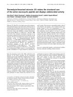
Figure 1. Claudin-1 is not expressed solely in the nucleus even when under the direction of a nuclear localization
signal. A) Cytoplasmic and nuclear extracts of G361 melanoma cells (low claudin-1 expressers) were transfected with wild
type CLDN1 (CLDN1). By Western analysis, these transfectants demonstrate the presence of claudin-1 protein in the
cytoplasm, with small amounts in the nucleus. When cells are transfected with CLDN1 under the control of a nuclear
localization signal (CLDN1-NLS), there is increased expression of claudin-1 in the nucleus, but there are still large amounts
of claudin-1 in the cytoplasm as well. B) All of the claudin-1 in the nucleus can be shuttled into the cytoplasm by treatment
with the PKC activator PMA (phorbol ester). C) This implies that active PKC may exist in the nucleus of melanoma cells, so
cells were stained with an antibody to pan-PO4-PKC. Confocal microscopy demonstrates that active PKC can be found in
the nucleus of G361 melanoma cells. D) To determine if claudin-1 was a potential PKC substrate, cell lysates from claudin-1
high UACC647 cells, and claudin-1 low G361 cells were subjected to immunoprecipitation using a PKC substrate antibody,
and western analysis for claudin-1 was performed. GO6983, a PKC inhibitor, decreases the amount of claudin-1 that is
immunoprecipitated by the PKC substrate antibody. G361 cells have very little claudin-1 and thus, none is precipitated by
the PKC substrate antibody, indicating the specificity of this immunoprecipitation.
Int. J. Med. Sci. 2009, 6
96
PKC and PKA play distinct roles in the expres-
sion and localization of claudin-1
To better characterize the phosphorylation of
claudin-1 by PKC, we performed an in silico analysis
of the putative PKC phosphorylation sites for
claudin-1. We found that there are five putative PKC
phosphorylation sites on the claudin-1 protein (212
amino acids long), but all of these are also sites of PKA
phosphorylation (Table 1). There are however, three
unique sites of PKA phosphorylation, at amino acid
residues 65-69, and 189-192 and 201-205. It should be
noted that the aa189-192 site overlaps with a
PKC/PKA phosphorylation site (aa188-191). To assess
whether PKC or PKA was responsible for the phos-
phorylation and nuclear exclusion of claudin-1, we
used site-directed mutagenesis of our
PCDNA3.1-CLDN1 vector to make mutants that ei-
ther mimicked a constitutively phosphorylated state
(conversion of the phosphorylation site to an aspartic
acid residue, referred to as “D” mutants) or mutants
that are non-phosphorylable (conversion of the phos-
phorylation site to an alanine residue, referred to as
“A” mutants). This vector, when transfected into
melanoma cells shows both cytoplasmic and nuclear
expression of claudin-1, as compared to empty vector
controls (Figure 2A). The sites of phosphorylation
close to the end of the protein sequence (aa200-212)
did not provide us with successful mutants. Muta-
tions to alanine in PKC/PKA sites did not affect the
subcellular localization of claudin-1, but alanine mu-
tations of the two unique PKA sites caused nuclear
localization of claudin-1 (Figure 2B). Mutations to
aspartic acid in both PKA only as well as PKC/PKA
sites caused cytoplasmic redistribution of claudin-1
with complete exclusion from the nucleus (Figure 2C).
Nuclear localization of claudin-1 upon mutation
of the PKA sites could be mimicked using PKA in-
hibitors. Untreated M93-047 cells have high levels of
claudin-1 and exhibit a diffuse, largely cytoplasmic
pattern of claudin-1 (Figure 3A). Upon treatment with
PKA inhibitors for 15 minutes, claudin-1 shuttles into
the nucleus (Figure 3B). After 1 hour of treatment with
PKA inhibitor, claudin-1 is still highly nuclear, al-
though some claudin-1 has started to shuttle out of
the nucleus (Figure 3C). This is unlike the situation
with PKC inhibitors, which cause a general down-
regulation of claudin-1 expression (4). Furthermore, it
is interesting to note that all of the cell lines have
similar levels of phospho-PKA (Figure 3D) which
explains why the transfected claudin-1 is shuttled out
of the nucleus even when transfected into less metas-
tatic G361 cells, which have low amounts of phos-
pho-PKC (4,13). Taken together these data indicate
that PKA is likely contributing to the subcellular lo-
calization of claudin-1.
Nuclear claudin-1 does not increase melanoma
cell motility.
We have previously shown that increasing the
levels of CLDN-1 increases the invasion of melanoma
cells (4). To determine if the increase in claudin-1
needs to be cytoplasmic and not nuclear to affect the
ability of melanoma cells to invade, we first per-
formed a stable transfection of our G361 cells with the
S69A (PKA, non-phosphorylatable) claudin-1 mu-
tants. Pooled stable clones were analyzed for the ex-
pression of claudin-1 and its subcellular localization.
As can be seen the PCDNA3.1-CLDN1 transfected
cells have plenty of cytoplasmic claudin as compared
to the stable empty vector clones (Figure 4A, B)
whereas the S69A mutants have largely nuclear ex-
pression of claudin-1 (Figure 4C). To test their inva-
sive capacity, stable clones were allowed to invade
through a Matrigel-coated invasion chamber. As
compared to empty vector controls, cells transfected
with the PKA deactivating S69A mutation did not
show any increase in invasion. However, G361 cells
transfected with the claudin-1 overexpressing vector
showed a nearly 2-fold increase in invasion as com-
pared to the empty vector control (Figure 4D). These
data appear to support the hypothesis that nuclear
overexpression of claudin-1 does not increase the in-
vasive capacity of melanoma cells, where cytoplasmic
expression of claudin-1 does.
Table 1. Site-directed mutagenesis. Putative sites of PKC and PKA phosphorylation on the claudin-1 protein, and the
primers used to perform site-directed mutagenesis of these sites. The first 10 rows represent mutations to alanine, and the
last ten rows represent mutations to aspartic acid.
AMINO ACID
SEQUENCE
MOTIF (PO4) PKA/PKC DNA BASE CHANGE PRIMER
31-34 RIYS RXXpS PKA T100G
31-34 RIYS [R/K]XX[pS/pT] PKC T100G
F: 5'-cagtggaggatttacgcctatgccggcgaca-3'
R: 5'-tgtcgccggcataggcgtaaatcctccactg-3'
65-69 KVFDS* KXX[pS/pT] PKA T205G F: 5'-agtgcaaagtctttgacgccttgctgaatctgagc-3'
R: 5'-gctcagattcagcaaggcgtcaaagactttgcact-3'
188-190 RKT [R/K]X[pS/pT] PKA A568T F: 5'-tgttcctgtccccgaaaatcaacctcttacccaac-3'
Int. J. Med. Sci. 2009, 6
97
AMINO ACID
SEQUENCE
MOTIF (PO4) PKA/PKC DNA BASE CHANGE PRIMER
188-190 RKT [R/K]X[pS/pT] PKC A568T R: 5'-gttgggtaagaggttgattttcggggacaggaaca-3'
188-191 RKTT [R/K]XX[pS/pT] PKC A571G
188-191 RKTT [R/K][R/K]X[pS/
pT]
PKA A571G
F:5'-gctgttcctgtccccgaaaaacagcctcttacccaa-3'
R:5'-ttgggtaagaggctgtttttcggggacaggaacagc-3'
189-192 KTTS* KXX[pS/pT] PKA A568T_A571G F:5'-ctgttcctgtccccgaaaatcagcctcttacccaacac-3'
R:5'-gtgttgggtaagaggctgattttcggggacaggaacag-3'
195-197 TPR [pS/pT]X[R/K] PKA A589G_G590C
195-197 TPR [pS/pT]X[R/K] PKC A589G_G590C
F:5'-aacaacctcttacccaacaccagcgccctatccaaaacc-3'
R:5'-ggttttggatagggcgctggtgttgggtaagaggttgtt-3'
31-34 RIYS RXXpS PKA T100G_C102A
31-34 RIYS [R/K]XX[pS/pT] PKC T100G_C102A
F:5'-gccccagtggaggatttacgcatatgccggcgaca-3'
R:5'-tgtcgccggcatatgcgtaaatcctccactggggc-3'
65-69 KVFDS KXX[pS/pT] PKA T205G_C206A
F:5'-ccagtgcaaagtctttgacgacttgctgaatctgagcagc-3'
R:5'-gctgctcagattcagcaagtcgtcaaagactttgcactgg-3'
188-190 RKT [R/K]X[pS/pT] PKA A568G_C569A_A570C
188-190 RKT [R/K]X[pS/pT] PKC A568G_C569A_A570C
F:5'-ctttgctgttcctgtccccgaaaagacacctcttacccaacacca-3'
R:5'-tggtgttgggtaagaggtgtcttttcggggacaggaacagcaaag-3'
188-191 RKTT [R/K]XX[pS/pT] PKC A571G_C572A
188-191 RKTT [R/K][R/K]X[pS/
pT]
PKA A571G_C572A
F:5'-ttcctgtccccgaaaaacagactcttacccaacaccaagg-3'
R:5'-ccttggtgttgggtaagagtctgtttttcggggacaggaa-3'
189-192 KTTS KXX[pS/pT] PKA A568G_C569A_A570C_
A571G_C572A
F:5'-actttgctgttcctgtccccgaaaagacgactcttacccaacaccaaggccc-3'
R:5'-gggccttggtgttgggtaagagtcgtcttttcggggacaggaacagcaaagt-3'
195-197 TPR [pS/pT]X[R/K] PKA A589G_G590A_G591C
195-197 TPR [pS/pT]X[R/K] PKC A589G_G590A_G591C
F:5'-aaaacaacctcttacccaacaccagacccctatccaaaacctgca-3'
R:5'-tgcaggttttggataggggtctggtgttgggtaagaggttgtttt-3'
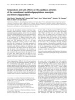
Figure 2. Site-directed mutagenesis
of PKA/PKC in CLDN1. A) G361
melanoma cells which have very little
endogenous claudin-1 were transfected
with either an empty vector or CLDN-1
and localization was observed using
confocal microscopy. Site directed
mutagenesis was performed to render
the potential sites of PKC and PKA
phosphorylation on the claudin-1 pro-
tein non phosphorylatable or to mimic
constitutive activation of PKC or PKA.
B) Rendering the serine at position 69
on the claudin-1 protein
non-phosphorylatable causes nuclear
localization of claudin-1. C) Mutations
mimicking constitutive activation of its
phosphorylatable sites cause exclusively
cytoplasmic localizationof claudin-1.