Báo cáo y học: "Fibrolipomatous hamartoma in the median nerve in the arm - an unusual location but with MR imaging characteristics: a case report" pps
Bạn đang xem bản rút gọn của tài liệu. Xem và tải ngay bản đầy đủ của tài liệu tại đây (930.05 KB, 6 trang )
CAS E REP O R T Open Access
Fibrolipomatous hamartoma in the median nerve
in the arm - an unusual location but with MR
imaging characteristics: a case report
Jessica Nilsson
1†
, Kristina Sandberg
1†
, Lars B Dahlin
1*
, Nina Vendel
2
, Eva Balslev
3
, Lone Larsen
4
, Niels Søe Nielsen
5
Abstract
Fibrolipomatous hamartoma of the median nerve are usua lly located distally in the forearm and may have charac-
teristic features on MR imaging. Here we report a patient with an extensive fibrolipomatous hamartoma at an unu-
sual location proximally in the arm, where a preoperative MR imaging was pathognomonic and diagnosis was
verified by an incisional biopsy. We suggest that MRI should be performed in cases with nerve dysfunction without
an obvious cause after a thorough clinical examination.
Background
The two most common nerve tumours in the upper
extremity are Schwannoma and neurofibroma [1,2].
More rare is a fibrolipomatous hamartoma, a benign,
slow-growing mass, which is usually located in the med-
ian nerv e distally in the forearm [3- 7] and in its digital
branches [1,4,5]. With MR imaging it is not always pos-
sible to make a diagnosis of a nerve tumour [2], but the
MR imaging characteristics of fibrolipomatous hamar-
toma are considered to b e pathognomonic [3]. In coro-
nal plane, the nerve tumour is characterised by
serpiginous structures [4,6] (thickened nerve fascicles),
which are surrounded by fat (high signal intensity on
T1-weighted images, low signal intensity on fat-sup-
pressed T2-weighted images) [3]. In most of the cases
the fat is distributed between the nerve fascicles making
the nerve tumour looking like a coaxial cable in the
axial plane [3-5,8,9]. Even if the nerve tumour has a
characteristic feature on MRI the suspicion of a nerve
tumour is not always obvious for the clinician. Here we
report a case with obscure clinical symptoms and signs
of isolated median nerve dysfunction, where the MR
imaging showed the characteristic features of a fibroli-
pom ato us hamartoma in the arm and the diagnosis was
verified by an incisional biopsy.
Case presentation
A 57 year right-handed secretary was referred to our
hospital July 2008. She described symptoms since
November 2002 with paresthesia in the right index, long
and ring (half of it) fingers. Furthermore, she told about
fibrillations in the interphalangeal joint of the right
thumb and the index finger, loss of FPL and FDP func-
tion to the index finger followed by atrophy of the the-
nar muscles a year lat er. She was operated with carpal
tunnel release at another hospital April 2007 due to a
suspicion of a carpal tunnel syndrome, but no neurogra-
phy or electromyography (EMG) was performed. In
addition, she was operated with division of the A1
pulley on the right thumb due to a susp icion of a right-
sided trigger thumb, but with no improvement. In Feb-
ruary 2008, after the carpal tunnel release, neurography
and EMG were performed. These investigations showed
a severe loss of nerve fibres, but with remaining nerve
fibers, in the right median nerve. E lectrophysiolo gically,
signs of reinnervation were seen, but no nerve compres-
sion lesion was found.
At the clinical examination in July 2008 at our hospi-
tal she had atrophy of the thenar muscles and clear
signs of affection of the anterior interosseous nerve with
impaired function of the FDP to the index finger and
FPL and decreased grip strength. She described a slight
pain at palpation in the middle part of the forearm
along the median nerve. She had paraesthesia located
only in the long finger. The circumference of the right
arm was 1.5 cm short er than the left forearm indicating
* Correspondence:
† Contributed equally
1
Hand Surgery, Department of Clinical Sciences in Malmö, Lund University,
Malmö, Sweden
Nilsson et al. Journal of Brachial Plexus and Peripheral Nerve Injury 2010, 5:1
/>JOURNAL OF BRACHIAL PLEXUS AND
PERIPHERAL NERVE INJURY
© 2010 Nilsson et al; licensee BioMed Central Ltd. This is an Open Access article distributed under the ter ms of the Creative Commons
Attribu tion License ( which permits unrestricted use, distribution, and reproduction in
any medium, provided the original work is properly cited.
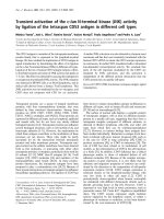
Figure 1 Magnetic resonance investigation (MRI) of the patient showing specific characteristics of a fibrolipomatous hamartoma
(arrows) in sagittal sections (T1 weighted in A and B) and in axial sections (T1-weighted in C and D; T2-weighted in E and F) at the
elbow region.
Nilsson et al. Journal of Brachial Plexus and Peripheral Nerve Injury 2010, 5:1
/>Page 2 of 6
atrophy of some of the forearm muscles. She had no
other clinical or neurographical signs of motor or sen-
sory dysfunction. There was no obvious cause of the
longstanding, severe nerve fibre loss in the median
nerve. An MRI was performed of the right median
nerve at axilla level and distally. The MRI showed a
median nerve with serpiginous appearance. Single nerve
fascicles in a well defined ti ssue mass containing fat was
found (Figure 1). The condition extended from the
proximal humerus to the wrist and thereafter the med-
ian nerve had a normal appearance. The diameter of the
tumour was 1.7-2.5 cm. Due to the typical MRI ch anges
(Figure 1) the diagnosis was suggested as a fibrolipoma-
tous hamartoma of the median nerve. An incisional
biopsy after exploration of the median nerve was done
in February 2009 under microscopical dissection. The
median nerve had a diameter up to 2 cm and there
were no adhesions to the surrounding tissue. The
macroscopical appearance of the nerve tumour indicated
a fibrolipomatous hamartoma (Figure 2) and five
incisional biopsies were taken from representative areas.
Neurolysis of the median nerve was done from the mid-
dle part of the arm down to the middle part of the fore-
arm. Microscopy showed a fibrolipomatous tissue that
surrounded and splayed apart the peripheral nervous tis-
sue, which was also fibrotic and atrophic (Figure 3). The
postoperative follow-up was uneventful. She r eturned to
her original profession. She still has dysfunction of t he
FPL, and thenar atrophy although she can do an opposi-
tion of the thumb. She felt some improvement after the
exploration and neurolysis of the nerve around the
elbow. A tendon transfer procedure may be considered
in the future. She is f ollowed regularly with particularly
clinical examinations.
Discussion
Of all tumours in the upper extremity, 2% are nerve
tumours [10]. Schwanno ma is probably the most fre-
quent one with a known incidence of less than 1/
100000 inhabitants and year in Swede n [1]. Usually, the
Figure 2 At exploration of the median nerve at the elbow region a thickened nerve (arrows in A) was shown where the incisional
biopsy showed a fibrolipomatous hamartoma (see Figure 3). Close up of the condition in B.
Nilsson et al. Journal of Brachial Plexus and Peripheral Nerve Injury 2010, 5:1
/>Page 3 of 6
diagnosis of a nerve tumour has to be based on micro-
scopical findings since MRI is not sufficient for a precise
diagnosis [2]. In contras t, a fibrolipomatous hamartoma,
which is even more rare, has very distinct characteristics
in MRI with serpiginous hypotense structures represent-
ing t hickened fascicles w hich are surrounded by evenly
distributed fat [4] (high T1-weighted signal intensity and
low fat-suppressed T2-weighted signal intensity) [3,4,6].
Our case showe d such MRI characteristics and, further-
more, the diagnosis was confirmed by microscopy. We
did not perform any ultrasound examination of the
patient due to the lack of palpable tumour befo re
exploration. However, such an investigation may be con-
sidered when there is a suspicion of a nerve tumour,
although MRI is frequently used [2].
The origin of fibrolipomatous hamartoma is still
obscure and is mainly affecting and found in young per-
sons [4-7]. That might indicate a congenital aetiology
[7], although a few cases have been reported in older
people as seen here. Another theory is that the lesion
Figure 3 Microscopical pictures of sections from the incisional biopsy showing (A) fibrolipomatous tissue with few atrophic peripheral
nerve fascicles and fibrotic tissue (HE; × 10) and (B) atrophic and fibrotic nerve fibers stained with S-100 immunohistochemical
staining (S-100; × 20).
Nilsson et al. Journal of Brachial Plexus and Peripheral Nerve Injury 2010, 5:1
/>Page 4 of 6
can be caused by trauma [3-5]. The re are debates of the
relationship between fibrolipomatous hamartoma and
macrodactyli [3-5,7]. However, our patient had no signs
of this clinical presentation.
Our patient had a long history with motor distur-
bances, such as thenar atrophy and loss of FPL function.
Most cases present with a longstanding painless mass.
Nerve compression of the affected nerve with paresthe-
sia, motor deficit and pain are known late symptoms
[4,5,7,11-14]. It is suggested that it may become sympto-
matic only in the median nerve due to encroachment by
the flexor retinaculum; thus causing carpal tunnel syn-
drome [4,5]. However, in our case the tumour was
located more proximally extending from the upper arm
almost down to wrist level; thus, presenting a more
proximal located fibrolipomatous hamartoma than pre-
viously described. A hypothesis coul d be that you rarely
find these fibrolipomatous hamartoma more proximal
because of spatial relations.
Only a limited number of cases of fibrolipomatous
hamartoma are reported in the literature showing the
uncertainty of the optimal treatme nt suggestion and
that treatment should be guided by the severity of
symptoms [5,15]. Our case was treated only by explora-
tion and release of potentia l narrowing structures, parti-
cularly around the elbow, which improved her
symptoms, but excision of the tumour is not recom-
mendab le [3-7,16,17]. Due to the extensive fatty infiltra-
tion of the nerve fascicles, surgical excision may cause
catastrophic motor and sensory deficits. We performed
five incisional biopsies from different locations to be
sure of adequate material for the neuropathological
examination. When an incisional biopsy is gently per-
formed it is our experience that no further dysfunction
is added to the patient. We will follow our p atient
regarding any progression of the t umour and further
consider additional treatment, such as tendon transfers,
for the impaired function of FPL and FDP to the index
finger. However, the long-term r esults are obscure of
fibrolipomatous hamartoma. Meticulous information to
the patient and a regular follow-up are recommended.
The present case emphasizes the need for a thorough
history f rom the patient and a careful and meticulous
clinical examination of cases with symptoms from the
peripheral nervous system. For example, a nerve tumour
can be the cause of symptoms as the present case. A
MRI may reveal a nerve structure with a coaxial-cable-
like appearance; thus with a high suspicion of the diag-
nosis of fibrolipomatous hamartoma.
Conclusions
Although history of patients with symptoms from the
peripheral nervous system as well as a meticulous clini-
cal examination is recommended, an MRI is an
additional t ool to reveal a fibrolipomatous hamartoma
at an unusual location.
Consent
Written informed consent was obtained from the patient
for publication of this case report and any accompany-
ing images. A copy of the written consent is available
for review by the Editor-in-Chief of this journal.
Acknowledgements
The research on nerve tumours was supported by grants from the Swedish
Research Council (Medicine), Region Skåne and Funds from the University
Hospital Malmö, Sweden. The article is the result of collaboration between
the Panum Institute in Copenhagen and authors in the Öresund region.
Author details
1
Hand Surgery, Department of Clinical Sciences in Malmö, Lund University,
Malmö, Sweden.
2
Department of Anesthesiology, Intensive Care and
Operations, Gentofte Hospital, Copenhagen, Denmark.
3
Department of
Pathology, Herlev Hospital, Denmark.
4
Department of Radiology, Herlev
Hospital, Denmark.
5
Department of Orthopaedics at Herlev Hospital, Division
of Hand Surgery, Gentofte Hospital, Hellerup, Denmark.
Authors’ contributions
The medical students JN and KS and senior author LD have done literature
review and written the draft of the manuscript. The patient was operated by
NSN (senior author) and NV (nurse; literature review). MRI was performed by
LL and the microscopical examination by EB. All authors have contributed in
different important ways to the present manuscript.
All authors have read and approved the final manuscript.
Competing interests
The authors declare that they have no competing interests.
Received: 5 October 2009
Accepted: 12 January 2010 Published: 12 January 2010
References
1. Sandberg K, Nilsson J, Soe Nielsen N, Dahlin LB: Tumours of peripheral
nerves in the upper extremity: a 22-year epidemiological study. Scand J
Plast Reconstr Surg Hand Surg 2009, 43:43-49.
2. Nilsson J, Sandberg K, Nielsen NS, Dahlin LB: Magnetic resonance imaging
of peripheral nerve tumours in the upper extremity. Scand J Plast
Reconstr Surg Hand Surg 2009, 1-7.
3. Marom EM, Helms CA: Fibrolipomatous hamartoma: pathognomonic on
MR imaging. Skeletal Radiol 1999, 28:260-264.
4. De Maeseneer M, Jaovisidha S, Lenchik L, Witte D, Schweitzer ME,
Sartoris DJ, Resnick D: Fibrolipomatous hamartoma: MR imaging findings.
Skeletal Radiol 1997, 26:155-160.
5. Guthikonda M, Rengachary SS, Balko MG, van Loveren H: Lipofibromatous
hamartoma of the median nerve: case report with magnetic resonance
imaging correlation. Neurosurgery 1994, 35:127-132.
6. Khanna G, Sundaram M, Rotman M, Janney CG: Fibrolipomatous
hamartoma of the nerve. Orthopedics 2001, 24:919-820.
7. Silverman TA, Enzinger FM: Fibrolipomatous hamartoma of nerve. A
clinicopathologic analysis of 26 cases. Am J Surg Pathol 1985, 9:7-14.
8. Cavallaro MC, Taylor JA, Gorman JD, Haghighi P, Resnick D: Imaging
findings in a patient with fibrolipomatous hamartoma of the median
nerve. AJR Am J Roentgenol 1993, 161:837-838.
9. Boren WL, Henry RE Jr, Wintch K: MR diagnosis of fibrolipomatous
hamartoma of nerve: association with nerve territory-oriented
macrodactyly (macrodystrophia lipomatosa). Skeletal Radiol 1995, 24:296-
297.
10. Adani R, Baccarani A, Guidi E, Tarallo L: Schwannomas of the upper
extremity: diagnosis and treatment. Chir Organi Mov 2008, 92:85-88.
11. Kakitsubata Y, Theodorou SJ, Theodorou DJ, Shibata M, Yuge M, Yuki Y,
Hatakeyama K, Yokouchi T: MR imaging of uncommon recurrence of
Nilsson et al. Journal of Brachial Plexus and Peripheral Nerve Injury 2010, 5:1
/>Page 5 of 6
fibrolipomatous hamartoma of the ulnar nerve. Acta Radiol 2003, 44:326-
328.
12. Canga A, Abascal F, Cerezal L, Bustamante M, Perez-Carro L, Vazquez-
Barquero A: Fibrolipomatous hamartoma of the median nerve. Case
illustration. J Neurosurg 1998, 89:683.
13. Oleaga L, Florencio MR, Ereno C, Grande J, Terrones J, Legorburu A,
Grande D: Fibrolipomatous hamartoma of the radial nerve: MR imaging
findings. Skeletal Radiol 1995, 24:559-561.
14. Sondergaard G, Mikkelsen S: Fibrolipomatous hamartoma of the median
nerve. J Hand Surg Br 1987, 12:224-226.
15. Chatillon CE, Guiot MC, Jacques L: Lipomatous, vascular, and
chondromatous benign tumors of the peripheral nerves: representative
cases and review of the literature. Neurosurg Focus 2007, 22:E18.
16. Louis DS, Hankin FM, Greene TL, Dick HM: Lipofibromas of the median
nerve: long-term follow-up of four cases. J Hand Surg [Am] 1985, 10:403-
408.
17. Price AJ, Compson JP, Calonje E: Fibrolipomatous hamartoma of nerve
arising in the brachial plexus. J Hand Surg Br 1995, 20:16-18.
doi:10.1186/1749-7221-5-1
Cite this article as: Nilsson et al.: Fibrolipomatous hamartoma in the
median nerve in the arm - an unusual location but with MR imaging
characteristics: a case report. Journal of Brachial Plexus and Peripheral
Nerve Injury 2010 5:1.
Publish with Bio Med Central and every
scientist can read your work free of charge
"BioMed Central will be the most significant development for
disseminating the results of biomedical research in our lifetime."
Sir Paul Nurse, Cancer Research UK
Your research papers will be:
available free of charge to the entire biomedical community
peer reviewed and published immediately upon acceptance
cited in PubMed and archived on PubMed Central
yours — you keep the copyright
Submit your manuscript here:
/>BioMedcentral
Nilsson et al. Journal of Brachial Plexus and Peripheral Nerve Injury 2010, 5:1
/>Page 6 of 6