Báo cáo y học: "Advanced radiological work-up as an adjunct to decision in early reconstructive surgery in brachial plexus injuries" pptx
Bạn đang xem bản rút gọn của tài liệu. Xem và tải ngay bản đầy đủ của tài liệu tại đây (1.98 MB, 7 trang )
RESEARC H ARTIC LE Open Access
Advanced radiological work-up as an adjunct
to decision in early reconstructive surgery
in brachial plexus injuries
Kasim Abul-Kasim
1
, Clas Backman
2
, Anders Björkman
2
, Lars B Dahlin
2,3*
Abstract
Background: As neurophysiologic tests may not reveal the extent of brachial plexus injury at the early stage, the
role of early radiological work-up has become increasingly important. The aim of the study was to evaluate the
concordance between the radiological and clinical findings with the intraoperative findings in adult patients with
brachial plexus injuries.
Methods: Seven consecutive male patients (median age 33; range 15-61) with brachial plexus injuries, caused by
motor cycle accidents in 5/7 patients, who underwent extensive radiologi cal work-up with magnetic resonance
imaging (MRI), computed tomography myelography (CT-M) or both were included in this retrospective study. A
total of 34 spinal nerve roots were evaluated by neuroradiologists at two different occasions. The degree of
agreement between the radiological findings of every individual nerve root and the intraoperative findings was
estimated by calculation of kappa coefficient (К-value). Using the operative findings as a gold standard, the
accuracy, sensitivity, specificity, positive predictive value (PPV) and negative predictive value (NPV) of the clinical
findings and the radiological findings were estimated.
Results: The diagnostic accuracy of radiological findings was 88% compared with 65% for the clinical findings. The
concordance between the radiological find ings and the intraoperative findings was substantial (К = 0.76) compared
with only fair (К = 0.34) for the clinical findings. There were two false positive and two false negative radiological
findings (sensitivity and PPV of 0.90; specificity and NPV of 0.87).
Conclusions: The advanced optimized radiological work-up used showed high reliability and substantial
agreement with the intraoperative findings in adult patients with brachial plexus injury.
Introduction
The most common cause of closed brachial plexus
injuries in adults is a motorcycle accident (70%) [1,2].
The generally agreed mechanism of a brachial plexus
injury is traction stress on the plexus as the head and the
shoulder are forced apart [3]. Up to 2/3 of high energy
brachial plexus injuries may need surgical intervention
[2]. Thus, the preoperative planning to determine type,
level and extent of the injury is crucial for optimal selec-
tion of patients that benefit from surgical reconstruction
and to plan the surgical procedure. Early reconstructive
surgery of nerve injuries encourages rapid regeneration
and repair [4-7]. Neurophysiological tests may not reveal
the extent of injury at the early stage [7]. Therefore, the
role of imaging studies performed early has beco me
increasingly important.
The choice of the radiological modality in the work-up
of brachial plexus injury has been continu ously changed
in last decades. Although myelography was the reliable
[8]andthemostusedmethodsintheradiological
work-up of brachial plexus injuries prior to the era of
sectional imaging, its use nowadays should only be
restricted to patients with contraindication t o magnetic
resonance imaging (MRI). Nowadays, MRI is the
imaging method of choice in the work-up of brachial
plexus injuries [9]. New MR sequences, e.g. 3D CISS (3-
dimensional constructive interference in steady state),
enable acquisition of thin slices with the possibility to
* Correspondence:
2
Department of Hand Surgery, Skåne University Hospital, S-205 02 Malmö,
Sweden
Abul-Kasim et al. Journal of Brachial Plexus and Peripheral Nerve Injury 2010, 5:14
/>JOURNAL OF BRACHIAL PLEXUS AND
PERIPHERAL NERVE INJURY
© 2010 Abul-Kasi m et al; licensee BioMed Central Ltd. This is an Open Access article distributed under the terms of the Creative
Commons Attribution License ( which permits unrestricted use, distribution, and
reproduction in any mediu m, provided the original work is properly cited.
perform reconstruction in three different planes and
hopefully can contribute to increase the diagnostic accu-
racy. The disadvantages o f MRI are the long acquisition
time for every individual sequence and the sensitivity to
movement, and thus demand for the patient to lay still.
New MRI technique that recently has been recom-
mended in the work-up of brachial plexus injury is the
diffusion weighted MR neurography [10]. However, the
main limita tion of this technique is lack of depiction of
cervical nerves above the lev el of the C5 nerve. Another
radiological modality that i s used in the evaluation of
brachial plexus is the computed tomography following
myelography (CT-M). As CT-M is invasive and means
exposing these, often young, patients to high doses of
ionising radiation, this type of imaging should also be
reserved to patients with contraindication to MRI. A
new modality that recently showed high feasibility in the
assessment of cervical nerve roots is Bezier surface tech-
nique, which enables reformatting volumetric data
obtained at CT-myelography to depict the individual
nerve root in a single image [11,12]. However, most of
these modalities are new and their role in the work-up
of brachial plexus injury is not yet well established.
The main purpose of the radiological examination
prior to brachial plexus surgery is to determine the loca-
tion of the injury in relation to the dorsal root ganglion
and categorize injuries into preganglionic avulsion or
postganglionic rupture or stretching. The aim of this
study was to evaluate the accuracy of the radiological
findings and the clinical signs with the intraoperative
findings in adult patients with brachial plexus injuries.
Methods
Seven consecutive male pati ents with brachial plexus
injuries who underwent MRI, CT-M or both were
included in this retrospective analysis. The median age
for the patients was 33 years (mean age 29 ± 17 years;
range 15-61 years). All patients were evaluated by the
same surgeons preoperatively and the extent of the
lesion was determine d clinically (e.g. evaluation of pain,
Tinel sign, presence of Horner syndrome, loss of muscle
function and sensory deficit). Preoperatively, all patients
underwent MRI, within 15 days of injury in average
(median 7 days). Two patients had also been examined
using CT-M because of motion artefacts in MRI in one
patient (patient No 6) and because of hematoma and
fibrosis at the root exit which resulted in a significant
signal drop on MRI in another patient (patient No 2).
One patient (patient No 5) underwent MRI two times
(one at the hospital where the patient was initially
admitted and one in our institution). All patients were
examined with sagittal T1-weighted images (WI), axial
T1WI,axialT2WI,axialturboflash(TF)gradient
echo images, and coronal short TI inversion recovery
(STIR)-images. In three cases ( case 1, 2 and 4) the
patients were also examined with a dual excitation
sequence called 3D CISS. The images were evaluated at
two different occasio ns, one a t the time of injury and
one at the time of analysis of this study. In cases of dis-
agreement the fina l results w ere reached by consensus
at joint evaluation of two radiologists. The reader was
blinded to the clinical and the intraoperative findings.
The radiological signs of brachial plexus injuries sought
for were the following: (a) signa l changes in the spinal
cord near the nerve root exit, (b) bleeding near the
nerve root exit, (c) failure of visualisation of the nerve
root (dorsal, ventral or both), (d) discontinuity in the
course of the nerve root (dorsal, ventral or bo th), (e)
CSF leakage along the nerve root, and (f) pseudomenin-
gocele. In 6 patients the spinal roots C5-T1 were exam-
ined and in the seventh patient only C6-T1 were
examined. In all patients, the brachial plexus lateral to
ganglion (trunks, divisions, and cords) was also evalu-
ated. For the purpose of evaluation the aforementioned
structures (trunks, divisions, and cords) were considered
as postganglionic plexus. The total number of the evalu-
ated spinal nerve roots was 34. The agreement between
the radiologica l findings of every individual spinal nerve
root and the preoperative findings of each root at the
time of the surgical exploration was estimated.
All patient s were operated on by the same surgeons in
average 2 6 days (median 17 days) after the injury when
the extent and location of the lesion was determined.
All patients were operated on in general anaesthesi a
with a supraclavicular approach extending along the
infra clavicular plexus, usually using an ost eotomy of the
clavicle, throu gh a longitud inal incisio n in the deltopec-
toral groove from approximately the middle of the clavi-
cle to the cranial border of the tendon of pectorali s
major. Appropriate nerve reconstructive procedures
were done based o n the findings in the individual
patients.
The study was approved by the local Ethics committee
of Lund University. The study was done in accordance
with the Helsinki declaration.
Statistical analysis
Statistical analysis was performed using SPSS 17. The
degree of agreement between the clinical findings and
radiological findings of every individual spinal nerve
root on one hand and the intraoperative findings on the
other hand was estimated by cross tabulation and calcu-
lation of kappa coefficient (K-value). The interpretation
of kappa values was done according to the method pro-
posed by Landis [13]. A 2-way contingency table was
generated comparing the cl inical findings and radiologi-
cal findings on one hand with the operative findings on
the other hand. The contingency table was used to
Abul-Kasim et al. Journal of Brachial Plexus and Peripheral Nerve Injury 2010, 5:14
/>Page 2 of 7
calculate the accuracy, sensitivity, specificity, positive
predictive value (PPV) and negative predictive value
(NPV) of the clinical findings and the radiological find-
ings with the operative findings as a gold standard.
Results
Patients’ characteristics
Motor cycle acci dent was the cause of the injury in five
patients. The remaining patients were subjected a ski
accident (n = 1), and a trauma of a falling tree (n = 1)
(Table 1). Fi ve patients showed i njuries of the right
sided brachial plexus. Four out of seven cases were clini-
cally suspected to have total damage of plexus brachialis
(C5-T1-injury). Six patients had other serious associated
injuries of which three were suspected to have total
damage of the brachial plexus (Table 2). The preopera-
tive clinical signs of the patients are summarized in
Table 1. The clinical signs of the extent of the lesion
(pre- or postganglionic injury) showed a suspicion of
preganglionic (based on no Tinel sign, character of the
pain, presence of Horner syndrome) or of a postganglio-
nic injury (presence of Tinel sign, remaining motor
function in serratus anterior muscle).
Radiological work-up
Outof34spinalrootssubjectedforradiological
evaluation, the diagnosis was the same as the intrao-
perative diagnosis for 30 of the explored nerve roots.
This resulted in diagnostic accuracy of 88%. The con-
cordance between the radiological findings and the
intraoperative findings was substantial (К value 0.76;
95% CI 0.54-0.98). There were two false positive and
two false negative radiological findings, which resulted
in sensitivity and positive predictive value of 0.90 (95%
CI 0.76-0.96), and specificity and negative predictive
value of 0.87 (95% CI 0.70-0.95), (Table 3). The accu-
racy of clinical diagnosis was 65% (in 22 of the 34
explored nerve roots the clinical diagnosis was the
same as the intraoperative findings), which resulted in
only fair agreement (К value 0.34; 95% CI 0.11-0.56).
At the surgical exploration, 12 roots that the clinical
examination raised a suspicion of root injury were
found to be intact (false positive clinical findings). This
resulted in a specificity of 0.56 and positive predictive
valueof0.37(Table3).Figure1showexamplesofthe
radiological findings in two different patients included
in this study.
Table 1 Patient characteristics and summary of the clinical, radiological and intraoperative findings in seven patients
with a traumatic brachial plexus injury
No Age
(yr)
Injury
mechanism
Imaging
modality
Clinical
findings
Imaging findings Operative findings Side
affected
1 15 MC MRI C5-C6 C6 C5-C6 Right
2 15 MC MRI + CT-M C5-T1 C5-C7 C5-C7 Right
3 34 MC MRI C5-T1 Postgangl. rupture at the level of
the cord
Postgangl. rupture at the level of
the cord
Right
4 34 MC MRI C5-T1 C6-T1, C5-T1 Left
C5 not included on axial images
5 14 Ski injury MRI C5-C6 C5-C6 Intact roots (axonotmesis) Right
6 61 Falling tree MRI + CT-M C5-C8 C6 Postgangl. C5, avulsion C6 Left
7 33 MC MRI C5-T1 C5-C8 C5-C8 Right
No = Patient number. yr = year. MRI = Magnetic Resonance Imaging. CT-M = Computed Tomography- Myelography. MC = Motor cycle. C indicates cervical roots
and T thoracic roots. Postgangl. = Postganglionic injury.
Table 2 Time between injury and radiological examination and surgery in seven patients with a traumatic brachial
plexus injury expressed in days
No Injury-Radiological
work-up
Injury-
Surgery
Associated injury
1 5 16 Metacarpal V fracture
2 7 17 None
3 26 48 Metatarsal injury, ankle fracture, radius and ulna fractures, supracondylar humerus fracture, and radial nerve
injury at elbow level.
4 23 27 Shoulder dislocation, metacarpal II-V fracture, radius fractures, and ligament injury left knee.
5 4 14 Lung contusion, skull base fracture, mandibular fracture, orbital fracture.
6 33 42 Hemo-/pneumothorax, scapular-, clavicular-, and rib fractures
7 4 17 Clavicle fracture, unstable T12 fracture, multiple rib fractures with flail chest, hemo-pneumothorax,
compartment syndrome forearm, metacarpal V fracture, and right subclavian artery injury.
Abul-Kasim et al. Journal of Brachial Plexus and Peripheral Nerve Injury 2010, 5:14
/>Page 3 of 7
Operative findings
In two cases (cases 4 and 7) there was a need fo r division
oftheclavicleinordertovisualizeallnerveendingsand
roots. The roots were evaluated as avulsed or ruptured.
The texture and looseness of the nerve roots were con-
sidered in the decision as to if the nerve could possibly
be avulsed but still in the spinal canal or intact. In cases
of scarred tissue over the plexus the area was explored
and meticulously dissected and ruptures were defined.
The clinical signs at evaluation of the patients indicated
upper trunk injuries in case 1, which was confirmed
at surgery as we found C5 and C6 avulsions. On
Table 3 2-way contingency table comparing the radiological and clinical findings on one hand with intraoperative
findings on the other hand
P-value Sensitivity Specificity PPV NPV
Root injuries on MRI
No Yes
Root injuries No 17 2
at operation Yes 2 13 <0.001 0.90
(0.76-0.96)
0.87
(0.70-0.95)
0.90
(0.76-0.96)
0.87
(0.70-0.95)
Root injuries suspected clinically
Root injuries No 7 12
at operation Yes 0 15 0.011 1
(0.69-1)
0.56
(0.48-0.56)
0.37
(0.25-0.37)
1
(0.86-1)
PPV indicates positive predictive value. NPV indicates negative predictive value
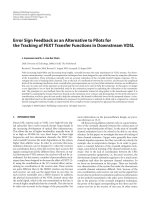
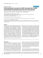
Figure 1 (A-C) MRI 3D CISS of pati ent No 1. The Coronal image (A) shows avulsion of C6 root on the right side. The intact roots are marked
with arrows. Axial images (B-C) show normal C5 roots (arrows, B) and avulsion of C6 roots on the right side (arrow, C). However, exploration
revealed avulsion of both C5 and C6 on the right side (false negative MRI-finding at C5). (D-F) Images of patient No 2. The coronal STIR (D)
shows edema around the supra- and infraclavicular plexus. (E) Axial turbo flash image shows extremely low signal at the C5 root exit indicating
bleeding. (F) Axial CT-M shows hematoma at the site of dorsal root exit (arrow head) and absence of ventral root. Black arrows show the normal
C5 roots on the left side. Similar findings were revealed at the level of C6 and C7. MRI findings were concordant with the intraoperative findings.
Abul-Kasim et al. Journal of Brachial Plexus and Peripheral Nerve Injury 2010, 5:14
/>Page 4 of 7
expl oration, case 2 showed C5-C7 rupture and the lower
roots were soft in texture, but were considered partially
injured. However, at a later follow-up there was M 3 for
flexor digitorum profundus (FDP) muscles and M2 for
flexor pollicis longus (FPL). In case 3, the preoperative
findings showed partial functio n in C5 and C7, while the
other roots were considered ruptured or avulsed. At sur-
gery there was rupture of the whole plexus at the infracla-
vicular level. In case 4, a total avulsion of the entire plexus
was found. Case 5 showed clinical signs of partial rupture
in upper trunk, however, at surgery the plexus was found
intact (axonotmesis). In case 6, there were, apart from
clinical signs of upper trunk rupture, ruptu re or avulsion
of C7-C8, while a partial function was seen in T1 inner-
vated muscles. At surgery C5 was found ruptured and C6
avulsed, while C7 and C8 were evaluated as intact. At a
later follow up there was some recove ry in the forearm
flexors and M1-2 in wrist extensors and extensor pollicis
longus (EPL), indicating a partial rupture in the latter
nerve roots. In case 7, there was rupture of C5, C6 and
C8, and avulsion of C7 while T1 was not visualized.
Discussion
The present study showed that the radiological work-up
in adult patients with brachial plexus injuries contribu-
ted to a better preoperative diagnosis with increased
diagnostic accuracy as compared to a clinical examina-
tionaloneandroutineMRI,whichmaybeusefulfor
the surgeon for the preoperative decision making of
possible reconstruction possibilities. The radiological
diagnostic accuracy was clearly better than the clinical
diagnostic accuracy. This may depend on the fact that
patients with brachial plexus injuries usually are severely
injured with multiple associated injuries that make the
clinical evaluation difficult to perform and interpret.
Furthermore, the patients may be severely injured or
treated in a respirator making a proper clinical evalua-
tion impossible to perform. Radiological work-up
showed a high accuracy (88%), a high sensitivity (90%),
and a high specificity (87%) compared to the intraopera-
tive findings. Carvalho et al reported a diagnostic accu-
racy of the preoperative CT myelography and MRI of
85% and 52%, respectively [14], while Hems et al
reported a sensitivity of 81% for MRI [15]. We believe
that higher accuracy and sensitivity in our s tudy, com-
pared w ith the aforementioned studies, depends on the
followings: (a) use of new MR-sequences, such as 3D
CISS, which enables acquisition of thin slices, recon-
struction in three planes and generation of images that
resemble myelography, (b) use of gradient echo
sequences (turbo flash), which is very sensitive to mini-
mal bleedings at e.g. the nerve root exit, and (c) inclu-
sion of CT-myelography whenever MRI provides
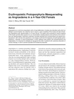
insufficient preoperative data. Specificity and PPV could
have been increased to 1 if there were no false positive
result (patient No 5). However, such clear and distinct
MRI-findings that were radiologically confirmed in case
5 (hematoma at the root exit C5 and C6 and subsequent
development of pseudomeningocele) should be reported
and regarded as signs highly suggestive of root avulsi on
(Figure 2).
Of course, no study is without limitations. Two major
limitations of this study are its retrospective nature a nd
the limited number of patients included in the analysis.
The limited number of patients may have make it diffi-
cult to keep the radiological reader totally blinded a s
there is a small , but existing, possibility that the reader
remembered the findings in some of the evaluated
images. However, t he evaluation of the images in our
study was focused on the individual roots rather than
on individual patients. We believe that our findings of
high reliability of the optimized radiological work-up
with addit ion of special MRI sequences o r performance
of CT-myelography to reveal the precise extent of the
brachial plexus injury is worth to report. In addition,
the analyses have been done by the same surgeons and
radiologist in all patients which is strength of the study.
We performed all brachial plexus explorations and
reconstructions early in most of the cases (within 27
days in 5 out of 7 case s); a decision based on neurobio-
logical knowledge indicating that alterati ons after injury
in neurons and non-neuronal cells are rapid with
respect to cell death and signal transduction. Motor and
sensory neurons die after a nerve injury [4,5]. In addi-
tion, a nerve injury induces rapid, sometimes transient,
upregulation of transcription factors in various signal
transduction pathways, a phenomenon which can not be
utilized if nerve repair or reconstruction is delayed and
may lead to impaired axonal outgrowth [16-20]. The
neurobiological data is supported by a recent clinical
study indicating b etter functional o utcome if brachial
plexus injuries in adults are reconstructed without a
long delay [1,6].
As our radiological work-up showed high accuracy,
sensitivity, and a high specificity as well high concor-
dance w ith the intraoperati ve findings, we strongly
recommend the use of new MR-sequences, such as 3D
CISS (3-dimensional constructive interference in steady
state) or complementary CT-M, to reveal the extent of
the brachial plexus injury.
Conclusion
We conclude that radiological investigation plays an
important role in the preoperative work-up of adult
patients with a brachial plexus injury, where early recon-
struction of the injury may be decisive for an improved
outcome. Advanced and optimized radiological work-up
of this study showed high reliability and substantial
Abul-Kasim et al. Journal of Brachial Plexus and Peripheral Nerve Injury 2010, 5:14
/>Page 5 of 7
agreement with the intraoperative findings. We strongly
recommend the use of new MR-sequences, such as 3D
CISS (3-dimensional constructive interference in steady
state) or addition of CT-myelography, to precisely reveal
the extent of the brachial plexus injury.
Acknowledgements
The study and research done by the authors are supported by Swedish
Research Council (Medicine), Skåne University Hospital, Lund University and
Region Skåne (ALF).
Author details
1
Department of Radiology, Skåne University Hospital, S-205 02 Malmö,
Sweden.
2
Department of Hand Surgery, Skåne University Hospital, S-205 02
Malmö, Sweden.
3
Department of Clinical Sciences Malmö - Hand Surgery,
Lund University, S-205 02 Malmö, Sweden.
Authors’ contributions
KAK performed the radiological evaluation. All surgery has been done by CB,
AB, and LBD. All authors have equally in different ways contributed to the
manuscript. All authors read and approved the final manuscript.
Competing interests
The authors declare that they have no competing interests.
Received: 13 April 2010 Accepted: 8 July 2010 Published: 8 July 2010
References
1. Narakas AO: The treatment of brachial plexus injuries. Int Orthop 1985,
9(1):29-36.
2. Songcharoen P: Brachial plexus injury in Thailand: a report of 520 cases.
Microsurgery 1995, 16(1):35-39.
3. Songcharoen P: Management of brachial plexus injury in adults. Scand J
Surg 2008, 97(4):317-323.
4. McKay Hart A, Brannstrom T, Wiberg M, Terenghi G: Primary sensory
neurons and satellite cells after peripheral axotomy in the adult rat:
timecourse of cell death and elimination. Exp Brain Res 2002,
142(3):308-318.
5. Ma J, Novikov LN, Kellerth JO, Wiberg M: Early nerve repair after injury to
the postganglionic plexus: an experimental study of sensory and motor
neuronal survival in adult rats. Scand J Plast Reconstr Surg Hand Surg 2003,
37(1):1-9.
6. Jivan S, Kumar N, Wiberg M, Kay S: The influence of pre-surgical delay on
functional outcome after reconstruction of brachial plexus injuries. J
Plast Reconstr Aesthet Surg 2009, 62(4):472-479.
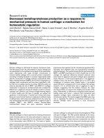
Figure 2 (A-B) axial T1WI and turbo flash image. (C) sagittal T1WI of the initial MRI of patient No 5 show methemoglobin at the C5 root exit
with high signal intensity on T1WI and extremely low signal intensity on turbo flash images (arrows). Prior to surgery a new MRI (D-F) coronal
STIR, sagittal T2WI and axial turbo flash image showed development of pseudomeningocele (intradural cysts) along the nerve roots at several
levels (arrows). Despite these findings the roots were found to be intact (axonotmesis) on exploration (false positive MRI-finding).
Abul-Kasim et al. Journal of Brachial Plexus and Peripheral Nerve Injury 2010, 5:14
/>Page 6 of 7
7. Wiberg M, Backman C, Wahlstrom P, Dahlin LB: Brachial plexus injuries in
adults. Early reconstruction for better clinical results. Lakartidningen 2009,
106(9):586-590.
8. Nagano A, Ochiai N, Sugioka H, Hara T, Tsuyama N: Usefulness of
myelography in brachial plexus injuries. J Hand Surg Br 1989, 14(1):59-64.
9. van Es HW: MRI of the brachial plexus. Eur Radiol 2001, 11(2):325-336.
10. Takahara T, Hendrikse J, Yamashita T, Mali WP, Kwee TC, Imai Y, Luijten PR:
Diffusion-weighted MR neurography of the brachial plexus: feasibility
study. Radiology 2008, 249(2):653-660.
11. Yoshikawa T, Hayashi N, Yamamoto S, Tajiri Y, Yoshioka N, Masumoto T,
Mori H, Abe O, Aoki S, Ohtomo K: Brachial plexus injury: clinical
manifestations, conventional imaging findings, and the latest imaging
techniques. Radiographics 2006, 26(Suppl 1):133-143.
12. Yoshioka N, Hayashi N, Akahane M, Yoshikawa T, Takeshita K, Ohtomo K:
Bezier surface reformation: an original visualization technique of cervical
nerve roots on myelographic CT. Radiat Med 2006, 24(8):600-604.
13. Landis JR, Koch GG: The measurement of observer agreement for
categorical data. Biometrics 1977, 33(1):159-174.
14. Carvalho GA, Nikkhah G, Matthies C, Penkert G, Samii M: Diagnosis of root
avulsions in traumatic brachial plexus injuries: value of computerized
tomography myelography and magnetic resonance imaging. J Neurosurg
1997, 86(1):69-76.
15. Hems TE, Birch R, Carlstedt T: The role of magnetic resonance imaging in
the management of traction injuries to the adult brachial plexus. J Hand
Surg Br 1999, 24(5):550-555.
16. Kataoka K, Kanje M, Dahlin LB: Induction of activating transcription factor
3 after different sciatic nerve injuries in adult rats. Scand J Plast Reconstr
Surg Hand Surg 2007, 41(4):158-166.
17. Saito H, Dahlin LB: Expression of ATF3 and axonal outgrowth are
impaired after delayed nerve repair. BMC Neurosci 2008, 9:88.
18. Saito H, Kanje M, Dahlin LB: Delayed nerve repair increases number of
caspase 3 stained Schwann cells. Neurosci Lett 2009, 456(1):30-33.
19. Martensson L, Gustavsson P, Dahlin LB, Kanje M: Activation of extracellular-
signal-regulated kinase-1/2 precedes and is required for injury-induced
Schwann cell proliferation. Neuroreport 2007, 18(10):957-961.
20. Lindwall C, Dahlin L, Lundborg G, Kanje M: Inhibition of c-Jun
phosphorylation reduces axonal outgrowth of adult rat nodose ganglia
and dorsal root ganglia sensory neurons. Mol Cell Neurosci 2004,
27(3):267-279.
doi:10.1186/1749-7221-5-14
Cite this article as: Abul-Kasim et al.: Advanced radiological work-up as
an adjunct to decision in early reconstructive surgery in brachial plexus
injuries. Journal of Brachial Plexus and Peripheral Nerve Injury 2010 5:14.
Submit your next manuscript to BioMed Central
and take full advantage of:
• Convenient online submission
• Thorough peer review
• No space constraints or color figure charges
• Immediate publication on acceptance
• Inclusion in PubMed, CAS, Scopus and Google Scholar
• Research which is freely available for redistribution
Submit your manuscript at
www.biomedcentral.com/submit
Abul-Kasim et al. Journal of Brachial Plexus and Peripheral Nerve Injury 2010, 5:14
/>Page 7 of 7