Báo cáo y học: " Spontaneous bleeding of an Abrikossoff''''s tumor - a case report." pps
Bạn đang xem bản rút gọn của tài liệu. Xem và tải ngay bản đầy đủ của tài liệu tại đây (549.44 KB, 3 trang )
BioMed Central
Open Access
Page 1 of 3
(page number not for citation purposes)
Journal of Cardiothoracic Surgery
Case report
Spontaneous bleeding of an Abrikossoff's tumor - a case report
Philipp Honigmann*
1
, Alexander Walz
2
, Christian Bussmann
3
and
Bruno Lerf
1
Address:
1
Surgical Department, Cantonal Hospital Zug, Switzerland,
2
Department of Internal Medicine, Cantonal Hospital Zug, Switzerland and
3
Pathological Department, Cantonal Hospital Lucerne, Switzerland
Email: Philipp Honigmann* - ; Alexander Walz - ;
Christian Bussmann - ; Bruno Lerf -
* Corresponding author
Abstract
Abrikossoff tumors are a rare tumor entity. The complication of a hemothorax has not been
described in the literature so far. A 24-year-old patient presented with repeated hemoptysis and
right thoracic pain. The initial CT-scan revealed a solid tumor mass in the right lower bronchus.
After further diagnostics, the patient was discharged and surgical intervention was planned. He was
readmitted 4 days after discharge with a spontaneous hemothorax. After the right lower lobectomy
and an uneventful course the patient recovered well.
Case presentation
Our 24-year-old non-smoking male patient presented
with repeated hemoptysis in May 2008 with 4 days of con-
comitant right thoracic pain which intensified while
breathing. During holidays in his home country, this
Cuban patient suffered from a cold with fever and a strong
cough. The strong dry cough persisted after recovery from
the cold. The patient did not report any loss of weight.
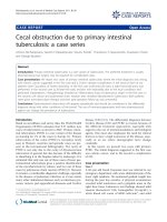
The initial CT scan of the thorax showed a 12 × 4 cm solid
mass paravertebral right in the lower thorax without any
signs of metastases (Figure 1). The bronchoscopy (Figure
2) with non-bleeding biopsy revealed a mass of the lower
right bronchus which histologically and immunohisto-
logically provided evidence of a granular cell or Abrikos-
soff tumor [1]. The bronchial lavage which followed was
negative for malignant cells. The patient was discharged
and surgical intervention was planned.
Four days after discharge a spontaneous hemothorax
developed. The patient needed to be readmitted and the
hemothorax was drained. No malignant cells were
detected in the cytological examination of the drained liq-
uid. After an uneventful course and decreasing of the
hematoma, the tumor was excised by performing a lower
right lobectomy 6 months after the initial admission. The
final histological examination confirmed a peribronchial
and infiltrating S100 positive tumor supporting the
Schwann cell origin theory with very low growth rate of
2% and a size of 15 mm (Figure 3).
About 130 cases of pulmonary occurrence of Abrikossoff's
tumor have been described in the literature up until now.
Van der Maten et al. [2] reported an incidence of this
mostly benign and slow-growing tumor in the tracheo-
bronchial system in the Netherlands of 2:100,000. In this
retrospective case series, the upper tracheobronchial sys-
tem was more frequently affected than the lower part, and
65% of the patients were smokers. Valenstein [3] reported
a more frequent occurrence on the right than on the left
side, and most commonly with a cough as the presenting
symptom. This kind of tumor can occur anywhere in the
Published: 28 October 2009
Journal of Cardiothoracic Surgery 2009, 4:57 doi:10.1186/1749-8090-4-57
Received: 9 June 2009
Accepted: 28 October 2009
This article is available from: />© 2009 Honigmann et al; licensee BioMed Central Ltd.
This is an Open Access article distributed under the terms of the Creative Commons Attribution License ( />),
which permits unrestricted use, distribution, and reproduction in any medium, provided the original work is properly cited.
Journal of Cardiothoracic Surgery 2009, 4:57 />Page 2 of 3
(page number not for citation purposes)
body, but mainly in the head and neck region, mostly
intraoral [4-7]. Other localizations are the skin, thoracic
region, breast and GI-tract [8,9]. Only 10% are located in
the pulmonary system and of these, 25% are multiple
occurrences. Deavers [10] presented a slight trend for a
predilection of dark-skinned patients. He also reported on
the infiltrative nature of this tumor and described a peri-
bronchial tissue extension of 48% which often makes it
impossible to excise the tumor bronchoscopically. Daniel
et al. [11] reported that tumors with a diameter of 8 mm
or greater are likely to invade the full-thickness bronchial
wall, with infiltration into the peribronchial tissue. They
recommend a lobectomy or pneumonectomy for the
treatment of bronchial tumors with extensive destruction
of distal tissue. If there is no extensive distal suppuration
or tissue destruction the tumors can be excised broncho-
scopically as long as they are less than 8 mm in diameter.
Bronchoscopical treatment of larger tumors is associated
with a significant increase in the recurrence rate. In addi-
tion, the hemorrhage rate is also increased [12,13].
Our patient recovered fully from the surgical intervention
and presented in very good condition during follow-up.
Conclusion
Vascular arrosions of this tumor entity have not been
described in the literature so far. The occurrence of a
hemothorax is a rare complication but one which has to
be kept in mind by the treating surgeon.
CT-reconstructionFigure 1
CT-reconstruction.
Tumor mass (bronchoscopy)Figure 2
Tumor mass (bronchoscopy).
Immunohistological image (zoom 20 ×; S100)Figure 3
Immunohistological image (zoom 20 ×; S100).
Publish with BioMed Central and every
scientist can read your work free of charge
"BioMed Central will be the most significant development for
disseminating the results of biomedical research in our lifetime."
Sir Paul Nurse, Cancer Research UK
Your research papers will be:
available free of charge to the entire biomedical community
peer reviewed and published immediately upon acceptance
cited in PubMed and archived on PubMed Central
yours — you keep the copyright
Submit your manuscript here:
/>BioMedcentral
Journal of Cardiothoracic Surgery 2009, 4:57 />Page 3 of 3
(page number not for citation purposes)
Competing interests
The authors declare that they have no competing interests.
Authors' contributions
PH is the author of the manuscript, AW was the initial
doctor in charge, CB is the pathologist, BL performed the
lobectomy as head of surgical department. All authors
have read and approved the final version of this manu-
script.
Consent
Written informed consent was obtained from the patient
for this publication including any accompanying images.
A copy of the signed consent is available for review by the
Editor-in-Chief of this journal.
References
1. Abrikosov AA: Über Myome ausgehend von der quergestreif-
ten willkürlichen Muskulatur. Virchows Arch 1926, 260:215-33.
2. Maten J van der, Blaauwgeers JL, Sutedja TG, Kwa HB, Postmus PE,
Wagenaar SS: Granular cell tumors of the tracheobronchial
tree. J Thorac Cardiovasc Surg 2003, 126(3):740-3.
3. Valenstein SL, Thurer RJ: Granular cell myoblastoma of the
bronchus. Case report and literature review. J Thorac Cardio-
vasc Surg 1978, 76(4):465-8.
4. Buley ID, Garter KC, Kelly PMA, Heryet A, Millard PR: Granular
cell turnouts revisited. An immunhistochemical and
ultrastructural study. Histopathology 1988, 12:263-274.
5. Enzinger FM, Weiss SW: Soft tissue tumors. St. Louis-Baltimore-Ber-
lin-Boston-Carlsbad-Chicago-London-Madrid-Naples-NewYork-Philadelphia-
Sydney-Tokyo-Toronto, Mosby-Year Book 1995:864-875.
6. Lack EE, Worsham GF, Callihan MD, Crawford BE, Klappenbach S,
Rowden G, Chun B: Granular Cell Tumor: A Clinicopathologic
Study of 110 Patients. J Surg Oncol 1980, 13:301-316.
7. Peterson LJ: Granular cell tumor. Review of the literature and
report of a case. Oral Surg 1974, 37:728-735.
8. Koch M, Hanke S, Dittert J, Stoelben E: Die thorakoskopische
Excision eines unklaren intrumuralen Oesophagustumors
(Granularzell-tumor/Abrikossof Tumor). Chirurg 1998,
69:981-984.
9. Orlowska J, Pachlewski J, Gugulski A, Butruk E: A conservative
approach to granular cell tumors of the esophagus: four case
reports and literature review. Aln J Gastrnenterol 1993,
88:311-315.
10. Deavers M, Guinee D, Koss MN, Travis WD: Granular cell tumors
of the lung. Clinicopathologic study of 20 cases. Am J Surg
Pathol 1995, 19(6):627-35.
11. Daniel TM, Smith RH, Faunce HF, Sylvest VM: Transbronchoscopic
versus surgical resection of tracheobronchial granular cell
myoblastomas. Suggested approach based on follow-up of all
treated cases. J Thorac Cardiovasc Surg 1980,
80(6):898-903.
12. Ramsey JH: Bronchial granular cell myoblastomas. Arch
Otolaryngol 1955, 62:81-83.
13. Kommel RM, Bernstein J: Granular cell myoblastoma of the
bronchus. Report of a case. Harper Hosp Bull 1960, 18:20-24.