báo cáo khoa học: " Overexpression of Pim-1 in bladder cancer" pdf
Bạn đang xem bản rút gọn của tài liệu. Xem và tải ngay bản đầy đủ của tài liệu tại đây (607.23 KB, 7 trang )
RESEARC H Open Access
Overexpression of Pim-1 in bladder cancer
Shengjie Guo
1†
, Xiaopeng Mao
1†
, Junxing Chen
1
, Bin Huang
1
, Chu Jin
2,3
, Zhenbo Xu
2,4
, Shaopeng Qiu
1*
Abstract
Background: Pim-1 is a serine-threonine kinase which promotes early transformation, cell proliferation and cell
survival during tumorigenesis. Several studies have demonstrated that Pim-1 kinase play a role in different cancer
types, however, the function of Pim-1 in bladder cancer is poorly understood.
Methods: Expression and localization of Pim-1 in human normal and malignant bladder specimens were examined
by Immunohistochemistry and Pim-1 staining score was compared with several clinicopathologic parameters. To
further demonstrate the biological function of Pim-1 in bladder cancer, its expression was validated in five bladder
cancer cell lines by western blot and immunohistochemistry analyses. Subsequent knockdown of Pim-1 was
achieved by lentivirus encoding small interfering RNA, and the effect of Pim-1 on bladder cell survival and drug
sensitivity were further assessed by colony formation and cell proliferation assays.
Results: When compared with normal epithelium, Pim-1 was overexpressed in bladder cancer epithelium, and the
expression level was higher in invasive bl adder cancer than Non-invasive bladder cancer specimens. Pim-1 was also
detected in all the bladder cancer cell lines examined in our study. Moreover, the knockdown of Pim-1 significantly
inhibited bladder cancer cell growth and also sensitized cells to chemotherapeutic drugs in vitro.
Conclusions: Our results in this study suggest that Pim-1 may play a role in bladder cancer initiation and
progression. Since Pim-1 is also involved in bladder cancer cell surviv al and drug resistance, Pim-1 is a potential
candidate for targeted therapy in bladder cancer.
Background
Bladder cancer is one of the most common types of
cancer globally, with approximately 75% of the diag-
nosed tumors classified as Non-invasive tumor (Ta, Tis,
or T1). Treatment of Non-invasive tumor includes
transurethral resection (TUR) with or without intravesi-
cal instillation therapy, but the recurrence rate is high,
ranging from 50% to 70%. In addition, an average of
10% to 20% for Non-invasive tu mors may fu rther pro-
gress to muscle-invasive disease, thus lead to eventual
radical Cystectomy and urinary diversion [1-3]. In this
context, clinicians face challenges to identify the novel
therapeutic targets for bladder cancer.
Pim-1 is overexpressed in several types of cancer,
including lymphoid and haematopoietic malignancies
[4], prostate cancer [5], squamous cell carcinomas [6],
gastric carcinoma and colorectal carcinomas [7].
Currently a vailable studies have demonstrated that the
expression of Pim-1 can be predictive of tumor outcome
following chemotherapy and surgery, and it is correlated
with the enhanced metastatic potential of the tumor [8].
As a member of serine/threonine kinase family, Pim-1
has multiple roles in tumorigenesis such as promoting
transformation and cell proliferation partly through reg-
ulation of cell cycle and transcription by phosphorylat-
ing of number of substrates including cdc25A/C, HP1,
and p100 [9-11]. Moreover, it has bee n shown that Pim-
1 may play a role in the regulation of the survival signal-
ing through the modulation of Bcl-2 family member
including Bad, Bcl-2 and Bcl-XL [12-14]. However, the
expression and significance of Pim-1 in bladder cancer
remains unknown. Therefore, the aims of the present
study are to investigate the expression level of Pim-1 in
bladder cancer tissue and study its function in the
pathogenesis and progression of bladder cancer.
* Correspondence:
† Contributed equally
1
Department of Urology, the First Affiliated Hospital, Sun Yat-Sen University,
Guangzhou, 510080 China
Full list of author information is available at the end of the article
Guo et al. Journal of Experimental & Clinical Cancer Research 2010, 29:161
/>© 2010 Guo et al; licensee BioMed Central Ltd. This is an Open Access article distributed under the terms of the Creative Commons
Attribution License ( which permits unrestr icted use, distribution, and reproduction in
any medium, provided the original work is properly ci ted.
Methods
Patient samples
Sixty-six clinical bladder samples isolated fr om the First
Affiliated Hospital of the Sun Yat-Sen University
(Guangzhou, China), were examin ed in the present
study. All patients including forty-eight men (72.3%)
and eighteen women (27.7%), had been treated for
urothelial carcinoma of the bladder by transurethral
resection of bladder (TUR) or Cystectomy and were
diagnosed with a bladder cancer for the first time at an
average age of 56 years (range, 33-78 years). Pathologic
staging and grading were performed according to the
2002 TNM classification systemandWorldHealth
Organization criteria, respectively. The use of the
human tissue in this study was approved by the Ethics
Council of the Sun Yat-Sen University f or Approval of
Research Involving Human Subjects.
Immunohistochemistry
All 5μm thick paraffin sections were deparaffinized
with xylene a nd rehydrated through graded alcohol
washes, followed by antigen retrieval by heating sec-
tions in sodium citrate buffer (10 mmol/L, pH6.0) for
30 minutes. Endogenous peroxidase activity was
blocked with 30 min incubation in 0.03% H
2
O
2
in
methanol. The slides were then blocked by incubation
in normal goat serum (dilution 1:10) in PBS (pH 7.4)
and subsequently incubated for monoclonal mouse
IgG1 anti-Pim-1 antibody(sc-13513; Santa Cruz Bio-
technology, Santa Cruz, CA, USA) with 1:30 dilution
at 4°C overni ght. Following this step, slides were trea-
ted with biotin-labeled anti-IgG and incubated w ith
preformed avidin -biotin pero xidase complex. Con trol
staining of the same sections was performed with the
preimmune primary antibody, and no Pim-1 immunos-
taining was observed in t hese sections. The sections
were briefly counter-stained with hematoxylin. IHC
reactions for all samples were repeated at least three
times, and typical results were illustrated.
Scoring and Statistical analyses
The staining of Pim-1 was graded in each samp le based
on the intensity of the immunoreactivity in the cancer
cells and was stratified a s strong staining (3), modera te
staining (2), weak staining (1) and negative (0). Using
these criteria, the immunostaining results were evaluated
independentlybyXPMandBH.Thecorrelationof
interobserver was calculated from the independent eva-
luations. For cases with discrepancy, a consensus was
reached during a common evaluation session. The sta-
tistical analyses w ere carried out by u sing SAS version
9.0 statistics software (SAS Institute, Inc., Cary, NC).
Cell culture and lentiviral infection
Bladder cancer cell lines T24, UM-UC-3, 5637, J82 and
RT-4 w ere purchased from the American Type Culture
Collection. UM-UC-3 an d T24 cells were grown in Dul-
becco’s modified Eagle’ s medium. 5637, J82 and RT-4
cells were maintained in R PMI 1640 with 10% fetal
bovine serum and 1% (v/v) penicillin a nd streptomycin
(100 μg/ml) and maintained at 37°C in a 5% CO
2
atmo-
sphere. The inf ection of len tivirus of Pi m-1 siRNA was
carried out as reported previously [15].
Western Blot
Western blot was performed as described previously
[16]. Briefly, the equal amounts of sample were resolved
on a SDS polyacrylamide gel and transferred to a polyvi-
nylidene difluoride membrane. Blots were incubated
with the indicated primary antibodies overnight at 4°C
and followed by detection with horseradish peroxidase-
conjugated secondary antibody. The monoclonal anti-
Pim-1 antibody was used at the dil ution of 1:300,
whereas anti-tubulin, Bcl-2, Bad and p-Bad (Ser112)
(Santa Cruz Biotechnology, Santa Cruz, CA, USA) were
used at the dilution of 1:2,000.
Cell immunoperoxidase staining
Bladder cancer cells were plated onto the glass slides.
After 24 h, cells were fixed with ice-cold a cetone. The
endogenous peroxides activity was inactivated by incu-
bating cells with 0.03% H
2
O
2
for 10 min. Slides were
then incubated with Pim-1 antibody at room tempera-
ture for 1 hour and followed by horseradish peroxides-
conjugated anti-mouse Ig (Chemicon; 1:500 dilutions).
Finally, slides were incubated with biotin- labeled anti-
IgG avidin-biotin peroxidase complex and developed
with DAB Solution.
Colony formation assay
The cells (1 × 10
4
) were seeded in 6-well plate and
infected with the lentivirus expressing control siRNA or
Pim-1 siRNA. Cell culture was maintained in complete
medium for two weeks. The cell colonies were then
visualized by Coomassie blue staining.
Drug-sensitivity assay
Cells were infected with lentivirus encoding control
siRNA or Pim-1 siRNA. At 48 h post-infection, cells
were seeded on 96-well plate at a density of 6 × 10
3
cells/well. After 24 h , cells were treated with various
doses of Doxorubicin or Docetaxel (Sigma, St Louis,
MO, USA) for another 48 h. The cells viability was mea-
sured by the WST-1 (Roche) assay follo wing the manu-
facturer’s instructions.
Guo et al. Journal of Experimental & Clinical Cancer Research 2010, 29:161
/>Page 2 of 7
Results
Overexpression of Pim-1 in human bladder cancer
specimens
To validate the expression of Pim-1 protein in bladder
cancer, human bladder specimens containing normal
epithelium (n = 21) and malignant tissues (n = 45)
were studied by immunohistochemistry using Pim-1
antibody. The staining data showed that Pim-1 expres-
sion is weakely detect in the epithelial cells of normal
bladder epithelium, however, most of the malignant
bladder epithelial cells exhibited Pim-1 immunoreactiv-
ity in both cytoplasm and nuclear (Figure 1). For
further analysis, the immunoreactivity of Pim-1
was divided into negative (score 0-1) vs. positive (score
2-3) subgroups. Detailed staining scores in normal and
malignant bladder specimens are presented in Table 1,
which showed that Pim-1 expr ession is significantly
higher in bladder cancer specimens (84.4%) than in
normal specimens (9.5%) (p < 0.001), suggesting an
overexpression of Pim-1 at the translational level in
bladder cancer.
To explore potential correlations between the
expression of Pim-1 and tumor progression, malignant
bladder specimens were further cla ssified into Non-
invasive (Tis, Ta and T1) and invasive (≥T2) groups.
Thedata(Table2)showsthatthestainingintensityof
Pim-1 is increased in invasive bladder carcinoma
samples (95%) when compared with Non-invasive blad-
der cancer specimens (76%)(p < 0.01). However,
correlation of Pim-1 within different tumor grades was
not observed (data not shown). Taken together, Pim-1
may be associated with bladder cancer initiation and
progression.
Expression profile of Pim-1 in bladder cancer cell lines
In o rder to further demonstrate the role and function of
Pim-1 in bladder cancer, the expression level of Pim-1
was validated in bladder cancer cell lines using western
blot. As shown in Figure 2A, Pim-1 is expressed in a ll
five bladder cancer cell lines at variable levels, with the
maximum l evel in highly invasive cancer cell lines T24
and UM-UC-3.
The localization of Pim-1 in bladder cancer cells was
confirmed by immunoperoxidase staining and as the
results showed that Pim-1 was detected in all human
bladder cell lines examined, including T24, UM-UC-3,
5637, J82 and RT-4. Representative images are pre-
sented in Figure 2B. The positive signals were primar-
ily immunolocalized in both cell cytoplasm and
nucleus, while some cell membrane staining is also
detected.
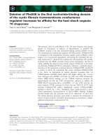
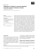
Figure 1 Overexpression of Pim-1 in human bladder cancer specimens. Pim-1 is overexpressed in both cytoplasm and nucleus of bladder
cancer cells. Normal bladder epithelium cells show no or minimal staining (A&D). Bladder cancer cells show cytoplasm and nucleus positive
staining (B&E). Invasive bladder cancer cells show strong staining(C&F). Magnification × 200 (A, B, C), or × 400 (D, E, F).
Table 1 Pim-1 immunostaining intensity in human
normal and maligancy bladder tissues
groups n negtive positive
Normal 21 19(90.5%) 2(9.5%)
Malignancy 45 7(15.6%) 38(84.4%)
p < 0.001
Guo et al. Journal of Experimental & Clinical Cancer Research 2010, 29:161
/>Page 3 of 7
Pim-1 is essential for bladder cancer cell survival
To examine the biological si gnificance of Pim-1, targeted
knockdown of Pim-1 was achieved by lentivirus encoding
siRNA specific for Pim-1 in T24 and UM-UC-3 cells,
which express relatively high levels of Pim-1. The Pim-1
siRNA using in our experiments has been previously
shown to specific knockdown Pim-1 in multiple prostate
cancer cell lines [17,18]. As shown in Figure 3A, downre-
gulation of Pim-1 decreased Phospho-Bad and Bcl-2
levels that are known to be regulated by Pim-1. Further-
more, downregulation of Pim-1 could also inhibit the cell
growth and proliferation in vitro (Figure 3B ), suggesting
that Pim-1 may be important for the growth and survival
of bladder cancer cells.
Knockdown of Pim-1 sensitizes bladder cancer cells to
chemotherapy in vitro
As Pim-1 is involved in drug resistance in some cancer
types and adjuvant intravesical chemotherapy is one of
the most common treatments in bladder cancer, we
tested whether Pim-1 is also involved in drug response
of bladder cancer cells. T24 and UM-UC-3 cells were
treated with lentivirus encoding the siRNA specific for
vector control or Pim-1 an d then were tested for their
responses to chemotherapeutic drugs. As sho wn in
Figure 3C, downregulation of Pim-1 sensitized T24 and
UM-UC-3 cells to Dox orubicin (DOX) and Docetaxel
(DTX) when compared to the vector control. Our data
implied that Pim-1 may contribute to the resistance of
apoptosis and survival of bladder cancer cells in
response to cytotoxic drugs.
Discussion
Inthepresentstudywedemonstratedforthefirsttime
that, Pim-1 was increased in human bladder cancer
epithelium as compared with that in normal bladder tis-
sue. When the tumors were stratified by Non-invasive
and invasive, a statistically significant increase of Pim-1
expression was found in the subgroup of invasive tumor
when compared with that in the Non-invasive tumor.
Pim-1 was also detected in all human bladder cancer
cell lines tested in our study. Knockdown Pim-1 led to
decreased phosphorylation of Bad and reduced expres-
sion of Bcl-2. Furthermore, downregulation of Pim-1
inhibited the bladder cancer cells growth and sensitized
them to chemotherapy in vitro. Further evaluation of
the prognostic significance of Pim-1 in a larger cohort
with sufficient follow-up times will allow better under-
stand of the clinical significance of Pim-1.
Table 2 Pim-1 immunostaining intensity in No-invasive
and Invasive bladder tumors
groups n negtive positive
Non-invasive 25 6(24.0%) 19(76.0%)
Invasive 20 1(5%) 19(95.0%)
p < 0.01
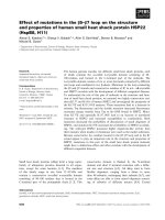
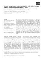
Figure 2 Expression profile of Pim-1 in bladder cancer cell lines. A. Expression profile of Pim-1 in bladder cancer cell lines. Cell lysate from
five bladder cancer cell lines were examined by western blot for Pim-1. Tubulin is as the loading control. B. The expression and localization of
Pim-1 in human bladder cancer cell lines. Cells were immunoperoxidase stained with Pim-1 antibody as described as methods. Original
magnification ×400.
Guo et al. Journal of Experimental & Clinical Cancer Research 2010, 29:161
/>Page 4 of 7
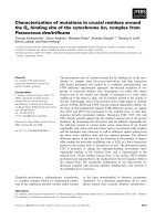
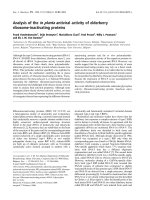
Figure 3 Downregula tion of Pim-1 inhibited the bladder cells growth an d sensi tized them to Dox orubicin and Docetaxel treatme nt.
A. Knockdown of Pim-1 decreased the phosphorylation of Bad and the expression of Bcl-2. The cells were infected lentivirus siRNA specific for
Pim-1(si Pim-1) or vector control. At 48 h postinfection, cells were lysed and the lysates were subjected to western blot with indicated antibody.
B. Downregulation of Pim-1 inhibited the bladder cancer cell growth. Total of 1 × 10
4
T24 and UM-UC-3 cells were plated in each well of a 6-
well plate and infected with lentivirus encoding Pim-1 siRNA or vector control siRNA. The cell culture was maintained in complete medium for
two weeks. Finally, the cell colonies were visualized by Coomassie blue staining. C. Decreased expression of Pim-1 sensitized bladder cancer cells
to Doxorubicin and Docetaxel treatment. The cells were plated on 96 wells and infected with lentivirus encoding Pim-1 siRNA or vector control
siRNA. At postinfection for 48 h, cells were treated with DOX (T24, 2.5 and 5μg/ml; UM-UC-3, 1.25 and 2.5 μg/ml) and DTX (T24, 25 and 50 nm;
UM-UC-3, 2.5 and 5 nm) for another 48 h. The cell viability was assessed by WST-1 assay.*, p < 0.05 compared with the control; **, p < 0.01
compared with control.
Guo et al. Journal of Experimental & Clinical Cancer Research 2010, 29:161
/>Page 5 of 7
Overexpression of the Pim-1 protein has been
reported in hematolymphoid malignancies and solid
cancers [4,5]. Pim-1 has been asserted to promote
tumorigenesis through multiple mechanisms, including
its interaction with other proteins such as c-myc,
p27
KIP1
,p21
Cip1/WAF1
, Bad, Cdc25A/C dual specificity
phos phates, androgen receptors and its ability to induce
genomic instability [19-22]. The oncogenic effect of
Pim-1 on non-haematopoietic malignancies is currently
under investigation. Ellwood-Yen et al demonstr ated
that the overexpression of Pim-1, in cooperation with
increased levels of c-myc, could lead to murine prostatic
intraepithelial neoplasia and invasive adenocarcinoma in
c-myc transgenic mice [23]. Taking into account the
biological role of Pim-1 as an oncoprotein involved in
cell cycle regulation and proliferative processes, our
results suggested possible implication of Pim-1 in the
initiation of bladder carcinogenesis. Moreover, upregula-
tion of Pim-1 in invasive bladder cancer compar ed with
Non-invasive tumors indicated that Pim-1 al so may also
contribute to bladder cancer progression.
Pim-1 has been cons idered as a survival kinase. Inhibi-
tion of Pim-1 results in a significant growth repression of
prostate cancer cell [24]. Several inhibitors of Pim-1 have
been shown to inhibit the growth of cancer cells, such as
leukemic cells as well as prostate cancer cells. There are
clinical trials to explore the safety of one of the Pim-1
inhibitor, SGI-1776, for the treatment of refractory non-
Hodgkin’s lymphoma and prostate cancer [25,26]. It also
has been demonstrated t hat Pim-1 monoclonal antibody
(mAb) could induce apoptosis in cancers cells of the
prostate, breast and colon. Furthermore, the inhibition of
Pim-1 function by treatment with Pim-1 siRNA, Pim-1
inhibitors or Pim-1 mAb sensitizes cancer cells to che-
motherapy [15,27-29]. It is notewor thy that Pim-1 inter-
acted and phosphorylated Bad, Etk and BCRP leading to
antagonism of drug-induced apoptosis [14,17,18]. In
bladder cancer, af ter an initial transurethral resection of
bladder tumor (TURBT), adjuvant intravesical therapy is
another treatment strategy used to reduce the risk of
recurrence. However, the cancer recurrence rate is still
high and the recurring canc er cells can become more
resistant to further intravesical chemotherapy. It is neces-
sary to identify an effective strategy to counter act chal-
lenges associ ated with clini cal managem ent of bladder
cancer patients. In this regard, Pim-1 might be one of the
potential therapeutic targets for the treatme nt of bladder
cancer and further studies examining Pim-1 as a target of
therapeutics are worthy of investigation.
Conclusions
To the best of our knowledge, this is the first report
showing ov erexpression of Pim-1 in bladder cancer and
its association with bladdercancercellsurvival,drug
resistance and tumor progression. The current study
offers significant information on the role and functions
of Pim-1 in bladder cancer, and may aid in the develop-
ment of novel therapy.
Acknowledgements
We would like to thank Dr Qiu (University of Maryland) for supplying the
necessary experimental material (such as lentivirus of Pim-1 siRNA). This
work was supported by grants from the National Natural Science
Foundation(30872584); Guangdong Natural Science Foundation
(8251008901000018); Doctoral Program of Guangdong Natural Science
Foundation (9451008901002062), Preceptorial Program of Higher Education
(20090171120062), Preceptorial Program of Sun Yat-Sen University (2009038)
and International program fund of 985 project of Sun Yat-Sen University,
China.
Author details
1
Department of Urology, the First Affiliated Hospital, Sun Yat-Sen University,
Guangzhou, 510080 China.
2
School of Food Science and Nutrition, Leeds
University, Leeds LS2 9JT, UK.
3
Colleges of Light Industry and Food Sciences,
South China University of Technology, Guangzhou, China.
4
Department of
Microbial Pathogenesis, Dental School, University of Maryland, Baltimore,
MD-21201, USA.
Authors’ contributions
XPM and BH evaluated the immunostainings. JXC and ZBX performed the
statistical analysis. SJG and SPQ drafted the manuscript. JC revised the
manuscript. All authors read and approved the final manuscript.
Competing interests
The authors declare that they have no competing interests.
Received: 12 November 2010 Accepted: 11 December 2010
Published: 11 December 2010
References
1. Epstein JI, Amin MB, Reuter VR, Mostofi FK: The World Health
Organization/International Society of Urological Pathology consensus
classification of urothelial (transitional cell) neoplasms of the urinary
bladder. Bladder Consensus Conference Committee. Am J Surg Pathol
1998, 22(12):1435-1448.
2. Edwards BK, Ward E, Kohler BA, et al: Annual report to the nation on the
status of cancer, 1975-2006, featuring colorectal cancer trends and
impact of interventions (risk factors, screening, and treatment) to reduce
future rates. Cancer 2010, 116(3):544-573.
3. Jemal A, Siegel R, Xu J, Ward E: Cancer statistics 2010. CA Cancer J Clin
2010, 60(5):277-300.
4. Meeker TC, Nagarajan L, ar-Rushdi A, Croce CM: Cloning and
characterization of the human PIM-1 gene: a putative oncogene related
to the protein kinases. J Cell Biochem 1987, 35(2):105-112.
5. Dhanasekaran SM, Barrette TR, Ghosh D, et al: Delineation of prognostic
biomarkers in prostate cancer. Nature 2001, 412(6849):822-826.
6. Chiang WF, Yen CY, Lin CN, et al: Up-regulation of a serine-threonine
kinase proto-oncogene Pim-1 in oral squamous cell carcinoma. Int J Oral
Maxillofac Surg 2006, 35(8):740-745.
7. Warnecke-Eberz U, Bollschweiler E, Drebber U, et al: Prognostic impact of
protein overexpression of the proto-oncogene PIM-1 in gastric cancer.
Anticancer Res 2009, 29(11):4451-4455.
8. Shah N, Pang B, Yeoh KG, et al: Potential roles for the PIM1 kinase in
human cancer - a molecular and therapeutic appraisal. Eur J Cancer 2008,
44(15):2144-2151.
9. Mochizuki T, Kitanaka C, Noguchi K, Muramatsu T, Asai A, Kuchino Y:
Physical and functional interactions between Pim-1 kinase and Cdc25A
phosphatase. Implications for the Pim-1-mediated activation of the c-
Myc signaling pathway. J Biol Chem 1999, 274(26):18659-18666.
10. Bhattacharya N, Wang Z, Davitt C, McKenzie IF, Xing PX, Magnuson NS:
Pim-1 associates with protein complexes necessary for mitosis.
Chromosoma 2002, 111(2):80-95.
Guo et al. Journal of Experimental & Clinical Cancer Research 2010, 29:161
/>Page 6 of 7
11. Leverson JD, Koskinen PJ, Orrico FC, et al: Pim-1 kinase and p100
cooperate to enhance c-Myb activity. Mol Cell 1998, 2(4):417-425.
12. Lilly M, Sandholm J, Cooper JJ, Koskinen PJ, Kraft A: The PIM-1 serine
kinase prolongs survival and inhibits apoptosis-related mitochondrial
dysfunction in part through a bcl-2-dependent pathway. Oncogene 1999,
18(27):4022-4031.
13. Yan B, Zemskova M, Holder S, et al: The PIM-2 kinase phosphorylates BAD
on serine 112 and reverses BAD-induced cell death. J Biol Chem 2003,
278(46):45358-45367.
14. Aho TL, Sandholm J, Peltola KJ, Mankonen HP, Lilly M, Koskinen PJ: Pim-1
kinase promotes inactivation of the pro-apoptotic Bad protein by
phosphorylating it on the Ser112 gatekeeper site. FEBS Lett 2004, 571(1-
3):43-49.
15. Kim O, Jiang T, Xie Y, Guo Z, Chen H, Qiu Y: Synergism of cytoplasmic
kinases in IL6-induced ligand-independent activation of androgen
receptor in prostate cancer cells. Oncogene 2004, 23(10):1838-1844.
16. Cao KY, Mao XP, Wang DH, et al: High expression of PSM-E correlated
with tumor grade in prostate cancer: a new alternatively spliced variant
of prostate-specific membrane antigen. Prostate 2007, 67(16):1791-1800.
17. Xie Y, Xu K, Dai B, et al: The 44 kDa Pim-1 kinase directly interacts with
tyrosine kinase Etk/BMX and protects human prostate cancer cells from
apoptosis induced by chemotherapeutic drugs. Oncogene 2006,
25(1):70-78.
18. Xie Y, Xu K, Linn DE, et al: The 44-kDa Pim-1 kinase phosphorylates BCRP/
ABCG2 and thereby promotes its multimerization and drug-resistant
activity in human prostate cancer cells. J Biol Chem 2008,
283(6):3349-3356.
19. Zhang Y, Wang Z, Magnuson NS: Pim-1 kinase-dependent
phosphorylation of p21Cip1/WAF1 regulates its stability and cellular
localization in H1299 cells. Mol Cancer Res 2007, 5(9):909-922.
20. Morishita D, Katayama R, Sekimizu K, Tsuruo T, Fujita N: Pim kinases
promote cell cycle progression by phosphorylating and down-regulating
p27Kip1 at the transcriptional and posttranscriptional levels. Cancer Res
2008, 68(13):5076-5085.
21. Bachmann M, Kosan C, Xing PX, Montenarh M, Hoffmann I, Moroy T: The
oncogenic serine/threonine kinase Pim-1 directly phosphorylates and
activates the G2/M specific phosphatase Cdc25C. Int J Biochem Cell Biol
2006, 38(3):430-443.
22. Wang J, Kim J, Roh M, et al: Pim1 kinase synergizes with c-MYC to induce
advanced prostate carcinoma. Oncogene 2010, 29(17):2477-2487.
23. Ellwood-Yen K, Graeber TG, Wongvipat J,
et al: Myc-driven murine prostate
cancer shares molecular features with human prostate tumors. Cancer
Cell 2003, 4(3):223-238.
24. Zhang T, Zhang X, Ding K, Yang K, Zhang Z, Xu Y: PIM-1 gene RNA
interference induces growth inhibition and apoptosis of prostate cancer
cells and suppresses tumor progression in vivo. J Surg Oncol 2010,
101(6):513-519.
25. Chen LS, Redkar S, Bearss D, Wierda WG, Gandhi V: Pim kinase inhibitor,
SGI-1776, induces apoptosis in chronic lymphocytic leukemia cells. Blood
2009, 114(19):4150-4157.
26. Mumenthaler SM, Ng PY, Hodge A, et al: Pharmacologic inhibition of Pim
kinases alters prostate cancer cell growth and resensitizes
chemoresistant cells to taxanes. Mol Cancer Ther 2009, 8(10):2882-2893.
27. Li J, Hu XF, Xing PX: Pim-1 expression and monoclonal antibody
targeting in human leukemia cell lines. Exp Hematol 2009,
37(11):1284-1294.
28. Hu XF, Li J, Vandervalk S, Wang Z, Magnuson NS, Xing PX: PIM-1-specific
mAb suppresses human and mouse tumor growth by decreasing PIM-1
levels, reducing Akt phosphorylation, and activating apoptosis. J Clin
Invest 2009, 119(2):362-375.
29. Teh BG: [Pim-1 induced by hypoxia is involved in drug resistance and
tumorigenesis of solid tumor cells]. Hokkaido Igaku Zasshi 2004,
79(1):19-26.
doi:10.1186/1756-9966-29-161
Cite this article as: Guo et al.: Overexpression of Pim-1 in bladder
cancer. Journal of Experimental & Clinical Cancer Research 2010 29:161.
Submit your next manuscript to BioMed Central
and take full advantage of:
• Convenient online submission
• Thorough peer review
• No space constraints or color figure charges
• Immediate publication on acceptance
• Inclusion in PubMed, CAS, Scopus and Google Scholar
• Research which is freely available for redistribution
Submit your manuscript at
www.biomedcentral.com/submit
Guo et al. Journal of Experimental & Clinical Cancer Research 2010, 29:161
/>Page 7 of 7