báo cáo khoa học: "Enhancive effects of Lewis y antigen on CD44-mediated adhesion and spreading of human ovarian cancer cell line RMG-I" pps
Bạn đang xem bản rút gọn của tài liệu. Xem và tải ngay bản đầy đủ của tài liệu tại đây (580.05 KB, 8 trang )
RESEARCH Open Access
Enhancive effects of Lewis y antigen on
CD44-mediated adhesion and spreading
of human ovarian cancer cell line RMG-I
Lili Gao
1
, Limei Yan
1
, Bei Lin
1*
, Jian Gao
1
, Xiuyun Liang
1
, Yanyan Wang
1
, Juanjuan Liu
1
, Shulan Zhang
1
,
Iwamori Masao
2
Abstract
Background: This study aimed to investigate the molecular structural relationship between cell adhesive molecule
CD44 and Lewis y antigen, and determine the effects of Lewis y antigen on CD44-mediated adhesion and
spreading of ovarian cancer cell line RMG-I and the Lewis y antigen-overexpressed cell line RMG-I-H.
Methods: The expression of CD44 in RMG-I and RMG-I-H cells before and after treatment of Lewis y monoclonal
antibody was detected by immunocytochemistry; the expression of Lewis y antigen and CD44 was detected by
Western Blot. The structural relationship between Lewis y antigen and CD44 was determined by
immunoprecipitation and confocal laser scanning microscopy. The adhesion and spreading of RMG-I and RMG-I-H
cells on hyaluronic acid (HA) were observed. The expression of CD44 mRNA in RMG-I and RMG-I-H cells was
detected by real-time RT-PCR.
Results: Immunocytochemistry revealed that the expression of CD44 was significantly higher in RMG-I-H cells than
in RMG-I cells (P < 0.01), and its expression in both cell lines was significantly decreased after treatment of Lewis y
monoclonal antibody (both P < 0.01). Western Blot confirmed that the content of CD44 in RMG-I-H cells was 1.46
times of that in RMG-I cells. The co-location of Lewis y antigen and CD44 was confirmed by co-
immunoprecipitation. The co-expression of CD44 and Lewis y antigen in RMG-I-H cells was 2.24 times of that in
RMG-I cells. The adhesion and spreading of RMG-I-H cells on HA were significantly enhanced as compared to those
of RMG-I cells (P < 0.01), and this enhancement was inhibited by Lewi s y monoclonal antibody (P < 0.01). The
mRNA level of CD44 in both cell lines was similar (P > 0.05).
Conclusion: Lewis y antigen strengthens CD44-mediated adhesion and spreading of ovarian cancer cells.
Background
Glycosylated antigens , important compone nts of glycoli-
pids and glycoproteins, are widely expressed on cell
membrane and are involved in cell adhesion, recogni-
tion, and signal transduction [1]. The alterations of type
II sugar chains, such as Lewis × and Lewis y, are com-
mon in ovarian cancer: 75% of epithelial ovarian cancers
have overexpression of Lewis y antigen which shows
obvious relationship with prognosis; tumor marker
CA125 in epithelial ovarian cancer also contains Lewis y
structure [2,3]. Alpha1, 2-fucosyltransferase (a1, 2-FT)
is a key enzyme for synthes izing Lewis y antigen. In our
previous study, we successfully transferred a1, 2-FT
gene into ovarian cancer cell line RMG-I and established
a cell line RMG-I-H with stable high expression of
Lewis y antigen, which showed obviously enhanced
malignant behaviors [4-6].
CD44, one of important adhesive molecules on cells, is
involved in the adhesion and metastasis of tumor cells
and plays an important role in tumor development
[7-10], but the regulatory mechanism is unclear yet. The
molecule CD44 is abundant of a-L-fucose, and is an
important a1, 2-fucose antigen-containing protein on
the surface of cells [11]. CD44 is expressed on several
* Correspondence:
1
Department of Obstetrics and Gynecology, Shengjing Hospital Affiliated to
China Medical University, Shenyang, 110004, P R of China
Full list of author information is available at the end of the article
Gao et al. Journal of Experimental & Clinical Cancer Research 2011, 30:15
/>© 2011 Gao et al; licensee BioMed Cen tral L td. This i s an Open Access article distributed under the t erms of t he Creative Co mmons
Attribution License ( which permits unrestricte d use, distribution, and reproduction in
any medium, provided the original work is properly cited.
tissue cells, binds to receptors in extracellular matrix
such as hyaluronic acid (HA) and laminin, and mediates
cell-cell and cell-matrix adhesion [12,13]. The present
study aimed to determine the impact of a1, 2-FT gene
trans fection on the expression of CD44 on cells and the
effects of Lewis y antigen on CD44-mediated cell adhe-
sion and spreading.
Methods
Materials
Lewis y monoclonal antibody was purchased from
Abcam Co.; CD44 monoclonal antibody from Santa
Cruz Co. and Wuhan Boster Co.; Protein A-agarose,
ECL chromogenic agent, and 5× SDS-PAGE loading
buffer from Shangha i Beyotime Institute of Biotechnol-
ogy; SABC kit from Beijing ZhongshanGoldenBridge
Biotechnology Co., Ltd; HA f rom Hefei Bomei Biotech-
nology Co., Ltd; DMEM culture medium from Gibco
Co.; fetal bovine serum (FBS) from Shenyang Boermei
Reagent Co.; Coomassie brilliant blue from Beijing
Solarbio Science & Technology Co., Ltd; Trizol reagent,
PrimeScript™RT reagent k it, and SYBR
®
Premix Ex
Taq™from Dalian TaKaRa Biotechnology Co. The
sequences of primers were synthesized by Shanghai Invi-
trogen Co.
Cell line and cell culture
The cell line RMG-I was originated from ovarian clear
cell cancer tissues. The cell line RMG-I-H with high
expression of a1, 2-FT and Lewis y antigen was estab-
lished in our lab [14]. RMG-I and RMG-I-H cells
were cultured in DMEM medium containing 10% FBS
at 37°C in 5% CO
2
and saturated humidity. Cells are
grouped in immunocytochemistry, cell spreading, cell
adhesion as follows: negative groups, Lewis y antibody-
untreated groups, Lewis y antibody-treated groups
(single layer cells were treated with 10 μg/mL Lewis y
monoclonal antibody at 37°C in 5% CO
2
for 60 min),
irrelevant isotype-matched control(10 μ g/mL normal
mouse IgM).
Immunocytochemistry
RMG-I-H and RMG-I cells at exponential phase of
growth were digested by 0.25% trypsin and cultured in
DMEM medium containing 10% FBS to prepare single-
cell suspension. Cells were washed twice with cold PBS
when growing in a single layer, and fixed with 4% paraf-
ormaldehyde for 30 min. The expression of CD44 on
cells was detected according to the SABC kit instruc-
tions. The concentration of CD44 monoclonal antibody
was 1:100. The primary antibody was replaced by PBS
for negative c ontrol. 10 μg/mL normal mice IgM acted
as irrelevant isotype-matched control. The average opti-
cal densities were measured under a microscope with
image processing, being presented as the means ± stan-
dard deviation for three separate experiments.
Confocal laser scanning microscopy
After fixing with 4% paraformaldehyde, RMG-I-H cells
were treated by the one-step immunofluorescence dual-
labeling method. In brief, mouse anti-human Lewis y
antibody and rabbit anti-human CD44 antibody were
diluted to 1:100 as primary antibody s olutions; goat
anti-rabbit TRITC-labeled secondary antibody and goat
anti-mouse FITC-labeled secondary antibody were
diluted to 1:200. Cells were blocked by normal goat
serum for 30 min, added w ith primary antibody solu-
tions at 37°C for 1 h, then cultured at room temperature
overnight. After washing with PBS, cells were added
with secondary antibody solutions at 37°C for 1 h,
stained with 4, 6-diamidino-2-phenylindole (PI) for
5 min, then observed under the confocal laser scanning
microscope. The data were colleted by a computer for
digital imaging. The experiment was repeated 3 times.
Western Blot
RMG-I-H and RMG-I cells at exponential phase of
growth were washed twice with cold PBS, added with
cell lysis buffer (0.2 mL/bottle), placed on ice for
15 min, then centrifuged at 14,000 rpm for 15 min. The
protein concentration in the supernatant was detected
by the method of Coomassie brilliant blue. The superna-
tant was cultured with 1× SDS-PAGE loading buffer at
100°C for 5 min for protein denaturation. Then, 50 μg
of the protein was used for SDS-PAGE gel electrophor-
esis. The protein was transferred onto PVDF membrane,
blocked by 5% fat-free milk powder at room tempera-
ture for 2 h, added w ith primary mouse anti-human
CD44 monoclonal antibody (1:200) and mouse anti-
human Lewis y monoclonal antibody (1:1000) and
cultured at 4°C overnight, then added with secondary
HRP-labeled goat anti-mouse IgG (1:5000) and cultured
at room temperature for 2 h, and finally visualized by
ECL reagent. The experiment was repeated 3 times.
Co-immunoprecipitation
The protein was extracted from cells before and after
transfection with the method described in Western Blot
section. After protein quantificatio n, 500 μg of each cell
lysis was added with 1 μg of CD44 monoclonal antibody
and shaken at 4°C overnight, then added with 40 μLof
Protein A-agarose and shaken at 4°C for 2 h, finally cen-
trifuged at 2500 rpm for 5 min and washed to collect
the precipitation. The precipitated protein was added
with 20 μL of 1× SDS-PAGE loading buffer at 100°C for
5 min for denaturation. The supernatant was subjected
to SDS-PAGE gel electrophoresis. Lewis y monocl onal
antibody (1:1000) was used to detect Lewis y antigen.
Gao et al. Journal of Experimental & Clinical Cancer Research 2011, 30:15
/>Page 2 of 8
Other steps were the same as described in Western Blot
section.
Cell spreading
The 2 mg/mL HA-coated 35-mm culture dishes were
placed at 37°C for 1 h, and then blocked by 1% bovine
serum albumin (BSA) for 1 h. The single-cell suspension
(15,000/mL) prepared with serum-free DMEM was
added to the dishes (1 mL/well) and cultured at 37°C in
5% CO
2
for 90 min. Under the inverted microscope, 3
to 5 visual fields ( ×200) were randomly selected to
count 200 cells: the round and bright cells were counted
as non-spreading cells; the oval cells with pseudopods
were counted as spreading cells. Irrelevant contro l anti-
bodies (10 mg/ml) are used to evalua te the specificit y of
the inhibitions. The experiment was repeated 3 times.
Cell adhesion
The 96-well plates were coated with 2 mg/ml HA (50
μL/well). The plate coated with 3 mg/mL polylysine
and 1% BSA was used as maximal and minimal adhe-
sion controls, respectively. After 2-hour coating at
37°C, the plates were washed twice with PBS, and
blocked again with 1% BSA for 2 h. The cells were
digested by 0.25% trypsin, centrifuged at 1000 rpm for
5 min, and then added with serum-free DMEM cul-
ture medium to prepare single-cell suspension. Cells
were diluted t o 5 × 10
4
/mL,addedtocoatedplates
(100 μL/well) and cultured at 37°C in 5% CO
2
for 2 h.
After washing off the un-adhered cells, the 96-well
plates were fixed by 4% paraformaldehyde for 30 min,
stained with 0.5% crystal violet (100 μL/well) for 2 h,
and then washed twice with cold PBS. The absorbance
at 597 nm (A
597
absorbance represents the adhesive
cells) was detected by a microplate reader. Irrelevant
control antibodies (10 mg/ml) are used to evaluate the
specificity of the inhibitions. The experiment was
repeated 3 times.
Detecting CD44 mRNA in RMG-I and RMG-I-H cells by
real-time PCR
RMG-I and RMG-I-H cells at exponential phase of
growth were added with Trizol reagent (1 mL per 1 × 10
7
cells) to extract total RNA. The concentration and purity
of RNA were detected by an ultraviolet spectrometer.
cDNA was synthesized according to the RNA reverse
transcription kit instructions (TaKaRa Co.). The reaction
system contained 4 µL of 5× PrimeScript™Buffer, 1 µL
of PrimeScript™RT Enzyme Mix I, 1 µL of 50 µmol/L
Oligo dT Primer, 1 µL of 100 µmol/L Random 6 mers,
2µLoftotalRNA,and11µLofRNase-freedH
2
O. The
reaction conditions were 37°C for 15 min, 85°C for 5 s,
and 4°C for 5 min. The sequences of CD 44 gene primers
were 5’-CCAATG CCTTTGATGGACCA-3’ for forward
primer and 5’-TGTGAGTGTCCATCTGATTC-3’ for
reverse primer. The sequences of a1,2-FT gene primers
were 5’-AGGTCATCCCTGAGCTGAAACGG-3’ for for-
ward primer and 5’-CGCCTGCTTCACCA CCTTCTTG-
3’ for reverse primer. The sequences of b-actin gene
primers were 5’-GGACTTCGAGCAAGAGATGG-3’ for
forward primer and 5’-ACATCTGCTGGAAGGTG-
GAC-3’ for reverse primer. The reaction system for
real-time fluorescent PCR contained 5 µL of 2× SYBR
®
Premix Ex Taq™ ,0.5μLof5μmol/L PCR forward
primer, 0.5 μLof5μmol/L PCR reverse primer, 1 µL
ofcDNA,and3µLofdH
2
O. The reaction conditions
were 45 cycles of denaturation at 95°C for 20 s and
annealing at 60°C for 60 s. The Light Cycler PCR sys-
tem (Roche Diagnostics, Mannheim, Germany) was
used for real-time PCR amplification and Ct value
detection. The melting curves were analyzed after
amplification. PCR reactions of each sample were done
in triplicate. Data were analyzed through the compara-
tive threshold cycle (CT) method.
Statistical analyses
All data are expressed as mean ± standard deviation and
were processed by the SPSS17.0 software. Raw data
were analyzed by the variance analysis. A value of P <
0.05 was considered to be statistically significant.
Results
The expression of CD44 in RMG-I and RMG-I-H cells
Immunocytochemistry showed that the positive CD44
staining presented as light yellow particles in the cyto-
plasm of RMG-I cells and brown- yellow particles in the
cytoplasm and on the membrane of RMG-I-H cells
(Figure 1). The relative level of CD44 expression was
significantlyhigherinRMG-I-HcellsthaninRMG-I
cells (P < 0.01) (Table 1).
After treatment of Lewis y monoclonal antibody, the
expression of CD44 was decreased in both RMG-I-H
cells and RMG-I cells (P <0.01),moreovershowedno
significant difference between the two cell lines (P >
0.05); after treatment of normal mouse IgM, the expres-
sion of CD44 did not change in RMG-I-H cells and
RMG-I cells, compared with Lewis y antibody-untreated
groups(Figure 1 Table 1).
Co-location of CD44 and Lewis y antigen on RMG-I-H cells
Under the confocal laser scanning microscope, CD44
presented red fluoscence mainly on cell membrane and
partly in cytoplasm; Lewis y antigen presented green
fluoscence mainly on cell membrane (Figure 2). Both
red fluoscence and green fluoscence were accumulated
at the margin of cell clusters and overlapped as yellow
fluoscence, indicating the co-location of CD44 and
Lewis y antigen.
Gao et al. Journal of Experimental & Clinical Cancer Research 2011, 30:15
/>Page 3 of 8
The expression of CD44 and Lewis y antigen in RMG-I
and RMG-I-H cells
Western Blot showed that the expression of CD44 in
RMG-I-H cells was significantly increased by 1.46 times
of that in RMG-I cells (P < 0.01) (Figur e 3.BD), and the
expression of Lewis y antigen was signifi cantly increased
by 2.98 times (P < 0.01) (Figure 3.AD). Immunoprecipi-
tation showed that, using the ratio of Lewis y antigen
expression to CD44 expression to represent the relative
expression of Lewis y antigeninCD44,theexpression
of Lewis y antigen in RMG-I-H cells was increased by
2.24 times of that in RMG-I cells (P <0.01)(Figure3.
CD).
The mRNA levels of CD44 and a1,2-FT in RMG-I and
RMG-I-H cells
The 2
-ΔΔCT
value of mRNA level of CD44 in RMG-I-H
cells is 79% of that in RMG-I cells, which had no sig-
nificant difference (P > 0.05), whereas the mRNA level
of a1,2-FT in RMG-I-H cells was increased by 3.07
times of that in RMG-I cells detected by Real-time
PCR (P < 0.01). (Figure 4).
HA-mediated cell adhesion and spreading
The adhesion of RMG-I-H cells to HA was significantly
stronger than that of RMG-I cells (P < 0.01) (Table 2).
The adhesion of RMG-I-H and RMG-I cells to HA after
Lewis y antigen blocking was decreased respectively by
62.31% and 70.34% of irrelevant isotype-matched control
(P < 0.01), and no difference was observed between
these two cell lines (P > 0.05). Cell adhesion did not
change after treatment of normal mouse IgM, compared
with Lewis y antibody-untreated groups (P > 0.05).
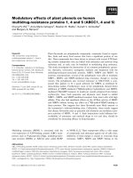
Figure 1 The expression of CD44 in RMG-I and RMG-I-H cells detected by immunocytochemistry (×400). Panels 1 and 5 are negative
controls; panels 2 and 6 are Lewis y antibody-untreated cells; panels 3 and 7 are Lewis y antibody-treated cells; panels 4 and 8 are cells treated
by irrelevant isotype-matched control. The expression of CD44 was detected by SABC methods in RMG-I and RMG-I-H cells, and brown color
degree by DAB staining indicated the expression level of CD44. It can be seen from the figure that the expression of CD44 in the RMG-I-H cells
was stronger than that in RMG-I cells, which was decreased after Lewis y antibody blocking.
Table 1 The average optical density on
immunocytochemical staining with CD44 antibodies
Group RMG-I RMG-I-H
Negative control 0.02 ± 0.03 0.03 ± 0.01
Lewis y antibody-untreated 0.28 ± 0.02 0.49 ± 0.02*
Lewis y antibody-treated 0.11 ± 0.01** 0.11 ± 0.01**
Irrelevant isotype-matched control 0.26 ± 0.01 0.46 ± 0.01
* P < 0.01, vs. RMG-I cells; ** P < 0.01, vs. Irrelevant isotype-matched control.
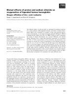
Figure 2 Co-location of CD44 and Lewis y antigen on RMG-I-H
cells observed under confocal laser scanning microscope. Red
fluoscence on the upper left panel indicates CD44 expression; green
fluoscence on the upper right panel indicates Lewis y antigen
expression; blue fluoscence on the upper right panel indicates cell
nuclear location; the lower right panel is a merged image of the
other three panels. Lewis y antigen CD44 mainly expressed in the
cell membrane observed under the confocal laser scanning
microscope, and it were seen as yellow fluorescence after the two
overlap, suggesting that Lewis y antigen and CD44 co-localizated in
the cell membrane.
Gao et al. Journal of Experimental & Clinical Cancer Research 2011, 30:15
/>Page 4 of 8
On HA-coated plates, spreading RMG-I-H cells were
significantly more than spreading RMG-I cells (P < 0.01)
(Table 2). Cell spreading showed similar changes as cell
adh esio n after Lewis y antigen blocking, suggesting that
Lewis y antigen was involved in the interaction of CD44
and HA.
Discussion
This article mainly found that Lewis y antigen, as a
structure in CD44 molecule, strengthens CD44-
mediated adhesion and s preading of ovarian cancer
cells. Inhibiting the expression of CD44 or blocking its
binding to receptors and downstream signal molecules
can inhibit the progression of ovarian cancer.
Glycoconjugates, an important component of cell
membrane, are involved in cell growth and differentia-
tion [15]. Fucose, the terminal residue of synthesized
sugar chains, is involved in constructing the sugar chain
structure of some important growth factor receptors
and plays a n important role in tumorigenesis [16]. Stu-
dies showed that fucosylated antigens expressed in
tumor cells are involved in several cellular functions and
related to some mal ignant cell behaviors, including
adhesion, recogniti on, and signal transduction, and that
the increased fucosylated antigens benefit the invasion
and migration of tumor cells [17,18]. Ovarian cancer
mostly has changes of type II glycosylated antigens, such
as Lewis x, Lewis y and H antigens, which mainly
depend on the a1, 2-FT-catalyzed fucosylation of
galactose residues at the non-reducing terminal [19].
Our previous study showed that ovarian canc er cel l line
RMG-I mainly expressed L ewis × antigen, and
confirmed that the enhanced adhesion of Lewis × a nti-
gen-overexpressed cells to peritoneal mesothelia was
weakened after Lewis × antigen blocking in nude mouse
experiments, suggesting that Lewis × antigen is related
to the intraperitoneal dissemination of RMG-I cells [20].
We transfected wild type a1,2-FT gene into ovarian
cancer cell line RMG-I to establish the a1,2- FT-overex-
pressed cell line RMG-I-H, and found that the activity
of a1,2-FT in RMG-I-H cells was enhanced by 20 to
30 times [5]. We also found that only Lewis × and
Lewis y antigens in the type II lactose chain family were
expressed, 42.6% of Lewis × antigen in RMG-I-H cells
transformed into Lewis y antigen, and that the
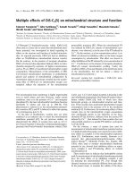
Figure 3 The expression of CD44 and Lewis y antigen in RMG-I and RMG-I-H cells. Panel A shows the expression of Lewis y antigen in
RMG-I-H cells was higher than that in RMG-I; panel B shows the expression of CD44 in RMG-I-H cells was higher than that in RMG-I; panel C
shows that Lewis y antigen, which in RMG-I-H cells was higher than that in RMG-I, was expressed both in RMG-I and RMG-I-H cells after CD44
immunoprecipitation; panel D Quantitative data were expressed as the intensity ratio target genes to beta-actin. (P < 0.01).
Figure 4 The mRNA express ion of CD44 and a1, 2-FT in RMG-I
and RMG-I-H cells were tested by quantitative Real-Time RT-
PCR. The mRNA level of a1, 2-FT was significantly increased, but the
mRNA level of CD44 was almost the same in RMG-1-hFUT cells and
RMG-1 cells. (**P < 0.01, * P > 0.05).
Gao et al. Journal of Experimental & Clinical Cancer Research 2011, 30:15
/>Page 5 of 8
concentration of Lewis y antigen in RMG-I-H cells was
increased by about 20 times of that in RMG-I cells[5].
After transfection of a1, 2-FT gene, while the expression
ofLewisyantigeninRMG-I-H cells was increased,
the malignant behaviors of cells were also enhanced, for
examples, the G1 phase of meiosis was shortened, the
colony formation rate on soft agar was increased,
the growth of subcutaneous and intraperitoneal xeno-
grafts in nude mice was accelerated, and the drug-
resistance was enhanced [6,21-23]. Lewis y antigen has
dual fucosylations–one more fucose than Lewis × anti-
gen. Lewis y monoclonal antibody or a-L-fucosidase can
significantly inhibit the proliferation and adhesion of
RMG-I-H cells [6,24], indicating that the effect of Lewis
y antigen on cell behaviors is stronger that that of Lewis
× antigen, which may due to the number of fucoses.
CD44, an important a1, 2-FT-containing protein on
cell surface, is involved in the adhesion and metastasis
of tumor cells, and plays an important role in tumor
progression [9]. Our present study showed that after
transfection of a1,2-FT gene, the expression of CD44 in
RMG-I-H ce lls was significantly increased together with
the increase of Lewis y antigen (P < 0.01). Confocal
laser sca nning microscopy confirmed the co-location of
CD44 and Lewis y antigen, interpreted that Lewis y
antigen was a structure in CD44. In 2010, Lin et al. [25]
reported that both CD173(H2) and Lewis y(CD174)
could immunoprecipitate with CD44 in breast cancer
cells. Our results showed that the increase of Lewis y
antigen was more obvious, which increased by 2.24
times after a1, 2-FT gene transfection (P < 0.0 5). Lewis
y antibody can block the increase of CD44 expression.
We used gene chip to detect the differential expression
of genes in cells before and after transfection, and
found that 88 genes were differentially expressed after
transfection, which were involved in cell proliferation
and adhesion, signal transduction, protein phosphoryla-
tion, transcription, apoptosis, and so on[22]. However,
the change o f CD44 after transfection was mainly at
protein level, with no obvious change at mRNA level
(P > 0.05). Yuan et al. [26] also believed that CD44 and
its several subtypes have post-transcriptional modifica-
tion, including the addition of glycosaminoglycan and
glycosylation.
The functions of a1, 2-FT in CD44 molecule are
unclear yet. Studies found that it can prevent decompo-
sition by proteolytic enzyme, enhance cell-cell adhesion,
and inhibit cell apoptosis [11]. Labarrière et al. [27] also
found that CD44v6 in mouse colon cancer cells contains
H antigen. Its fucose structure is involved in cell adhe-
sion, and the increase of its expression is related to the
decrease of the sensitivity to natural killer cells or the
decrease of the cytotoxicity of lymphoc yte-activated
killer cells. Therefore, CD44v6 helps mouse colon can-
cer cells to escape from the recognition and killing by
the immune system, prone to invade lymph nodes and
form metastasis. Our study confirmed that the adhesion
and spreading of RMG-I-H cells to HA in extracellular
matrix were significantly enhanced (all P <0.01).After
Lewis y antigen blocked, the expression of CD44 in cells
was decreased, cell adhesion and spreading were also
significantly decreased (all P < 0.01), suggesting that
Lewis y antigen plays an important role in mediating
theadhesionofCD44toHAin extracellular matrix.
Yuan et al. [26] used a-L-fucosidase to treat breast can-
cer cells, and found that the expression of CD44 was
decreased; the adhesion of tumor cells to matrix was
decreased, resulting in a decrease of cell invasion. This
finding confirms our deduction.
The interaction of CD44 and HA a ctivates R hoA
signals and Rho kinase, enhances serine/threonine phos-
phorylation on Gab-1 (Grb2-associated binder-1),
induces PI3K activati on, triggers the PI3K/Akt pathway,
andisinvolvedintheprogressionofbreastcancer[28].
It is also confirmed that the binding of CD44 to HA
induces c-Src kinase activation, and is involved in the
metastasis of ovarian cancer cells by activating the c-Src
kinase pathway [29] . Our previous study showed that
the expression of Akt total protein in Lewis y antigen-
overexpressed ovarian cancer cells did not change, but it
phosphorylation was significantly enhanced; ZD1839
and Lewis y antibody decreased the level of phosphory-
lated Akt in Lewis y antigen-overexpressed cells, but
showed no effect in the ovarian cancer cells with low
Lewis y antigen expression. MTT assay showed that
PI3K-specific inhibitor LY294002 can significantly i nhi-
bit the proliferation of Lewis y antigen-overexpressed
ovarian cancer cells [30].
Table 2 HA-mediated adhesion and spreading of RMG-I and RMG-I-H cells
Cell adhesion Cell spreading
Group RMG-I RMG-I-H RMG-I RMG-I-H
Lewis y antibody-untreated 1.41 ± 0.20 2.57 ± 0.58* 34 ± 5 57 ± 6*
Lewis y antibody-treated 0.53 ± 0.03** 0.76 ± 0.27** 16 ± 5** 14 ± 4**
Irrelevant isotype-matched control 1.36 ± 0.15 2.44 ± 0.67 35 ± 6 59 ± 8
* P < 0.01, vs. RMG-I cells; ** P < 0.01, vs. Irrelevant isotype-matched control.
Gao et al. Journal of Experimental & Clinical Cancer Research 2011, 30:15
/>Page 6 of 8
Ovarian cancer cells adhere to peritoneal mesothelia
via the formation o f several compounds: CD44/HA,
b1-integrin/fibron ectin, CA125/mesothelin, and so on
[31,32]. HA and fibronectin are components of extracel-
lular matrix. HA in extracellular matrix is a major
ligand of CD44. Many studies proved the importance of
CD44 and its receptors in the biological behaviors of
ovarian cancer [33]. Stu dies found that oncosta tin M
and transforming growth factor 1 (TGF1) could mediate
the binding of HA to CD44 in tumor cells originated
from lung epithelia, leading to the glycosylation and
phosphatization of CD44 [34]. CD44 and HA med iate
the overexpression and activation of integrin as well as
the adhesion of tumor cells to epithelia, and enhance
the migration and metastasis of tumor cells [35]. Wie-
lenga et al. [36] reported that, in colorectal cancer,
heparin sulfate-modified CD44 showed increased ability
of binding to hepatocyte growth factor/scatter factor
(HGF/SF), thus presenting HGF/SF to c-Met and lead-
ing to c-Met phosphorylation, and triggering the c-Met
signal pathway to activate lymphocyte function-asso-
ciated antigen-1 (LFA-1), therefore, affecting the biolo-
gical activities of tumor cells, such as angiogenesis and
cell motivation. Zhang et al. [37] found that the binding
of HA to CD44 affected the adhesi on of tumor cells via
some signal transduction pathways (such as the kinase
C pathway), and played an important role in tumor
metastasis. Kim et al. [38] used CD44 ant ibody to com-
petitively inhibit the binding of HA to CD44, and found
that the invasion of colorectal cancer cells to basement
membranes was decreased by 95%. The above findings
indicate that CD44 is involved in several signal trans-
duction pathways related to tumor cell metastasis, and
that inhibiting the expression of CD44 or blocking its
binding to receptors can inhibit the metastasis of tumor
cells. Our previous study showed that the expression of
EGFR, TGF-bR, a5b1, and a5b3 was also increased in
Lewis y antigen-overexpressed cells, and that Lewis y
antigen, as an important structure in EGFR, TGF-bR,
a5b1, and a5b3 (unpublished data), affected the biologi-
cal behaviors of cells by activating the Raf/MEK/MAPK,
PI3K/Akt, TGF-b/Smads, and FAK signal pathways
[39,40].
In summary, Lewis y antigen is overexpressed on
ovarian cancer cells, and is homogeneous in primary
and metastatic lesions; hence, it has become a target
antigen of immune therapy.
Conclusions
We have transfected the alfa1, 2-fucosyltransferase gene
into cultured cells from an ovarian carcinoma and
showed that the transfected cells have elevated expres-
sion of CD44 with Lewis y resulting in their increased
ability to adhere and to spread via the CD44-hyaluronic
acid interaction. The paper demonstrates a novel role of
Lewis y in regulating the CD44- hyaluronic interaction.
Acknowledgements
This work is supported by the National Natural Science Foundation of China
(No. 30170980, 30571958, 30872757, 81072118); Natural Science Foundation
of Liaoning Province, China (No. 20052107); Ph. D. Programs Foundation of
Ministry of Education of China (No. 20070159023); Key Laboratory
Foundation from Education Department of Liaoning Province, China (No.
2008S247); Shengjing Free Researcher Project (No. 200807); Science
Committee Foundation of Shenyang City, China (No. F10-14-9-9-52).
Author details
1
Department of Obstetrics and Gynecology, Shengjing Hospital Affiliated to
China Medical University, Shenyang, 110004, P R of China.
2
Departments of
Biochemistry, Faculty of Science and Technology, Kinki University, Osaka,
577-8502, Japan.
Authors’ contributions
LG carried out most parts of the experiment; LY, JG, XL, YW, JL and SZ
participated in the exp eriment; BL participated in the design of the study; LY
performed the statistical analysis; IM participated in its design and
coordination and helped to draft the manuscript. All authors read and
approved the final manuscript.
Competing interests
The authors declare that they have no competing interests.
Received: 15 January 2011 Accepted: 7 February 2011
Published: 7 February 2011
References
1. Ugorski M, Laskowska A: Sialyl Lewis a: a tumor-associated carbohydrate
antigen involved in adhesion and metastatic potential of cancer cells.
Acta Biochim Pol 2002, 49 :303-311.
2. Diao B, Lin B: Lewis y antigen and its applications to tumor diagnosis
and treatment. J Modern Oncol 2009, 17:132-134.
3. Rodríguez-Burford C, Barnes MN, Berry W, Partridge EE, Grizzle WE:
Immunohistochemical expression of molecular markers in an avian
model: a potential model for preclinical evaluation of agents for ovarian
cancer chemoprevention. Gynecol Oncol 2001, 81:373-379.
4. Hao YY, Lin B, Zhao Y, Zhang YH, Li FF, Diao B, Ou YL, Zhang SL: α1, 2-
Fucosyltransferase gene transfection influences on biological behavior
of ovarian carcinoma-derived RMG-I cells. Fen Zi Xi Bao Sheng Wu Xue
Bao 2008, 41:435-442.
5. Iwamori M, Tanaka K, Kubushiro K, Lin B, Kiguchi K, Ishiwata I, Tsukazaki K,
Nozawa S: Alterations in the glycolipid composition and cellular
properties of ovarian carcinoma-derived RMG-I cells on transfecton of
the alpha 1,2-fucosyltransferase gene. Cancer Sci 2005, 96:26-30.
6. Li FF, Lin B, Hao YY, Liu JJ, Zhang F, Zhang SL: Inhibitory effect of anti-
Lewis y antibody on α1,2-fucosyltransferase gene transfected human
ovarian cancer cells in vitro. Xi Bao Yu Fen Zi Mian Yi Xue Za Zhi 2008,
24:267-269.
7. Sy MS, Mori H, Liu D: CD44 as a marker in human cancers. Curr Opin
Oncol 1997, 9:108-112.
8. Matsumura Y, Tarin D: Significance of CD44 gene products for cancer
diagnosis and disease evaluation. Lancet 1992, 340:1053-1058.
9. Isacke CM, Yarwood H: The hyaluronan receptor, CD44. Int J Biochem Cell
Biol 2002, 34:718-721.
10. Alaniz L, Cabrera PV, Blanco G, Ernst G, Rimoldi G, Alvarez E, Hajos SE:
Interaction of CD44 with different forms of hyaluronic acid. Its role in
adhesion and migration of tumor cells. Cell Commun Adhes 2002,
9:117-130.
11. Goupille C, Marionneau S, Bureau V, Hallouin F, Meichenin M, Rocher J, Le
Pendu J: α1,2-Fucosyltransferase increases resistance to apoptosis of rat
colon carcinoma cells. Glycobiology 2000, 10:375-382.
12. Roa I, Villaseca M, Araya J, Roa J, de Aretxabala X, Ibacache G, García M:
CD44 (HCAM) expression in subserous gallbladder carcinoma. J Rev Med
Chil 2001, 129:727-734.
Gao et al. Journal of Experimental & Clinical Cancer Research 2011, 30:15
/>Page 7 of 8
13. Murai T, Miyazaki Y, Nishinakamura H, Sugahara KN, Miyauchi T, Sako Y,
Yanagida T, Miyasaka M: Engagement of CD44 promotes rac activation
and CD44 eleavage during tumor cell migration. J Biol Chem 2004,
279:4541-4550.
14. Lin B, Hao YY, Wang DD, Zhu LC, Zhang SL, Saito M, Iwamori M:
Transfection of α1,2-fucosyltransferase gene increase the antigenic
expression of Lewis y in ovarian cancer cell line RMG-I. Zhongguo Yi Xue
Ke Xue Yuan Xue Bao 2008, 30:284-289.
15. Nonaka M, Ma BY, Murai R, Nakamura N, Baba M, Kawasaki N, Hodohara K,
Asano S, Kawasaki T: Glycosylation-dependent interactions of C-Type
lectin DC-SIGN with colorectal tumor-associated lewis glycans impair the
function and differentiation of monocyte-derived dendritic cells. J
Immunol 2008, 180:3347-3356.
16. Roseman S: Reflections on glycobiology. J Biol Chem 2001,
276:41527-41542.
17. Wang X, Gu J, Ihara H, Miyoshi E, Honke K, Taniguchi N: Core fucosylation
regulates epidermal growth factor receptor-mediated intracellular
signaling. J Biol Chem 2006, 281:2572-2577.
18. Orczyk-Pawiłowicz M: The role of fucosylation of glycoconjugates in
health and disease. Postepy Hig Med Dosw 2007, 61:240-252.
19. Baldus SE, Hanisch FG, Pütz C, Flucke U, Mönig SP, Schneider PM, Thiele J,
Hölscher AH, Dienes HP: Immunoreactivity of Lewis blood group and
mucin peptide core antigens: correlations with grade of dysplasia and
malignant transformation in the colorectal adenomaecarcinoma
sequence. Histol Histopathol 2002, 17:191-198.
20. KiguchiK,IwamoriM,MochizukiY,KishikawaT,TsukazakiK,SagaM,
Amemiya A, Nozawa S: Selection of human ovaria n carcinoma
cells with high dissemi nation potential by repeated passage of
the cells in vivo into nude mice, and involvement of Le(x)-
determinant in the dissemination potential. Jpn J Cancer Res 1998 ,
89:923-932.
21. Iwamori M, Iwamori Y, Kubushiro K, Ishiwata I, Kiguchi K: Characteristic
expression of Lewis-antigenic glycolipids in human ovarian carcinoma-
derived cells with anticancer drug-resistance. J Biol Chem 2007,
141:309-317.
22. Zhu LC, Lin B, Hao YY, Li FF, Diao B, Zhang SL: Impact of α1,2-
fucosyltransferase gene transfection on cancer-related gene expression
profile of human ovarian cancer cell line RMG-I. Ai Zheng 2008,
27:934-941.
23. Yue ZHAO, Bei LIN, Ying-Ying HAO, Li-Mei YAN, Juan-Juan LIU, Lian-
Cheng ZHU, Shu-Lan ZHANG: The effects of Lewis(y) antigenic content
on drug resistance to Carboplatin in ovarian cancer line RMG-I. Prog
Biochem Biophys 2008, 35:1175-1182.
24. Juan-juan LIU, Bei LIN, Yue QI, Fei-fei LI, Ying-ying HAO, Da-wo LIU,
Yue ZHAO, Fan ZHANG, Lian-cheng ZHU, Shu-lan ZHANG: Inhibitory effect
of α-L-fucosidase on Lewis y antigen overexpressed human ovarian
cancer cells in vitro.
J China Med Univ 2010, 39:321-324.
25. Lin WM, Karsten U, Goletz S, Cheng RC, Cao Y: Co-expression of CD173
(H2) and CD174 (Lewis Y) with CD44 suggests that fucosylated histo-
blood group antigens are markers of breast cancer-initiating cells.
Virchows Arch 2010, 456:403-409.
26. Yuan K, Listinsky CM, Singh RK, Listinsky JJ, Siegal GP: Cell Surface
Associated Alpha-L-Fucose Moieties Modulate Human Breast Cancer
Neoplastic Progression. Pathol Oncol Res 2008, 14:145-156.
27. Labarrière N, Piau JP, Otry C, Denis M, Lustenberger P, Meflah K, Le Pendu J:
H Blood Group Antigen Carried by CD44V Modulates Tumorigenicity of
Rat Colon Carcinoma Cells. J Cancer Res 1994, 54:6275-6281.
28. Bourguignon LY, Singleton PA, Zhu H, Diedrich F: Hyaluronan-mediated
CD44 interaction with Rho GEF and Rho kinase promotes Grb2-
associated binder-1 phosphorylation and phosphatidylinositol 3-kinase
signaling leading to cytokine (macrophage-colony stimulating factor)
production and breast tumor progression. J Biol Chem 2003,
278:29420-29434.
29. Bourguignon LY, Zhu H, Shao L, Chen YW: CD44 Interaction with c-Src
Kinase Promotes Cortactin-mediated Cytoskeleton Function and
Hyaluronic Acid-dependent Ovarian Tumor Cell Migration. J Biol Chem
2001, 276:7327-7336.
30. Liu J, Lin B, Hao Y, Qi Y, Zhu L, Li F, Liu D, Cong J, Zhang S, Iwamori M:
Lewis y antigen promotes the proliferation of ovarian carcinoma-derived
RMG-I cells through the PI3K/Akt signaling pathway. J Exp Clin Cancer Res
2009, 28:154-165.
31. Gardner MJ, Jones LM, Catterall JB, Turner GA: Expression of cell adhesion
molecules on ovarian tumour cell lines and mesothelial cells, in relation
to ovarian cancer metastasis. Cancer Lett 1995, 91:229-234.
32. Kaneko O, Gong L, Zhang J, Hansen JK, Hassan R, Lee B, Ho M: Binding
Domain on Mesothelin for CA125/MUC16. J Biol Chem 2009,
284:3739-3749.
33. Makrydimas G, Zagorianakou N, Zagorianakou P, Agnantis NJ: CD44 family
and gynaecological cancer. In Vivo 2003, 17:633-640.
34. Pure E: Cytokines regulate the affinity of solube CD44 for hyaluronan.
FEBS Lett 2004, 556:69-74.
35. Fujisaki T, Tanaka Y, Fujii K, Mine S, Saito K, Yamada S, Yamashita U,
Irimura T, Eto S: CD44 stimulation induces integrin-mediated adhesion of
colon cancer cell lines to endothelial cells by up-regulation of integrins
and c-Met and activation of integrins. J Cancer Res 1999, 59:4427-4434.
36. Wielenga VJ, van der Voort R, Taher TE, Smit L, Beuling EA, van Krimpen C,
Spaargaren M, Pals ST: Expression of c-Met and heparan-sulfate
proteoglycan forms of CD44 in colorectal cancer. Am J Pathol 2000,
157:1563-1573.
37. Zhang L, Wang YW, Lang SX: Research of the signal pathway of CD44-HA
in colorectal carcinoma. China Med Engineering 2006, 14:586-589.
38. Kim HR, Wheeler MA, Wilson CM, Iida J, Eng D, Simpson MA, McCarthy JB,
Bullard KM: Hyaluronan facilitates invasion of colon carcinoma cells in
vitro via interaction with CD44. J Cancer Res
2004, 64:4569-4576.
39. Yan LM, Lin B, Zhu LC, Hao YY, Qi Y, Wang CZ, Gao S, Liu SC, Zhang SL,
Iwamori M: Enhancement of the adhesive and spreading potentials of
ovarian carcinoma RMG-1 cells due to increased expression of integrin
alpha5beta1 with the Lewis Y-structure on transfection of the alpha1,2-
fucosyltransferase gene. Biochimie 2010, 92:852-857.
40. Liu JJ, Lin B, Hao YY, Li FF, Liu DW, Qi Y, Zhu LC, Zhang SL, Iwamori M:
Lewis(y) antigen stimulates the growth of ovarian cancer cells via
regulation of the epidermal growth factor receptor pathway. Oncol Rep
2010, 23:833-841.
doi:10.1186/1756-9966-30-15
Cite this article as: Gao et al.: Enhancive effects of Lewis y antigen on
CD44-mediated adhesion and spreading of human ovarian cancer cell
line RMG-I. Journal of Experimental & Clinical Cancer Research 2011 30:15.
Submit your next manuscript to BioMed Central
and take full advantage of:
• Convenient online submission
• Thorough peer review
• No space constraints or color figure charges
• Immediate publication on acceptance
• Inclusion in PubMed, CAS, Scopus and Google Scholar
• Research which is freely available for redistribution
Submit your manuscript at
www.biomedcentral.com/submit
Gao et al. Journal of Experimental & Clinical Cancer Research 2011, 30:15
/>Page 8 of 8