báo cáo khoa học: " Rapamycin potentiates cytotoxicity by docetaxel possibly through downregulation of Survivin in lung cancer cells" ppsx
Bạn đang xem bản rút gọn của tài liệu. Xem và tải ngay bản đầy đủ của tài liệu tại đây (556.09 KB, 8 trang )
RESEARC H Open Access
Rapamycin potentiates cytotoxicity by docetaxel
possibly through downregulation of Survivin in
lung cancer cells
Huiyan Niu, Jiahe Wang, Hui Li, Ping He
*
Abstract
Background: To elucidate whether rapamycin, the inhibitor of mTOR (mammalian target of rapamycin), can
potentiate the cytotoxic effect of docetaxel in lung cancer cells and to probe the mechanism underlying such
enhancement.
Methods: Lung cancer cells were treated with docetaxel and rapamycin. The effect on the proliferation of lung
cancer cells was evaluated using the MTT method, and cell apoptosis was measured by flow cytometry. Protein
expression and level of phosphorylation were assayed using Western Blot method.
Results: Co-treatment of rapamycin and docetaxel was found to favorably enhance the cytotoxic effect of
docetaxel in four lung cancer cell lines. This tumoricidal boost is associated with a reduction in the expression and
phosphorylation levels of Survivin and ERK1/2, respectively.
Conclusion: The combined application of mTOR inhibitor and docetaxel led to a greater degree of cancer cell
killing than that by either compound used alone. Therefore, this combination warrants further investigation in its
suitability of serving as a novel therapeutic scheme for treating advanced and recurrent lung cancer patients.
Background
Despite recent advancement in the multidisciplinary
treatment of cancer, the prognosis for lung cancer
remains poor in more advanced stages and recurrent
cases. According to World Health Organization, lung
cancer ranks at the top in cancer-related mortalities in
humans, killing more than one million people each year.
Mammalian target of rapamycin (mTOR), a serine/
threonine protein kinase of 289 kDa, is critically
involved in cellular signal transduc tion mediated by
phosphatidylinositol 3 kinase (PI3K) [1]. The activation
of mTOR results in changes in multiple cellular pro-
cesses, e.g., catabolism, anabolism, proliferation, growth
and apoptosis [2,3]. Although mTOR is expressed in vir-
tually all mammalian cells, it is believed to play a parti-
cularly important role in cancer cells [4-7]. Recent
reports have sugges ted that PI3K/Akt/mTOR pathway is
often activated in various forms of lung cancer and t hat
this pathway is considered to be important for cancer
cells’ survival, proliferation, angiogenesis and resistance
to chemotherapy. This pathway can, therefore, be
regarded as an attractive target of molecular targeting
therapy [8].
Docetaxel (DTX) is one of the most effective che-
motherapeutic agents used in the treatment of advanced
non-small cell l ung cancer (NSCLC). Its a nticancer
effect is believed to be associated with its ability to
induce the polymerization of tubulin, which in turn
leads to mitotic arrest. In clinical applications involving
lung cancer patients, docetaxel could be either taken
together with a platinum compound such as cistaplatin
for the first-l ine treatment or used alone in th e seco nd-
line treatment of advance stages of NSCLC [9-11]. How-
ever, it appears that cancer cells can ada pt to become
resistant to docetaxel. This currently poses a major clin-
ical problem, because it reduces markedly the effective-
ness of docetaxel in the treatment of cancers.
Docetaxel has also been th e standard of care for other
solid tumors such as breast, head and neck, ovarian and
prostate cancers, etc. It was reported that the activation
* Correspondence:
Department of Geriatrics, Shengjing Hospital, China Medical University,
Shenyang 110004, China
Niu et al . Journal of Experimental & Clinical Cancer Research 2011, 30:28
/>© 2011 Niu et al; license e BioMed Central Ltd. This is an Open Access article distributed under the terms of the Creative Commons
Attribution License (http://creati vecommons.org/licenses/by/2.0), which permits unrestricted use , distribution, and reproduction in
any medium, provid ed the original wor k is properly cited.
of the PI3K/Akt/mTOR signalling pathway can cause
ovarian cancer cells to develop resistance to taxane dur-
ing the course of the therapy [12]. H owever, a combina-
tion treatment using specific PI3K inhibitor and
paclitaxel seemed more effective than using paclitaxel
alone not only in the reduction of tumor growth, but
also in minimizing side effects [12].
Rapamycin and related compounds are molecular tar-
geting agents that specifically inhibit t he mammalian
target of rapamycin (mTOR). Orig inally intended for
use in transplantation procedures to prevent organ or
graft rejection, rapamycin has recently become of signifi-
cant interest as a potential anti-cancer drug. It has been
reported that rapamycin can exert antitumor activity
with cytostatic activities such as G1 phase arrest and
that it can exhibit anti-an giogenesis properties [13,14 ].
Rapamycin was also demonstrated to have synergistic
cytotoxic effect in conjunction with other chemothera-
peutic agents on several cancer cell types [15-19]. Sev-
eral rapamycin analogues have been synthesized and put
under evaluation in phase Ⅰ/Ⅱ clinical trials, showing a
promising antitumor effect in se veral types of refractory
or advanced tumors. This evidence prompted us to
examine whether the administration of rapamycin will
result in some beneficial modulation of the cancer kill-
ing properties of docetaxel in lung cancer cells [20,21].
To the best of our kno wledge, the effect of including
rapamycin in c ombination therapies intended to treat
advanced stage lung cancer has not been reported in the
literature. This prompted us to examine whether juxta-
posed administration of rapamycin will result in some
beneficial modulation of the c ancer killing properties of
docetaxel in lung cancer cells. Our results showed that
rapamycin can sensitize lung cancer cells for more effec-
tive killing by docetaxel and suggested that such
enhancement may involve down-regulation of the
expression of Survivin and the inactivation of ERK
signalling.
Materials and methods
Therapeutic compounds and reagents
Lung cancer cell lines A549, SPC-A-1, 95D and NCI-
H446 were purchased from Shanghai Institue of Bio-
chemistry and Cell Biology, Chinese Academy of
Sciences. Rapamycin, DM SO and MTT were purchased
from Sigma (St Louis, MO, USA). Docetaxel was pur-
chased from Shanghai Sanwei Pharmaceutical Company
(Shanghai, China). Annexin V-FITC apoptosis detection
kit was from Jingmei Biotech (Shenzhen, China). RPMI
tissue culture medium and fetal bovine serum (FBS)
were purchased from GIBCO (USA). Anti-Survivin,
anti-caspase-3, anti-ERK1/2, anti-p-ERK1/2, anti-
GAPDH and HRP-conjugated secondary antibodies were
purchased from Santa Cruz Biotechnology (CA, USA).
Chemiluminescence (ECL) reagent kit was purchased
from Pierce Biotechnology (Rockford, IL, USA).
Cell culture
A549, SPC-A-1, 95D and NCI-H446 cells were cultured
in RPMI-1640 medium containing 1 0% fetal bovine
serum, 100 IU/ml penicillin and 100 μg/ml s treptomy-
cin. The cells were grown in a humidified incubator at
37°C and in an atmosphere of 5% CO
2
in air. Cells were
grown on sterile tissue culture petri dis hes and passaged
once every 2 to 3 days.
MTT cell viability assay
Cell were seeded in a 96-well plate at a density of 1 ×
10
6
/ml and cultured in medium for 24 h. Cell viability
was determined using the conversion of MTT to forma-
zan via mitochondrial oxidation. Var ious treatments of
cells included the addition of rapamycin (12.5 nM, 25
nM, 50 nM, 100 nM), docetaxel (1 nM, 10 nM, 50 nM,
100 nM) and the combination of docetaxel and 20 nM
rapamycin for 24 h. Cells in the control group were
treated with only the DMSO solution used to dilute
rapamycin. MTT solution was then added to each well
at a final concentration of 1 mg/ml per well and the
plates were incubated at 37°C for another 4 h. After
incubation, 150 μl DMSO was added to each well to dis-
solve the formazan formed and the absorbance was read
at 490 nm using a spectrophotometer.
Flow cytometry apoptosis assay
Cellular apoptosis was determined using the Annexin V-
FITC and propidium io dide (PI) double staining kit
according to the manufacturer’s protocol. Briefly, 95D
cells were seeded in six-well plates and allowed to attach
overnight; they were then treated with 20 nM rapamycin
(Rapa), 10 nM docetaxel (DTX) alone or a combination
(20 nM Rapa + 10 nM DTX). After 48 h, cells were har-
vested, washed twice with cold PBS, resuspended in
250 μl of binding buffer, and stained with staining solu-
tion containing Annexin V/FITC and PI. After incuba-
tion in the dark for 30 min, cells were analyzed by
FACSCalibur flow cytometry (BD Biosciences).
Western blot
Western Blotting was performed using standard techni-
ques as previously described [22]. Briefly, cells were
washed twice with PBS buffer and lysed in RIPA lysis buf-
fer (50 mM Tris-Cl pH 7.4, 150 mM NaCl, 0.5% sodium
deoxycholate, 1% NP-40, 0.1% SDS, 1 mM EDTA, 100
mM NaF, 1 mM Na
3
VO
4
,1mMPMSF,and2μg/ml
aprotinin) on ice. 50 μg total proteins were subjected to
sodium dodecyl sulfate-polyacrylamide gel electrophoresis
(SDS-PAGE) and transferred to polyvinylidene difluoride
(PVDF) membranes. PVDF membranes were blocked with
Niu et al . Journal of Experimental & Clinical Cancer Research 2011, 30:28
/>Page 2 of 8
5% nonfat milk in TBST (10 mM Tris, pH 7.4, 150 mM
NaCl and 0.1% Tween-20) at room temperature for 2 h
and incubated with the indicated primary antibodies at 4°
C overnight with gentle rocking. After was hing with
TBST, the membranes were reacted with appropriate
horseradish peroxidase (HRP)-conjugated secondary anti-
bodies for 1 h at room temperature. After extensive wash-
ing with TBST, the presence of proteins was visualized by
the enhanced chemilumines cence (ECL) detection kit in
accordance with the manufacture’srecommendation.
Statistical analysis
Each experiment involving tissue culture was performed in
triplicates. All analyses were performed using the SPSS
13.0 software. Results are expressed as mean ± SD. The
one-way analysis of variance (ANOVA) was used to com-
pare the difference between treatment groups. Differences
were considered significant if the p value is less than 0.05.
Results
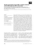
Growth inhibitory effect of rapamycin on lung cancer
cells
Wefirstsetouttoexaminewhetherandatwhatlevels
rapamycin inhibits the growth of four different lung
cancer cell lines (NCI-H446, A549, SPC-A-1 and 95D).
As shown in Figure 1, rapamycin treatment exerted
modest inhibitory effect on lung cancer cell proliferation
in a dose-dependent manner in all cell lines tested. In
addition, the effect of rapamycin seems to level off with
its increasing concentration, achieving about 30 - 40%
reduction in cell proliferation at 100 nM vs. ~ 10%
reduction at 12.5 nM. Finally, the inhibitory effect and
its saturating trend towards higher doses of rapamycin
are the same for all four cancer cell lines, suggesting
rapamycin may act on some targets/pathways common
in all of them.
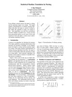
Growth inhibitory effect of rapamycin with docetaxel on
lung cancer cells
Next we checked the effect of rapamycin on docetaxel-
induced growth inhibition in lung cancer cells. It was
found that 20 nM rapamycin can potentiate the growth
inhibition activity of docetaxel in all four cancer cell lines
(Figure 2). This enhancing effect of rapamycin is espe-
cially pronounced at low docetaxel concentration (1 nM),
having led to an additional 20 - 40% of reduction in cell
growth. Although rapamycin does not cha nge the maxi-
mum level of cell growth inhibition elicited by docetaxel
(e.g., at 100 nM), the co-treatment of rapamycin with
docetaxel effectively lowered the EC50 (concentration
needed to achieve 50% of maximal effect) of the latter.
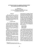
Rapamycin induces apoptosis in synergy with docetaxel
To further investigate whether the enhanc ing effect that
rapamycin showed in docetaxel-co-trea ted cancer cells
is associated with an increased level of apoptosis, we
performed flow cytomety analysis using Annexin V/pro-
pidium iodide-stained cells. As shown in Figure 3, rapa-
mycin enhances the effects of docetaxel in promoting
cancer cell death. Discounting the basal apoptosis lev el
as shown in the control samp le, the level of apoptosis in
the Rapa+DTX group is close to the sum of those in the
two monotreaments using either compound alone.
These findings indicate that rapamycin may further
enhance the e fficacy of docetaxel by inducing a higher
degree of apoptosis.
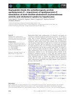
Combination treatment of rapamycin with docetaxel
decreases the expression of Survivin
As we wondered whether the enhancing effect of rapa-
mycin might come from its ability to block cellular
pathways that can counteract the cytotoxic activity of
docetaxel, the effect of rapamycin on the expression of
Survivin was next examined. Treatment of 95D cells
with either rapamycin or docetaxel alone resulted in
moderate but significant reduction on the level of Survi-
vin expression compared with that of the untreated
cells. Moreover, the co-treatment resulted in an even
bigger reduction in the Survivin protein level than those
of the two single drug treatments added to gether
(Figure 4). In contrast, the expression of a key marker
in the apoptotic pathway , caspase-3, is largely unaffected
by these treatments.
Combination treatment of rapamycin with docetaxel
decreases the phosphorylation level of ERK1/2 in 95D cell
lines
To further clarify t he cell growth inhibitory mechanism
of rapamycin with docetaxel, we examined the changes
in the expression levels of the enzymes involved in cell
growth signal transduction pathways. 95D cells were
Figure 1 Rapamycin exerts growth inhibitory effects in four
lung cancer cell lines in a dose-dependent fashion. Cells were
treated with increasing levels of rapamycin for 24 hours before cell
viability was examined by MTT assay. Control group received
treatment of DMSO solution of the same volume and concentration
used to dissolve rapamycin.
Niu et al . Journal of Experimental & Clinical Cancer Research 2011, 30:28
/>Page 3 of 8
exposed to rapamycin (10 nM, 20 nM) and docetaxel
(1 nM, 10 nM) alone or in combination (Rapa 20 nM+
DTX 10 nM). After 24 hr of incubation, the expression
and the phosphorylation levels of ERK1/2 were exam-
ined. As presented in Figure 5, a 24-hr exposure to
rapamycin or d ocetaxel alone did not significantly alter
the level of expression or phosphorylation of ERK1/2,
whereas cells treated with the combination of rapamycin
Figure 2 Rapamycin administered at 20 nM was able to potentiate the growth inhibitory effect of docetaxel in four lung cancer cells.
Figure 3 Rapamycin enhances the apoptosis effect of
docetaxel in lung cancer cells. *P < 0.05, significantly different
from untreated control; **P < 0.05, significantly different from either
rapamycin or docetaxel monotherapy.
Figure 4 Rapamycin and docetaxel decrease the level of
Survivin expression while the expression of caspase-3 is
unaffected. (A) The presence of various proteins was detected by
Western blot. (B) The relative level of Survivin and caspase-3
expression to GAPDH is shown in bar graph.
Niu et al . Journal of Experimental & Clinical Cancer Research 2011, 30:28
/>Page 4 of 8
with docetaxel exhibited a marked reduction in the
phosphorylation levels of ERK1/2. This suggests that
there may exist positive interacti ons between rapamycin
and docetaxel in the suppression o f ERK1/2 pathway in
95D cells.
Discussion
The prognosis for inoperable or recurrent lung cancer
patients has not been much improved despite the advent
of new chemotherapeutic agents. Although early stage
lung cancer is potentially curable, most l ung cancer
patients were already at advanced stages when diag-
nosed. Moreover, most advanced lung cancer patients
have a history of smoking thus suffer concurrent com-
plications in both cardiovascular and pulmonary sys-
tems, rendering aggressive surgery and multimodality
therapy unfeasible.
Docetaxel is a common second-line therapeutic agent
used for advanced NSCLC. In several randomized clini-
cal tries, combination cytotoxic chemotherapy regimens
for second-line therapy of advanced NSCLC failed to
establish patient survival benefit, although there was
report of higher cytotoxic effect [23]. It has been
thought that the clinical benefit of present second-line
therapies for advanced NSCLC has reached its peak.
More recently, combinations of molecularly targeted
agents with standard chemotherapy are being investi-
gated clinically with the hope to surpass the current
therapeutic threshold of second-line therapies [24].
Activation of PI3K-Akt-mTOR pathway has been
detected in many types of tumor s including lung cancer,
which is considered to be important for the survival,
proliferation, angiogenesis and resistance of cancer cells
to chemotherapy [25]. Consequently, this pathway has
been regarded as an attractive target of molecular tar-
geting therapy. Indeed, rapamycin treatment has shown
some promising antitumor effect in tissue culture sys-
tems [19]. However, as evidenced in clinical phase stu-
dies, rapamycin analogue monotherapy exerted a
modest but limited antitumor effect [26,27]. In order to
achieve a greate r therapeutic b enefit, several combina-
tion therapies of rapamycin and other cytotoxic or
molecular targeting agents have been under clinical
study. Encouragingly, rapamycin has clearly shown
either synergistic or additive effects in these treatments
[28-30]. In the present study, rapamycin treatment alone
exerted modest inhibition on cell proliferation of several
lung cancer cell lines in a dose-dependent manner.
However, when applied together, the proliferation inhi-
bition effect of docetaxel was significantly potentiated by
rapamycin. This observation is in line with previous
reports that regarded the mTOR pathway as a promising
target of therapy in the treatment of other solid tumors
refractory to conventional chemotherapies [31,32].
Apoptosis, induced by chemotherapy, radiation and
cytokines, seems to be the main mechanism to kill
tumor cells. We suspected that the rapamycin may also
enhance the apoptosis-inducing effect of docetaxel in
cancer cells. We used flow cytometry analysis to show
that rapamycin and docetaxel combination indeed
induced higher degree of apoptosis in lung cancer cell
lines than that by either compound alone. This led us to
further ponder upon the potential downstream effectors
of rapamycin and docetaxel-induced signaling pathways
in lung cancer cell lines. As a first step, we exami ned
the expression and ph osphorylation levels of some pro-
teins known to be involved in cell proliferation and
apoptosis. Interestingly, Survivin and ERK1/2 were
found to be down-regulated in expression and phos-
phorylation, respectively, especially by the combination
treatment of rapamycin and docetaxel. In comparison,
the expression of caspase-3, an apoptosis effector down-
stream of mitochondrial cytochrome c release, was
found to be unaffected.
Survivin is a member of the inhibitor of apoptosis pro-
teins (IAP) family that is typically absent in most normal
adult differentiated tissues. However, its mRNA and
protein are found in abundance in fetal tissue, most
transformed cell lines and cancers. Survivin suppresses
apoptosis and promotes angiogenesis, proliferation and
metastasis in cancer cells [33-37]. Survivin can block
apoptosis by inhibiting terminal apoptotic effectors cas-
pase-3 and caspase-7, and by suppressing both the pro-
teolytic activation and the activity of caspase-9 in the
context of Surv ivin-IAP complexes [38-40]. Clinically,
incre ased expression of Survivin is often associated with
elevated resistance of cancer cells to apoptotic stimuli
during chemotherapy an d is negatively correlated with
response to proapoptotic drugs and/or radiotherapy in
patients with bladder cancer, breast cancer, lymphoma
and multiple myeloma [41-46]. Furthermore, overex-
pression of Survivin is a prognostic biomarker for
decreased patient survival in multiple cancers, e.g.,
Figure 5 Combination treatment of rapamycin and docetaxel
decreases phosphorylation of ERK in 95D cell lines. 95D cells
were treated with 1 nM and 10 nM docetaxel alone, 10 nM and 20
nM rapamycin alone and a combination with 10 nM docetaxel and
20 nM rapamycin for 24 hr. After incubation, levels of ERK1/2 and p-
ERK1/2 (phosphorylated Tyr204) were examined. Con: control, Rapa:
rapamycin, DTX: docetaxel.
Niu et al . Journal of Experimental & Clinical Cancer Research 2011, 30:28
/>Page 5 of 8
breast cancer, colorectal and gastric carcinomas, neuro-
blastoma and NSCLC. All these findings on Survivin
indicate that it could be an attractive cancer target. In
this study, we were intrigued to find that co-treatment
with rapamycin and docetaxel significantly down-regu-
lates the expression of Survivin, as shown in Figure 4.
Although the underlying mechanism for this down-regu-
lation is currently unclear, our finding is consist ent with
a previous report that found rapamycin reduced IGF-
induced Survivin expression in prostate cancer cells
[47]. Similarly, Vaira et al. also reported that treatment
of rapamycin with taxol at suboptimal concentra tion
resulted in a bigger reduction in Survivin expression
than that by either treatment alone [47]. It is possible
that when co-treatment of rapamycin and docetaxel
synergistically reduced Survivin level beyond the thresh-
old for its antiapoptotic activity in cancer cells, the cyto-
toxic effect of docetaxel becomes more effective in
cancer treatment. In addition, our result suggests that
Survivin is essentially involved in lung cancer mainte-
nance and progression rather than initiation, which is in
agreement with the prevailing hypothesis. Finally,
because Survivin is selectively expressed at the G2/M
phase of the cell cycle and is a known mito tic regulator
of microtubule assembly, the target of action by doce-
taxel, it is tempting to speculate an antagonistic inter-
play between Survivin and docetaxel [48,49].
Interestingly, recent studies are converging on the
notion that inhibition of Surviv in in conjunction with
docetaxel treatment delivers better cancer-killing effect
by reversing the resistance to docetaxel in cancer
[50,51].
Activation of the MEK/ERK axis is often associated
with cell proliferation and survival [52,53]. Similar to
Survivin’ s role in cancer, the phosphorylation level of
ERK1/2 is often found upregulated in cancer cel ls and
inhibitors against MEK are currentl y in Phase II clinical
trials. In our study, we found that while monotherapies
with either rapamycin or d ocetaxel did not significantly
affect the phosphor ylation level of ERK1/2, the combi-
nat ion of the two led to a considerable reduction in the
amount of phosphorylated ERK1/2(Figure 5). This is sig-
nificant, because ERK1/2 activation was known to coun-
teract the cancer-killing activity of docetaxel in some
malignancies such as leukemia and melanoma [54-56].
It follows that if ERK1/2 activation is blocked due to the
combined effects of rapamycin and docetaxel-induced
events, cancer cells may be more sensitized to proapop-
totic chemotherapeutics.
Conclusion
In conclusion, the present study demonstrates that mTOR
inhibition by rapamycin suppresses lung cancer cell
growth and sensitizes tumor cells to docetaxel-induced
cytotoxicity. The rapamycin-dependent enhancement of
cancer-killing effects by docetaxel is associated with down-
regulation of Survivin expression. Although the precise
mechanism of interactions between rapamycin and doce-
taxel is not presently clear, their proliferation inhibitory
and apoptosis-inducing effects may be exerted through
down-regulating Survivin expression, either directly or
indirectly. Our results suggest that a therapeutic strategy
combining specific inhibitor of mTOR with cytotoxic
agents may be a promising approach to an improved treat-
ment of advanced lung cancer.
Acknowledgements
This work was supported by a grant from the Natural Science Funds of
Liaoning Province (No.20082104) and a grant from the Science and
Technology Plan Projects of Liaoning Province (No. 2009225008-10).
Authors’ contributions
HYN participated in research design, the writing of the paper, the
performance of the research and data analysis. JHW participated in the
performance of the research and data analysis. HL participated in the
performance of the research. PH participated in research design and data
analysis. All authors read and approved the final manuscript.
Competing interests
The authors declare that they have no competing interests.
Received: 18 January 2011 Accepted: 10 March 2011
Published: 10 March 2011
References
1. Hay N: The Akt-mTOR tango and its relevance to cancer. Cancer Cell
2005, 8:179-183.
2. Bjornsti MA, Houghton PJ: The TOR pathway: A target for cancer therapy.
Nature Reviews Cancer 2004, 4:335-348.
3. Vignot S, Faivre S, Aguirre D, Raymond E: MTOR-targeted therapy of
cancer with rapamycin derivatives. Annals of Oncology 2005, 16:525-537.
4. Sparks CA, Guertin DA: Targeting mTOR: prospects for mTOR complex 2
inhibitors in cancer therapy. Oncogene 2010, 29:3733-3744.
5. Guertin DA, Sabatini DM: Defining the role of mTOR in cancer. Cancer Cell
2007, 12:9-22.
6. Guertin DA, Sabatini DM: An expanding role for mTOR in cancer. Trends
Mol Med 2005, 11:353-361.
7. Strimpakos AS, Karapanagiotou EM, Saif MW, Syrigos KN: The role of mTOR
in the management of solid tumors: an overview. Cancer Treat Rev 2009,
35:148-159.
8. Shaw RJ, Cantley LC: Ras, PI(3)K and mTOR signalling controls tumour cell
growth. Nature 2006, 441:424-430.
9. Ramalingam SS, Khuri FR: The role of the taxanes in the treatment of
older patients with advanced stage non-small cell lung cancer.
Oncologist 2009, 14:412-424.
10. Chu Q, Vincent M, Logan D, Mackay JA, Evans WK: Taxanes as first-line
therapy for advanced non-small cell lung cancer: a systematic review
and practice guideline. Lung Cancer 2005, 50:355-374.
11. Ramalingam S, Belani CP: Taxanes for advanced non-small cell lung
cancer. Expert Opin Pharmacother 2002, 3:1693-1709.
12. Hu L, Hofmann J, Lu Y, Mills GB, Jaffe RB: Inhibition of
phosphatidylinositol 3’-kinase increases efficacy of paclitaxel in in vitro
and in vivo ovarian cancer models. Cancer Res 2002, 62:1087-1092.
13. Brown EJ, Albers MW, Shin TB, Ichikawa K, Keith CT, Lane WS, Schreiber SL:
A mammalian protein targeted by G1-arresting rapamycin-receptor
complex. Nature 1994, 369:756-758.
14. Hashemolhosseini S, Nagamine Y, Morley SJ, Desrivieres S, Mercep L,
Ferrari S: Rapamycin inhibition of the G1 to S transition is mediated by
effects on cyclin D1 mRNA and protein stability. J Biol Chem 1998,
273:14424-14429.
Niu et al . Journal of Experimental & Clinical Cancer Research 2011, 30:28
/>Page 6 of 8
15. Lei W, Jia T, Su Z, Wen W, Zhu X: Combined effect of rapamycin and
cisplatin on survival of Hep-2 cells in vitro. Oncol Res 2009, 18:73-81.
16. Calabro A, Tai J, Allen SL, Budman DR: In-vitro synergism of m-TOR
inhibitors, statins, and classical chemotherapy: potential implications in
acute leukemia. Anticancer Drugs 2008, 19:705-712.
17. Xu RH, Pelicano H, Zhang H, Giles FJ, Keating MJ, Huang P: Synergistic
effect of targeting mTOR by rapamycin and depleting ATP by inhibition
of glycolysis in lymphoma and leukemia cells. Leukemia 2005,
19:2153-2158.
18. Takeuchi H, Kondo Y, Fujiwara K, Kanzawa T, Aoki H, Mills GB, Kondo S:
Synergistic augmentation of rapamycin-induced autophagy in malignant
glioma cells by phosphatidylinositol 3-kinase/protein kinase B inhibitors.
Cancer Res 2005, 65:3336-3346.
19. Mondesire WH, Jian W, Zhang H, Ensor J, Hung MC, Mills GB, Meric-
Bernstam F: Targeting mammalian target of rapamycin synergistically
enhances chemotherapy-induced cytotoxicity in breast cancer cells. Clin
Cancer Res 2004, 10:7031-7042.
20. Zeng Q, Yang Z, Gao YJ, Yuan H, Cui K, Shi Y, Wang H, Huang X, Wong ST,
Wang Y, et al: Treating triple-negative breast cancer by a combination of
rapamycin and cyclophosphamide: an in vivo bioluminescence imaging
study. Eur J Cancer 2010, 46:1132-1143.
21. Yang Z, Lei Z, Li B, Zhou Y, Zhang GM, Feng ZH, Zhang B, Shen GX,
Huang B: Rapamycin inhibits lung metastasis of B16 melanoma cells
through down-regulating alphav integrin expression and up-regulating
apoptosis signaling. Cancer Sci 2010, 101:494-500.
22. Niu H, Li H, Xu C, He P: Expression profile of RhoGDI2 in lung cancers
and role of RhoGDI2 in lung cancer metastasis. Oncol Rep 2010,
24:465-471.
23. Di Maio M, Chiodini P, Georgoulias V, Hatzidaki D, Takeda K, Wachters FM,
Gebbia V, Smit EF, Morabito A, Gallo C, et al: Meta-analysis of single-agent
chemotherapy compared with combination chemotherapy as second-
line treatment of advanced non-small-cell lung cancer. J Clin Oncol 2009,
27:1836-1843.
24. Ramalingam SS, Harvey RD, Saba N, Owonikoko TK, Kauh J, Shin DM,
Sun SY, Strychor S, Tighiouart M, Egorin MJ, et al: Phase 1 and
pharmacokinetic study of everolimus, a mammalian target of rapamycin
inhibitor, in combination with docetaxel for recurrent/refractory
nonsmall cell lung cancer. Cancer 2010, 116:3903-3909.
25. Nicholson KM, Anderson NG: The protein kinase B/Akt signalling pathway
in human malignancy. Cell Signal 2002, 14:381-395.
26. Pandya KJ, Dahlberg S, Hidalgo M, Cohen RB, Lee MW, Schiller JH,
Johnson DH: A randomized, phase II trial of two dose levels of
temsirolimus (CCI-779) in patients with extensive-stage small-cell lung
cancer who have responding or stable disease after induction
chemotherapy: a trial of the Eastern Cooperative Oncology Group
(E1500). J Thorac Oncol 2007, 2
:1036-1041.
27.
Hudes G, Carducci M, Tomczak P, Dutcher J, Figlin R, Kapoor A,
Staroslawska E, Sosman J, McDermott D, Bodrogi I, et al: Temsirolimus,
interferon alfa, or both for advanced renal-cell carcinoma. N Engl J Med
2007, 356:2271-2281.
28. O’Reilly KE, Rojo F, She QB, Solit D, Mills GB, Smith D, Lane H, Hofmann F,
Hicklin DJ, Ludwig DL, et al: mTOR inhibition induces upstream receptor
tyrosine kinase signaling and activates Akt. Cancer Res 2006,
66:1500-1508.
29. Cejka D, Preusser M, Fuereder T, Sieghart W, Werzowa J, Strommer S,
Wacheck V: mTOR inhibition sensitizes gastric cancer to alkylating
chemotherapy in vivo. Anticancer Res 2008, 28:3801-3808.
30. Hahn M, Li W, Yu C, Rahmani M, Dent P, Grant S: Rapamycin and UCN-01
synergistically induce apoptosis in human leukemia cells through a
process that is regulated by the Raf-1/MEK/ERK, Akt, and JNK signal
transduction pathways. Mol Cancer Ther 2005, 4:457-470.
31. Fan QW, Knight ZA, Goldenberg DD, Yu W, Mostov KE, Stokoe D,
Shokat KM, Weiss WA: A dual PI3 kinase/mTOR inhibitor reveals
emergent efficacy in glioma. Cancer Cell 2006, 9:341-349.
32. Shapira M, Kakiashvili E, Rosenberg T, Hershko DD: The mTOR inhibitor
rapamycin down-regulates the expression of the ubiquitin ligase subunit
Skp2 in breast cancer cells. Breast Cancer Res 2006, 8:R46.
33. Altieri DC: The molecular basis and potential role of survivin in cancer
diagnosis and therapy. Trends Mol Med 2001, 7:542-547.
34. Marioni G, Bertolin A, Giacomelli L, Marchese-Ragona R, Savastano M,
Calgaro N, Marino F, De Filippis C, Staffieri A: Expression of the apoptosis
inhibitor protein Survivin in primary laryngeal carcinoma and cervical
lymph node metastasis. Anticancer Res 2006, 26:3813-3817.
35. Osaka E, Suzuki T, Osaka S, Yoshida Y, Sugita H, Asami S, Tabata K,
Hemmi A, Sugitani M, Nemoto N, Ryu J: Survivin as a prognostic factor for
osteosarcoma patients. Acta Histochem Cytochem 2006, 39:95-100.
36. Tran J, Rak J, Sheehan C, Saibil SD, LaCasse E, Korneluk RG, Kerbel RS:
Marked induction of the IAP family antiapoptotic proteins survivin and
XIAP by VEGF in vascular endothelial cells. Biochem Biophys Res Commun
1999, 264:781-788.
37. Harfouche R, Hassessian HM, Guo Y, Faivre V, Srikant CB, Yancopoulos GD,
Hussain SN: Mechanisms which mediate the antiapoptotic effects of
angiopoietin-1 on endothelial cells. Microvasc Res 2002, 64:135-147.
38. Altieri DC: Survivin, versatile modulation of cell division and apoptosis in
cancer. Oncogene 2003, 22:8581-8589.
39. Marusawa H, Matsuzawa S, Welsh K, Zou H, Armstrong R, Tamm I, Reed JC:
HBXIP functions as a cofactor of survivin in apoptosis suppression. EMBO
J 2003, 22:2729-2740.
40.
Dohi T, Okada K, Xia F, Wilford CE, Samuel T, Welsh K, Marusawa H, Zou H,
Armstrong R, Matsuzawa S, et al: An IAP-IAP complex inhibits apoptosis.
J Biol Chem 2004, 279:34087-34090.
41. Als AB, Dyrskjot L, von der Maase H, Koed K, Mansilla F, Toldbod HE,
Jensen JL, Ulhoi BP, Sengelov L, Jensen KM, Orntoft TF: Emmprin and
survivin predict response and survival following cisplatin-containing
chemotherapy in patients with advanced bladder cancer. Clin Cancer Res
2007, 13:4407-4414.
42. Hinnis AR, Luckett JC, Walker RA: Survivin is an independent predictor of
short-term survival in poor prognostic breast cancer patients. Br J Cancer
2007, 96:639-645.
43. Nakagawa Y, Abe S, Kurata M, Hasegawa M, Yamamoto K, Inoue M,
Takemura T, Suzuki K, Kitagawa M: IAP family protein expression
correlates with poor outcome of multiple myeloma patients in
association with chemotherapy-induced overexpression of multidrug
resistance genes. Am J Hematol 2006, 81:824-831.
44. Watanuki-Miyauchi R, Kojima Y, Tsurumi H, Hara T, Goto N, Kasahara S,
Saio M, Moriwaki H, Takami T: Expression of survivin and of antigen
detected by a novel monoclonal antibody, T332, is associated with
outcome of diffuse large B-cell lymphoma and its subtypes. Pathol Int
2005, 55:324-330.
45. Schlette EJ, Medeiros LJ, Goy A, Lai R, Rassidakis GZ: Survivin expression
predicts poorer prognosis in anaplastic large-cell lymphoma. J Clin Oncol
2004, 22:1682-1688.
46. Adida C, Haioun C, Gaulard P, Lepage E, Morel P, Briere J, Dombret H,
Reyes F, Diebold J, Gisselbrecht C, et al: Prognostic significance of survivin
expression in diffuse large B-cell lymphomas. Blood 2000, 96:1921-1925.
47. Vaira V, Lee CW, Goel HL, Bosari S, Languino LR, Altieri DC: Regulation of
survivin expression by IGF-1/mTOR signaling. Oncogene 2007,
26:2678-2684.
48. Shin S, Sung BJ, Cho YS, Kim HJ, Ha NC, Hwang JI, Chung CW, Jung YK,
Oh BH: An anti-apoptotic protein human survivin is a direct inhibitor of
caspase-3 and -7. Biochemistry 2001, 40:1117-1123.
49. Li F, Ambrosini G, Chu EY, Plescia J, Tognin S, Marchisio PC, Altieri DC:
Control of apoptosis and mitotic spindle checkpoint by survivin. Nature
1998, 396:580-584.
50. Wang T, Wei J, Qian X, Ding Y, Yu L, Liu B: Gambogic acid, a potent
inhibitor of survivin, reverses docetaxel resistance in gastric cancer cells.
Cancer Lett 2008, 262:214-222.
51. Giaccone G, Zatloukal P, Roubec J, Floor K, Musil J, Kuta M, van Klaveren RJ,
Chaudhary S, Gunther A, Shamsili S: Multicenter phase II trial of YM155, a
small-molecule suppressor of survivin, in patients with advanced,
refractory, non-small-cell lung cancer. J Clin Oncol 2009, 27:4481-4486.
52. Friday BB, Adjei AA: Advances in targeting the Ras/Raf/MEK/Erk mitogen-
activated protein kinase cascade with MEK inhibitors for cancer therapy.
Clin Cancer Res 2008,
14:342-346.
53.
Roberts PJ, Der CJ: Targeting the Raf-MEK-ERK mitogen-activated protein
kinase cascade for the treatment of cancer. Oncogene 2007,
26:3291-3310.
54. Mhaidat NM, Zhang XD, Jiang CC, Hersey P: Docetaxel-induced apoptosis
of human melanoma is mediated by activation of c-Jun NH2-terminal
kinase and inhibited by the mitogen-activated protein kinase
extracellular signal-regulated kinase 1/2 pathway. Clin Cancer Res 2007,
13:1308-1314.
Niu et al . Journal of Experimental & Clinical Cancer Research 2011, 30:28
/>Page 7 of 8
55. Yu C, Wang S, Dent P, Grant S: Sequence-dependent potentiation of
paclitaxel-mediated apoptosis in human leukemia cells by inhibitors of
the mitogen-activated protein kinase kinase/mitogen-activated protein
kinase pathway. Mol Pharmacol 2001, 60:143-154.
56. Wang S, Guo CY, Castillo A, Dent P, Grant S: Effect of bryostatin 1 on
taxol-induced apoptosis and cytotoxicity in human leukemia cells
(U937). Biochem Pharmacol 1998, 56:635-644.
doi:10.1186/1756-9966-30-28
Cite this article as: Niu et al.: Rapamycin potentiates cytotoxicity by
docetaxel possibly through downregulation of Survivin in lung cancer
cells. Journal of Experimental & Clinical Cancer Research 2011 30:28.
Submit your next manuscript to BioMed Central
and take full advantage of:
• Convenient online submission
• Thorough peer review
• No space constraints or color figure charges
• Immediate publication on acceptance
• Inclusion in PubMed, CAS, Scopus and Google Scholar
• Research which is freely available for redistribution
Submit your manuscript at
www.biomedcentral.com/submit
Niu et al . Journal of Experimental & Clinical Cancer Research 2011, 30:28
/>Page 8 of 8