báo cáo khoa học: " Silibinin induces apoptosis via calpain-dependent AIF nuclear translocation in U87MG human glioma cell death" potx
Bạn đang xem bản rút gọn của tài liệu. Xem và tải ngay bản đầy đủ của tài liệu tại đây (563.31 KB, 8 trang )
RESEARC H Open Access
Silibinin induces apoptosis via calpain-dependent
AIF nuclear translocation in U87MG human
glioma cell death
Ji C Jeong
1
, Won Y Shin
1
, Thae H Kim
2
, Chae H Kwon
2
, Jae H Kim
2
, Yong K Kim
2
and Ki H Kim
3*
Abstract
Background: Silibinin, a natural polyphenolic flavonoid, has been reported to induce cell death in various cancer
cell types. However, the molecular mechanism is not clearly defined. Our previous study showed that silibinin
induces glioma cell death and its effect was effectively prevented by calpain inhibitor. The present study was
therefore undertaken to examine the role of calpain in the silibinin-induced glioma cell death.
Methods: U87MG cells were grown on well tissue culture plates and cell viability was measured by MTT assay. ROS
generation and △ψ
m
were estimated using the fluorescence dyes. PKC activation and Bax expression were
measured by Western blot analysis. AIF nuclear translocation was determined by Western blot and
immunocytochemist ry.
Results: Silibinin induced activation of calpain, which was blocked by EGTA and the calpain inhibitor Z-Leu-Leu-
CHO. Silibinin caused ROS generation and its effect was inhibited by calpain inhibitor, the general PKC inhibitor GF
109203X, the specific PKC
δ
inhibitor rottlerin, and catalase. Silibinin-induce cell death was blocked by calpain
inhibitor and PKC inhibitors. Silibinin-induced PKC
δ
activation and disruption of △ψ
m
were prevented by the calpain
inhibitor. Silibinin induced AIF nuclear translocation and its effect was prevented by calpain inhibitor. Transfection
of vector expressing microRNA of AIF prevented the silibinin-induced cell death.
Conclusions: Silibinin induces apoptotic cell death through a calpain-dependent mechanism involving PKC, ROS,
and AIF nuclear translocation in U87MG human glioma cells.
Background
Glioblastoma is the most lethal and frequent primary
brain tumors [1]. It is comprised of poorly differentiated
heterogeneous neoplastic astrocytes with aggressive pro-
liferation and highly invasive properties. After diagnosis
of glioblastoma, the median survival time of 9-12
months has remained unchanged despite aggressive
treatment including surgery, radiation, and chemother-
apy [2,3]. Thus, new effective strategies f or controlling
glioblastoma are required. Because glioblastoma cells
avoid differentiation and apoptosis, the i nduction of dif-
ferentiation and apoptosis in glioblastoma cells may be
considered as a potential treatment strategy.
Silibinin, a natural polyphenolic flavonoid, is a major
bioactive component of silymarin which is isolated from
the plant milk thistle (Silybum marianum), and has
been extensive ly used for its hepatoprotective ef fects in
Asia and Europe. It has been reported that silibinin has
anticancer activities in various cancers including pros-
tate cancer in b oth in vitro and in vivo models [4-7].
Recently, we observ ed that s ilibinin induces apoptosis
through Ca
2+
/ROS-dependent mechanism in human
glioma cells [8]. The study showed that silibinin-induced
cell death was prevented by calpain inhibitor, suggesting
involvement of calpain activation in apoptosis induced
by silibinin. Therefore, the present study was undertaken
to examine r ole of calpain in the s ililbinin-induced
glioma cell death. The present study demonstrated that
silibinin induces human glioma cell death via a calpai n-
dependent AIF nuclear translocation involving ROS and
PKC.
* Correspondence:
3
Department of Obstetrics and Gynecology, College of Medicine, Pusan
National University, and Medical Research Institute and Pusan Cancer Center,
Pusan National University Hospital, Pusan, 602-739, Korea
Full list of author information is available at the end of the article
Jeong et al. Journal of Experimental & Clinical Cancer Research 2011, 30:44
/>© 2011 Jeong et al; licensee BioMed Central Ltd. This is an Open Access article distributed und er the terms of the Creative Commons
Attribution Lice nse (http://c reativecommons.org/licenses/by/2.0), which permits unrestricted use, distribution, and reproduction in
any medium, provided the original work is properly cited.
Materials and methods
Reagents
Silibinin, GF 109203X, rottlerin, c atalase, MTT, propi-
dium iodide was purchased from Si gma-Aldrich Chemi-
cal(St.Louis,MO,USA).Z-Leu-Leu-CHOwas
purchased from BIOMOL International LP (Plymouth
Meeting, PA, USA). DCFH-DA and DiOC
6
(3) were
obtained from Molecular Probes (Eugene, OR, USA).
Antibodies were obtained from Cell Signaling Technol-
ogy Inc. (Beverly, MA, USA). All other chemicals were
of the highest commercial grade available.
Cell culture
U87MG cells were obtain ed from the American Type
Culture Collection (Rockville, MD, USA) and maintained
by serial passages in 75-cm
2
culture flasks (Costar, Cam-
bridge, MA, USA). The cells were grown in Dulbecco’ s
modified Eagle’ s medium (DME M, Gibco BRL, Invitro-
gen, Carsbad, CA, USA) containing 10% heat inactivated
fetal bovine serum (HyClone, Logan, UT, USA) at 37°C
in humidified 95% air/5% CO
2
incubator. When the cul-
tures reached confluence, subculture was prepared usi ng
a 0.02% EDTA-0.05% trypsin solution. The cells were
grown on well tissue culture plates and used 1-2 days
after plating when a confluent monolayer culture was
achieved. Unless otherwise stated, cells were treated with
silibinin in serum-free medium. Test reagents were
added to the medium 30 min before silibinin exposure.
Measurement of cell viability
Cell viability was evaluated using a MTT assay [9]. Cul-
ture medium containing 0.5 mg/ml of MTT was added
to each well. The cells were incubated for 2 h at 37°C,
the supernatant was removed and the formed formazan
crystals in viable cells were solubilized with 0.11 ml of
dimethyl sulfoxide. A 0.1 ml aliquot of each sample was
then translated to 96-well plates and the absorbance of
each well was measured at 550 nm with ELISA Reader
(FLUOstar OPTIMA, BM G LABTECH, Offenburg, Ger-
many). Data were expressed as a percentage of cont rol
measured in the absence of silibinin.
Measurement of calpain activity
Calpain activity was measured by calpain assay kit (Bio-
Vision Research Products, CA, USA) according to the
manufacturer’sinstructions.Cellsweregrownin6-well
plates and were treated as indicated. Detached cells from
the bottom of culture plates by trypsin were pelleted b y
centrifugation and washed with phosphate-buffered sal-
ine (PBS). The pellet were suspended in extraction buffer
and incubated on ice for 20 min then centrifuged at
10,000 × g for 10 min at 4°C. The supernatant repre-
sented the cytosolic protein. Add 10 μl o f 10× reaction
buffer and 5 μl of calpain substrate, Ac-LLY-AFC, to
each assay. Incubate at 37°C for 1 h in the dark. After
incubation, production of free AFC was fluorometrically
measured suing a Victor 3 Multilabel Counter with an
excitation filter of 400 nm and an emission filter of 505
nm (PerkinElmer, Boston, MA, USA).
Measurement of reactive oxygen species (ROS)
The intracellular generation of ROS was measured using
DCFH-DA. The nonfluorescent ester penetrates into the
cells and is hydrolyzed to DCFH by the cellular
esterases. The probe (DCFH) is rapidly oxidized to the
highly fluorescent compound DCF in the presence of
cellular peroxidase and ROS such as hydrogen peroxide
or fatty acid peroxides. Cells cultured in 24-well plate
were preincubated in the culture medium with 30 μM
DCFH-DA for 1 h at 37°C. After the preincubation, the
cells were exposed to 30 μM silibinin for various times.
Changes in DCF fluorescence was assayed using FAC-
Sort Becton Dickinson Flow Cytometer (Becton-Dickin-
son Bioscience, San Jose, CA, USA) and data were
analyzed with CELLQuest Software.
Measurement of △ψ
m
The △ψ
m
was measured with DiOC
6
(3), a fluorochrome
that is incorporated into cells depending upon the mito-
chondrial membrane potential [10]. Loss in DiOC
6
(3)
staining indicates disruption of the △ψ
m
.Cellswere
stained with DiOC
6
(3) at a final concentration of 50 nM
for 20 min at 37°C in the dark. Cells were washed and
resuspended in Hank’s balanced salts solution containing
Ca
2+
and Mg
2+
. The fluorescence intensity was analyzed
with a FACScan flow cytometer using the fluorescence
signal 1 channel.
Western blot analysis
Cells were harvest at various times after silibinin treatment
and disrupted in lysis buffer (1% Triton X-100, 1 mM
EGTA, 1 mM EDTA, 10 mM Tris-HCl, pH 7.4). Cell deb-
ris was removed by centrifugation at 10,000 g for 10 min
at 4°C. The resulting supernatants were resolved on a 10%
SDS-PAGE under denatured reducing conditions and
transferred to nitrocellulose membranes. The membranes
wereblockedwith5%non-fatdriedmilkatroomtem-
perature for 30 min and incubated with different primary
antibodies. The membranes were washed and incubated
with horseradish peroxidase-conjugated secondary antibo-
dies. The sig nal was visualized using an enhanced chemi-
luminescence (Amersham, Buckinghamshire, UK).
Measurement of AIF nuclear translocation
Cells were harvested and washed twice with PBS. The cells
were incubated with extraction buffer (10 mM Hepes,
Jeong et al. Journal of Experimental & Clinical Cancer Research 2011, 30:44
/>Page 2 of 8
250mMsucrose,10mMKCl,1.5mMMgCl
2
,1mM
EDTA, 1 mM EGTA, 0.05% digitonin, and 1 mM phenyl-
methylsulfonyl fluoride) at 4°C for 10 min, then centri-
fuged at 100000 g for 10 min at 4°C. The supernatant
cytosolic protei n wa s re moved and the pellet was incu-
bated in the nuclear extra ction buffer (3 50 mM NaCl, 1
mM EGTA, 1 mM EDTA, 10 mM Tris-HCl, pH 7.4, and
protease inhibitors) at 4°C for 10 min, then centrifuged at
10000 g for 10 min at 4°C. Protei ns were loaded onto a
12% SDS-polyacrylamide gels and transferred to nitrocel-
lulose membranes. After blocking in 5% non-fat dried
milk at room temperature for 30 min, membranes were
probed with rabbit polyclonal anti-AIF antibody, followed
by horseradish peroxidase-conjugated secondary antibo-
dies. Bands were visualized using the ECL detection sys-
tem (Amersham, Buckinghamshire, UK).
AIF nuclear translocation was further confirmed by
immunofluorescence analysis. Cells were cultured on
glass coverslips a nd treated with silibinin. Cells were
washed twice with PBS, fixed with 4% paraformadehyde
in PBS for 10 min, permeabilized with 0.5% Triton X-
100 in PBS for 10 min. After washing twice with PBS,
cells were blocked with 8% BSA in Tris-buffered saline
Triton X-100 (TBST). Cells were incubated w ith rabbit
polyclonal anti-AIF overnight 4°C and washed twice
with TBST. Cells were incubated with FITC-conjugated
secondary antibody (Jackson I mmunoResearch Labora-
tories, PA, USA) for 1 h, and the nuclei w ere counter-
stained with propidium iodide to ascertain AIF unclear
localization. Cell were washed twice and visualized by
using the confocal microscope (Leica, Wetzlar,
Germany).
RNA interference (RNAi)
For AIF targeting, we used The BLOCK-iT™ Pol miR
RNAi Expression Vector Kits (Invitrogen, Carlsbad, CA,
USA) to facilitate the expression of micr oRNA (miRNA).
miRNA sequences for AIF were designed using online
software (BLOCK-iT RNAi Designer from Invitrogen).
Thetargetsequencewas5’-GTGCCTATGCCTACAA-
GACTA-3’. This single-stranded oligonucleotide gener-
ated a d ouble-stranded oligonucleotide, which instructed
into pcDNA™ 6.2-GW/EmGFP-miR vector. This vector
contains EmGFP that allow identifying of the transfection
efficiency using fluorescen ce microscopy. The construct
pcDNA™ 6.2-GW/EmGFP-miR-LacZ was used as a con-
trol. Cells were transiently transfected with these plas-
mids using lipofectamine (Invitrogen).
Statistical analysis
The data are expressed as means ± SEM and the differ-
ence between two groups was evaluated using Student’s
t-test. Multiple group comparison was done using one-
way analysis of variance followed by the Tukey post hoc
test. A probability level of 0.05 was used to establish
significance.
Results and Discussion
Effect of calpain inhibitor on silibinin-induced cell death
Calpains are cytosolic Ca
2+
-activated neutral cysteine
proteases and ubiquitously distributed in all animal
cells, which play a critical role i n regulating cell viability
[11,12]. Accumulating evidence suggests that calpain
activation may contribute to cell death in certain cell
types including thymocytes, monocytes, cardiomyocytes,
and neuronal cells [13]. Since our previous study
showed that the calpain inhibitor Z-Leu-Leu-CHO at
0.5 μ M significantly protected effectively against the sili-
binin-induced cell death [8], we observed in the present
study the dose-dependency of the inhibitor effect. The
results showed that the calpain inhibitor exerted protec-
tive effect against the silibinin-induced cell death in a
dose-dependent manner with maximum potency at 0.5-
1 μM (Figure 1A). Silibinin also induced calpain activa-
tion, which was blocked by EGTA and calpain inhibitor
(Figure 1B). These results indicate that calpain activation
plays a critical role in the silibinin-ind uced cell death in
human glioma cells.
Role of calpain and protein kinase C (PKC) activation in
ROS generation and cell death induced by silibinin
The silibinin-induced cell death was associated with
ROS generation mediated by intracellular Ca
2+
[8]. To
determine therefore whether ROS production by silibi-
nin is attributed to calpain activation, cells were exposed
to silibinin in the presence of calpain inhibitor and ROS
generation was measured. As shown in Figure 2A, the
silibinin-induced ROS generation was blocked by the
calpain inhibitor with potency similar to that of catalase.
PKCs are a family of serine/threonine kinases which are
involved in tumor formation and progression [14] . PKC
isoforms cooperate or exert opposite effects on the process
of apoptosis [15,16]. PKC isoforms such as PKCa, ε,andξ
inhibit apoptosis, whereas PKC
δ
is involved in the process
of apoptosis [16,17]. Although previous studies have
shown that flavonoids can induce activation of PKC
[18,19], it is unclear whether PKC is involved in the signal-
ing cascade of silibinin-induced cell death. Although PKCs
are activated by ROS [20,21], it has been reported that
PKC activation can also cause ROS generation [22,23].
Therefore, we examined involvement of PKC in the silibi-
nin-ind uced RO S generation. The general PKC inhibi tor
GF 109203X and the sele ctive PKC
δ
inhibitor rottlerin
blocked the ROS generation (Figure 2A). The silibinin-
induced cell death was also prevented by the general PKC
inhibitor GF 109203X and rottlerin (Figure 2B), indicating
that silibinin induces ROS generation and cell death
through PKC activation. We next examined whether
Jeong et al. Journal of Experimental & Clinical Cancer Research 2011, 30:44
/>Page 3 of 8
silibinin induces PKC
δ
phosphorylation, an index of PKC
δ
activation. Silibinin induced a transient phosphorylation of
PKC
δ
after 10 min of treatment, which was inhibited by
treatment of calpain inhibitor (Figure 2C and 2D), suggest-
ing that PKC
δ
may be a downstream of calpain in the sili-
binin-induced cell death. Similar results are reported in
human U-937 leukemia cells in which the flavonoid wogo-
nin induces cell arrest through PKC
δ
activation [18].
Role of Bax expression and mitochondria in silibinin-
induced cell death
Since numerous death signals converge on mitochondria
through the activation of pro-apoptotic members of the
Bcl-2 family such a s Bax [24], calpain activation may
induce the silibinin-induced cell death through a Bax-
dependent pathway. To test this possibility, the effect of
silibinin on Bax expressi on was examined. Silibinin
increased Bax expression after 3 h of treatment, which
was blocked by the calpain inhibitor (Figure 3).
The increase in Bax expression may cause disruption
of △ψ
m
to induce cell death. To test the pos sibility, cells
were exposed to silibinin and the △ψ
m
was measured
using the fluorescence dye. After silibinin treatment, dis-
ruption of △ ψ
m
was o bserved as evidenced by an
increase in the proportion of cells with lower fluores-
cence intensity (Figure 4A). The reduction in △ψ
m
was
observed after 3 h of silibini n treatment an d remained
unchanged even after 12 h (Figure 4B).
Disruption of △ψ
m
by silibini n may be associated with
ROS generation. To test the possibility, cells were
exposed to silibinin in the presence of the antioxidant
catalase and △ψ
m
was measured. Figure 4C shows that
the silibinin-induced reduction in △ψ
m
was blocked by
catalase, suggesting that the △ψ
m
disruption b y silibinin
is mediated by ROS generation.
-EGTAZ-CHO
100
120
140
160
180
200
C
alpain activity
(% Control)
S
ili
b
inin
(A)
(B)
(A)
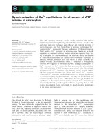
Figure 1 Role of calpain in silibinin-induced cell death. (A) Cells
were exposed to 30 μM silibinin for 36 h in the presence of various
concentrations of calpain inhibitor (Z-CHO). Cell viability was
estimated by MTT assay. Data are mean ± SEM of four independent
experiments performed in duplicate. *p < 0.05 compared with
silibinin alone. (B) Cells were exposed to 30 μM silibinin for 24 h in
the presence of 2 mM EGTA and 0.5 μM Z-CHO. Calpain activity was
measured by calpain assay kit. Data are mean ± SEM of four
independent experiments performed in duplicate. *p < 0.05
compared with silibinin alone.
(
A
)
(B)
C-CHOGFRoCat
0
20
40
60
80
100
120
R
OS
generation
(fluorescence intensity)
Silibinin
C-GFRo
0
20
40
60
80
100
Cell v iability (%)
Silibinin
0 0.2 0.5 1 3 6 12 24
p-PKC
G
ȕ-actin
(C)
C - CHO
p-PKC
G
ȕ-actin
(D)
Silibilnin
Silibilnin
(
h
)
Figure 2 Role of calpain and PKC in ROS generation and cell
death induced by silibinin. (A) Effect of inhibitors of calpain and
PKC on silibinin-induced ROS generation. Cells were exposed to 30
μM silibinin in the presence or absence of 0.5 μM calpain inhibitor
(CHO), 1 μM GF 109203X (GF), 1 μM rottlerin (Ro), and 800 units/ml
catalase (Cat) and ROS generation was estimated by measuring
changes in DCF fluorescence using FACS analysis. Data are mean ±
SEM of five independent experiments performed in duplicate. *p <
0.05 compared with silibinin alone. (B) Effect of PKC inhibitors on
silibinin-induced cell death. Cells were exposed to 30 μM silibinin in
the presence or absence of 1 μM GF 109203X (GF) and 1 μM
rottlerin (Ro) and cell viability was measured by MTT assay. Data are
mean ± SEM of four independent experiments performed in
duplicate. *p < 0.05 compared with silibinin alone. (C) Effect of
silibinin on PKC
δ
activation. Cells were exposed to 30 μM silibinin
for various times and PKC
δ
phosphorylation was estimated by
Western blot analysis. (D) Effect of calpain inhibitor on PKC
δ
phosphorylation. Cells were exposed to 30 μM silibinin for 10 min in
the presence or absence of 0.5 μM calpain inhibitor (CHO) and PKC
δ
phosphorylation was estimated by Western blot analysis.
Jeong et al. Journal of Experimental & Clinical Cancer Research 2011, 30:44
/>Page 4 of 8
As shown above, since the silibinin-induced ROS gen-
eration was blocked by inhibitors of calpain and PKC, the
silibinin-induced disruption of △ψ
m
would be prevented
by these inhibitors. As expected, the reduction in △ψ
m
was
blocked by Z-Leu-Leu-CHO, GF 109203X , and rottlerin,
with similar potency to that by catalase (Figure 4C).
Role of AIF nuclear translocation in silibinin-induced cell
death
The mitochondrial apoptotic pathway is initiated by
the cytosolic release of mitochondrial intermembrane
space proteins that can trigger either caspase-activation
or caspase-independent apoptotic pathways [25,26].
Mitochondrial proteins that cau se caspase-dependent
Bax
0 0.5 1 3 6 12 24 36
Silibinin (h)
E
-actin
Bax
E
-actin
C - CHO
S
ili
b
inin
0 6 12 18 24 30 36
Time (h)
0
1
2
3
4
5
6
Bax expression
(fold-increase)
(
A
)
(B)
(C)
Figure 3 Effect of silibinin on Bax expression. Cells were
exposed to 30 μM silibinin for various times and Bax expression was
estimated by Western blot analysis. Representative (A) and
quantitative (B) results of four independent experiments. (C) Cells
were exposed to 30 μM silibinin for 24 h in the presence or
absence of 0.5 μM calpain inhibitor (CHO) and Bax expression was
estimated by Western blot analysis.
036912
Time (h)
40
60
80
100
120
MMP
(fluoroscence intensity)
Control
Silibinin
(A)
(B)
(C)
C-CHOGFRoCat
0
20
40
60
80
100
120
MMP
(fluorescence intensity)
S
ili
b
inin
Figure 4 Effect of silibinin on mitochondrial membrane
potential (MMP). Cells were exposed to 30 μM silibinin for 6 h (A)
and various times (B). The MMP was estimated by the uptake of a
membrane potential-sensitive fluorescence dye DiCO
6
(3). The
fluorescence intensity was analyzed using FACS analysis. Data in (B)
are mean ± SEM of three independent experiments performed in
duplicate. *p < 0.05 compared with control. (C) Effect of inhibitors
of calpain and PKC and antioxidant on silibinin-induced disruption
of MMP. Cells were exposed to 30 μM silibinin for 6 h in the
presence or absence of 0.5 μM calpain inhibitor (CHO), 1 μMGF
109203X (GF), 1 μM rottlerin (Ro), and 800 units/ml catalase (Cat).
The MMP was measured as described above. Data are mean ± SEM
of four independent experiments performed in duplicate. *p < 0.05
compared with silibinin alone.
Jeong et al. Journal of Experimental & Clinical Cancer Research 2011, 30:44
/>Page 5 of 8
Cytosol- AIF
Nuclear-AIF
C 1 3 6 12 24 36 (h)
Silibinin
(
A
)
(B)
Control Silibinin Silibinin + CHO
AIF
ȕ-actin
LacZ mi-AIF
Silibinin
(C)
(D)
Figure 5 Role of AIF nuclear translocation in silibinin-induced cell death. (A) Cells were exposed to with 30 μM silibinin for various times
and cytosolic and nuclear fractions were prepared. AIF expression was estimated by Western blot using antibodies specific against AIF. (B) Cells
were exposed to 30 μM silibinin for 36 h in the presence or absence of 0.5 μM calpain inhibitor (CHO). AIF nuclear translocation was estimated
by immunofluorescence using antibody specific against AIF. Nuclei were counterstained with propidium iodide (PI). Images were captured by
confocal microscope and presented. Arrows indicate AIF nuclear localization. (C) Cells were transfected with mipcDNA vector for LacZ or AIF
micro-RNA (mi-AIF). The expression levels of AIF were determined by Western blotting. (D) Cells transfected with LacZ or mi-AIF were exposed
to 30 μM silibinin for 36 h and cell viability was estimated by MTT assay. Data are mean ± SEM of four independent experiments performed in
duplicate. *p < 0.05 compared with LacZ control; #p < 0.05
compared with LacZ silibinin.
Jeong et al. Journal of Experimental & Clinical Cancer Research 2011, 30:44
/>Page 6 of 8
cell death include cytochrome c w hich triggers cas-
pase-9 activation through Apaf-1. The activate d cas-
pase-9 then activates the downstream caspase-3
[26-28]. Mitochondria have also been reported to con-
tain AIF, which can cleave directly DNA and intracel-
lular substrates when released into the cytosol. During
apoptosis, AIF translocates into the nucleus where it
causes oligonucleosomal DNA fragmentation [29,30].
The present study showed that s ilibinin causes AIF
nuclear translocation, which was inhibited by t he cal-
pain inhibitor (Figure 5A and 5B). To determine if sili-
binin induced cell death through AIF nuclear
translocation, effect of silibini n on the cell death in
cells transfected with AIF mi-RNA was measured.
Transfection of AIF mi-RNA was decreased AIF pro-
tein levels (Figure 5C) and effectively prevented t he
silibinin-in duced cell death (Figur e 5D). These data
suggest that calpain activation induces AIF-de pendent
cell death in silibinin-treated ce lls. This is the first
report showing involvement of calpain-dependent AIF
nuclear translocation in the silibinin-induced glioma
cell death.
Conclusion
The present study demonstrated that silibinin induces
apoptosis through AIF nuclear translocation mediated
by a calpain-dependent pathway in U87MG human
glioma cells. This pathway involves PKC activation and
ROS generation. These data suggest that silibinin may
be considered a potential candidate in prevention and
treatment of human malignant gliomas.
List of abbreviations
AIF: apoptosis-inducing factor; DCF: 2’,7’-dichlorofluorescein; DCFH-DA: 2’,7’-
dichlorofluorescein diacetate; DiOC
6
(3): 3,3’-dihexyloxacarbocyamide; MTT: 3-
[4,5-dimethylthiazol-2-yl]-2,5-diphenyltetrazolium bromide; PBS: phosphate
buffer solution; PKC: protein kinase C; ROS: reactive oxygen species; △ψ
m
:
mitochondrial membrane potential.
Acknowledgements
This research was supported by Basic Science Research program through
the National Research Foundation of Korea (NRF) funded by the Ministry of
Education, Science and Technology (2010-0003690) and a grant from the
National R&D Program for Cancer Control, Ministry for Health, Welfare and
Family affairs (0920050).
Author details
1
Department of Oriental Medicine, Dongguk University, Kyung Ju, 780-714,
Korea.
2
Department of Physiology, College of Medicine, Pusan National
University, Yangsan, Gyeongsangnam-do, 626-770, Korea.
3
Department of
Obstetrics and Gynecology, College of Medicine, Pusan National University,
and Medical Research Institute and Pusan Cancer Center, Pusan National
University Hospital, Pusan, 602-739, Korea.
Authors’ contributions
JJ carried out cell viability and apoptosis assay, participated in drafted the
manuscript. WS and TK carried out mitochondrial membrane potential, ROS
generation, and statistical analyses. CK and YK carried out Western blot,
calpain activity, and AIF nuclear translocation. KK and JK participated in
experiment design and the draft preparation. All authors read and approved
the final manuscript.
Competing interests
The authors declare that they have no competing interests.
Received: 6 January 2011 Accepted: 19 April 2011
Published: 19 April 2011
References
1. Ohgaki H, Kleihues P: Population-based studies on incidence, survival
rates, and genetic alterations in astrocytic and oligodendroglial gliomas.
J Neuropathol Exp Neurol 2005, 64(6):479-489.
2. DeAngelis LM: Brain tumors. N Engl J Med 2001, 344(2):114-123.
3. Sanai N, Alvarez-Buylla A, Berger MS: Neural stem cells and the origin of
gliomas. N Engl J Med 2005, 353(8):811-822.
4. Singh RP, Gu M, Agarwal R: Silibinin inhibits colorectal cancer growth by
inhibiting tumor cell proliferation and angiogenesis. Cancer Res 2008,
68(6):2043-2050.
5. Singh RP, Mallikarjuna GU, Sharma G, Dhanalakshmi S, Tyagi AK, Chan DC,
Agarwal C, Agarwal R: Oral silibinin inhibits lung tumor growth in
athymic nude mice and forms a novel chemocombination with
doxorubicin targeting nuclear factor kappaB-mediated inducible
chemoresistance. Clin Cancer Res 2004, 10(24):8641-8647.
6. Ramasamy K, Agarwal R: Multitargeted therapy of cancer by silymarin.
Cancer Lett 2008, 269(352-362.
7. Kaur M, Agarwal R: Silymarin and epithelial cancer chemoprevention:
how close we are to bedside? Toxicol Appl Pharmacol 2007,
224(3):350-359.
8. Kim KW, Choi CH, Kim TH, Kwon CH, Woo JS, Kim YK: Silibinin inhibits
glioma cell proliferation via Ca2+/ROS/MAPK-dependent mechanism in
vitro and glioma tumor growth in vivo. Neurochem Res 2009,
34(8):1479-1490.
9. Denizot F, Lang R: Rapid colorimetric assay for cell growth and survival.
Modifications to the tetrazolium dye procedure giving improved
sensitivity and reliability. J Immunol Methods 1986, 89(2):271-277.
10. Pastorino JG, Chen ST, Tafani M, Snyder JW, Farber JL: The overexpression
of Bax produces cell death upon induction of the mitochondrial
permeability transition. J Biol Chem 1998, 273(13):7770-7775.
11. Orrenius S, Zhivotovsky B, Nicotera P: Regulation of cell death: the
calcium-apoptosis link. Nat Rev Mol Cell Biol 2003, 4(7):552-565.
12. Huang Y, Wang KK: The calpain family and human disease. Trends Mol
Med 2001, 7(8):355-362.
13. Vanags DM, Porn-Ares MI, Coppola S, Burgess DH, Orrenius S: Protease
involvement in fodrin cleavage and phosphatidylserine exposure in
apoptosis. J Biol Chem 1996, 271(49):31075-31085.
14. Koivunen J, Aaltonen V, Peltonen J: Protein kinase C (PKC) family in
cancer progression. Cancer Lett 2006, 235(1):1-10.
15. Musashi M, Ota S, Shiroshita N: The role of protein kinase C isoforms in
cell proliferation and apoptosis. Int J Hematol 2000,
72(1):12-19.
16.
Gutcher I, Webb PR, Anderson NG: The isoform-specific regulation of
apoptosis by protein kinase C. Cell Mol Life Sci 2003, 60(6):1061-1070.
17. Basu A, Miura A: Differential regulation of extrinsic and intrinsic cell
death pathways by protein kinase C. Int J Mol Med 2002, 10(5) :541-545.
18. Zhang HW, Yang Y, Zhang K, Qiang L, Yang L, Hu Y, Wang XT, You QD,
Guo QL: Wogonin induced differentiation and G1 phase arrest of human
U-937 leukemia cells via PKCdelta phosphorylation. Eur J Pharmacol 2008,
591(1-3):7-12.
19. Ogborne RM, Rushworth SA, O’Connell MA: Epigallocatechin activates
haem oxygenase-1 expression via protein kinase Cdelta and Nrf2.
Biochem Biophys Res Commun 2008, 373(4):584-588.
20. Gopalakrishna R, Jaken S: Protein kinase C signaling and oxidative stress.
Free Radic Biol Med 2000, 28(9):1349-1361.
21. Wu WS: The signaling mechanism of ROS in tumor progression. Cancer
Metastasis Rev 2006, 25(4):695-705.
22. Frey RS, Gao X, Javaid K, Siddiqui SS, Rahman A, Malik AB:
Phosphatidylinositol 3-kinase gamma signaling through protein kinase
Czeta induces NADPH oxidase-mediated oxidant generation and NF-
kappaB activation in endothelial cells. J Biol Chem 2006,
281(23):16128-16138.
Jeong et al. Journal of Experimental & Clinical Cancer Research 2011, 30:44
/>Page 7 of 8
23. Rahman A, Bando M, Kefer J, Anwar KN, Malik AB: Protein kinase C-
activated oxidant generation in endothelial cells signals intercellular
adhesion molecule-1 gene transcription. Mol Pharmacol 1999,
55(3):575-583.
24. Birbes H, Bawab SE, Obeid LM, Hannun YA: Mitochondria and ceramide:
intertwined roles in regulation of apoptosis. Adv Enzyme Regul 2002, 42
(113-129.
25. Gross A, McDonnell JM, Korsmeyer SJ: BCL-2 family members and the
mitochondria in apoptosis. Genes Dev 1999, 13(15):1899-1911.
26. Green DR, Reed JC: Mitochondria and apoptosis. Science 1998,
281(5381):1309-1312.
27. Zou H, Henzel WJ, Liu X, Lutschg A, Wang X: Apaf-1, a human protein
homologous to C. elegans CED-4, participates in cytochrome c-
dependent activation of caspase-3. Cell 1997, 90(3):405-413.
28. Chandra D, Liu JW, Tang DG: Early mitochondrial activation and
cytochrome c up-regulation during apoptosis. J Biol Chem 2002, 52
(50842-50854.
29. Joza N, Susin SA, Daugas E, Stanford WL, Cho SK, Li CY, Sasaki T, Elia AJ,
Cheng HY, Ravagnan L, Ferri KF, Zamzami N, Wakeham A, Hakem R,
Yoshida H, Kong YY, Mak TW, Zuniga-Pflucker JC, Kroemer G, Penninger JM:
Essential role of the mitochondrial apoptosis-inducing factor in
programmed cell death. Nature 2001, 410(6828):549-554.
30. Otera H, Ohsakaya S, Nagaura Z, Ishihara N, Mihara K: Export of
mitochondrial AIF in response to proapoptotic stimuli depends on
processing at the intermembrane space. Embo J 2005, 24(7):1375-1386.
doi:10.1186/1756-9966-30-44
Cite this article as: Jeong et al.: Silibinin induces apopt osis via calpain-
dependent AIF nuclear translocation in U87MG human glioma cell
death. Jo urnal of Experimental & Clinical Cancer Research 2011 30:44.
Submit your next manuscript to BioMed Central
and take full advantage of:
• Convenient online submission
• Thorough peer review
• No space constraints or color figure charges
• Immediate publication on acceptance
• Inclusion in PubMed, CAS, Scopus and Google Scholar
• Research which is freely available for redistribution
Submit your manuscript at
www.biomedcentral.com/submit
Jeong et al. Journal of Experimental & Clinical Cancer Research 2011, 30:44
/>Page 8 of 8