báo cáo khoa học: " Statin-induced apoptosis via the suppression of ERK1/2 and Akt activation by inhibition of the geranylgeranyl-pyrophosphate biosynthesis in glioblastoma" potx
Bạn đang xem bản rút gọn của tài liệu. Xem và tải ngay bản đầy đủ của tài liệu tại đây (7.94 MB, 8 trang )
RESEARC H Open Access
Statin-induced apoptosis via the suppression of
ERK1/2 and Akt activation by inhibition of the
geranylgeranyl-pyrophosphate biosynthesis in
glioblastoma
Masashi Yanae
1,2
, Masanobu Tsubaki
1
, Takao Satou
3
, Tatsuki Itoh
3
, Motohiro Imano
4
, Yuzuru Yamazoe
5
and
Shozo Nishida
1*
Abstract
Background: Statins are inhibitors of 3-hydroxy-3-methylglutaryl-coenzyme A reductase, the rate-limiting enzyme
in cholesterol synthesis. The inhibition of this key enzyme in the mevalonate pathway leads to suppression of cell
proliferation and induction of apoptosis. However, the molecular mechanism of apoptosis induction by statins is
not well understood in glioblastoma. In the present study, we attempted to elucidate the mechanism by which
statins induce apoptosis in C6 glioma cells.
Methods: The cytotoxicity of statins toward the C6 glioma cells were evaluated using a cell viability assay. The
enzyme activity of caspase-3 was determined using activity assay kits. The effects of statins on signal transduction
molecules were determined by western blot analyses.
Results: We found that statins inhibited cell proliferation and induced apoptosis in these cells. We also observed
an increase in caspase-3 activity. The apoptosis induced by statins was not inhibited by the addition of farnesyl
pyrophosphate, squalene, ubiquinone, and isopentenyladenine, but by geranylgeranyl-pyrophosphate (GGPP).
Furthermore, statins decreased the levels of phosphorylated extracellular signal-regulated kinase 1/2 (ERK1/2) and
Akt.
Conclusions: These results suggest that statins induce apoptosis when GGPP biosynthesis is inhibited and
consequently decreases the level of phosphorylated ERK1/2 and Akt. The results of this study also indicate that
statins could be used as anticancer agents in glioblastoma.
Keywords: statins, C6 glioma, ERK, Akt
Background
Glioblastoma is the most common type of malignant
brain tumor and its prognosis is very poor. Surgical
resection and chemotherapy are common treatments
[1]. Despite recent advances in the understanding of the
molecular mechanism of tumorigenesis, the outcome of
malignant glioma remains poor [ 2]. Thus, it is impera-
tive that new effective forms of therapy are developed
for its treatment.
Statins are chol esterol -lowering agents that inhibit 3-
hydroxy-3-methylglutaryl-coenzyme A (HMG-CoA)
reductase, which catalyzes the conversion of HMG-CoA
into mevalonate. Mevalonate is converted into farnesyl
pyrophosphate (FPP) or geranylgeranyl pyrophosphate
(GGPP) that can be anchored onto intracellular proteins
through prenylat ion, thereby ensuring the relocalization
of the target proteins in the cell membranes [3-5]. Inhi-
bition of H MG-CoA reductase results in alteration of
the prenylation of small G proteins such as Ras, which
regulates cell growth and survival via the downstream
signaling pathways [3-5]. Accordingly, inhibiti on of
HMG-CoA reductase by statins was found to trigger
* Correspondence:
1
Division of Pharmacotherapy, Kinki University School of Pharmacy, Kowakae,
Higashi-Osaka 577-8502, Japan
Full list of author information is available at the end of the article
Yanae et al. Journal of Experimental & Clinical Cancer Research 2011, 30:74
/>© 2011 Yanae et al; licensee BioMed Central Ltd. This is an Open Access article distributed under the terms of the Creative Commons
Attribution License ( g/li censes/by/2.0), which permits unrestricted use, distribution, and reproduction in
any medium, provided the original wor k is properly cited.
apoptosis in several cancer cells [3-5]. We recently
showed that statins decreased the activation of the Ras/
extracellular regulated kinase 1/2 (ERK1/2) pathway and
Ras/phosphoinositol-3 kinase/Akt pathway [3,4]. In
malignant glioma cells, statins induce apoptosis by the
activation of c-Jun N-terminal kinase 1/2 (JNK1/2) or by
increasing the expre ssion of Bi m [6,7]. However, several
aspects of the mechanism by which statins induce apop-
tosis in glioma cells remain unclear. In the present
study, we investigated the mechanism by which statins
induce apoptosis in rat C6 glioma cells.
Materials and methods
Materials
Mevastatin was purchased from Sigma (St. Louis, MO,
USA), fluvastatin from Calbiochem (San Diego, CA,
USA), and simvastatin from Wako (Osaka, Japan).
These reagents were dissolved in dimethyl sulfoxide
(DMSO) and filtered through syringe filters (0.45 μm;
Iwaki Glass, Tokyo, Japan). The dissolved reagents
were resuspended in phosphate-buffered saline (PBS,
pH 7.4) and used in the various assays described
below.
Mev alonic acid lactone (MVA ), FPP, GGPP, squalen e,
ubi quinone, isopentenyladenine, and dolicho l wer e pur-
chased from Sigma. These reagents were dissolved in
DMSO. These dissolved reagents were then resuspended
in PBS (0.05 M; pH 7.4) and filtered through syringe fil-
ters (0.45 μm; Iwaki Glass) before use.
Cell culture
C6 glioma cells were supplied by Dr. Takashi Masuko
(Kinki University, Osaka, Japan) and cultured in Dul-
becco’s Modified Eagle’s Medium (Sigma) supplemen-
ted with 10% fetal calf serum (FCS) (Gibco, Carlsbad,
CA, USA), 100 μg/ml penicillin (Gibco), 100 U/ml
streptomycin (Gibco), and 25 mM HEPES (pH 7.4;
Wako)inanatmospherecontaining5%CO
2
. U251MG
cells were provided by Health Science Research
Resources Bank (Osaka, Japan) and cultured in mini-
mum essential medium (Sigma) supplemented with
10% fetal calf serum (Gibco), 100 μg/ml penicillin
(Gibco), 100 U/ml streptomycin (Gibco), and 25 mM
HEPES (pH 7.4; Wako) in an atmosphere containing
5% CO
2
.
Cell viability
Cell viability was quantified by using a trypan blue dye
assay. The cells (2000 cells/well) were plated in 96-
well plates and incubated with various concentrations
of mevastatin, fluvastatin, and simvastatin for 24, 48,
and 72 h. After incubation, the cells were stained with
trypan blue, and the number of stained cells was
counted.
Measurement of caspase-3 proteolytic activity
We measured the caspase-3-like enzyme activity by
monitoring proteolytic cleavage of the fluorogenic sub-
strate Asp-Glu-Val-Asp-7-Amino-4-trifluoromethylcou-
marin (DEVD-AFC) using the ApoTarget caspase-3
proteas e assay kit (BioSource International Inc., Camar-
illo, CA). The C6 glioma cells were incubated with or
without mevastatin, fluvastatin, and simvastatin for 24 h.
The cells were then collected, washed in PBS, and lysed
in the lysis buffer provided in the aforementioned k it.
Theassaywasperformedbyincubatingasolutionof
cell lysates containing a 50 μM substrate at 37°C for 1
h. We fluorometrically measured the release of 7-
amino-4-methylcoumarin f rom the substrate by using a
fluorescence spectrophotometer (F-4010, Hitachi) at an
emission wavelength of 505 nm and an excitation wave-
length of 400 nm. Caspase-3 activity (measured on the
basis of proteolytic cleavage of the caspase-3 substrate
DEVD-AFC) was expressed in terms of change in sub-
strate concentration (in pM) per h per mg of protein,
after correction for the protein content of the lysates;
the protein content of the cell lysate was determined by
using the bicinchoninic acid (BCA) protein assay kit
(Pierce, Rockford, IL, USA).
Western blotting
C6 glioma cells treated with statins were lysed with a
lysis buffer containing 20 mM Tris-HCl (pH 8.0), 150
mM NaCl, 2 mM EDTA, 100 mM NaF, 1% NP-40, 1
μg/ml leupeptin, 1 μg/ml antipain, and 1 mM phenyl-
methylsulfonyl fluoride. T he protein co ntent in th e cell
lysates was determined using a BCA protein-assay kit.
The extracts (40 μg pro tein) were fractionated on polya-
crylamide-S DS gels and transferred to polyvinylidene
difluoride (PVDF) membranes (Amersham, Arlington
Heights, IL, USA). The membranes were bl ocked with a
solution containing 3% skim milk and incubated over-
night at 4°C with each of the following antibodies: anti-
phospho-ERK1/2 (Thr202/Tyr204), anti-phospho-Akt
(Ser473), anti-phospho-JNK1/2 (Thr183/Tyr185), anti-
ERK1/2, anti-Akt, and anti-JNK1/2 (Cell Signaling Tech-
nology, Beverly, MA, USA). Subsequently, the mem-
branes were incubated for 1 h at room temperature with
horseradish peroxidase-coupled anti-rabbit IgG sheep
antibodies (Amersham). The reactive proteins were
visualized using ECL-plus (Amersham) according to the
manufacturer’s instructions.
Statistical analysis
All r esults are expressed as mean ± SD of several inde-
pendent experiments. Multiple comparisons of the dat a
were performed by analysis o f variance (ANOVA) w ith
Dunnett’stest.P values less than 5% were regarded as
significant.
Yanae et al. Journal of Experimental & Clinical Cancer Research 2011, 30:74
/>Page 2 of 8
Results
Effects of statins on C6 glioma cell proliferation and
viability
To examine the cytotoxic effects of mevastatin, fluvasta-
tin, or simvastatin on C6 glioma cells, C6 glioma cell
proliferation was assessed in the presence of mevastatin
(1-10 μ M), fluvastatin (1-10 μM), or simvas tatin (2.5-20
μM). We found that statins inhibited the C6 glioma cell
proliferation in a concentration- and time-dependent
manner (Figure 1A-C).
We also determined the cell survival rate, which was
defined as the number of living cells at 24, 48, and 72 h
aft er exposure to these agents at various concentrations
compared with the number of live control (0.1%
DMSO-treated) cells. The survival rates on exposure to
1, 2.5, 5, and 10 μM of mevastatin were 83.82%, 58.23%,
4.41%, and 0.52%, respectively, at 72 h (Figure 1D).
Thus, the number of C6 glioma cells significantly
decreased at 72 h aft er the administration of 5 and 10
μM mevastatin. The survival rates on exposure to 1, 2.5,
5, and 10 μM of fluvastatin were 69.70%, 54.71%, 9.71%,
and 0.88%, respectively, at 72 h (Figure 1E). Thus, the
number of C6 glioma cells significantly decreased at 72
h after the administration of 5 and 10 μM fluvastatin.
The survival rates on exposure to 2.5, 5, 10, and 20 μM
of simvastatin were 96.17%, 53.82%, 1.76%, and 0.49%,
respectively, at 72 h (Figure 1F). Thus, the number of
C6 glioma cells significantly decreased at 72 h after the
administration of 10 and 20 μM simvastatin. On the
basis of these results, 5, 5, and 10 μM were determined
to be the cytotoxic concentrations of mevastatin, fluvas-
tatin, and simvastatin, respectively.
To examine the cytotoxic effects of mevastatin, fluvas-
tatin, or simvastatin on U251MG cells, the survival of
these cells was assessed in the presence of mevastatin
(1-10 μ M), fluvastatin (1-10 μM), or simvas tatin (2.5-20
μM). We determined the cell survival rate, which was
defined as the ratio of the number of living cells after
24, 48, and 72 h of incubation with 1, 2.5, 5, 10 μM
mevastatin, 1, 2.5, 5, and 10 μM fluvastatin or 2.5, 5, 10,
and 20 μM simvastatin to the number of living cells in
the control (0.1% DMSO-treated) samples. The survival
rates on e xposure to 1, 2.5, 5, and 10 μM of mevastatin
were 81.44%, 58.41%, 31.81%, and 16.93%, respectively,
at 72 h (Figure 2A). Thus, the number of U251MG cell s
significantly decreased at 72 h after the administration
of 5 and 10 μM mevastatin. The survi val rates on expo-
sure to 1, 2.5, 5, and 10 μM of fluvastatin were 63.37%,
53.71%, 25.45%, and 24.08%, respectively, at 72 h (Figure
2B). Thus, the number of U251MG cells significantly
decreased at 72 h aft er the administration of 5 and 10
μM fluvastatin. The survival rates on exposure to 2.5, 5,
10, and 20 μM of simvasta tin were 65.57%, 57.59%,
25.11%, and 21.87%, respectively, at 72 h (Figure 2C).
Thus, the number of U251MG cells significantly
decreased at 72 h after the administration of 10 and 20
μM simvastatin.
Statins-mediated activation of caspase-3
The cytotoxic effects of statins on C6 glioma cells were
attributed to the induction of apoptosis, as demon-
strated by the results of the following biochemical
assays. We investigated the involvement of statins in
caspase-3 activation. Caspase-3 activity was measured at
24 h after the addition of 5 μM mevastatin, 5 μM fluvas-
tatin, 10 μM simvastatin to the C6 glioma cells. We
observed that t he addition of statins resulted in a
marked increase in caspase-3 activity in comparison
with that in the control (0.1% DMSO-treated cells) (Fig-
ure 3A).
Combined effects of intermediate in the mevalonate
pathway on the apoptosis-inducing effect of statins
To study the combined effects of MVA, FPP, GGPP,
squalene, isopentenyladenine, dolichol, and ubiquinone
on the apoptosis-inducing effect of statins, C6 glioma
cells were pre-administered 1 mM MVA, 10 μMFPP,
10 μM GG PP, 300 μMsqualene,30μM isopentenylade-
nine, 30 μM dolichol, and 30 μM ubiquinone. Mevasta-
tin, fluvastatin, or simvastatin were added to cell
suspensions to a concentration of 5, 5, or 10 μM. After
72 h, the cell viability was measured by the trypan blue
dye method described above. The statins did not show
any significant difference in cell viability in the presence
of FPP, squalene, isopentenyladenine, doli chol, and ubi-
quinone. However, pretreatment with MVA and GGPP
caused the statin-induced apoptosis to be significantly
inhibited (Figure 3B-D).
Statin-induced decrease in the expressions of
phosphorylated ERK1/2 and Akt
To identify the molecules involved in statin-induced
apoptosis, we investigated the Ras downstream cascade
that statins may inhibit in order to induce apoptosis.
Statins inhibited t he expression of phosphorylated
ERK1/2 and Akt, as downstream Ras. There was no sub-
stantial change in the level of phosphorylated JNK1/2 in
the statins-treated cells relative to that of the control
cells (0.1%DMSO-treated cells) (Figure 4A).
We then administered statins in combination with
MVA, FPP, or GGPP to investigate whether the inhibi-
tion of ERK1/2 and Akt activation in C6 glioma cells
was due to the inhibitory action of statins on FPP or
GGPP biosynthesis via their mechanism of action. Sta-
tins inhibited the activation of ERK1/2 and Akt, whereas
in combination with GGPP, the activation levels of these
signal transduction molecules were restored to the
degree observed in control cells (0.1% DMSO-treated)
Yanae et al. Journal of Experimental & Clinical Cancer Research 2011, 30:74
/>Page 3 of 8
Figure 1 Effects of statins on C6 glioma cell proliferation and viability. (A-C) C6 glioma cells were incubated at a concentration of 2 × 10
4
cells/ml for 24 h in a 96-well plate. These cells were treated with various concentrations of statins. After incubation for 24, 48, or 72 h, the
number of viable cells was counted by trypan blue staining. The results are representative of 5 independent experiments. *p < 0.01 vs. controls
(ANOVA with Dunnett’s test). (D-F) C6 glioma cells were treated with various concentrations of statins and trypan blue exclusion test was
performed after 24, 48, or 72 h. The results are representative of 5 independent experiments. *p < 0.01 vs. controls (ANOVA with Dunnett’s test).
Yanae et al. Journal of Experimental & Clinical Cancer Research 2011, 30:74
/>Page 4 of 8
(Figure 4B). These observatio ns suggest that t he inhibi-
tion of ERK1/2 and Akt activation in C6 glioma cells
treated with statins was due to the inhibition of GGPP
biosynthesis.
Discussion
In the present study, we have demonstrated that statins
inhibit C6 glioma cell proliferation. We have also found
that statins induce apoptosis by activation of caspase-3
through inhibition of GGPP biosynthesis. It has been
reported that statins inhibit prenylation of small G pro-
teins by suppressing the production of GGPP [4,8].
Lovastatin is known to inhibit the mevalonic acid and
MAPK pathways, thereby inducing apoptosis [ 9,10]. It
has been reported that the mechanism of action is inhi-
bition of GGPP biosynthesis [10,11]. These findings sug-
gest that statins induce apoptosis by activation of
caspase-3 through suppression of GGPP biosynthesis.
GGPP is an important membrane-anchoring molecule
of Ras protein. A shortage of GGPP facilitates dissocia-
tion of Ras f rom the inner s urface of the membrane,
and decreases the Ras-mediated growth signal, thereby
inhibiting cellular proliferation [12,13]. Our results
clearly demonstrate that statins induce a decrease in
ERK1/2 and Akt activation of Ras downstream, but the
activation of JNK1/2 was not altered. We previously
reported that mevastatin induces a decrease in phos-
phorylated ERK [3]. We also demonstrated that fluvasta-
tin and simvastatin decrease the activation of ERK1/2
Akt [4]. These findings are in agreement with the results
of the present study and indicate that statins induce
apoptosis via suppression of Ras/ERK and Ras/Akt path-
ways in our experimental model (Figure 5).
As described above, statins are known to affect the
functions of Ras by inhibiting prenylation through the
inhibition of GGPP synthesis; this enables localizati on
of Ras at the plasma membrane [14,15]. Ras is involved
in the activation of the MEK/ERK and PI3K/Akt path-
ways [14,16], suggesting the mechanism of action of
statins.
The treatment of C6 glioma cells with 5 μM mevasta-
tin, 5 μMfluvastatinor10μM simvastatin for 72 h in
vitro inhibited GGPP synthesis. We also found t hat the
treatment of C6 glioma cells with 2.5 μM mevastatin, 1
μMfluvastatinor5μM simvastatin for 72 h inhibited
cell proliferation. The peak plasma concentrations of
fluvastatin or simvastatin achieved with standard doses
were ≤ 1 μM or 2.7 μM, respectively [17,18]. It has been
reported that peak plasma concentration of fluvastatin
achieved with high dose were ≤ 2 μM [ 19]. These find-
ings indicate that 2 μMand2.5μM of fluvastatin and
simvastatin, respectively, are within the peak plasma
values of fluvastatin or simvastatin that are likely to be
achieved in vivo. In addition, we found that 2.5 μMflu-
vastatin induced the apoptosis. Therefore, fluvastatin
may be potentially useful as anti-cancer agents in the
treatment of glioblastoma.
Figure 2 Effects of statins on U251MG cell v iability. U251MG
cells were treated with various concentrations of statins and trypan
blue exclusion test was performed after 24, 48, or 72 h. The results
are representative of 5 independent experiments. *p < 0.01 vs.
controls (ANOVA with Dunnett’s test).
Yanae et al. Journal of Experimental & Clinical Cancer Research 2011, 30:74
/>Page 5 of 8
Conclusion
In conclusion, these results provide evidence of the
specific molecular pathways via which statins induce
apoptosis by increasing the activation of caspase-3
through inhibition of Ras/ERK and Ras/Akt pathways.
The findings indicate that statins may act more eff ec-
tively on tumors that have spread on Ras-variable
tumors. This further suggests that statins may be
potentially useful as anti-cancer agents in the treat-
ment of glioblastom a.
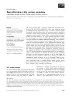
Figure 3 Inhibition of statin-induced apoptosis in C6 glioma cells by intermediates of the mevalonate pathway. (A) Induction of
caspase-3-like activity associated with statin-induced cell death. Caspase-3 activity is expressed as pM of proteolytic cleavage of the caspase-3
substrate Asp-Glu-Val-Asp-7-Amino-4-trifluoromethylcoumarin (DEVD-AFC) per h per mg of protein. The results are representative of 5
independent experiments. *p < 0.01 vs. controls (ANOVA with Dunnett’s test). (B-D) C6 glioma cells were pretreated with 1 mM mevalonic acid
lactone (MVA), 10 μM farnesyl pyrophosphate (FPP), 10 μM geranylgeranyl pyrophosphate (GGPP), 30 μM squalene, 30 μM isopentenyladenine,
30 μM ubiquinone, or 30 μM dolichol for 4 h and then treated with (B) 5 μM mevastatin, (C) 5 μM fluvastatin, or (D) 10 μM simvastatin for 72 h.
These results are representative of 5 independent experiments. *p < 0.01 vs. the controls (ANOVA with Dunnett’s test).
Yanae et al. Journal of Experimental & Clinical Cancer Research 2011, 30:74
/>Page 6 of 8
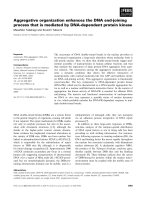
Figure 4 Statins specifically suppress the acti vation of Ra s/extracellu lar signal-regulated kinase (ERK) and Ras/Akt pathways in C6
glioma cells. (A) C6 glioma cells were treated with 5 μM mevastatin, 5 μM fluvastatin, or 10 μM simvastatin for 1, 3, 6, 12, or 24 h. Control cells
were treated with 0.1% DMSO and cultured in serum-containing medium for 24 h. Whole-cell lysates were generated and immunoblotted with
antibodies against phosphorylated ERK1/2 (phospho-ERK1/2), phosphorylated Akt (phospho-Akt), phosphorylated c-Jun N-terminal kinase 1/2
(phospho-JNK1/2), ERK1/2, Akt, and JNK1/2. (B) ERK1/2 and Akt activation in C6 cells to which statins were administered with or without the
addition of MVA, FPP, and GGPP. Phospho-ERK1/2, phospho-Akt, ERK1/2, and Akt levels were determined by immunoblotting analysis of the
whole-cell lysate.
Yanae et al. Journal of Experimental & Clinical Cancer Research 2011, 30:74
/>Page 7 of 8
Acknowledgements
This work was supported by the High-Tech Research Center Project for
Private Universities and a matching fund subsidy from MEXT (Ministry of
Education, Culture, Sports, Science and Technology), Japan, 2007-2011.
Author details
1
Division of Pharmacotherapy, Kinki University School of Pharmacy, Kowakae,
Higashi-Osaka 577-8502, Japan.
2
Department of Pharmacy, Sakai Hospital,
Kinki University School of Medicine, Sakai, Osaka 590-0132, Japan.
3
Department of Pathology, Kinki University School of Medicine,
Osakasayama, Osaka 589-8511, Japan.
4
Department of Surgery, Kinki
University School of Medicine, Osakasayama, Osaka 589-8511, Japan.
5
Department of Pharmacy, Kinki University Hospital, Osakasayama, Osaka
589-8511, Japan.
Authors’ contributions
MY and MT carried out cell viability assay, caspase-3 activity assay, statical
analysis, and drafted the manuscript. TS, TI, MI, and YY carried out western
bolotting analysis. TS, TI, and MI contributed to statistical analyses. SN
designed the experiments and revised the manuscript. All authors read and
approved the final manuscript.
Competing interests
The authors declare that they have no competing interests.
Received: 9 May 2011 Accepted: 10 August 2011
Published: 10 August 2011
References
1. DeAngelis LM: Brain tumors. N Engl J Med 2001, 344:114-123.
2. Reardon DA, Wen PY: Therapeutic advances in the treatment of glioblastoma:
rationale and potential role of targeted agents. Oncologist 2006, 11:152-164.
3. Nishida S, Matsuoka H, Tsubaki M, Tanimori Y, Yanae M, Fujii Y, Iwaki M:
Mevastatin induces apoptosis in HL60 cells dependently on decrease in
phosphorylated ERK. Mol Cell Biochem 2005, 269:109-114.
4. Tsubaki M, Yamazoe Y, Yanae M, Satou T, Itoh T, Kaneko J, Kidera Y,
Moriyama K, Nishida S: Blockade of the Ras/MEK/ERK and Ras/PI3K/Akt
pathways by statins reduces the expression of bFGF, HGF, and TGF-β as
angiogenic factors in mouse osteosarcoma. Cytokine 2011, 54:100-107.
5. Wu J, Wong WW, Khosravi F, Minden MD, Penn LZ: Blocking the Raf/MEK/
ERK pathway sensitizes acute myelogenous leukemia cells to lovastatin-
induced apoptosis. Cancer Res 2004, 64:6461-6468.
6. Jiang Z, Zheng X, Lytle RA, Higashikubo R, Rich KM: Lovastatin-induced
up-regulation of the BH3-only protein, Bim, and cell death in
glioblastoma cells. J Neurochem 2004, 89:168-178.
7. Koyuturk M, Ersoz M, Altiok N: Simvastatin induces proliferation inhibition
and apoptosis in C6 glioma cells via c-jun N-terminal kinase. Neurosci
Lett 2004, 370:212-217.
8. Fujiwara K, Tsubaki M, Yamazoe Y, Nishiura S, Kawaguchi T, Ogaki M,
Nishinobo M, Shimamoto K, Moriyama K, Nishida S: Fluvastatin induces
apoptosis on human tongue carcinoma cell line HSC-3. Yakugaku Zasshi
2008, 128:153-158.
9. Bouterfa HL, Sattelmeyer V, Czub S, Vordermark D, Roosen K, Tonn JC:
Inhibition of Ras farnesylation by lovastatin leads to downregulation of
proliferation and migration in primary cultured human glioblastoma
cells. Anticancer Res 2000, 20:2761-2771.
10. Cerezo-Guisado MI, García-Román N, García-Marín LJ, Alvarez-Barrientos A,
Bragado MJ, Lorenzo MJ: Lovastatin inhibits the extracellular-signal-
regulated kinase pathway in immortalized rat brain neuroblasts. Biochem
J 2007, 401:175-183.
11. Taylor-Harding B, Orsulic S, Karlan BY, Li AJ: Fluvastatin and cisplatin
demonstrate synergistic cytotoxicity in epithelial ovarian cancer cells.
Gynecol Oncol 2010, 119:549-556.
12. Lee MV, Fong EM, Singer FR, Guenette RS: Bisphosphonate treatment
inhibits the growth of prostate cancer cells. Cancer Res 2001, 61:2602-2608.
13. Shipman CM, Rogers MJ, Apperley JF, Russell RG, Croucher PI:
Bisphosphonates induce apoptosis in human myeloma cell lines: a novel
anti-tumour activity. Br J Haematol 1997, 98:665-672.
14. Morgan MA, Sebil T, Aydilek E, Peest D, Ganser A, Reuter CW: Combining
prenylation inhibitors causes synergistic cytotoxicity, apoptosis and
disruption of RAS-to-MAP kinase signalling in multiple myeloma cells. Br
J Haematol
2005, 130:912-925.
15.
Ts
ubaki M, Kato C, Nishinobo M, Ogaki M, Satou T, Ito T, Kusunoki T, Fujiwara K,
Yamazoe Y, Nishida S: Nitrogen-containing bisphosphonate, YM529/ONO-
5920, inhibits macrophage inflammatory protein 1 alpha expression and
secretion in mouse myeloma cells. Cancer Sci 2008, 99:152-158.
16. Park IH, Kim JY, Jung JI, Han JY: Lovastatin overcomes gefitinib resistance
in human non-small cell lung cancer cells with K-Ras mutations. Invest
New Drugs 2010, 28:791-799.
17. Horiguchi A, Sumitomo M, Asakuma J, Asano T, Asano T, Hayakawa M: 3-
hydroxy-3-methylglutaryl-coenzyme a reductase inhibitor, fluvastatin, as
a novel agent for prophylaxis of renal cancer metastasis. Clin Cancer Res
2004, 10:8648-8655.
18. Sondergaard TE, Pedersen PT, Andersen TL, Søe K, Lund T, Ostergaard B,
Garnero P, Delaisse JM, Plesner T: A phase II clinical trial does not show
that high dose simvastatin has beneficial effect on markers of bone
turnover in multiple myeloma. Hematol Oncol 2009, 27:17-22.
19. Skottheim IB, Gedde-Dahl A, Hejazifar S, Hoel K, Asberg A: Statin induced
myotoxicity: the lactone forms are more potent than the acid forms in
human skeletal muscle cells in vitro. Eur J Pharm Sci 2008, 33:317-325.
Figure 5 Schematic representation of interacellular effects of
statins in C6 glioma cells.
Yanae et al. Journal of Experimental & Clinical Cancer Research 2011, 30:74
/>Page 8 of 8