báo cáo khoa học: " Effects of RNA interference-mediated gene silencing of JMJD2A on human breast cancer cell line MDA-MB-231 in vitro" pptx
Bạn đang xem bản rút gọn của tài liệu. Xem và tải ngay bản đầy đủ của tài liệu tại đây (2.09 MB, 9 trang )
RESEARC H Open Access
Effects of RNA interference-mediated gene
silencing of JMJD2A on human breast
cancer cell line MDA-MB-231 in vitro
Bei-Xu Li, Ming-Chang Zhang, Cheng-Liang Luo, Peng Yang, Hui Li, Hong-Mei Xu, Hong-Fei Xu, Yi-Wen Shen,
Ai-Min Xue and Zi-Qin Zhao
*
Abstract
Previous data demonstrate that JMJD2A is a cancer-associated gene and may be involved in human breast cancer
by demethylation of H3K9me3. The aim of this study was to investigate depressive effects on JMJD2A by
transfection with JMJD2A-sepcific siRNA in human breast cancer cell line MDA-MB-231 and effects on cell
proliferation, invasion and migration. JMJD2A-specific siRNA was chemi cally synthesised and transfected into
human breast cancer cell line MDA-MB-231. Expression levels of JMJD2A were detected by quantitative real-time
PCR and Western blot analysis. Cells proliferation was evaluated by using flow cytometric anlysis and MTT assay.
The abilities of invasion and migration were evaluated by cell migration and invasion assay with Boyden chambers.
The results showed that the transfection was successful and expression levels of JMJD2A mRNA and protein in
siRNA group were both down-regulated. By MTT assay, the mean actual absorbance in siRNA group was
significantly lower than that in blank control group (P < 0.05) and negative control group (P < 0.05). In addition,
the percentage of cells in G0/G1 phase in siRNA group was significantly more than that in blank control group (P
< 0.05) and negative control group (P < 0.05). Furthermore, by cell invasion and migration assay, the decreased
number of migrated cells in siRNA group was observed (P < 0.05). These data imply that silencing JMJD2A gene
could result in cell cycle change and proliferation inhibition, and lead to suppress tumor cell invasion and
migration. It provides a new perspective in understanding the pleiotropic functions of JMJD2A and its contribution
to human breast cancer.
Keywords: JMJD2A, transfection, proliferation, invasion, migration
Background
Human breast cancer is one of the most frequent malig-
nant tumors with the incidence rate increasing year by
year. Based on the GLOBOCAN 2008 estimates, breast
cancer is the most frequently diagnosed cancer and the
leading cause of cancer death among females, account-
ing for 23% of the total cancer cases and 14% of the
cancer deaths [1]. The prognosis of the patients with
advanced stage breast cancer is poor, because of the
progression and metastasis of the disease, even surgical
removal, chemotherapy and endocrine therapy were
employed for most cases. Prevention and treatment of
breast cance r require a better understanding of the
molecular mechanisms underlying the progression of
breast cancer.
Gene therapies for tumor were focused on in recent
years, including gene replacement, antisense nucleic acid
technique, cytokine gene therapy and RNA interfer ence
(RNAi) technique. RNAi is a post-transcriptional regula-
tion and provides a rapid means of depleting mRNAs by
introducing double-stranded RNA homologous to a par-
ticular message leading to its sequence-specific degrada-
tion. It is simple, specific and effective to use small
interfering RNA (siRNA) to silence target gene [2].
Jumonji Domain Containing 2A (JMJD2A, also known
as JHDM3 or KDM4A) was identified and ch aracterized
in 2004 [3]. JMJD2A belongs to the JmjC domain-con-
taining family JMJD2 proteins, which are lysine tri-
methyl-specific histone demethylases catalyzing the
* Correspondence:
Department of Forensic Medicine, Shanghai Medical College, Fudan
University, Shanghai 200032, PR China
Li et al. Journal of Experimental & Clinical Cancer Research 2011, 30:90
/>© 2011 Li et al; licensee BioMed Central Ltd. This is an Open Access article distributed under the terms of the Creative Commons
Attribution License ( which permits unr estricted use, distribution, and reproduction in
any medium, provided the original work is properly cited.
demethylation of trimethylated H3K9 (H3K9me3) and
H3K36 (H3K36me3) [4-6]. JMJD2 family genes are can-
cer -associated genes [3]. JMJD2A is widely expressed in
human tissues and cell lines, and high endogenous
expression of JMJD2A mRNA was found in several cell
types, including human T-cell lymphotropic virus 1-
infected cell lines, the HT1376 bladder carcinoma cell
line, the U2OS osteosarcoma cell line and the prostate
cancer cell line [7,8]. However, there are rare literatures
focusing on the relationship between JMJD2A and
breast cancer.
In this study, JMJD2A-specific siRNA was chemically
synthesised and transfected into human breast c ancer
cell line MDA-MB-231. The levels on JMJD2A mRNA
and its protein expression, and biological characteristics
of MDA-MB-231 cells including proliferation, migration
and invasion were investigated.
Materials and methods
JMJD2A siRNA synthesis
JMJD2A siRNA was chemically synthesised by Qiagen
Technolog y Co. Ltd (Shanghai, China). siRNA was
diluted to 20 μmol/L with free-RNase water. siRNA
duplexes were synthesised as follows: Sense sequen ce: 5’-
GAGUUAUCAACUCAAGAUA-3’, Antisense sequence:
5’-UAUCUUGAGUUGAUAACUC-3’.
Cell transfection
Human breast cancer cell line MDA-MB-231 in this
research was preserved in our laboratory. At 24 h
before transfection, MDA-MB-231 cells in logarithmic
growth phase were seeded into 6-well plates, at a den-
sity of 5 × 10
5
cells per well and incubated in RPMI
1640 medium (GIBCO, Invitrogen, USA) conta ining
10% FBS (GIBCO, Invitrogen, USA). RPMI 1640 med-
ium containing 10% FBS was replaced by serum-free
Opti-MEM (GIBCO, Invitrogen, USA) at 8 h later.
HiPerFect Transfection Reagent and Negative control
siRNA were purchased from Qiagen Technology Co.
Ltd (Shanghai, China). Transfection compounds were
prepared in three groups as follows: siRNA group (100
μl Opti-MEM, 6 μl HiPerFect Transfection Reagent
and 5 μl JMJD2A siRNA), negative control group (100
μl Opti-MEM, 6 μl HiPerFect Transfection Reagent
and 5 μl negative control siRNA) and blank control
group (100 μl Opti-MEM). Transfection c ompounds
were placed at room temperature for 10 minutes and
then dropped onto 6-well plates. Bulk volume of the
compounds was 2200 μl per well. Both Opti-MEM and
transfection compounds were replaced by complete
medium at 24 h after transfection. FAM-siRNA was
transfected to measure the efficiency of transfection
simultaneously according to the manufacturer’ s
instructions.
Quantitative real-time PCR
Total RNA of three groups was extracted respectively with
the RNAiso Reagent kit (TaKaR a, Dalian, China) at 48 h
after transfection. cDNA was generated by reverse tran-
scription of 2 μg of total RNA using random primers and
PrimeScript RT Master Mix Perfect Real Time (TaKaRa,
Dalian, China) in a total reaction volume of 40 μl accord-
ing to the manufact urer’s inst ructions. The seq uences of
forward and reverse oligonucleotide primers, specific to
JMJD2A and housekeeping genes, were designed using
Primer5 software. The primers used are: 5’ -TGTGC
TGTGCTCCTGTAG -3’ and 5’ -GTCTCCTTCCTCTC
CATCC -3’ for JMJD2A; 5’-TGACGCTGGGGCTGG-
CATTG -3’ and 5’-GCTCTTGCTGGGGCTGGTGG -3’
for GAPDH. Primers were synthesised by Shanghai
Daweike Biotechnology Co. Ltd (Shanghai, China).
Real-time quantitative PCR was performed in an ABI
PRISM 7500 Real-Time System. A 10-fold dilution of
each cDNA was amplified in a 20-μl volume, using the
SYBR Premix Ex TaqTM Perfect Real Time (TaKaRa,
Dalian, China), with 0.2 μM final concentrations of each
primer. PCR cycle conditions were 95°C for 30 s, and
40 cycles of 95°C for 5 s and 60°C for 34 s. The amplifi-
cation specificity was evalu ated with melting curve ana-
lysis. Threshold cycle Ct, which correlates inversely with
the target mRNA levels, was calculated using the second
derivative maximum algorithm provided by the iCycler
software. For JMJD2A, the mRNA levels were normal-
ized to GAPDH mRNA levels [9].
Western blot
At 72 h after transfection, cells in different treatment
groups were homogenized in Western blot analysis buf-
fer containing 10 mM Tris-HCl (pH 7.4), 150 mM
NaCl, 1% (v/v) Triton X-100, 1% sodium deoxycholat e,
0.1% SDS, 5 mM EDTA, 1 mM P MSF, 0.28 kU/L apro-
tinin, 50 mg/L leupeptin, 1 mM benzamidine and 7 mg/
L pepstain A. The homogenate was then centrifuged at
12, 000 rpm for 10 min at 4°C and the supernatant was
retained and preserved at -80°C for later use. Protein
concent ration was determined using a BCA kit (Pierce).
Twenty micrograms of protein from each group were
subject to electrophoresis on 1 0% SDS-PAGE gel using
a constant current. Proteins were transferred to nitrocel-
lulose membranes on a semidry electrotransferring unit
and incubated with monoclonal rabbit anti-human
JMJD2A antibody (Cell Signaling Technology, USA,
1:1000) in Tris-buffered saline containing 0.1% Tween-
20(TBST)and5%nonfatdrymilkovernightat4°C.
After the o vernight incubation with the primary antibo-
dies, membranes were washe d and incubated with HRP-
labelled goat anti-rabbit second antibody (Santa Cruz
Biotechnology Inc., USA) in TBST for 2 h. Immunoreac-
tivity was detected with enhanced chemoluminescent
Li et al. Journal of Experimental & Clinical Cancer Research 2011, 30:90
/>Page 2 of 9
autoradiography (ECL kit, Amersham), according to the
manufacturer’ s instructions. The membranes were
reprobed wi th GAPDH (Cell Si gnaling Technology,
USA, 1:1000) after striping. The signal intensity of pri-
mary antibody binding was quantitatively analyzed with
Sigma Scan Pro 5 and was normalize d to a loading con-
trol, GAPDH [10].
Flow cytometric anlysis (FCM)
At 72 h after transfection, cells in different treatment
groups were collected with trypsinization, then washed
with PBS twice. Cells were fixed in 70% ethanol for 1 h
at room temperature. After centrifugation, the cell pellet
was resuspended in PBS (pH 7.4), containing 100 μL
RNase A (1 mg/mL) and 400 μL propidium iodide (50
μg/mL). The cells were incubated for 30 min at room
temperature, and DNA content was determined by flow
cytometry using a FACScan flow cytometer at 4 88 nm
and the data were input to compute r and analyzed by
software Light cycle. The experiment was performed
three times in triplicate [11]. Proliferation indexes (PI)
was calculated as follows: PI = (S+G2/M)/(G0/G1+S
+G2/M)×100%.
MTT assay
MDA-MB-231 cells were seeded into 96-well plates at a
density of 1 × 10
4
cells per well and incubated in RPMI
1640 medium containing 10% FBS. RPMI 1640 medium
containing 10% FBS was replaced by serum-free Opti-
MEM 8 h later. These cells were grouped as indicated
above (cell transfection).Thebulkvolumeofthetrans-
fect ion compounds was 100 μl per well. Opti-MEM and
transfection compounds were r eplaced by complete
medium at 24 h after transfection. After 72 h of incuba-
tion, MDA-MB-231 cells were incubated for an addi-
tional 4 hours with 20 μl MTT (Sigma Chemical Co.,
USA, 5 mg/ml). Then the supernatant was removed,
and 150 μl DMSO was added. Absorbance at 570 nm
(A570) of three groups and DMSO (Sigma Chemical
Co., USA) was measured with a microplate reader
(Model 550, Bio-Rad, USA) [11]. All experiments were
carried out eight times. Actual absorbance = absorbance
of the experimental group-absorbance of DMSO.
In vitro cell migration and invasion assay
At 24 h after transfection, the cells in different groups
were treated with trypsin and re-suspended as single-
cell solutions. A total of 2 × 10
5
cells in 0.5 ml of
serum-free RPMI 1640 medium were seeded on a 8 μm-
pore polycarbonate membrane Boyden chambers insert
in a transwell apparatus (Costar, Cambridge, MA), either
coated with (invasion) or without (migration) Matrigel
(BDBiosciences,SanJose,CA).600μlRPMI1640con-
taining 20% FBS was added to the lower chamber. After
the cells were incubated for 72 h (inv asion) or 36 h
(migration) at 37°C in a 5% CO
2
incubator, the cells on
the top surface of the insert were removed by wiping
with a cotton swab. The cells that migrated to the bot-
tom surface of the insert were fixed in 100% methanol
for 2 min, stained in Giemsa for 2 min, rinsed in PBS
and then subjected to microsc opic inspection (×200).
Values for invasion and migration were obtaine d by
counting five fields per membrane and represented the
average of three independent experiments [12].
Statistics analysis
The data were presented as means-standard errors (SE)
for MDA-MB-231 cells in each group. Statistical analysis
was carried out by one-way ANOVA followed by Dun-
nett t-test or Student t-test (two means comparison).
Statistical analysis was given using the related programs
in SPSS 12.0. Differences were considered significant
when P < 0.05.
Results
JMJD2A siRNA synthesis
The sequence of chemically synthesized JMJD2A siRNA
was consistent with the requirements, and the purity
reached to 98%. This met the experiment requirements.
Observation of cell transfection results
MDA-MB-231 cells transfected with FAM-siRNA were
subjected to Fluorescence microscopy at 8 h after trans-
fect ion. The green fluorescence cells were considered to
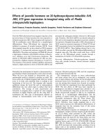
be transfected successfully. As shown in Figure 1A, cell
transfection was successful and HiPerFect Transfect ion
Reagent was effective. The transfection efficiency was
about 72.3%.
Transfection with JMJD2A-specific siRNA down-regulated
JMJD2A mRNA levels to silence JMJD2A gene
According to the results of quantitative real-time PCR
(Figure 1B), no significant difference (P > 0.05) was
detected in the levels o f JMJD2A mRNA between blank
control group (0.998 ± 0.170) and negative control
group (0.997 ± 0.150). The mRNA expression of siRNA
group (0.386 ± 0.108) were significantly lower than that
in blank control group (P < 0.05) and negative control
group (P < 0.05), respectively. These data suggested that
JMJD2A mRNA levels in MDA-MB-231 cells decreased
significantly after transfection with JMJD2A siRNA.
Transfection with JMJD2A-specific siRNA could result
in JMJD2A mRNA degradation to silence JMJD2A gene.
Transfection with JMJD2A-specific siRNA inhibited
JMJD2A protein expression in MDA-MB-231 cells
Western blot analysis showed that, the levels of JMJD2A
protein expression in the siRNA group (0.093 ± 0.051)
Li et al. Journal of Experimental & Clinical Cancer Research 2011, 30:90
/>Page 3 of 9
were significantly lower than that in blank control group
(0.203 ± 0.042) and nega tive control group (0.210 ±
0.050), respectively (P < 0.05; Figure 1C and 1D), while
the difference between blank control group and negative
control group was not significant (P > 0.05; Figure 1C
and 1D). These data indicated that JMJD2A-specific
siRNA silencing mRNA could significantly reduce the
levels of JMJD2A protein expression in MDA-MB-231
cells.
Silencing JMJD2A gene resulted in cell cycle changes and
proliferation inhibition in MDA-MB-231 cells
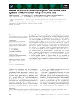
Cell cycle analysis by FCM revealed that JMJD2A siRNA
could induce changes in cell cy cle of MDA-MB-231
cells. The mean value of the experiments was shown in
Figure 2A, B and 2C. There were no significant differ-
ences (P > 0.05) in the percentages of cells at each
phase between blank control group and negative control
group. Compared with blank control group (30.3 ±
2.7%) a nd negative control group (34.2 ± 2.3%) respec-
tively, there was a significant difference (P < 0.05) in the
percentage of cells in G0/G1 phase in s iRNA group
(44.3 ± 1.6%). Similarly, there was a s ignificant differ-
ence (P < 0.05) in the percentage of cells in S phase in
siRNA group (43.4 ± 2.3%), versus blank control group
(58.4 ± 2.1%) and negative control group (52.8 ± 2.2%),
respectively. However, there was no significant differ-
ence (P > 0.05) in the percentage of cells in G2/M
phase in siRNA group (12.1 ± 2.2%), relative to blank
control group (11.0 ± 1.2%) and negative control group
(13.3 ± 1.8%), respectively. Silencing JMJD2A gene
could increase the percentage of cells at G0/G1 phase
and decrease the percentage of cell s at S phase. The
results suggested that the treatment could arrest cells at
the G1/S checkpoint and delay cell cycle into S phase.
Furthermore, proliferation indexes (PI) of each group
were calculated. We found that there was a significant
difference (P < 0.05) in PI of siRNA group (55.6 ±
2.1%), versus blank control group ( 69.6 ± 2.1%) and
negative control group (65.9 ± 2.2%), respectively. Our
results revealed a change in cell cycle with transfection
and indicated that cell proliferation could be inhibited
by transfection.
Additionally, MTT assay was performed to test the
effects of transfection with JMJD2A siRNA on the pro-
liferation of MDA-MB-231 cells treated in three differ-
ent groups. As shown in Figure 2D, there was no
significant difference (P > 0.05) in the average actual
absorbance between blank control group (2.136 ± 0.135)
and negative control group (2.089 ± 0.115). The average
actual absorbance in siRNA group (1.711 ± 0.087) was
significantly lower than that in blank control group (P <
0.05) and negative control group (P < 0.05), respectively.
Absorbance represents cell proliferation in MTT assay.
Figure 1 Transfection was successful and levels of JMJD2A
mRNA and protein were both down-regulated. A. The green
fluorescence cells transfected with FAM-siRNA under fluorescence
microscope (Note: ×100). B. Column diagram analysis for mRNA
levels of JMJD2A. JMJD2A-specific siRNA resulted in the reduction of
JMJD2A mRNA levels in MDA-MB-231 cells. C. Western blot analysis
for JMJD2A protein. D. Column diagram analysis for optical density
by Western blotting. JMJD2A protein levels were down-regulated in
siRNA group. (*P < 0.05, compared with blank control group and
negative control group respectively)
Li et al. Journal of Experimental & Clinical Cancer Research 2011, 30:90
/>Page 4 of 9
Figure 2 Knock down of JMJD2A resulted in cell cycle change and proliferation inhibition. A. DNA content s of MDA-MB-231 cells treated
in blank control group, negative control group and siRNA group by FCM. B. Column diagram analysis for the percentages of cells at each phase
in three different groups: G0/G1 phase, S phase and G2/M phase. At G0/G1 phase, there was a significant difference in the percentage of cells in
siRNA group compared with blank control group and negative control group respectively. At S phase, there was a significant difference in the
percentage of cells in siRNA group compared with blank control group and negative control group respectively, while no significant differences
in the percentages of cells at G2/M phase in the three groups. C. Column diagram analysis for the proliferation indexes (PI) calculated in three
different groups. PI in siRNA group was significantly lower than that in blank control group and negative control group respectively. D. Column
diagram analysis for the actual absorbance of three different groups, the mean actual absorbance of siRNA group was significantly lower than
that of the blank control group and the negative control group, respectively. (*P < 0.05, compared with blank control group and negative
control group respectively)
Li et al. Journal of Experimental & Clinical Cancer Research 2011, 30:90
/>Page 5 of 9
The MTT assay results consistented with FCM results.
These data indicated that transfection with JMJD2A
siRNA could significantly reduce the proliferation of
MDA-MB-231 cells.
Silencing JMJD2A gene suppressed MDA-MB-231 cell
migration and invasion in vitro
As displayed in Figure 3, cell migration was significantly
decreased in siRNA group than in blank control group
(P < 0.05) and negative control group (P < 0.05), respec-
tively. Cells in siRNA group showed significantly
decreased invasiveness, compared with blank control
group (Figure 4; P < 0.05) and negative control group
(Figure 4; P < 0.05). These results demonstrated that
transfection with JMJD2A siRNA could reduce the
migration and invasion of MDA-MB-231 cells.
Discussion
As leading cause of cancer death among females, human
breast cancer has the features of powerful invasive abil-
ity and early metastatic prope rty. Human breast cancer
with the incidence rate increasing is the threat to
human health. It is significantl y meaningful to u nder-
stand the pathologic mechanism of breast cancer and
find treatment target site. Recent researches indicate
that not only gene dysfunction but also histone modifi-
cations are involved in breast tumorigenesis [13].
Recent studies have implicated H3K9 modifications in
numerous biological phenomena including germ cell
development, × chromosome inactivation, DNA damage
repair and apoptosis [14]. Recent reports also link
deregulated histone methylation to tumorigenesis
[15,16]. An H3K9 histone methyltransferase, Suv39H1,
Figure 3 Knock down of JMJD2A resulted in suppressing tumor cell migration. A. Cells in blank control group transversed the Transwell
membrane. B. Cells in negative control group. C. Cells in siRNA group. D. Column diagram analysis for the number of MDA-MB-231 cells in
migration assay. The number of siRNA group (67 ± 10.2) was decreased compared with that of blank control group (173 ± 17.7) and negative
control group (168 ± 16.4), respectively. (*P < 0.05, compared with blank control group and negative control group respectively) (Note: ×200)
Li et al. Journal of Experimental & Clinical Cancer Research 2011, 30:90
/>Page 6 of 9
has been shown to function as a tumor suppressor by
maintaining H3K9 methylation levels [17,18]. These data
imply that H3K9me3 demethylases JMJD2A protein may
take part in tumorigenesis through demethylation of
H3K9me3.
Here we hypothesized that down-regulation of
JMJD2A expression in MDA-MB-231 cell line would
affect breast tumorigenesis and tumor biological charac-
teristics. To test this hypothesis, JMJD2A-specific siRNA
was transfected into human breast cancer cell line
MDA-MB-231 to observe the effects. It was proved that
JMJD2A gene could be silenced efficiently in MDA-MB-
231 cell line by transfection w ith JMJD2A-specific
siRNA and HiPerFect Transfection Reagent in this
study. According to the results of Quantitative real-time
PCR and Western blot analysis, the levels of JMJD2A
mRNA and protein expression were both down-regu-
lated based on the transfection. Further, FCM and MTT
assay results showed cell cycle changes and proliferation
inhibition existed in MDA-MB-231 c ell line, and
migration and invasion in vitro were both suppressed.
These data imply tumor growth and metastasis may be
restrained by silencing JMJD2A, and JMJD2A may be
associated with breast cancer cell line MDA-MB-231,
thus JMJD2A might be the potential therapeutic target
in breast cancer.
However, the mechanism of JMJD2A in breast cancer
is not very clear, h ere we discuss the probable role of
JMJD2A in breast cancer based on our own recent
data and the literature. Local chromatin architecture
which is strongly influenced by post-translational mod-
ifications of histones like methylation is now g enerally
recognized as an important factor in the regulation of
gene expression [19,20]. The combination of diff erent
modifications and the incorporation of different his-
tone variants which have distinct roles in gene regula-
tion, have led to the proposition of a regulatory
histone code which determines, at least partly, the
transcriptional potential for a specific gene or a geno-
mic region [21]. High endogenous expression of
Figure 4 Knock down of JMJD2A resulted in suppressing tumor cell invasion. A . Cells in blank control group transversed the Transwell
membrane. B. Cells in negative control group. C. Cells in siRNA group. D. Column diagram analysis for the number of MDA-MB-231 cells in
invasion assay. The number of siRNA group (175 ± 14.4) was decreased compared with that of blank control group (327 ± 20.8) and negative
control group (311 ± 15.3), respectively. (*P < 0.05, compared with blank control group and negative control group respectively) (Note: ×200)
Li et al. Journal of Experimental & Clinical Cancer Research 2011, 30:90
/>Page 7 of 9
JMJD2A protein catalyzes deme thylation of H3K9me3
excessively to break the balance between methylated
and demethylated histones. Genome-wide studies show
that H3K9me3 is enriched in heterochromatin, espe-
cially, as the mark with general repressive nature,
H3K9me3 is predominant in coding regions of some
active genes [22-25]. The intragenic permissive chro-
matin regions are flanked by the repressive mark,
H3K9me3, and the main tenance of the intragenic chro-
matin boundary appears to functions as a checkpoint
in elongation [26]. These data predict that the
H3K9me3 demethylase activities of JMJD2A protein
may act as transcriptional activators.
A recent research focusing on another member of
JMJD2 family proteins J MJD2B, which is considered to
have the similar function as JMJD2A in breast cancer
demonstrated that JMJD2B constitutes a key co mponent
of the estrogen signaling pathway and the establishment
of local epigenetic state and chromatin structure
required for proper induction of ER responsive genes.
JMJD2B which interacts with ERa and components of
the SWI/SNF-B chromatin remodeling complex was
recruited to ERa target sites, demethylated H3K9me3
and facilitated transcription of ER responsive oncoge nes
including MYB, MYC and CCND1, and knockdown of
JMJD2B severely impaired estrogen induced cell proli f-
eration and the tumor formation capacity of breas t can-
cer cells as a consequence [27]. Consisting with that
research, our data showed that silen cing of JMJD2A
coul d suppress the proliferation, migra tion and invasion
of MDA-MB-231 cell line, thereby indicating that
JMJD2A may be involved in the estrogen signaling
pathway.
Though JMJD2A and 2B exhibited robust interactions
with ER, in contrast to depletion of JMJD2B, depletion
of JMJD2A caused only a marginal defect in ER target
gene induction [27]. There maybeanotherpathway
JMJD2A involved in human breast cancer. It was
described that JMJD2A has molecular characterization
in binding both retinoblastom a protein (pRb) and
histone deacetylases (HDACs) [28]. JMJD2A maybe
associated with pRb recruits HDACs to t he pRB-E2F
complex, changes the chromatin structure at the E2F-
responsive promoter and induced suppression of target
gene E2F expression [29,30]. E2F1, 4 and their com-
plexes with HDAC play an important role in downregu-
lating the expression of the maternally imprinted tumor
suppressor gene ARHI in b reast cancer cells. Expression
of ARHI is markedly down-regulated in breast cancer,
and reactivation of ARHI expression in breast cancer
cells is associated with decreased H3K9me3 which is
demethylated by JMJD2A [31,32].
Together, JMJD2A may be, at least in part, involved in
human breast cancer by constituting a key component
of the estrogen signaling pathway or binding pRb and
HDACs to suppress E2F-induced ARHI expression.
However, the exact mechani sm of JMJD2A in human
breast cancer still remains elusive. The role of JMJD2A
may be diverse rather than single.
To date, this is the first report highlighting that the
suppression of proliferation, invasion and migration in
human breast cancer cell line MDA-MB-231, at least in
part, results from silencing of JMJD2A. The present
study sheds light on the novel role of JMJD2A in breast
cancer. However, our results were based on a single cell
line. Further researches to determine the differential
expression of JMJD2A between normal and cancer
breast tissue and the mechanism of JMJD2A in breast
cancer are required.
Acknowledgements
The work was supported by the National Science Foundation of China (No.
81172897 and No. 81072512).
Authors’ contributions
BX-L and MC-Z carried out experiments and drafted the manuscript. CL-L
and P-Y participated in study design and helped to draft the manuscript. H-
L, HM-X, HF-X, YW-S and AM-X participated in study design, performed
experiments and ZQ-Z participated in study design and revised manuscript.
All authors approved the final manuscript.
Competing interests
The authors declare that they have no competing interests.
Received: 10 August 2011 Accepted: 3 October 2011
Published: 3 October 2011
References
1. Jemal A, Bray F, Center MM, Ferlay J, Ward E, Forman D: Global cancer
statistics. CA Cancer J Clin CA Cancer J Clin 2011, 61:69-90.
2. Sen GL, Blau HM: A brief history of RNAi: the silence of the genes. FASEB
J 2006, 20:1293-1299.
3. Katoh M, Katoh M: Identification and characterization of JMJD2 family
genes in silico. Int J Oncol 2004, 24:1623-1628.
4. Trojer P, Reinberg D: Histone lysine demethylases and their impact on
epigenetics. Cell 2006, 125:213-217.
5. Whetstine JR, Nottke A, Lan F, Huarte M, Smolikov S, Chen Z, Spooner E, Li E,
Zhang G, Colaiacovo M, Shi Y: Reversal of Histone Lysine Trimethylation by
the JMJD2 Family of Histone Demethylases. Cell 2006, 125:467-481.
6. Nottke A, Colaiácovo MP, Shi Y: Developmental roles of the histone lysine
demethylases. Development 2009, 136:879-889.
7. Gray SG, Iglesias AH, Lizcano F, Villanueva R, Camelo S, Jingu H, Teh BT,
Koibuchi N, Chin WW, Kokkotou E, Dangond F: Functional Characterization
of JMJD2A, a Histone Deacetylase- and Retinoblastoma-binding Protein.
J Biol Chem 2005, 280:28507-28518.
8. Shin S, Janknecht R: Activation of androgen receptor by histone
demethylases JMJD2A and JMJD2D. Biochem Biophys Res Commun 2007,
359:742-746.
9. Zhang XD, Wang Y, Wang Y, Zhang X, Han R, Wu JC, Liang ZQ, Gu ZL,
Han F, Fukunaga K, Qin ZH: p53 mediates mitochondria dysfunction-
triggered autophagy activation and cell death in rat striatum. Autophagy
2009, 5:339-350.
10. Luo CL, Li BX, Li QQ, Chen XP, Sun YX, Bao HJ, Dai DK, Shen YW, Xu HF,
Ni H, Wan L, Qin ZH, Tao LY, Zhao ZQ: Autophagy is involved in traumatic
brain injury-induced cell death and contributes to functional outcome
deficits in mice. Neuroscience 2011, 184:54-63.
11. Dai HY, Liu L, Qin SK, He XM, Li SY: Lobaplatin suppresses proliferation
and induces apoptosis in the human colorectal carcinoma cell Line
LOVO in vitro. Biomed Pharmacother 2011, 65:137-141.
Li et al. Journal of Experimental & Clinical Cancer Research 2011, 30:90
/>Page 8 of 9
12. Li L, Zhang C, Li X, Lu S, Zhou Y: The candidate tumor suppressor gene
ECRG4 inhibits cancer cells migration and invasion in esophageal
carcinoma. J Exp Clin Cancer Res 2010, 29:133.
13. Jovanovic J, Rønneberg JA, Tost J, Kristensen V: The epigenetics of breast
cancer. Mol Oncol 2010, 4:242-254.
14. Martin C, Zhang Y: The diverse functions of histone lysine methylation.
Nat Rev Mol Cell Biol 2005, 6:838-849.
15. Müller-Tidow C, Klein HU, Hascher A, Isken F, Tickenbrock L, Thoennissen N,
Agrawal-Singh S, Tschanter P, Disselhoff C, Wang Y, Becker A, Thiede C,
Ehninger G, zur Stadt U, Koschmieder S, Seidl M, Müller FU, Schmitz W,
Schlenke P, McClelland M, Berdel WE, Dugas M, Serve H, Study Alliance
Leukemia: Profiling of histone H3 lysine 9 trimethylation levels predicts
transcription factor activity and survival in acute myeloid leukemia.
Blood 2010, 116:3564-3571.
16. Cloos PA, Christensen J, Agger K, Helin K: Erasing the methyl mark:
histone demethylases at the center of cellular differentiation and
disease. Genes Dev 2008, 22:1115-1140.
17. Peters AH, O’Carroll D, Scherthan H, Mechtler K, Sauer S, Schöfer C,
Weipoltshammer K, Pagani M, Lachner M, Kohlmaier A, Opravil S, Doyle M,
Sibilia M, Jenuwein T: Loss of the Suv39h histone methyltransferases
impairs mammalian heterochromatin and genome stability. Cell 2001,
107:323-337.
18. Braig M, Lee S, Loddenkemper C, Rudolph C, Peters AH, Schlegelberger B,
Stein H, Dörken B, Jenuwein T, Schmitt CA: Oncogene-induced
senescence as an initial barrier in lymphoma development. Nature 2005,
436:660-665.
19. Schübeler D, MacAlpine DM, Scalzo D, Wirbelauer C, Kooperberg C, van
Leeuwen F, Gottschling DE, O’Neill LP, Turner BM, Delrow J, Bell SP,
Groudine M: The histone modification pattern of active genes revealed
through genome-wide chromatin analysis of higher eukaryote. Genes
Dev 2004, 18:1263-1271.
20. Shilatifard A: Chromatin modifications by methylation and ubiquitination:
implications in the regulation of gene expression. Annu Rev Biochem
2006, 75:243-269.
21. Xu D, Bai J, Duan Q, Costa M, Dai W: Covalent modifications of histones
during mitosis and meiosis. Cell Cycle 2009, 8:3688-3694.
22. Mikkelsen TS, Ku M, Jaffe DB, Issac B, Lieberman E, Giannoukos G, Alvarez P,
Brockman W, Kim TK, Koche RP, Lee W, Mendenhall E, O’Donovan A,
Presser A, Russ C, Xie X, Meissner A, Wernig M, Jaenisch R, Nusbaum C,
Lander ES, Bernstein BE: Genome-wide maps of chromatin state in
pluripotent and lineage-committed cells. Nature 2007, 448:553-560.
23. Barski A, Cuddapah S, Cui K, Roh TY, Schones DE, Wang Z, Wei G,
Chepelev I, Zhao K: High-resolution profiling of histone methylations in
the human genome. Cell 2007, 129:823-837.
24. Brinkman AB, Roelofsen T, Pennings SW, Martens JH, Jenuwein T,
Stunnenberg HG: Histone modification patterns associated with the
human X chromosome. EMBO Rep 2006, 7:628-634.
25. Vakoc CR, Mandat SA, Olenchock BA, Blobel GA: Histone H3 lysine 9
methylation and HP1gamma are associated with transcription
elongation through mammalian chromatin. Mol Cell 2005, 19:381-391.
26. Gomes NP, Espinosa JM: Gene-specific repression of the p53 target gene
PUMA via intragenic CTCF-Cohesin binding. Genes Dev 2010,
24:1022-1034.
27. Kawazu M, Saso K, Tong KI, McQuire T, Goto K, Son DO, Wakeham A,
Miyagishi M, Mak TW, Okada H: Histone demethylase JMJD2B functions as
a co-factor of estrogen receptor in breast cancer proliferation and
mammary gland development. PLoS One 2011, 6:e17830.
28. Gray SG, Iglesias AH, Lizcano F, Villanueva R, Camelo S, Jingu H, Teh BT,
Koibuchi N, Chin WW, Kokkotou E, Dangond F: Functional characterization
of JMJD2A, a histone deacetylase- and retinoblastoma-binding protein. J
Biol Chem 2005, 280:28507-28518.
29. Takaki T, Fukasawa K, Suzuki-Takahashi I, Hirai H: Cdk-mediated
phosphorylation of pRB regulates HDAC binding in vitro. Biochem
Biophys Res Commun 2004, 316:252-255.
30. Lai A, Kennedy BK, Barbie DA, Bertos NR, Yang XJ, Theberge MC, Tsai SC,
Seto E, Zhang Y, Kuzmichev A, Lane WS, Reinberg D, Harlow E, Branton PE:
RBP1 recruits the mSIN3-histone deacetylase complex to the pocket of
retinoblastoma tumor suppressor family proteins found in limited
discrete regions of the nucleus at growth arrest. Mol Cell Biol 2001,
21:2918-2932.
31. Yu Y, Xu F, Peng H, Fang X, Zhao S, Li Y, Cuevas B, Kuo WL, Gray JW,
Siciliano M, Mills GB, Bast RC Jr: NOEY2 (ARHI), an imprinted putative
tumor suppressor gene in ovarian and breast carcinomas. Proc Natl Acad
Sci USA 1999, 96:214-219.
32. Lu Z, Luo RZ, Peng H, Huang M, Nishmoto A, Hunt KK, Helin K, Liao WS,
Yu Y: E2F-HDAC complexes negatively regulate the tumor suppressor
gene ARHI in breast cancer. Oncogene 2006, 25:230-239.
doi:10.1186/1756-9966-30-90
Cite this article as: Li et al.: Effects of RNA interference-mediated gene
silencing of JMJD2A on human breast cancer cell line MDA-MB-231 in
vitro. Journal of Experimental & Clinical Cancer Research 2011 30:90.
Submit your next manuscript to BioMed Central
and take full advantage of:
• Convenient online submission
• Thorough peer review
• No space constraints or color figure charges
• Immediate publication on acceptance
• Inclusion in PubMed, CAS, Scopus and Google Scholar
• Research which is freely available for redistribution
Submit your manuscript at
www.biomedcentral.com/submit
Li et al. Journal of Experimental & Clinical Cancer Research 2011, 30:90
/>Page 9 of 9