Handbook of Analytical Methods for Materials Part 1 docx
Bạn đang xem bản rút gọn của tài liệu. Xem và tải ngay bản đầy đủ của tài liệu tại đây (156.16 KB, 8 trang )
Handbook Of
Analytical
Methods For
Materials
Materials Evaluation and Engineering, Inc.
Practical Solutions to Materials ProblemsPractical Solutions to Materials Problems
Practical Solutions to Materials ProblemsPractical Solutions to Materials Problems
Practical Solutions to Materials Problems
Through Technology and InnovationThrough Technology and Innovation
Through Technology and InnovationThrough Technology and Innovation
Through Technology and Innovation
2
Materials Evaluation and Engineering, Inc.
13805 1st Avenue North, Suite 400
Plymouth, MN 55441-5447
Phone: (763) 449-8870
Fax: (763) 449-8699
Toll Free: (888) 349-8870
Website: www.mee-inc.com
Email:
3
Table of Contents
Introduction 5
Atomic Force Microscopy (AFM) 7
Auger Electron Spectroscopy (Auger) 11
Energy Dispersive X-ray Spectroscopy (EDS) 13
Fourier Transform-infrared Spectroscopy (FTIR) 15
Gas Chromatography / Mass Spectroscopy (GC-MS) 17
Ion Chromatography (IC) 19
Light Microscopy (LM) 21
Metallographic Study 23
Microindentation Hardness Testing 25
Nanoindentation Hardness Testing 27
Quantitative Chemical Analysis 29
Rockwell Hardness Testing 33
Scanning Electron Microscopy (SEM) 35
Secondary Ion Mass Spectrometry (SIMS) 39
Thermal Analysis (DSC, TGA) 41
X-ray Photoelectron Spectroscopy (XPS or ESCA) 43
Sample Preservation And Handling 45
About Materials Evaluation And Engineering, Inc 49
4
Blank Page
Handbook of Analytical Methods for Materials Copyright © 2001 by Materials Evaluation and Engineering, Inc.
6
Blank Page
Handbook of Analytical Methods for Materials Copyright © 2001 by Materials Evaluation and Engineering, Inc.
8
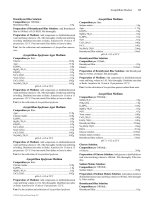
Intermittent Contact (Tapping Mode) AFM - In this mode, the probe cantilever is oscillated at or
near its resonant frequency. The oscillating probe tip is then scanned at a height where it barely
touches or “taps” the sample surface. The system monitors the probe position and vibrational ampli-
tude to obtain topographical and other property information. Accurate topographical information can
be obtained even for very fragile
surfaces. Optimum resolution is about
50 Å lateral and <1 Å height. Images
for phase detection mode, magnetic
domains, and local electric fields are
also obtained in this mode.
Lateral Force Microscopy - This
mode measures the lateral deflection
of the probe cantilever as the tip is
scanned across the sample in contact
mode. Changes in lateral deflection
represent relative frictional forces
between the probe tip and the sample
surface.
Phase Detection Microscopy -
With the system operating in Tapping
mode, the cantilever oscillation is
damped by interaction with the sample
surface. The phase lag between the drive signal and actual cantilever oscillation is monitored. Changes
in the phase lag indicate variations in the surface properties, such as viscoelasticity or mechanical
properties. A phase image, typically collected simultaneously with a topographical image, maps the
local changes in material’s physical or mechanical properties.
Magnetic Force Microscopy - This mode images local variations in the magnetic forces at the
sample’s surface. The
probe tip is coated with a
thin film of ferromagnetic
material that will react to
the magnetic domains on
the sample surface. The
magnetic forces between
the tip and the sample are
measured by monitoring
ATOMIC FORCE MICROSCOPY
Height and Phase Mode Image of a Polymer Sample
AFM Image of Defect on Coated Glass
Handbook of Analytical Methods for Materials Copyright © 2001 by Materials Evaluation and Engineering, Inc.
9
cantilever deflection while the probe is scanned at a constant height above the surface. A map of the
forces shows the sample’s natural or applied magnetic domain structure.
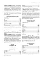
Image Analysis - Since the images are collected in
digital format, a wide variety of image manipulations
are available for AFM data. Quantitative topographical
information, such as lateral spacing, step height, and
surface roughness are readily obtained. Images can be
presented as two-dimensional or three-dimensional
representations in hard copy or as digital image files for
electronic transfer and publication.
Nanoindentation - A specialized probe tip is forced
into the sample surface to obtain a measure of the
material’s mechanical properties in regions as small as a
few nanometers. (See the Handbook section on Nanoindentation Hardness Testing.)
ATOMIC FORCE MICROSCOPY
Feature Measurements for CD Stamper
Top View AFM Image of Steel Microstructure
Handbook of Analytical Methods for Materials Copyright © 2001 by Materials Evaluation and Engineering, Inc.
10
ATOMIC FORCE MICROSCOPY
TYPICAL APPLICATIONS
• 3-dimensional topography of IC device
• Roughness measurements for chemical mechanical polishing
• Analysis of microscopic phase distribution in polymers
• Mechanical and physical property measurements for thin films
• Imaging magnetic domains on digital storage media
• Imaging of submicron phases in metals
• Defect imaging in IC failure analysis
• Microscopic imaging of fragile biological samples
• Metrology for compact disk stampers
SAMPLE REQUIREMENTS
No sample preparation is typically required. Samples can be imaged in air or liquid. Sample height is
limited to about 1.5 inches. Areas up to 8 inches in diameter can be fully traversed without reposition-
ing. Larger samples can be fixtured for imaging within a limited area. Total surface roughness in the
image area should not exceed about 6 µm.
AFM Images of Gold Plating for Wire Bond Failure Analysis