Carcinoma of the Esophagus - part 2 pdf
Bạn đang xem bản rút gọn của tài liệu. Xem và tải ngay bản đầy đủ của tài liệu tại đây (2.58 MB, 17 trang )
Etiology
Squamous cell carcinoma
Many of the environmental factors associated with a high incidence of SCC of the
esophagus relate to poor socioeconomic circumstances. A diet rich in preserved
and pickled foods and low in fresh fruit and vegetables, vitamin and mineral
deficiencies, and a thermal effect of hot food and beverages have all been impli-
cated. Alcohol intake and smoking are also strongly associated with an increased
risk of SCC of the esophagus [28,29,30,31]. The risk is thought to be dose related
and the genetic changes brought about by chronic exposure to causative agents
lead to a progression through epithelial dysplasia and carcinoma in situ to invasive
cancer [32]. After several years of cessation of exposure to irritant factors, such as
smoking and alcohol, risk is substantially reduced [33]. Conditions such as caustic
ingestion and achalasia of the cardia, which are associated with chronic mucosal
inflammation, also predispose to SCC of the esophagus.
Adenocarcinoma
The recent and rapid escalation in incidence of esophageal adenocarcinoma would
seem to suggest a mostly environmental rather than genetic effect. While a number
of factors including race, obesity, use of esophageal sphincter-relaxing drugs,
smoking, and alcohol consumption have all been incriminated as possible etiolo-
gical factors in esophageal adenocarcinoma [34,35,36,37,38], many cohort studies
have pointed strongly to gastroesophageal reflux and Barrett’s disease (Barrett’s
esophagus) as a causative factor [39,40].
There is a strong dose–response relationship between previous gastroesophageal
reflux symptoms and esophageal adenocarcinoma, but the relationship to cardia
cancer is weaker [40]. Wu et al. demonstrated a threefold increase in esophageal
cancer and a doubling in cardia cancer with reflux symptoms [41]. The relationship
between adenocarcinoma of the lower esophagus and adenocarcinoma of the cardia
is less clear. Siewert et al. have separated adenocarcinoma occurring at or near the
gastroesophageal junction into three groups depending on the anatomic relation to
the gastroesophageal junction. Type 1 tumors represent cancers of the lower eso-
phagus, mostly arising in Barrett’s esophagus. Type 2 and 3 tumors represent true
cardia and proximal gastric cancers, respectively [42], and while their incidence has
risen in recent d ecades, the changes are not as marked as for true lower esophageal
adenocarcinoma that are associated with Barrett’s esophagus, Type I. The clinical
4 S. S. Mudan and J Y. Kang
behavior and treatment of type 2 and 3 tumors are more like those of gastric
carcinoma [43,44].
Gastroesophageal reflux leads to columnar cell metaplasia in the distal esopha-
geal epithelium, a condition known as Barrett’s esophagus. This increases the risk
of developing esophageal adenocarcinoma 30- to 60-fold. The squamous cell
epithelium of the normal esophagus is replaced with a mature columnar-type
epithelium, with Barrett’s mucosa being derived from pleuripotential cells in the
basal layer of the esophageal epithelium [45,46,47]. The presence of goblet and
pregoblet cells is a requisite for intestinal metaplasia, which is associated with the
increased risk of malignant transformation. The probable driver toward metaplasia
is that the columnar epithelium is more tolerant of refluxate and the progression to
columnar metaplasia is a function of the refluxate content and periodicity [34].
Metaplasia of fundic- or cardiac-type gastric mucosa not involving the presence of
goblet cells is thought to carry a lower risk of malignant transformation. Most cases
of distal esophageal adenocarcinomas (90%) are thought to arise in the setting of
Barrett’s esophagus [48]. In other words, the risk of malignant transformation is
greatly elevated i n patients w ith Barrett’s esophagus a nd much less elevated in patients
with reflux esophagitis or nonerosive gastroesophageal reflux without Barrett’s
esophagus. Solaymani-Dodaran et al. reported relative risks for developing eso-
phageal adenocarcinoma of 29.8 for Barrett’s esophagus, 4.5 for reflux esophagitis,
and 3.1 for gastroesophageal reflux without Barrett’s esophagus or reflux esopha-
gitis [39]. A patient with Barrett’s esophagus has a 5% lifetime risk of developing
esophageal adenocarcinoma. The risk of transformation from benign intestinal
epithelium in Barrett’s esophagus to dysplasia and then adenocarcinoma is related to
the length of Barrett’s epithelium lining the esophagus, duration of reflux disease,
and presence of a hiatus hernia [49,50,51]. The risk of transformation may be
mitigated by antireflux surgery, but the evidence is not strong enough to recom-
mend this as a strategy for cancer prevention. Molecular markers of high risk are
recognized but do not as yet form part of routine practice [52,53,54,55].
Dysplasia is classified as low or high grade and is characterized by the degree of
hyperchromasia, nuclear : cytoplasm ratio, and glandular atypia. High-grade dys-
plasia is considered as indicative of at least an intraepithelial malignancy. About
one-third of patients with high-grade dysplasia at biopsy will have invasive disease
evident on a resection specimen. In population terms Barrett’s esophagus is a
common condition, occurring in 0.45–2.2% of all patients undergoing upper GI
endoscopy, about 12% of patients undergoing endoscopy for reflux symptoms,
and about 0.3% in unselected autopsy series [56]. While excess exposure to acid is
Epidemiology and Clinical Presentation in Esophageal Cancer 5
demonstrable in most patients with Barrett’s esophagus, progression to dysplasia is
more likely in patients with alkaline or bile-containing duodenogastric reflux
rather than those with pure acid reflux [57,58].
Several other potential causative factors have been evaluated. The incidence of
esophageal adenocarcinoma has increased since the introduction of powerful acid
suppressants such as histamine-2 receptor antagonists and proton pump inhibi-
tors, but the lead time for carcinogenesis probably precludes these agents as
etiological agents and the association is likely to reflect the use of these agents
to treat symptoms of reflux in patients already at increased risk of developing
esophageal adenocarcinoma. Drugs that reduce the lower esophageal sphincter
tone, e.g., anticholingergics, nitroglycerin, beta-adrenergic agonists, aminophyl-
line, and benzodiazepines, have all been implicated through increasing the poten-
tial for reflux [36]. Reduction in intragastric acidity through gastric mucosal
atrophy-induced hypochlorhydria from Helicobacter pylori infection may be
another factor in the promotion of distal esophageal SCC, while its carcinogenic
effect in noncardia gastric cancer is well recognized. By contrast, Helicobacter pylori
infection, especially of the cagA
þ
strain, may have a protective effect against esopha-
geal adenocarcinoma [59,60]. The role of diet is controversial, and while there appears
to be an association with noncardia gastriccancer,thelinktoesophagealcanceris
not so clear [61,62,63,64]. The relationship t o s moking is less clear t han t hat f or SCC.
Increased abdominal pressure brought about by central obesity, sedentary posture,
and tight belts has also been implicated [37,65], although a high body mass index
appears to be an independent risk factor for adenocarcinoma but not SCC [38].
Familial clustering has been demonstrated in Barrett’s esophagus and adeno-
carcinoma of the esophagus, but no ‘‘Barrett gene’’ has been identified, and it is not
clear whether the familial tendency represents a genetic predisposition or merely
the effect of similar lifestyle factors among family members [66].
The carcinogenic pathway from Barrett’s mucosa involves a multistep alteration
in the genotype, loss of regulatory function, induction of proinflammatory
enzymes such as cyclooxygenase-2, and angiogenesis. Consequently, chemopre-
vention and treatment through the use of therapies directed at specific molecular
targets has been postulated [67].
Clinical presentation
The majority of symptomatic patients turn out to have advanced disease. Presenting
symptoms are similar for SCC and adenocarcinoma. The most common are
6 S. S. Mudan and J Y. Kang
dysphagia and odynophagia (i.e., pain on swallowing). The pliability of the esopha-
gus is such that dysphagia occurs when the lumen is obstructed by about 75% of the
circumference, although a small tumor may cause a tight stenosis through intense
fibrosis. Chronic cough secondary to laryngopharyngeal reflux may be an early
marker of malignant transformation in Barrett’s esophagus [68]. Hoarseness or
Horner’s syndrome usually implies invasion of the recurrent laryngeal nerve or
cervical ganglia, and such patients are almost always inoperable. Cervical or supra-
clavicular lymphadenopathy is indicative of distant spread and indicates inoper-
ability in adenocarcinoma. It is present in about one-third of SCCs, and resection
with curative intent might still be considered in this disease with radical three-field
node dissection, in particular for mid- or upper-third tumors [68].
Prognosis
While some rare esophageal tumors such as lipomas or smooth muscle tumors
have a good outlook, the prognosis for SCC and adenocarcinoma of the esophagus
is poor with an overall tumor-specific lethality rate of $0.95 [69]. Survival appears
comparable across age groups, but females appear to have better outcomes. For
patients undergoing operations with curative intent, the 5-year survival ranges
from 5 to 20%. Large tumors, nodal involvement, and extracapsular nodal spread
are all strong prognostic factors for poor outcome [70]. Progression of nodal
disease to subdiaphragmatic sites is generally considered to carry the same prog-
nostic significance as distant metastases, although long-term survival with resec-
tion of celiac nodes is possible. It is likely that micrometastases in lymph nodes and
sites such as bone marrow behave in a way different from clinically obvious disease.
Whether the type of operation performed affects outcome is uncertain. Tumor
location in the upper esophagus predicates for a poor operative risk. Since most
SCCs are either mid or upper esophageal cancers and other comorbidities such as
age, chronic respiratory disease, liver disease, and poor nutrition are common, the
immediate results of surgery are consistently worse for SCC than for adenocarci-
noma [4,71,72]. Resections with microscopic positive surgical margins consis-
tently perform worse than those with negative margins.
Cancer-specific outcomes have improved [73] in the last two decades through
reduced surgical morbidity and mortality brought about by improvement in the
perioperative care, multidisciplinary collaboration, and the use of multimodal
therapies [74]. Although no survival advantage has been consistently demonstrated
by adjuvant chemotherapy in resected esophageal cancer and trials of neoadjuvant
Epidemiology and Clinical Presentation in Esophageal Cancer 7
chemotherapy or chemoradiotherapy are inconsistent, a large recently published
British Medical Research Council study (ST-02 MAGIC study) demonstrated a
significant tumor-specific survival advantage [75,76,77,78], and a distal esophageal
location and a measurable response to preoperative chemotherapy appear to
identify a favorable group [74,79,80,81]. Strategies based on identification of
high-risk individuals allowing surveillance by endoscopy or molecular markers
and for those progressing to cancer-targeted therapies with newer systemic agents
and pretreatment response prediction are awaited [67,82,83,84,85].
Conclusions
The epidemiology of esophageal cancer is rapidly changing. In Western countries,
adenocarcinoma of the lower esophagus has overtaken the previously more pre-
valent SCC. The divergent etiology and tumor behavior between the two diseases
require different prevention and treatment strategies. Until its etiology becomes
better understood, the continued rise in incidence of esophageal adenocarcinoma
presents a significant healthcare problem in Western countries. Better identifica-
tion of those at risk, e.g., individuals with Barrett’s esophagus, might allow more
effective screening policies. At present, surgery, when possible, represents the only
potentially curative modality, but results remain poor and we await improvements in
outcome through incorporation of therapies directed at novel cellular and molecular
targets.
REFERENCES
1. A. Jemal, R. Siegel, E. Ward, et al. Cancer statistics, 2006. CA Cancer J Clin, 56 (2006), 106–30.
2. R. Holmes and T. L. Vaughan. Epidemiology and pathogenesis of esophageal cancer. Semin Radiat
Oncol, 17 (2007), 2–9.
3. C. Mariette, L. Finzi, G. Piessen, et al. Esophageal carcinoma: prognostic differences between
squamous cell carcinoma and adenocarcinoma. World J Surg, 29 (2005), 39–45.
4. J. R. Siewert and K. Ott. Are squamous and adenocarcinomas of the esophagus the same disease?
Semin Radiat Oncol, 17 (2007), 38–44.
5. L. M. Brown, R. Hoover, D. Silverman, et al. Excess incidence of squamous cell esophageal cancer
among US black men: role of social class and other risk factors. Am J Epidemiol, 153 (2001),
114–22.
6. H. R. Wabinga, D. M. Parkin, F. Wabwire-Mangen, and S. Nambooze. Trends in cancer incidence
in Kyadondo County, Uganda, 1960–1997. Br J Cancer, 82 (2000), 1585–92.
8 S. S. Mudan and J Y. Kang
7. A. Kubo and D. A. Corley. Marked regional variation in adenocarcinomas of the esophagus and
the gastric cardia in the United States. Cancer, 95 (2002), 2096–102.
8. X. Wu, V. W. Chen, B. Ruiz, et al. Incidence of esophageal and gastric carcinomas among
American Asians/Pacific Islanders, whites, and blacks: subsite and histology differences.
Cancer, 106 (2006), 683–92.
9. S. Keeney and T. L. Bauer. Epidemiology of adenocarcinoma of the esophagogastric junction.
Surg Oncol Clin N Am, 15 (2006), 687–96.
10. A. P. Vizcaino, V. Moreno, R. Lambert, et al. Time trends incidence of both major histologic
types of esophageal carcinomas in selected countries, 1973–1995. Int J Cancer, 99 (2002),
860–8.
11. M. Pera. Trends in incidence and prevalence of specialized intestinal metaplasia, Barrett’s
esophagus, and adenocarcinoma of the gastroesophageal junction. World J Surg, 27 (2003),
999–1008.
12. M. Pera, C. Manterola, O. Vidal, and L. Grande. Epidemiology of esophageal adenocarcinoma. J
Surg Oncol, 92 (2005), 151–9 (Review).
13. A. Newnham, M. J. Quinn, P. Babb, J. Y. Kang, and A. Majeed. Trends in the subsite and
morphology of oesophageal and gastric cancer in England and Wales 1971–1998. Aliment
Pharmacol Ther, 17 (2003), 665–76.
14. H. Pohl and H. G. Welch. The role of overdiagnosis and reclassification in the marked increase
of esophageal adenocarcinoma incidence. J Natl Cancer Inst, 97 (2005), 142–6.
15. N. J. Shaheen. Advances in Barrett’s esophagus and esophageal adenocarcinoma. Gastroenterology,
128 (2005), 1554–66 (Review).
16. J. Lagergren. Adenocarcinoma of oesophagus: what exactly is the size of the problem and who
is at risk? Gut, 54:Suppl. 1 (2005), 1–5 (Review).
17. A. Kubo and D. A. Corley. Marked multi-ethnic variation of esophageal and gastric cardia
carcinomas within the United States. Am J Gastroenterol, 99 (2004), 582–8.
18. E. Bollschweiller, E. Wolfgarten, C. Gutschow, et al. Demographic variations in the rising
incidence of esophageal adenocarcinoma in white males. Cancer, 92 (2001), 549–55.
19. R. V. Lord, M. G. Law, R. L. Ward, et al. Rising incidence of oesophageal adenocarcinoma in
men in Australia. J Gastroenterol Hepatol, 13 (1998), 356–62.
20. A. Newnham, M. J. Quinn, P. Babb, J. Y. Kang, and A. Majeed. Trends in oesophageal and
gastric cancer incidence, mortality and survival in England and Wales 1971–1998/1999. Aliment
Pharmacol Ther, 17 (2003), 655–64.
21. S. S. Devesa, W. J. Blot, and J. F. Fraumeni. Changing patterns in the incidence of esophageal and
gastric carcinoma in the United States. Cancer, 83 (1998), 2049–53.
22. Thames Cancer Registry.
Cancer in South East England 1997 (London: Thames Cancer
Registry, 2000).
23. A. A. Botterweck, L. J. Schouten, A. Volovics, et al. Trends in incidence of adenocarcinoma of the
oesophagus and gastric cardia in ten European countries. Int J Epidemiol, 29 (2000), 645–54.
Epidemiology and Clinical Presentation in Esophageal Cancer 9
24. B. P. Wijnhoven, M. W. Louwman, H. W. Tilanus, and J. W. Coebergh. Increased incidence of
adenocarcinomas at the gastro-oesophageal junction in Dutch males since the 1990s. Eur J
Gastroenterol Hepatol, 14 (2002), 115–22.
25. L. E. Hansson, P. Sparen, and O. Nyren. Increasing incidence of both major histological types of
esophageal carcinomas among men in Sweden. Int J Cancer, 54 (1993), 402–7.
26. J. Powell, C. C. McConkey, E. W. Gillison, et al. Continuing rising trend in oesophageal adeno-
carcinoma. Int J Cancer, 102 (2002), 422–7.
27. M. Younes, D. E. Henson, A. Ertan, et al. Incidence and survival trends of esophageal carcinoma
in the United States: racial and gender differences by histological type. Scand J Gastroenterol, 37
(2002), 1359–65.
28. L. S. Engel, W. H. Chow, T. L. Vaughan, et al. Population attributable risks of esophageal and
gastric cancers. J Natl Cancer Inst, 95 (2003), 1404–13.
29. M. Farhadi, Z. Tahmasebi, S. Merat, et al. Human papillomavirus in squamous cell carcinoma of
esophagus in a high-risk population. World J Gastroenterol, 11 (2005), 1200–3.
30. S. Bahmanyar and W. Ye. Dietary patterns and risk of squamous-cell carcinoma and adeno-
carcinoma of the esophagus and adenocarcinoma of the gastric cardia: a population-based case-
control study in Sweden. Nutr Cancer, 54 (2006), 171–8.
31. H. Boeing, T. Dietrich, K. Hoffman, et al. I ntake of fruits and vegetables and ri sk of c ancer o f th e
upper aero-digestive tract: the prospective EPIC-study. Cancer Causes C ontrol, 17 (2006), 957–69.
32. H. Kuwano, H. Kato, T. Miyazaki, et al. Genetic alterations in esophageal cancer. Surg Today,
35 (2005), 7–18.
33. C. H. Lee, J. M. Lee, D. C. Wu, et al. Independent and combined effects of alcohol intake, tobacco
smoking and betel quid chewing on the risk of esophageal cancer in Taiwan. Int J Cancer, 113
(2005), 456–63.
34. S. R. DeMeester and T. R. DeMeester. Columnar mucosa and intestinal metaplasia of the
esophagus: fifty years of controversy. Ann Surg, 231 (2000), 303–21 (Review).
35. S. R. DeMeester. Adenocarcinoma of the esophagus and cardia: a review of the disease and its
treatment. Ann Surg Oncol, 13 (2006), 12–30.
36. J. Lagergren, R. Bergstrom, H. O. Adami, and O. Nyren. Association between medications that
relax the lower esophageal sphincter and risk for esophageal adenocarcinoma. Ann Intern Med,
133 (2000), 165–75.
37. J. Lagergren and C. Jansson. Use of tight belts and risk of esophageal adenocarcinoma. Int J
Cancer, 119 (2006), 2464–6.
38. J. Lagergren, R. Bergstrom, and O. Nyren. Association between body mass and adenocarcinoma
of the esophagus and gastric cardia.
Ann Intern Med, 130 (1999), 883–90.
39. M. Solaymani-Dodaran, R. F. Logan, J. West, et al. Risk of oesophageal cancer in Barrett’s
oesophagus and gastro-oesophageal reflux. Gut, 53 (2004), 1070–4.
40. J. Lagergren, R. Bergstrom, A. Lindgren, and O. Nyren. Symptomatic gastroesophageal reflux as a
risk factor for esophageal adenocarcinoma. N Engl J Med, 340 (1999), 825–31.
10 S. S. Mudan and J Y. Kang
41. A. H. Wu, C. C. Tseng, and L. Bernstein. Hiatal hernia, reflux symptoms, body size, and risk of
esophageal and gastric adenocarcinoma. Cancer, 98 (2003), 940–8.
42. J. R. Siewert and H. J. Stein. Classification of adenocarcinoma of the oesophagogastric junction.
Br J Surg, 85 (1998), 1457–9.
43. J. R. Siewert, M. Feith, and H. J. Stein. Biologic and clinical variations of adenocarcinoma at the
esophago-gastric junction: relevance of a topographic–anatomic subclassification. J Surg Oncol,
90 (2005), 139–46.
44. V. W. Rusch. Are cancers of the esophagus, gastroesophageal junction, and cardia one disease,
two, or several? Semin Oncol, 31 (2004), 444–9.
45. H. M. Shields, S. J. Rosenberg, F. R. Zwas, et al. Prospective evaluation of multilayered epithelium
in Barrett’s esophagus. Am J Gastroenterol, 96 (2001), 3268–78.
46. J. Mueller, M. Werner, and M. Stolte. Barrett’s esophagus: Histopathologic definitions and
diagnostic criteria. World J Surg, 28 (2004), 148–54.
47. P. A. Atherford and J. A. Jankowski. Molecular biology of Barrett’s cancer. Best Pract Res Clin
Gastroenterol, 20 (2006), 813–27.
48. J. Theisen, H. J. Stein, H. Feith, et al. Preferred location for the development of esophageal
adenocarcinoma within a segment of intestinal metaplasia. Surg Endosc, 20 (2006), 235–8.
49. N. J. Shaheen. Should we worry about the length of Barrett’s esophagus? Gastrointest Endosc, 62
(2005), 682–5.
50. P. J. F. De Jonge, E. W. Steyerberg, E. J. Kuipers, et al. Risk factors for the development of
esophageal adenocarcinoma in Barrett’s esophagus. Am J Gastroenterol, 101 (2006), 1421–9.
51. B. Avidan, A. Sonnenberg, T. G. Schnell, et al. Hiatal hernia size, Barrett’s length, and severity of
acid reflux are all risk factors for esophageal adenocarcinoma. Am J Gastroenterol, 7 (2002), 1930–6.
52. D. V. Gopal, D. A. Lieberman, N. Margaret, et al. Risk factors for dysplasia in patients with
Barrett’s esophagus. Dig Dis Sci, 48 (2003), 1537–41.
53. A. P. Weston, P. Sharma, S. Mathur, et al. Risk stratification of Barrett’s esophagus: Updated
prospective multivariate analysis. Am J Gastroenterol, 99 (2004), 1657–66.
54. S. Oberg, J. Wenner, J. Johansson, et al. Barrett’s esophagus. Risk factors for progression to
dysplasia and adenocarcinoma. Ann Surg, 242 (2005), 49–54.
55. B. J. Ried, D. S. Levine, G. Longton, et al. Predictors of progression to cancer in Barrett’s
esophagus: Baseline histology and flow cytometry identify low and high risk patient subsets.
Am J Gastroenterol,
95 (2000), 1669–76.
56. A. J. Cameron. Epidemiology of columnar lined esophagus and adenocarcinoma. Gastroenterol
Clin North Am, 92 (1997), 918–22.
57. P. Singh, R. H. Taylor, and D. G. Colin-Jones. Esophageal motor dysfunction and acid exposure
in reflux esophagitis are more severe if Barrett’s metaplasia is present. Am J Gastroenterol, 89
(1994), 349–56.
58. D. S. Oh, J. A. Hagen, M. Fein, et al. The impact of reflux composition on mucosal injury and
esophageal function. J Gastrointest Surg, 10 (2006), 787–96.
Epidemiology and Clinical Presentation in Esophageal Cancer 11
59. W. Ye, M. Held, J. Lagergren, et al. Helicobacter pylori infection and gastric atrophy: risk of
adenocarcinoma and squamous-cell carcinoma of the esophagus and adenocarcinoma of the
gastric cardia. J Natl Cancer Inst, 96 (2004), 388–96.
60. F. Kamanger, S. M. Dawsey, M. J. Blaser, et al. Opposing risks of gastric cardia and noncardia
gastric adenocarcinomas associated with Helicobacter pylori seropositivity. J Natl Cancer Inst, 98
(2006), 1432–4.
61. C. A. Gonza
´
lez, P. Jakszyn, G. Pera, et al. Meat intake and risk of stomach and esophageal
adenocarcinoma within the European Prospective Investigation into Cancer and Nutrition
(EPIC). J Natl Cancer Inst, 98 (2006), 345–54.
62. C. A. Gonza
´
lez, G. Pera, A. Agudo, et al. Fruit and vegetable intake and the risk of stomach
and oesophagus adenocarcinoma in the European Prospective Investigation into Cancer and
Nutrition (EPIC-EURGAST). Int J Cancer, 118 (2006), 2559–66.
63. P. Jakszyn and C. A. Gonzalez. Nitrosamine and related food intake and gastric and oesophageal
cancer risk: a systematic review of the epidemiological evidence. World J Gastroenterol, 12 (2006),
4296–303 (Review).
64. S. T. Mayne, H. A. Risch, R. Dubrow, et al. Nutrient intake and risk of subtypes of esophageal
and gastric cancer. Cancer Epidemiol Biomarkers Prev, 10 (2001), 1055–62.
65. C. La Vecchia, E. Negri, P. Lagiou, and D. Trichopoulos. Oesophageal adenocarcinoma: a
paradigm of mechanical carcinogenesis? Int J Cancer, 102 (2002), 269–70.
66. A. Chak, T. Lee, M. F. Kinnard, et al. Familial aggregation of Barrett’s oesophagus, oesophageal
adenocarcinoma, and oesophagogastric junctional adenocarcinoma in Caucasian adults. Gut,
51 (2002), 323–8.
67. W. P. Tew, D. P. Kelsen, and D. H. Ilson. Targeted therapies for esophageal cancer. Oncologist, 10
(2005), 590–601.
68. K. M. Reavis, C. D. Morris, D. V. Gopal, et al. Laryngopharyngeal reflux symptoms better predict
the presence of esophageal adenocarcinoma than typical gastroesophageal reflux symptoms. Ann
Surg, 239 (2004), 849–58.
69. S. M. Lagarde, F. J. W. ten Kate, J. B. Reitsma, et al. Prognostic factors in adenocarcinoma of the
esophagus or gastroesophageal junction. J Clin Oncol, 24 (2006), 4347–55.
70. T. Lerut, W. Coosemans, G. Decker, et al. Extracapsular lymph node involvement is a negative
prognostic factor in T3 adenocarcinoma of the distal esophagus and gastroesophageal junction. J
Thorac Cardiovasc Surg, 126 (2003), 1121–8.
71. A. Alexandrou, P. A. Davis, S. Law, et al. Squamous cell carcinoma and adenocarcinoma of the
lower third of the esophagus and gastric cardia: similarities and differences. Dis Esophagus, 15
(2002), 290–5.
72. H. Abunasra, S. Lewis, L. Beggs, et al. Predictors of operative death after oesophagectomy for
carcinoma. Br J Surg, 92 (2005), 1029–33.
73. T. Lerut, W. Coosemans, G. Decker, et al. Diagnosis and therapy in advanced cancer of the
esophagus and the gastroesophageal junction. Curr Opin Gastroenterol, 22 (2006), 437–41.
12 S. S. Mudan and J Y. Kang
74. M. Koshy, N. Esiashvilli, J. C. Landry, et al. Multiple management modalities in esophageal
cancer: combined modality management approaches. Oncologist, 9 (2004), 147–59.
75. M. Stahl. Adjuvant chemoradiotherapy in gastric cancer and carcinoma of the oesophago-gastric
junction. Onkologie, 27 (2004), 33–6.
76. R. A. Malthaner, R. K. Wong, R. B. Rumble, et al. Neoadjuvant or adjuvant therapy for resectable
esophageal cancer: a systematic review and meta-analysis. BMC Med, 2:1 (2004), 35.
77. S. E. Greer, P. P. Goodney, J. E. Sutton, and J. D. Birkmeyer. Neoadjuvant chemoradiotherapy for
esophageal carcinoma: a meta-analysis. Surgery, 137 (2005), 172–7.
78. D. Cunningham, W. H. Allum, S. P. Stenning, et al. Perioperative chemotherapy versus surgery
alone for gastroesophageal cancer. N Engl J Med, 355 (2006), 11–20.
79. M. Stahl, H. Wilke, M. Stuschke, et al. Clinical response to induction chemotherapy predicts local
control and long-term survival in multimodal treatment of patients with locally advanced
esophageal cancer. J Cancer Res Clin Oncol, 131 (2005), 67–72.
80. R. J. Korst, A. L. Kansler, J. L. Port, et al. Downstaging of T or N predicts long-term survival after
preoperative chemotherapy and radical resection for esophageal carcinoma. Ann Thorac Surg, 82
(2006), 480–4.
81. R. A. Malthaner, S. Collins, and D. Fenlon. Preoperative chemotherapy for resectable thoracic
esophageal cancer. Cochrane Database Syst Rev, 3 (2006), CD001556.
82. E. S. Dellon and N. J. Shaheen. Does screening for Barrett’s esophagus and adenocarcinoma of
the esophagus prolong survival? J Clin Oncol, 23 (2005), 4478–82.
83. J. Tabernero, T. Macarulla, F. J. Ramos, and J. Baselga. Novel targeted therapies in the treatment
of gastric and esophageal cancer. Ann Oncol, 16 (2005), 1740–8.
84. M. Akilu and D. H. Ilson. Targeted agents and esophageal cancer – the next step? Semin Radiat
Oncol, 17 (2007), 62–9.
85. R. Langer, K. Specht, K. Becker, et al. Association of pretherapeutic expression of chemotherapy-
related genes with response to neoadjuvant chemotherapy in Barrett carcinoma. Clin Cancer Res,
11 (2005), 7462–9.
Epidemiology and Clinical Presentation in Esophageal Cancer 13
2
Pathology of Esophageal Cancer
Harriet M. R. Deere
Introduction
Worldwide, squamous cell carcin oma is the most common malignant ep ithelial tumor
of the esopha gus. The m ajority of r emaining tumors are adenocarcinomas, t he inci-
dence of which has been increasing d ramatically i n t he last fe w decades in the W estern
world. Rarely, adenosquamo us carcinoma and small cell carcinoma may occur.
This chapter will focus on the morphologic features of esophageal carcinoma
and associated precursor lesions.
Histopathology of tumor types
Squamous cell carcinoma
Precursor lesions – hyperplasia and dysplasia
Squamous cell carcinoma is thought to develop through a multistep process from
basal hyperplasia due to chronic esophagitis through increasing severity of dyspla-
sia to invasion [1].
Dysplasia is defined as the presence of unequivocal neoplastic cells confined to
the epithelium. It is seen more commonly in high cancer risk areas, e.g., China [2],
is frequently seen adjacent to invasive carcinomas, and is often multifocal [3].
Traditionally, dysplasia has been classified as mild, moderate, or severe (and
carcinoma in situ). More recently, a two-grade system for dysplasia in the gastro-
intestinal tract is preferred, with mild and moderate atypia being classed as low
grade and severe dysplasia and carcinoma in situ as high grade.
The risk of carcinoma rises with increasing severity of dysplasia. A recent study
from China has shown a relative risk of 2.9 for mild dysplasia, 9.8 for moderate,
28.3 for severe, and 34.4 for carcinoma in situ at 13 years follow-up [4].
Carcinoma of the Esophagus, ed. Sheila C. Rankin. Published by Cambridge University Press. # Cambridge
University Press 2008.
Macroscopic appearance
At endoscopy, dysplastic epithelium may have an erythematous, friable appearance
or be associated with erosions, nodules, or plaques [5]; however, it may appear
normal.
Microscopic appearance
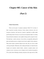
Dysplastic squamous epithelium shows cytological and architectural atypia, which
varies in severity according to the grade. Architectural atypia refers to disorganiza-
tion and loss of polarity of the cells and lack of surface maturation. Cytologically,
the cells exhibit nuclear hyperchromasia (dark staining due to increased DNA),
increased nuclear/cytoplasmic ratio, pleomorphism, and increased mitotic activity
(Figure 2.1a and b).
Invasive squamous cell carcinoma
About 50–60% of squamous cell carcinomas occur in the middle third of the
esophagus, approximately 30% occur in the lower third, and 10–20% in the upper
third [6]. Squamous cell carcinomas are separated into superficial (early) and
advanced tumors. Superficial tumors do not infiltrate beyond submucosa and
may or may not have lymph node metastases. The incidence of superficial carci-
noma is increasing, particularly in high-risk areas with screening programs. It
accounts for 10–20% of tumors in Japan and less than 1% in Europe [7].
Macroscopic appearance
Superficial tumors may be plaque-like, polypoid, depressed, or occult [8].
Advanced tumors are exophytic (60%), ulcerating (25%), or infiltrative (15%). A
(a) (b)
Figure 2.1 Squamous dysplasia of the esophagus: (a) mild dysplasia and (b) severe dysplasia.
Pathology of Esophageal Cancer 15
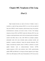
combination of these patterns may be seen. Intramural metastases due to intra-
mural lymphatic spread are found in 11–16% of cases [9,10], and multiple primary
tumors are found in 14–31% of patients [11,12] (Figure 2.2).
Microscopic appearance
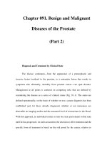
Squamous cell carcinoma is graded as well, moderately or poorly differentiated.
Well-differentiated tumors show well-formed cell nests, squamous pearls with
keratinization, and intercellular bridges. As tumors become less well differentiated
the proportion of basaloid cells increases, there is increased nuclear pleomorphism
and mitotic activity and loss of keratinization and prickle cells (Figure 2.3a and b).
Approximately two thirds of tumors are moderately differentiated, but it is
common for there to be a variation in the degree of differentiation within a tumor.
Approximately 20–30% of tumors show focal glandular differentiation [13,14].
Focal neuroendocrine (small cell) differentiation is also sometimes seen [15].
Rare variants
Basaloid squamous cell carcinoma is similar to the tumor that more often occurs in
the upper aerodigestive tract. Tumors are usually advanced at presentation.
Prognosis appears similar to typical squamous cell carcinoma [16].
Spindle c ell carcinoma (carcinosarcoma) usually p resents as a large polypoid mass
in the middle or lower third. Typically the tumors are biphasic, with conventional
Figure 2.2 Infiltrative ulcerated squamous cell
carcinoma. Case provided by Dr. F. Chang, London,
United Kingdom.
16 H. M. R. Deere
squamous cell carcinoma admixed with spindle cells showing variable differentiation .
Presentation is often at an earlier stage due to the intraluminal growth. Prognosis is
comparable to conventional squamous cell carcinoma of the same stage [17].
Verrucous squamous cell carcinoma presents as an exophytic papillary tumor. It is
a very well-differentiated tumor that is notoriously difficult to diagnose on biopsy.
Metastasis is rare; however; prognosis is poor as these tumors are locally aggressive
and fistula formation may occur [18].
Differential diagnosis
It may sometimes be difficult to distinguish between reactive atypia due to infla-
mmation and both dysplasia and invasive carcinoma, particularly on subopti-
mally orientated biopsy material. Chemoradiotherapy may also cause epithelial
atypia. Reactive stromal cells in areas of ulceration may be worrying for malig-
nancy, especially if there has been previous chemoradiotherapy. Cytokeratin stain-
ing is helpful in this situation as carcinoma is typically positive and mesenchymal
cells are negative.
Adenocarcinoma
Precursor lesions – Barrett’s esophagus and dysplasia
The most significant risk factor for adenocarcinoma is Barrett’s esophagus.
Rarely, esophageal adenocarcinoma may arise from heterotopic gastric tissue
[19,20] or the submucosal glands [21] and has a similar morphology to the
Barrett’s associated tumors.
(a) (b)
Figure 2.3 Squamous cell carcinoma: (a) well-differentiated squamous cell carcinoma and (b) poorly
differentiated squamous cell carcinoma composed mainly of basaloid cells.
Pathology of Esophageal Cancer 17
Barrett’s esophagus refers to replacement of the normal esophageal squamous
epithelium by metaplastic columnar epithelium as an acquired response to chronic
acid and bile reflux. Three main types of columnar mucosa may be present and
frequently coexist. The epithelium may be of junctional/cardiac type, gastric fundic
type, or intestinal type with goblet cells (Figure 2.4).
It is the intestinal metaplastic epithelium that is a significant risk factor for
malignancy.
The American College of Gastroenterology’s definition of Barrett’s esophagus is
‘‘a change in the esophageal epithelium of any length that can be recognized at
endoscopy and is confirmed to have intestinal metaplasia by biopsy’’ [22]. In the
United Kingdom, expert pathological opinion is that the identification of intestinal
metaplasia should not be required for diagnosis and that the eponym Barrett’s
esophagus should be replaced by the term columnar-lined esophagus (CLE) [23].
The British view is that the absence of intestinal metaplasia on biopsy may be due
to sampling error, as it has been shown that its demonstration is related to the
number of biopsies taken [24].
Histological diagnosis of CLE requires close correlation with the endoscopic find-
ings, as the microscopic features are not always pathognomonic. Biopsies are diag-
nostic for CLE when native esophageal structures, e.g., esophageal gland ducts are
present, but these are only seen in a pproximately 15% of biopsies [25]. In t heir absence
the pathologist can only corroborate the endoscopic diagnosis if columnar mucosa is
present. Biopsy from the gastroe sophageal junction or from a hiatus hernia, with or
without intestinal metaplasia, may have a similar a ppearance. It is also important to
distinguish between CLE and microscopic intestinal metaplasia at the cardia.
There have been conflicting results from studies looking at cytokeratin (CK7/20)
staining patterns that may help distinguish CLE from intestinal metaplasia at the
gastroesophageal junction/stomach [26,27,28].
Figure 2.4 Columnar-lined esophagus. Columnar-
lined esophagus with residual islands of squamous
epithelium and intestinal metaplasia.
18 H. M. R. Deere
The metaplastic epithelium is thought to originate from multipotential stem cells.
The precise location of these cells is unclear; however, the gland ducts and basal
epithelial layer are likely [29,30]. A distinctive multilayered epithelium with features
of both squamous and columnar epithelium has been described and may represent a
transitional phase in the conversion of squamous epithelium to CLE [31].
Approximately 5% of p atients with CLE develop d ysplas ia, which is classified as low
or high gra de. An ‘‘ indefinite for dysplasia’’ category is used for cases where it is not
possible to distinguish between true dysplasia and reactive atypia due to inflammation.
Adenocarcinoma is found in 30–50% of resection specimens from patients with
a biopsy diagnosis of high-grade dysplasia [32,33,34]. It is recommended that two
pathologists confirm the diagnosis of high-grade dysplasia prior to treatment, one
of whom should ideally be an expert gastrointestinal pathologist. Low-grade
dysplasia may persist, regress to nonneoplastic metaplasia, or progress to high-
grade dysplasia or carcinoma. High-grade dysplasia may also regress to low-grade
dysplasia, but the majority of cases progress to carcinoma within 4 years [35,36].
Recent research has led to a greater understanding of the molecular alterations
involved in neoplastic progression. Proliferation markers, aneuploidy, p53, p16,
and adenomatous polyposis coli (APC) mutations, and cyclin D1 overexpression
are some of the most common abnormalities investigated. However, currently
there are no molecular techniques that are sufficiently reliable for routine use
either for the diagnosis of dysplasia or to predict progression [23].
Macroscopic appearance
Columnar epithelium has a velvety red appearance. Often dysplastic epithelium
appears endoscopically normal, but granularity, plaques, or erosions are some-
times present [37] (Figure 2.5).
Microscopic appearance
Dysplastic epithelium displays architectural abnormalities such as a villiform sur-
face and crowding and budding of the glands. Cytological atypia includes nuclear
hyperchromasia and pleomorphism, nuclear stratification, increased mitotic activ-
ity, and atypical mitoses. There is lack of surface maturation, with atypical cells
extending onto the mucosal surface (Figure 2.6a and b).
Effects of treatment
Studies have shown that CLE may regress with antireflux surgery or medical
treatment [38,39].
Pathology of Esophageal Cancer 19
Endoscopic ablation techniques are increasingly used and also lead to squamous
reepithelialization. This occurs due to extension of adjacent squamous epithelium
and the formation of squamous islands arising from the submucosal gland ducts.
Squamous metaplasia is often seen within metaplastic glands unrelated to esopha-
geal ducts, lending support for the existence of multipotential stem cells [40].
(a)
Figure 2.5 Distal esophageal adenocarcinoma arising on a
background of columnar-lined esophagus. Case provided by
Dr. F. Chang, London, United Kingdom.
(a) (b)
Figure 2.6 Columnar-lined esophagus. (a) Low-grade dysplasia. The nuclei are elongated,
pseudostratified, and hyperchromatic. There is no surface maturation. (b) High-grade dysplasia. The
glands are closely packed together and show cytological atypia with round nuclei with prominent
nucleoli.
20 H. M. R. Deere