Emergencies and Complications in Gastroenterology - part 2 potx
Bạn đang xem bản rút gọn của tài liệu. Xem và tải ngay bản đầy đủ của tài liệu tại đây (176.23 KB, 10 trang )
10
Dig Dis 2003;21:6–15
Lata/Hulek/Vanasek
significantly decreases not only the portal pressure but
also the gastric mucosa blood flow (GMBF) [37], which is
potentially important in the bleeding from portal hyper-
tensive gastropathy. However, trial data are conflicting.
Meta-analyses have shown better control of bleeding com-
pared with vasopressin [38]. A meta-analysis did not show
significantly better efficacy in comparison to placebo [39].
Smaller studies, however, found a similar efficacy com-
pared to sclerotherapy [40], terlipressin [31] and found a
lesser need for blood transfusions and other urgent thera-
pies [41].
Octreotide
Octreotide is a synthetic octapeptide derivate of so-
matostatin, first described in 1982. Besides octreotide,
more than 20 synthetic analogues of the somatostatin are
known. Lanreotide was tested mainly in animal models.
Vapreotide was better in comparison with placebo and
was proved to increase the efficacy of endoscopic treat-
ment in variceal bleeding in humans [42]. None of these
other analogues are currently used in common clinical
practice.
Octreotide has a similar pharmacological effect as so-
matostatin. The differences are dependent on its binding
to three out of five somatostatin receptors. In comparison
to somatostatin, its advantages are its longer half-time
(90–120 min) and especially longer pharmacological ac-
tion (8–12 h). Octreotide (as well as somatostatin) de-
creases significantly the portal pressure in animals [43],
but its influence on hemodynamics in cirrhotics, including
decrease of the portal pressure, was not significantly
proved [44]. It probably also influences the mesenteric cir-
culation [45]. Meta-analysis studies using octreotide or
somatostatin have shown a lower rate of complications
and a similar effect as sclerotherapy or balloon tamponade
[46]. A newer meta-analysis comparing octreotide to other
medical therapy and placebo has shown a better effect of
the octreotide on the bleeding control compared to place-
bo and other drugs and side effects comparable to placebo
or no treatment [47]. The administration of the octreotide
after sclerotherapy decreases the portal pressure and re-
bleeding rate compared to sclerotherapy alone [48, 49]; the
effect on mortality, however, was not proved.
Nitrates
Intravenous nitrates are mostly used to counteract the
vasoconstriction effect of vasopressin, of which isosor-
bide-5-dinitrate is the most common. Its hypotensive
effects limits its use in the acute phase of the bleeding epi-
sode.
Mechanical-Balloon Tamponade of the Varices
The balloon tamponade may have a life-saving effect
but its inappropriate application has many complications.
The ability to place properly balloon tamponade is sur-
prisingly low outside specialized centers. Generally, now-
adays it is seldom indicated. Currently it is accepted as a
temporary measure after second unsuccessful endoscopic
treatment en route to portosystemic decompression (sur-
gical or TIPS). If indicated, the patient should be man-
aged in the specialized intensive care unit. Most common
is the three-lumen double-balloon (Sengstaken-Blake-
more). In case of bleeding from subcardial- fundal gastric
varices, the single-balloon (Linton-Nachlas) tamponade is
more appropriate. The Minnesota balloon is a modifica-
tion of the double-balloon device with four lumens; the
fourth is used for sucking from the space above the esoph-
ageal balloon, thus it prevents aspiration better. Balloons
must be inflated by the air, not liquid. Water, due to its
weight, changes the shape of the balloon, which results in
malfunction of the device, and is therefore not an appro-
priate filling medium. The gastric balloon is inflated first,
then traction is ensured and the esophageal balloon is
inflated. Its pressure should be higher than portal pres-
sure, 40 mm Hg is usually sufficient, overinflation is con-
traproductive and causes complications. Suction should
be provided for gastric content and swallowed saliva. The
correct location of the balloon tube should be checked by
X-ray.
The balloon should not be insufflated more than 24 h.
Some authors recommend deflation of the balloon every
4–6 h for 30 min [50]. Up to 50% of patients do have
rebleeding after balloon decompression. Thus this tempo-
rary measure should always be combined with other
methods [51]. The complications include aspiration, re-
trosternal pain, esophageal or gastric rupture and mainly
esophageal and gastric ulcerations. Overinflated or water-
filled balloons or dislocated balloons as well as multiple
sclerotization sessions cause significant damage to the
esophagus which replaces varices as bleeding source. Sel-
dom the upright movement of the inflated esophageal bal-
loon causes obstruction of the airways and suffocation,
most such cases are due to the rupture of the gastric bal-
loon. In this case the cross section of the lumen causing
immediate decompression of the balloon and subsequent
extraction are indicated.
Management of Acute Variceal Bleeding
Dig Dis 2003;21:6–15
11
Transjugular Intrahepatic Portosystemic Shunt
(TIPS)
TIPS is a calibrated portosystemic shunt which re-
duces quickly portosystemic gradient and opens access to
endovasal treatment of varices (endovasal obliteration by
sealants). Therefore, it is highly effective in stopping vari-
ceal bleeding [52]. TIPS is indicated only when first-line
methods (medical and endoscopic) have failed. This hap-
pens as ‘chronic’ or ‘acute’ failure. ‘Chronic’ means that
patients do have repeated bleeding episodes despite ade-
quate application of first-line treatment. An ‘elective’
TIPS may be indicated. ‘Acute’ failure means bleeding
refractory to other measures and ‘urgent – salvage’ TIPS is
often a life-saving procedure.
It is difficult to organize a study comparing the TIPS
procedure as ‘salvage treatment’ as there is difficulty in
setting up a comparable alternative. Even the first paper
reporting TIPS dealt with uncontrolled bleeding in Child-
Pugh class C patients and showed reasonably good results
[53]. Most relevant papers investigating ‘salvage TIPS’
showed immediate control of bleeding in 91–100% of
cases, 30-day rebleeding 7–30% and 1-month (or 42 days)
mortality 28–55%. Child class C patients formed in most
of them more than 60% of cases [54–56] and in one 41%
of cases [57]. Retrospective comparison with esophageal
transection [58] significantly favored TIPS (30-day mor-
tality was 42 vs. 79%, rebleeding 16 vs. 26%). The role of
TIPS is especially important in patients bleeding from
gastric varices, which have a worse response to sclerother-
apy and in bleeding portal hypertensive gastropathy
which cannot be treated endoscopically at all. Gastric var-
ices in rescue TIPS series form up to 73% of cases [55].
These impressive data show that rescue TIPS definitively
has its place in therapeutic algorithm for bleeding pa-
tients. Most of TIPS procedures in question are per-
formed with a combination of endovasal obliteration of
varices as ‘urgent’ operations. It was proved that uncon-
trolled bleeding can be effectively treated with TIPS, and
TIPS has lower morbidity and mortality compared to sur-
gery.
Indications of TIPS and TIPS-Related Procedures in
Bleeding Patients
In general, accepted indications are patients with
bleeding that is uncontrolled by pharmacological and
endoscopic therapy. This is true both for emergency situa-
tions (urgent TIPS) and for patients with repeated epi-
sodes of hemorrhage despite adequate preventive treat-
ment who are not surgical candidates (elective TIPS).
These conclusions were confirmed by both the Reston
and Baveno consensus meetings. Most patients appear
with gastroesophageal varices. Clinical situations as
chronic anemia due to portal hypertensive gastropathy,
prevention of rebleeding from large gastric or intestinal
varices, fresh portal vein thrombosis contributing to
bleeding can be added to the list. Rare indications pub-
lished include treatment of massive hemoptysis second-
ary to bronchial collaterals [59], bleeding from stomal var-
ices in patients after external enteric diversion [60], bleed-
ing from colonic variceal veins and intestinal varices [61]
and traumatic bleeding from cirrhotic liver [62].
Limitations of TIPS in Control of Bleeding
Not all cases with refractory or repeated bleeding are
indicated for TIPS. Contraindications are technical and
clinical. Technical contraindications are mainly due to
portal vein obstruction. However, successful placement of
TIPS is feasible also in selected cases of chronic occlusion
[63], sometimes with the use of local thrombolysis [64].
Favorable clinical outcome was reported in retrospective
studies and fairly good technical success reaching 75%
[65]. Even in patients with cavernomatous transforma-
tion of the portal vein, successful TIPS placement is feasi-
ble by combined percutaneous and intravasal approaches.
Further relative contraindication for TIPS placement is
polycystic liver disease. Rare conditions include extreme
obesity with body weight beyond the technical limits of
X-ray equipment.
Clinical contraindication means a situation where re-
lief of portal hypertension is likely to deteriorate the liver
function or the decrease of HVPG cannot improve the
general condition of the patient. Contraindication to elec-
tive TIPS is also sepsis and heart failure. It is obvious that
TIPS can treat the complications of portal hypertension
and not the liver disease. In a recent consensus confer-
ence, most investigators refused to perform TIPS with a
Child-Pugh score of 12 points or above, so a jaundiced
patient in coma with renal insufficiency and need of arti-
ficial ventilation is definitively not a candidate for TIPS
[66]. Others have searched for individual variables
and pointed out emergent TIPS, ALT level 1 100 IU/l
(1.7 Ìkat/l), bilirubin 1 3 mg/dl (51 Ìmol/l) and pre-TIPS
encephalopathy to predict overall mortality after TIPS
[67]. Another important factor is renal insufficiency [68].
One should have in mind, however, that in cirrhotics
protracted attack of esophageal bleeding has a deteriorat-
ing effect on liver function and the general status of the
patient. Marked improvement is usually seen after cessa-
tion of the bleeding period and therefore the exclusion of
12
Dig Dis 2003;21:6–15
Lata/Hulek/Vanasek
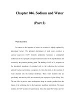
Fig. 1.
Suggested algorithm of treatment of acute variceal bleeding.
an individual from candidates to rescue TIPS because
ahigh Child-Pugh score should be based rather on the
evaluation prior to a bleeding catastrophe. Furthermore,
it appears that patients with varices due to alcoholic cir-
rhosis have the highest incidence of hemorrhage, especial-
ly if they continue to drink alcohol. The hepatocellular
dysfunction may improve in cases who abstain from alco-
hol [69].
Cases of portal vein obstruction are tricky not only
from a technical but also clinical point of view as the inci-
dence of hepatocellular carcinoma in this condition
reaches 35% [65] and is reported up to 22% even in cases
without clinical or imaging evidence of hepatoma if exam-
ined histologically [65, 70]. The survival is in such
patients limited to an average of 6 months and TIPS
brings the risk of systemic metastasis. On the other hand,
if portal blood is diverted by the thrombosis completely to
varices, the sclerotherapy is very likely to fail in case of
acute hemorrhage. Thus, TIPS is not contraindicated in
clinical conditions of immediate concern as acute variceal
or peritoneal hemorrhage, even if malignant portal vein
thrombosis is present.
If TIPS is indicated in refractory bleeding patients with
liver failure, it should be coordinated with a transplant
center. Cases with Child-Pugh score 1 11 and/or other risk
factors (emergent TIPS placement, elevated ALT levels,
pre-TIPS encephalopathy, elevated bilirubin levels), who
are not transplant candidates, have mortality reaching up
to 90% within few weeks after TIPS placement [67] and
therefore shunt is usually not appropriate. Bleeders who
are transplant candidates are transplanted according to
listing criteria.
Theoretically, TIPS has several advantages in trans-
plant candidates who require pre-transplant shunt inser-
tion because of the hemorrhage. All surgical shunts in-
crease the difficulty of dissection, and some permanently
reduce the available blood flow to the transplanted liver.
Shunts that divert flow from the original liver can result in
smaller, more fibrotic portal vein. On the contrary, TIPS
maintains high volume flow through the portal vein, pre-
vents portal vein thrombosis and could result in greater
portal flow to transplanted liver. The TIPS is removed
with the diseased liver entirely and there is no need for
further surgery to close the fibrotic and sometimes fragile
vascular shunt [71]. Published studies shown better re-
sults with TIPS than with surgical shunts [72, 73]. How-
ever, some surgeons do not prefer stenting prior to trans-
plantation (fig. 1).
Long-Term Follow-Up after TIPS
The technical limitation of TIPS from a long-time point
of view is dysfunction due to the clogging of the stent. That
is why patients with TIPS should be meticulously followed
up and the patency of TIPS regularly evaluated. Most cen-
ters use a 3-month interval as the minimal period for clini-
cal and Doppler check-up. Stent dysfunction should be
treated by balloon dilatation of the stent channel. Within
such a protocol, rebleeding due to shunt dysfunction can be
reduced to less than 5% within long-term follow-up and
mild forms of encephalopathy can be diagnosed and
treated before severe clinical consequences [74].
Surgery
In the modern era, surgeons were the first to cope with
bleeding varices. High mortality experienced in acutely
bleeding patients with impaired hepatic functions reach-
ing up to 80% forced accelerated introduction of non-
operative methods. The overall mortality of surgical pro-
cedures for all acutely bleeding patients refractory to med-
ical treatment remains generally high, ranging from 33 to
56%. Moreover, surgical shunting does not appear to
improve survival and is associated with a substantial inci-
dence of portosystemic encephalopathy [75].
Management of Acute Variceal Bleeding
Dig Dis 2003;21:6–15
13
Currently the first-line methods (vasoactive drugs and
endoscopic therapy) reach up to 90% success in cessation
of a bleeding episode. The remaining 10% of cases are one
of the most difficult groups to manage in hepatogastroen-
terology. In the pre-TIPS era, the only ‘salvage therapy’
accepted was surgery, but most patients with progressed
liver diseases are excluded as surgical candidates. In surgi-
cally treated patients, mortality reached 82% in patients
with Child class C [76]. Procedures as esophageal tran-
section plus gastric devascularization and variety of
shunt operations are technically possible. Portal-systemic
shunts can be separated into two basic types: nonselective
(total) shunts and selective shunts. Total shunts are de-
signed to divert portal blood away from the liver and
include end-to-side portacaval shunts, side-to-side porto-
caval shunts, interposition portocaval shunt, splenorenal
shunts and mesocaval shunts. End-to-side shunts anatom-
ically prevent any portal venous perfusion of the liver and
theoretically tends to more rapid liver failure, worsened
PSE and poor control of ascites, but this technique is tech-
nically simpler and is recommended in the emergency sit-
uation. Studies comparing different surgical shunting
techniques are difficult to interpret and still remain an
area of considerably controversy [77]. Randomized stud-
ies have shown that surgical shunts have a better hemo-
static effect than local surgical treatment of bleeding ves-
sels alone. In high-risk patients, sclerotherapy had a simi-
lar effect with fewer complications than transection of the
esophagus, thus transection does not seem to be a good
choice [78]. It can be concluded that surgery possibly still
has a place in the treatment of patients in otherwise good
condition, but practically it is rare for cirrhotics in good
condition to have refractory bleeding. The most impor-
tant objective measure for comparing invasive methods
treating refractory bleeding is the 30-day mortality. Un-
fortunately, at the moment no studies are available fulfill-
ing requirements for comparison of surgery and radioin-
terventions (TIPS). The only randomized study [79] is
questioned from the point of imbalanced distribution of
gender, Child class, and urgent timing disfavoring the
TIPS group. The results of this study showed comparable
30-day mortality in 6 of 35 patients of the TIPS group and
5 of the 35 patients treated by the H-graft. Another uncon-
trolled large study comparing TIPS and surgical shunt
[80] demonstrated 0% 30-day mortality in the surgical
group and 26% mortality in the TIPS group. Child-Pugh
class C patients were not operated at all, but received
exclusively TIPS and formed 57% of the TIPS group.
Comparison of this large surgical experience with results
of the Freiburg group [81] shows similar results in terms
of mortality and rebleeding for patients with less pro-
gressed disease (mortality 0% for Child A patients and
11% for Child B patients). The rebleeding from varices
was demonstrated by two meta-analyses [82, 83] to be
similar after TIPS (19%) and after surgical shunts (3–
45%) [1].
Orthotopic liver transplantation is not a treatment
measure of an acute bleeding episode but all bleeders
should be evaluated as transplant candidates and those
fulfilling standard criteria placed upon a waiting list.
Transplantation of the liver is the treatment option that
offers the best survival rates. The major mortality associ-
ated with the procedure occurs in the first year. The
reported survival rate of patients with liver transplanta-
tion because of variceal hemorrhage is 79% at 1 year and
71% at 5 years [84]. The greatest survival advantage is
conferred on the patient who falls in the Child’s C class.
Unfortunately, access to this procedure will never be open
to all patients due to limited sources of grafts, and ethical
and financial problems.
References
1 D’Amico G, Pagliaro L, Bosch J: The treatment
of portal hypertension: A meta-analytic review.
Hepatology 1995;22:332–354.
2 Power W: Contributions to pathology. MD
Med Surg J 1940;306–318.
3 Preble RB: Conclusions based on sixty cases of
fatal gastrointestinal hemorrhage due to cirrho-
sis of the liver. Am J Med Sci 1900;119:263–
268.
4 Terdiman JP: Update on upper gastrointestinal
bleeding. Postgrad Med 1998;103:43–64.
5 Mann NS, Hillis A, Mann SK, Buerk CA, Pra-
sad VM: In cirrhotic patients variceal bleeding
is more frequent in the evening and correlates
with severity of liver disease. Hepatogastroen-
terology 1999;46:391–394.
6 Emenike E, Srivastava S, Amoateng-Adjepong
Y, Al-Kharrat T, Zarich S, Manthous AC:
Myocardial infarction complicating gastroin-
testinal hemorrhage. Mayo Clin Proc 1999;74:
235–241.
7 De Franchis R (ed): Portal Hypertension. III.
Proceedings of the Third Baveno International
Consensus Workshop on Definitions, Method-
ology and Therapeutic Strategies. Oxford,
Blackwell Science, 2001.
8 De Franchis R (ed): Portal Hypertension. II.
Proceedings of the Second Baveno Internation-
al Consensus Workshop on Definitions, Meth-
odology and Therapeutic Strategies. Oxford,
Blackwell Science, 1996, pp 10–17.
9 Chalasani N, Patel K, Clark WS, Wilcox CM:
The prevalence and significance of leukocytosis
in upper gastrointestinal bleeding. Am J Med
Sci 1998;315:233–236.
10 Bernard B, Grange JD, Nyugen Khao E, et al:
Antibiotic prophylaxis for the prevention of
bacterial infections in cirrhotic patients with
gastrointestinal bleeding: A meta-analysis. He-
patology 1999;29:1655–1661.
14
Dig Dis 2003;21:6–15
Lata/Hulek/Vanasek
11 Bhasin DK, Malhi NJS: Variceal bleeding and
portal hypertension: Much to learn, much to
explore. Endoscopy 2002;43:119–128.
12 Waring JP, Sanowski RA, Pardy K, et al: Does
the addition of methylene blue to the sclerosant
improve the accuracy of endoscopic variceal
sclerotherapy? Am J Gastroenterol 1990;85:
1227.
13 Burroughs AK, McCormick PA, Hughes MD,
et al: Randomized, double-blind placebo-con-
trolled trial of somatostatin for variceal bleed-
ing. Gastroenterology 1990;99:1388–1395.
14 Lo GH, Lai KH, Cheng JS, et al: Emergency
banding ligation versus sclerotherapy for the
control of active bleeding from esophageal var-
ices. Hepatology 1997;25:1101–1104.
15 Hou MC, Chen WC, Lin HC, et al: A new
‘sandwich’ method of combined endoscopic
variceal ligation and sclerotherapy versus liga-
tion alone in the treatment of esophageal vari-
ceal bleeding: A randomized trial. Gastrointest
Endosc 2001;53:572–578.
16 Nakamura S, MItsunaga A, Murata Y, et al:
Endoscopic induction of mucosal fibrosis by
argon plasma coagulation (APC) for esophageal
varices: A prospective randomized trial of liga-
tion plus APC vs. ligation alone. Endoscopy
2001;33:210–215.
17 Bornman P, Terblanche J, Kahn D, et al: Limi-
tations of multiple injection sclerotherapy ses-
sions for acute variceal bleeding. S Afr Med J
1986;70:34–36.
18 Sung JY, Yor W, Suen R, et al: Cyanoacrylate
vs. sodium tetradecyl sulphate for the injection
of bleeding varices in patients with hepatocel-
lular carcinoma: A prospective randomized
study. Gastrointest Endosc 1997;45:AB85.
19 Maluf-Filho F, Sakai P, Ishioka S, et al: Endo-
scopic sclerosis versus cyanoacrylate endoscop-
ic injection for the first episode of variceal
bleeding: A prospective, control and random-
ized study in Child-Pugh class C patients. En-
doscopy 2001;33:421–427.
20 Lain L, Cook D: Endoscopic ligation compared
with sclerotherapy for treatment of esophageal
variceal bleeding: A meta-analysis. Ann Intern
Med 1995;22:663–665.
21 Merigan TC, Poltkin GR, Davidson CS: Effect
of intravenously administered posterior pitu-
itary extract on haemorrhage from bleeding
esophageal varices. N Engl J Med 1962;266:
134–135.
22 Gimson AES, Westaby D, Hegarty J, et al: A
randomized trial of vasopressin and vasopres-
sin plus nitroglycerin in the control of acute
variceal hemorrhage. Hepatology 1986;6:410–
413.
23 Jenkins SA, Baxter JN, Corbett WA, et al: A
prospective randomised controlled clinical trial
comparing somatostatin and vasopressin in
controlling acute variceal hemorrhage. Br Med
J 1985;290:275–278.
24 Cestari R, Graga M, Missale G, et al: Haemo-
dynamic effect of triglycyl-lysine-vasopressin
(glypressin) on intravascular oesophageal vari-
ceal pressure in patients with cirrhosis. A ran-
domized placebo-controlled trial. J Hepatol
1990;10:205–210.
25 Escorsell A, Bandi JC, Moitinho E, et al: Time
profile of the haemodynamic effects of terli-
pressin in portal hypertension. J Hepatol 1997;
26:621–627.
26 Moreau R, Cailmail S, Valla D, et al: Haemo-
dynamic responses to a combination of terli-
pressin and octreotide in portal hypertensive
rats. Aliment Pharmacol Ther 1997;11:993–
997.
27 Huang YT, Lin LC, Chern JW, et al: Portal
hypotensive effects of combined terlipressin
and DL-028, a synthetic ·
1
-adrenoreceptor an-
tagonist administration on anesthetized portal
hypertensive rats. Liver 1999;19:129–134.
28 Hansen EF, Strandberg C, Hojgaard L, et al:
Splanchnic haemodynamic after intravenous
terlipressin in anaesthetised healthy pig. J He-
patol 1999;30:503–510.
29 Soderlund C, Magnusson I, Torngren S, et al:
Terlipressin (triglycyl-lysine vasopressin) con-
trols acute bleeding oesophageal varices. A
double-blind, randomized, placebo-controlled
trial. Scand J Gastroenterol 1990;25:622–630.
30 Fort E, Sautereau D, Silvain C, et al: A ran-
domized trial of terlipressin plus nitroglycerin
vs. balloon tamponade in the control of acute
variceal hemorrhage. Hepatology 1990;11:
678–681.
31 Walker S, Kreichgauer HP, Bode JC: Terlipres-
sin vs. somatostatin in bleeding esophageal var-
ices: A controlled, double-blind study. Hepa-
tology 1992;15:1023–1030.
32 Pedretti G: Octreotide vs. terlipressin in acute
variceal haemorrhage in liver cirrhosis. Clin
Invest 1994;72:653–659.
33 Escorsell A, Ruiz del Arbol L, Planas R, et al:
Multicenter, randomised controlled trial of ter-
lipressin versus sclerotherapy in the treatment
of acute variceal bleeding: The TEST Study.
Hepatology 2000;32:471–476.
34 Levacher S, Letoumelin P, Paterson D, et al:
Early administration of terlipressin plus glyc-
eryl trinitrate to control active upper gastroin-
testinal bleeding in cirrhotic patients. Lancet
1995;346:865–868.
35 Cerini R, Lee SS, Hadengue A, Koshy A, et al:
Circulatory effects of somatostatin analogue in
two conscious rat models of portal hyperten-
sion. Gastroenterology 1988;94:703–708.
36 Hanisch E, Doertenbach J, Usadel KH: So-
matostatin in acute bleeding oesophageal var-
ices. Pharmacology and rational for use. Drug
1992;44(suppl):24–35.
37 Li MK, Sung JJ, Woo KS, et al: Somatostatin
reduces gastric mucosal blood flow in patients
with portal hypertensive gastropathy: A ran-
domized, double-blind crossover study. Dig
Dis Sci 1996;41:2440–2446.
38 Imperiale T, Teran J, McCullough AJ: A meta-
analysis of somatostatin versus vasopressin in
the management of acute esophageal variceal
hemorrhage. Gastroenterology 1995;109:
1289–1294.
39 Gotzsche P, Gjorup I, Bonnen H, et al: So-
matostatin vs. placebo in bleeding esophageal
varices – Randomised trial and meta-analysis.
BMJ 1995;310:1495–1498.
40 Shields R, Jenkins SA, Baxter JN, et al: A pro-
spective randomised controlled trial compar-
ing the efficacy of somatostatin with injection
sclerotherapy in the control of bleeding oesoph-
ageal varices. J Hepatol 1992;16:128–137.
41 Burroughs AK, McCormick PA, Hughes MD,
et al: Randomized, double-blind placebo-con-
trolled trial of somatostatin for variceal bleed-
ing. Gastroenterology 1990;99:1388–1395.
42 Cales P, Masliah C, Bernad B, et al: Early
administration of vapreotide for variceal
bleeding in patients with cirrhosis. N Engl J
Med 2001;344:23–28.
43 Jenkins SA, Baxter JN, Corbett WA, et al: The
effects of a somatostatin analogue SMS 201-
995 on hepatic haemodynamics in the cirrhotic
rat. Br J Surg 1985;72:864–867.
44 Moller S, Brinch K, Henriksen JH, et al: Effect
of octreotide on systemic, central and splanch-
nic haemodynamics in cirrhosis. J Hepatol
1997;26:1026–1033.
45 Escorsell A, Bandi JC, Andreu V, et al: Desen-
sitization to the effect of intravenous octreotide
in cirrhotic patients with portal hypertension.
Gastroenterology 2001;120:161–169.
46 Avgerinos A, Armonis A, Raptis S: Somato-
statin and octreotide in the management of
acute variceal hemorrhage. Hepatogastroenter-
ology 1995;42:145–150.
47 Corley D, Cello J, Adkisson W, et al: Octreo-
tide for acute esophageal variceal bleeding: A
meta-analysis. Gastroenterology 2001;120:
161–169.
48 Besson I, Ingrand P, Person B, et al: Sclerother-
apy with or without octreotide for acute vari-
ceal bleeding. N Engl J Med 1995;9:555–560.
49 Jenkins SA, Baxter JN, Critchley M, et al: Ran-
domised trial of octreotide for long-term man-
agement of cirrhosis after variceal haemor-
rhage. BMJ 1997;315:1338–1341.
50 Stanley AJ, Adrian J, Hayes PC: Portal hyper-
tension and variceal haemorrhage. Lancet
1997;350:1235–1239.
51 Vlavianos P, Gimson AES, Westaby D, Wil-
liams R: Balloon tamponade in variceal bleed-
ing: Use and misuse. BMJ 1989;298:1158–
1165.
52 Garcı´a-Villareal L, Martı´nez-Lagares F, Sierra
A, et al: Transjugular intrahepatic portosys-
temic shunt versus endoscopic sclerotherapy
for the prevention of variceal rebleeding after
recent variceal hemorrhage. Hepatology 1999;
29:27–32.
53 Richter GM, Palmaz JC, Nöldge G, et al: Der
transjugulare intrahepatische portosystemische
Stent-Shunt (TIPSS). Radiologie 1989;29:406–
411.
54 Chau TN, Patch D, Chan YW, Nagral A, Dick
R, Burroughs AK: ‘Salvage’ transjugular intra-
hepatic portosystemic shunts: Gastric fundal
compared with esophageal variceal bleeding.
Gastroenterology 1998;114:981–987.
55 Gerbes AL, Gülberg V, Waggershauser T, Holl
J, Reiser M: Transjugular intrahepatic porto-
systemic shunt (TIPS) for variceal bleeding in
portal hypertension: Comparison of emergency
and elective interventions. Dig Dis Sci 1998;
43:2463–2469.
Management of Acute Variceal Bleeding
Dig Dis 2003;21:6–15
15
56 McCormick PA, Dick R, Panagou EB, Chin
JK, Greenslade L, McIntyre N, Burroughs AK:
Emergency transjugular intrahepatic portosys-
temic stent shunting as salvage treatment for
uncontrolled variceal bleeding. Br J Surg 1994;
81:1324–1327.
57 Ban´ ares R, Casado M, Rodrı´guez-La´iz JM, et
al: Urgent transjugular intrahepatic portosys-
temic shunt for control of acute variceal bleed-
ing. Am J Gastroenterol 1998;93:75–79.
58 Jalan R, John TG, Redhead DN, Garden OJ,
Simpson KJ, Finlayson ND, Hayes PC: A com-
parative study of emergency transjugular intra-
hepatic portosystemic stent-shunt and esopha-
geal transection in the management of uncon-
trolled variceal hemorrhage. Am J Gastroen-
terol 1995;90:1932–1937.
59 Mansilla AV, Putman SG, Cohen GS, et al:
Massive hemoptysis secondary to bronchial
collaterals: Treatment with use of TIPS and
embolization. J Vasc Interv Radiol 1999;10:
372–374.
60 Ryu RK, Nemcek AA, Chrisman HB, et al:
Treatment of stomal variceal hemorrhage with
TIPS: Case report and review of the literature.
Cardiovasc Intervent Radiol 2000;23:301–
303.
61 Haskal ZJ, Scott M, Rubin RA, et al: Intestinal
varices: Treatment with the transjugular intra-
hepatic portosystemic shunt. Radiology 1994;
191:183–187.
62 Ludwig D, Borsa JJ, Maier RV: Transjugular
intrahepatic portosystemic shunt for trauma? J
Trauma 1999;48:954–956.
63 Radosevich PM, Ring EJ, LaBerge JM, et
al: Transjugular intrahepatic portosystemic
shunts in patients with portal vein occlusion.
Radiology 1993;186:523–527.
64 Blum U, Haag K, Rossle M, et al: Noncaverno-
matous portal vein thrombosis in hepatic cir-
rhosis: Treatment with transjugular intrahepat-
ic portosystemic shunt and local thrombolysis.
Radiology 1995;195:153–157.
65 Walser EM, McNees SW, DeLa Pena O, et al:
Portal venous thrombosis: percutaneous thera-
py and outcome. J Vasc Interv Radiol 1998;9:
119–127.
66 Bosch J: Transjugular intrahepatic portosys-
temic shunt (TIPS); in De Franchis R (ed): Por-
tal Hypertension. II. Oxford, Blackwell
Science, 1996, pp 127–137.
67 Chalasani N, Clark WS, Martin LG, et al:
Determinants of mortality in patients with ad-
vanced cirrhosis after transjugular intrahepatic
portosystemic shunting. Gastroenterology
2000;118:138–144.
68 Ochs A, Rössle M, Haag K, et al: The transju-
gular intrahepatic portosystemic stent-shunt
procedure for refractory ascites. N Engl J Med
1995;332:1192–1197.
69 De Franchis R, Primignani M: Why do varices
bleed? Gastroenterol Clin North Am 1992;21:
85–101.
70 Cedrona A, Rapaccini GL, Pompili M, et al:
Portal vein thrombosis complicating hepato-
cellular carcinoma: Value of ultrasound-guided
fine-needle biopsy of the thrombus in the thera-
peutic management. Liver 1996;16:94–98.
71 Reed MH: TIPS: A liver transplant surgeon’s
view. Semin Interv Radiol 1995;12:396–400.
72 Abouljoud MS, Levy MF, Rees CR, et al: A
comparison of treatment with transjugular in-
trahepatic portosystemic shunt or distal sple-
norenal shunt in the management of variceal
bleeding prior to liver transplantation. Trans-
plantation 1995;59:226–229.
73 Menegaux F, Kneefe EB, Baker E, et al: Com-
parison of transjugular and surgical portosys-
temic shunts on the outcome of liver transplan-
tation. Ann Surg 1994;129:1018–1024.
74 Zizka J, Elias P, Krajina A, et al: Value of
Doppler sonography in revealing transjugular
intrahepatic portosystemic shunt malfunction:
A 5-year experience in 216 patients. AJR 2000;
175:145–148.
75 Rikkers LF, Sorrell WT, Gongliang J: Which
portosystemic shunt is the best? Gastoenterol
Clin North Am 1992;21:179–196.
76 Willson PD, Kunkler R, Blair SD, Reynolds
KW: Emergency oesophageal transection for
uncontrolled variceal haemorrhage. Br J Surg
1994;81:992–995.
77 Holt DR, Klein AS: The surgical treatment of
portal hypertension: Patient and procedure se-
lection; in Perler B, Becker G (eds): A Clinical
Approach to Vascular Intervention. New York,
Thieme, 1996, pp 603–608.
78 Terés J, Baroni R, Bordas JM, Visa J, Pera C,
Rodés J: Randomized trial of portacaval shunt,
stapling transection and endoscopic sclerother-
apy in uncontrolled variceal bleeding. J Hepa-
tol 1987;4:2, 159–167.
79 Rosemurgy AS, Bloomston M, Zervos EE, et al:
Transjugular intrahepatic portosystemic shunt
versus H-graft portacaval shunt in the manage-
ment of bleeding varices: A cost-benefit analy-
sis. Surgery 1997;122:794–800.
80 Henderson JM, Nagle A, Curtas S, et al: Surgi-
cal shunts and TIPS for variceal decompres-
sion in the 1990s. Surgery 2000;128:540–547.
81 Rössle M: Is there still a need for surgical inter-
vention in portal hypertension? The internist’s
point of view; in Krajina A, Hulek P (eds): Cur-
rent Practice of TIPS, 2001, pp 202–204.
82 Luca A, D’Amico G, La Galla R, Midiri M,
Morabito A, Pagliaro L: TIPS for prevention of
recurrent bleeding in patients with cirrhosis:
Meta-analysis of randomized trials. Radiology
1999;212:411–421.
83 Papatheodoridis GV, Goulis J, Leandro G,
Patch D, Burroughs AK: Transjugular intrahe-
patic portosystemic shunt compared with en-
doscopic treatment for the prevention of vari-
ceal rebleeding: A meta-analysis. Hepatology
1999;30:612–622.
84 Millikan WJ Jr, Henderson JM, Galloway JR,
Dodson TF, Shires GT 3rd, Stewart M: Surgi-
cal rescue for failures of cirrhotic sclerotherapy.
Am J Surg 1990;160:117–121.
Review Article
Dig Dis 2003;21:16–18
DOI: 10.1159/000071334
Upper Gastrointestinal Hemorrhage –
Surgical Aspects
Lars Lundell
Department of Surgery, Huddinge University Hospital, Stockholm, Sweden
Lars Lundell, MD, PhD
Department of Surgery
Huddinge University Hospital
S–14186 Stockholm (Sweden)
Tel. +46 858 580 549, Fax +46 858 582 340, E-Mail
ABC
Fax + 41 61 306 12 34
www.karger.com
© 2003 S. Karger AG, Basel
0257–2753/03/0211–0016$19.50/0
Accessible online at:
www.karger.com/ddi
Key Words
Gastrointestinal hemorrhage
W Endoscopic therapy W
Peptic ulcer W Variceal bleeding W Acute surgery
Abstract
During the last decades, significant advantages have
been achieved with the use of emergency endoscopy
and respective hemostatic interventions. Rebleeding,
however, remains a significant clinical problem, and cur-
rently re-endoscopy or surgical intervention offers ad-
vantages and disadvantages. With the discovery of Heli-
cobacter pylori as a main causative factor behind peptic
ulcer disease, a more conservative surgical approach is
mandated even in situations with significant rebleeding.
In case of large gastric ulcer, however, resection is a wise
strategy depending on the risk of malignancy. Liver
transplantation has immensely improved the prognoses
for variceal bleeding in end-stage liver disease in careful-
ly selected patients.
Copyright © 2003 S. Karger AG, Basel
Acute upper gastrointestinal bleeding is a frequent
event with an incidence of around 40–50 cases per
100,000 persons per year. Since the early 1970s, emergen-
cy endoscopy has been widely used in the diagnosis and
management of upper gastrointestinal hemorrhage. Acid-
suppressive drugs have become available and since the
introduction of endoscopic intervention modalities in the
1980s, the mortality rate from this severe clinical mani-
festation has decreased slightly but still remains around
10%. One of the main reasons for the remaining high mor-
tality is probably the fact that the patients are at an
advanced age and have concomitant complicated dis-
eases. A quarter of the admitted patients are older than 80
years. Another factor might be the extensive use of
NSAIDs and anticoagulants [1–22].
If endoscopy is performed within 24 h of admission,
the cause of bleeding is identified in more than 90%.
However, in large epidemiological studies, the percent-
ages of undiagnosed patients vary widely between 0 and
25% (table 1). Gastroduodenal peptic ulcers account for
about 40% of the cases, where duodenal ulcers are most
frequently seen followed by hemorrhagic gastritis, vari-
ceal bleeding, esophagitis, duodenitis, Mallory-Weiss
tears and malignancies (1–5%). A meta-analysis showed
that endoscopic therapy, including injection therapy, was
effective in reducing the risk of rebleeding and need for
emergency surgery and mortality in patients with active
bleeding or non-bleeding visible vessels. Furthermore, the
routine use of a second endoscopic treatment in the case
of rebleeding has been suggested, although a more wide-
spread consensus and acceptance of this strategy has not
been achieved. Rebleeding and requirement for emergen-
cy and urgent surgical intervention remains and for
Upper Gastrointestinal Hemorrhage –
Surgical Aspects
Dig Dis 2003;21:16–18
17
Table 1.
Endoscopic diagnosis in patients presenting with upper gastrointestinal bleeding; review of the literature (mean and ranges are shown)
Years n DU GU Esopha-
gitis
Varices Mallory-
Weiss
Gastritis/
erosions
Malig-
nancies
Misc. Unclear
1973–1998 13,178 25% (12–53) 15.9% (9–26) 7.4% (4–13) 10.5% (1–23) 6.1% (0.5–12) 15.4% (4–41) 2.3% (1–5) 5.2% (0.5–15) 8.9% (3–22)
instance recent trials have shown a rebleeding rate of
around 20–25% with a 8–15% need for urgent surgery (ta-
ble 2). One trial has tried to assess whether elective endo-
scopic retreatment is better than early elective surgery
after initial endoscopic hemostasis, but the issue is far
from settled. Apparently endoscopic reintervention has
advantages over surgical intervention in terms of lower
morbidity.
Surgical Intervention
Depending on the timing of the operation, surgery for
hemorrhage can be divided into three main groups: emer-
gency surgery, elective early surgery and delayed surgery.
Emergency surgery carries a mortality rate between 10
and 20% but if surgery is inappropriately delayed, mortal-
ity increases rapidly. Therefore, patients who are likely to
rebleed are the best candidates for early elective surgery
after the initial bleeding has been stopped with endoscop-
ic therapy. Most surgical studies have been performed
before effective endoscopic therapy became available,
and it is therefore very difficult to compare the different
studies and strategies because of these methodological
weaknesses. Morris et al. [8] prospectively compared early
surgery with non-operative management in patients with
bleeding ulcers, and stratified them by age and ulcer loca-
tion. Over the age of 60 years, early surgery had a mortali-
ty rate of 7% compared to 43% for those with delayed
surgery. However, the different types of surgery were not
comparable in both groups and in those with delayed sur-
gery more patients received gastric resection, which car-
ries a higher procedure-related mortality. Overall mortali-
ty was 4% for early surgery and 15% for delayed surgical
management in all patients. In patients with ulcers in the
posterior wall of the duodenal bulb, with active bleeding
or a visible vessel, early surgery may be recommended.
Endoscopic hemostasis is difficult in these patients and
recurrence of bleeding is often fulminant because of large
side branches of the gastroduodenal artery being in-
volved.
Table 2.
Failure rates on modern endoscopic therapies for active-
ly bleeding ulcers; review of the literature (mean and ranges are
shown)
Patients Rebleed, % Urgent
surgery, %
Mortality, %
1,328 17.1 (0–40) 10.5 (0–32) 4.4 (0–16)
Gastric Ulcers
Gastric ulcers more frequently require surgery due to
uncontrolled bleeding than duodenal ulcers. At the time
of a laparotomy, each gastric ulcer has to be excised
including in most instances a formal resection. The main
reason for this strategy is that gastric ulcers always carry
the potential of being malignant. Concomitant duodenal
scaring and/or ulcers do not pose a significant problem in
the days of Helicobacter pylori eradication therapies.
Therefore, vagotomy procedures should only exceptional-
ly be added due to the associated morbidities.
Duodenal Ulcers
For bleeding duodenal ulcers, nowadays extensive
operations are almost never indicated, if ever, because
many patients are H. pylori infected and/or have the hem-
orrhage occurring as a consequence of NSAID usage.
Therefore, duodenal ulcer hemorrhage should mainly be
treated by under-running the ulcer which, if correctly
done, frequently elicits adequate hemostasis. If for any
specific reason surgical acid suppression is required, a
selected gastric vagotomy should be recommended due to
its lower morbidity and less frequent side effects.
18
Dig Dis 2003;21:16–18
Lundell
Variceal Bleeding
In many institutions, operative portosystemic shunts
are no longer used as treatment for variceal bleeding.
When the first-line options of non-selective ß-blockade or
endoscopic treatment fail to control bleeding, a transjugu-
lar intrahepatic portosystemic shunt (TIPS) is usually
placed. The advantages of TIPS are that it is non-opera-
tive, it effectively decompresses the portal venous circula-
tion during the short-term perspective and early compli-
cations and procedure-related mortality are infrequent.
However, late TIPS failure rates are high, with thrombo-
sis or stenosis developing in approximately in 50% of
patients within 1–2 years. Although TIPS revisions are
successful in many patients, in most series, rebleeding
rates after TIPS are considerably higher (10–30%) than
after surgically constructed shunts (! 10%). When patent,
TIPS is usually a non-selective shunt with encephalopathy
rates in most trials similar to those seen after a portocaval
shunt. Despite these disadvantages, TIPS is an excellent
option for patients in whom endoscopic treatment is
unsuccessful and who require relatively short-lasting por-
tal decompression while on the waiting list for a liver
transplant or whose anticipated survival is limited due to
the underlying liver disease.
Long-term survival has been particularly impressive
for patients undergoing surgery since the advent of liver
transplantation, especially for those who are potential liv-
er transplantation candidates and who can be salvaged by
this procedure when hepatic failure develops.
References and Suggested Reading
1 Vreeburg EM: Acute upper gastrointestinal
bleeding. A prospective valuation of diagnosis
ant therapy in the Amsterdam area; thesis, Am-
sterdam 1997.
2 Cook DJ, Guyatt GH, Salena BJ, Laine LA:
Endoscopic therapy for acute nonvariceal up-
per gastrointestinal hemorrhage: A meta-analy-
sis. Gastroenterology 1992;102:139–148.
3 Labenz J, Borsch G. Role of Helicobacter pylori
eradication in the prevention of peptic ulcer
bleeding relapse. Digestion 1994;55:19–23.
4 Langman MJ: Epidemiologic evidence on the
association between peptic ulceration and anti-
inflammatory drug use. Gastroenterology
1989;96(suppl):640–646.
5 Langman MJ, Morgan L, Worrall A: Use of
anti-inflammatory drugs by patients admitted
with small or large bowel perforations and
haemorrhage. Br Med J 1985;290:347–349.
6 Forrest JAH, Finlayson NDC, Sherman DJC:
Endoscopy in gastro-intestinal bleeding. Lan-
cet 1974;ii:391–397.
7 Hunt PS: Surgical management of bleeding
chronic peptic ulcer. A 10-year study prospec-
tive study. Ann Surg 1984;199:44–50.
8 Morris DI, Hawker PC, Brearly S, Simms M,
Dykes PW, Keighley MR: Optimal timing of
operation for bleeding peptic ulcer: Prospective
randomised trial. Br Med J 1984;288:1277–
1280.
9 Wheatley KE, Snyman JH, Brearley S, Keigh-
ley MR, Dykes PW: Mortality in patients with
bleeding peptic ulcer when those aged 60 or
over are operated on early. BMJ 1990;330:
272.
10 Pimpl W, Boeckl O, Heinerman M, Dapunt O:
Emergency endoscopy: A basis for therapeutic
decisions in the treatment of severe gastroduo-
denal bleeding. World J Surg 1989;13:592–
597.
11 Heldwein W, Schreiner J, Pedrazzoli J, Lehnert
P: Is the Forrest classification a useful tool for
planning endoscopic therapy of bleeding peptic
ulcers? Endoscopy 1989;21:258–262.
12 Schein M, Gecelter G: Apache II score in mas-
sive upper gastrointestinal hemorrhage from
peptic ulcer: Prognostic value and potential
clinical applications. Br J Surg 1989;76:733–
736.
13 Saperas E, Pique JM, Perez Ayuso R, Bordas
JM, Teres J, Pera C: Conservative manage-
ment of bleeding duodenal ulcer without a visi-
ble vessel: Prognostic randomised trial. Br J
Surg 1987;74:784–786.
14 Kubba AK, Choudari C, Rajgopal C, Palmer
KR: The outcome of urgent surgery for major
peptic ulcer hemorrhage following failed endo-
scopic therapy. Eur J Gastroenterol Hepatol
1996;8:1175–1178.
15 Qvist P, Arnesen KE, Jacobsen CD, Rosseland
AR: Endoscopic treatment and restrictive sur-
gical policy in the management of peptic ulcer
bleeding. Scand J Gastroenterol 1994;29:569–
576.
16 Jordan PH: Surgery for peptic ulcer disease.
Curr Probl Surg 1991;28:265–330.
17 Cochran TA: Bleeding peptic ulcer: Surgical
therapy. Gastroent Clin North Am 1993;22:
751–778.
18 Starlinger M, Becker HD: Upper gastrointesti-
nal bleeding – indications and results in sur-
gery. Hepatogastroenterology 1991;38:216–
219.
19 Hasselgren G: Peptic ulcer bleeding 2000: Im-
proved outcome; thesis, Gothenburg 1998.
20 Layton F, Rikkers MD: The changing spectrum
of treatment for variceal bleeding. Ann Surgery
1998;228:536–546.
21 Iwatsuki S, Starzl TE, Todo S, Gordon RD,
Tzakis AG, Marsh JW, Makowka L, Koneru B,
Stieber A, Klintmalm G, Husberg B, van Thiel
D: Liver transplantation in the treatment of
bleeding esophageal varices. Surgery 1988;104:
697–705.
22 Mercado MA, Orozco H, Ramirez-Cisneros FJ,
Hinojosa CA, Plata JJ, Alvarez-Tostado J: Di-
minished morbidity and mortality in portal hy-
pertension surgery: Relocation in the thera-
peutic armamentarium. J Gastroenterol Surg
2001;5:499–502.
Review Article
Dig Dis 2003;21:19–24
DOI: 10.1159/000071335
Lower Gastrointestinal Bleeding –
The Role of Endoscopy
Helmut Messmann
III. Medizinische Klinik, Klinikum Augsburg, Deutschland
Dr. H. Messmann, PD
III. Medizinische Klinik, Klinikum Augsburg
Postfach 1019 20, DE–86009 Augsburg (Germany)
Tel. +49 821 400 7351, Fax +49 821 400 3331
ABC
Fax + 41 61 306 12 34
www.karger.com
© 2003 S. Karger AG, Basel
0257–2753/03/0211–0019$19.50/0
Accessible online at:
www.karger.com/ddi
Key Words
Lower gastrointestinal bleeding
W Endoscopy
Abstract
Endoscopy is the method of choice in diagnosing the
cause of lower gastrointestinal bleeding, and it offers the
opportunity to treat patients suffering from lower gas-
trointestinal bleeding. Endoscopic procedures must be
integrated with other approaches to reach a correct diag-
nosis rapidly, safely, and economically. In all patients,
evaluation begins with a history and physical examina-
tion. The sequence of other tests depends on many fac-
tors, especially the rate of bleeding. New technologies
such as wireless capsule endoscopy will influence the
management of patients with lower gastrointestinal
bleeding.
Copyright © 2003 S. Karger AG, Basel
Definition
Lower intestinal bleeding is defined as acute or chronic
abnormal blood loss distal to the ligament of Treitz. 10–
20% of all gastrointestinal bleeding disorders occur distal
of this point, but bleeding of the small intestine is a rare
condition (3–5%).
Acute bleeding is arbitrarily defined as bleeding of ! 3
days’ duration resulting in instability of vital signs, ane-
mia, and/or need for blood transfusion [1, 2]. Hematoche-
zia is the most common clinical symptom in patients with
acute lower gastrointestinal bleeding (LGIB).
Chronic bleeding is defined as slow blood loss over a
period of several days or longer presenting with symptoms
of occult fecal blood, intermittent melena or scant he-
matochezia. Occult bleeding means that the amounts of
blood in the feces are too small to be seen but detectable
by chemical tests [3]. In 48–71% the source will be found
and an origin in the colorectum is to be expected in 20–
30% [3].
Obscure gastrointestinal bleeding often presents as
LGIB and means a bleeding from an unclear site, that per-
sists or recurs after a negative initial or primary endosco-
py. In 6% a repeat colonoscopy will identify the lesion in
the colon. Push enteroscopy will be helpful in 38–75% to
find the bleeding lesion, however, in two thirds the lesions
are detectable within the range of a conventional gastro-
scope [3].