Emergencies and Complications in Gastroenterology - part 3 pdf
Bạn đang xem bản rút gọn của tài liệu. Xem và tải ngay bản đầy đủ của tài liệu tại đây (290.56 KB, 10 trang )
20
Dig Dis 2003;21:19–24
Messmann
General Aspects of Lower Gastrointestinal
Bleeding
The incidence of lower gastrointestinal bleeding is only
one fifth of that of the upper gastrointestinal tract and is
estimated to be 21–27 cases per 100,000 adults/year [4,
5]. LGIB usually is chronic and self-limiting and can be
treated on an outpatient basis. Nevertheless, 21 of
100,000 adults/year require hospitalization due to severe
bleeding. Among those, male gender and older patients
suffer from more severe LGIB [4]. There is a 200-fold
increase from the third to the ninth decade due to diver-
ticulosis and angiodysplasia [6].
There is some evidence that upper gastrointestinal
bleeding (UGIB) differs in acuity and severity from
LGIB: Patients with LGIB are significantly less in shock
(19 vs. 35%, respectively), require fewer blood transfu-
sions (36 vs. 64%) and have a significantly higher hemo-
globin level (84 vs. 61%) [7, 8]. Similar to UGIB, the
majority of bleeding disorders (80–85%) in the lower gas-
trointestinal tract will stop spontaneously.
Mortality and morbidity increase with age. The overall
mortality rate varies between 2.0 and 3.6%. Those pa-
tients with bleeding episodes after hospital admission
have significantly higher mortality rates (23.1%) com-
pared to those who bleed before hospital admission [4].
Diagnosis
Endoscopy is the method of choice to diagnose and if
possible to treat lower gastrointestinal bleeding. While
colonoscopy has been accepted for years in patients with
chronic bleeding, urgent colonoscopy in acute bleeding
has been evaluated in the last few years and is meanwhile
also accepted as a safe method.
Before starting colonoscopy, history and clinical exam-
ination should lead to a tentative diagnosis in order to
plan the diagnostic procedures. In patients with chronic
LGIB, colonoscopy is the first diagnostic step. The time
point of colonoscopy is elective and optimal bowel prepa-
ration is standard. If the origin of bleeding cannot be
detected, further steps are necessary.
In contrast, patients with acute LGIB are a challenge
for optimal diagnostic procedures and there are still open
questions. It is generally accepted that in patients with
hematochezia, especially in combination with circulation
instability, an UGIB must be excluded, since in 11%
patients with suspected acute LGIB have their bleeding
source proximal to the ligament of Treitz. Although place-
ment of a nasogastric tube is safe and easy, it misses
UGIB in 7%. The rate might even be higher in patients
with duodenal ulcer since pylorospasm can prevent reflux
of blood into the stomach [9, 10].
While anoscopy and sigmoidoscopy were mandatory
procedures in the pre-colonoscopy era, their role is less
obvious in the era of emergency and early colonoscopy. In
recent years it could be demonstrated that in experienced
hands colonoscopy plays the same role in acute LGIB as
upper gastrointestinal endoscopy in acute UGIB.
All patients with acute LGIB must be stabilized and
contraindications for colonoscopy are severe active in-
flammation and also inadequate visual conditions. Fur-
thermore, the endoscopy should be aborted if the patient
becomes unstable, the bleeding is so severe that identifica-
tion of a bleeding source is impossible, or the risk of perfo-
ration is too high. It is unclear whether urgent unprepared
colonoscopy is more effective in detecting the bleeding
source as compared to prepared colonoscopy with a delay
of several hours, since no randomized trial exists to this
question.
The amount, location or pattern of blood are impor-
tant signs which make a detection of the bleeding source
in a circumscribed segment of the colon easier. Most stud-
ies, however, prefer bowel preparation before urgent co-
lonoscopy. Their arguments are the frequent spontaneous
bleeding stop and the improvement of visualization. The
bowel preparation can be performed by enemas and/or
polyethylene glycol solutions administered by mouth or
via a nasogastric tube. There exist no data that cleaning
the bowel might reactivate bleeding.
The detection rate of the bleeding source after bowel
preparation varies between 62 and 78%, and in patients
without preparation the urgent unprepared colonoscopies
could identify the bleeding source in 76% [8, 11, 12].
Therefore, urgent colonoscopy seems to be reasonable in
most patients.
In patients with intermittent or obscure gastrointesti-
nal bleeding, wireless capsule endoscopy may become an
interesting diagnostic approach. In two trials, capsule
endoscopy was compared to X-ray of the small bowel or
push enteroscopy.
Costamagna et al. [13] could demonstrate that in 13
patients with intermittent bleeding, the capsule was able
to detect the bleeding source in 11 cases while X-ray only
in 1 case, respectively. Ell et al. [14] examined 32 pa-
tients – the capsule detected a pathologic lesion in 66%
and the X-ray in 28%, respectively.
Lower Gastrointestinal Bleeding –
The Role of Endoscopy
Dig Dis 2003;21:19–24
21
Differential Diagnosis
Acute LGIB occurs most frequently in diverticular
(35%), followed by vascular malformation (21%), colitis
(16%), neoplasia/postpolypectomy (10%), anorectal dis-
eases (5%), and small bowel (5%). In 11% the acute UGIB
is falsely diagnosed as LGIB. Differential diagnosis of
severe acute LGIB is mainly dependent on the patient’s
age. While in children and young adults inflammatory
bowel disease and Meckel’s diverticulum are the main
bleeding sources, diverticula are predominantly found in
adults up to 60 years, and in the elderly, angiodysplasia is
the most common cause for severe LGIB.
Diverticular Disease
The true incidence of diverticular disease is difficult to
measure, mainly because most patients are asymptomat-
ic. The incidence however clearly increases with age from
10% under 40 years to an estimated 50–66% in patients
older than 80 [15, 16]. The estimated risk of a severe
bleeding has been reported to be 3–5% [16, 17], but
including milder forms of bleeding a risk up to 48% has
been described [18]. Among LGIB disorders, diverticula
are the cause in 15–27% [19]. The clinical presentation of
patients with diverticular bleeding is mostly abrupt with
a painless onset, associated with mild lower abdominal
cramps and the urge to defecate. The stool consists of red
voluminous or maroon blood or clots. Melena is uncom-
mon [16]. Approximately 80% of the bleeding episodes
stop spontaneously. The risk of a first rebleeding is 25%
but increases with definite bleeding stigmata (active
bleeding, nonbleeding visible vessel, adherent clot: 67, 50
and 43%, respectively) [19–21]. A third bleed after a sec-
ond episode will occur in 50%, therefore surgical resec-
tion is recommended after a second bleeding episode
[16].
Colitis
LGIB from IBD are rarely life-threatening (0.1% ulcer-
ative colitis, 1.3% Crohn’s disease), bleeding stops mostly
spontaneously and endoscopic treatment is not necessary
in most cases with diffuse bleeding. Bleeding from isch-
emic colitis occurs mainly in elderly patients (1 65 years)
and is associated with pain. Vascular diseases and atrial
fibrillation are risk factors which are associated with isch-
emic colitis. Patients with infectious colitis suffer mainly
from diffuse bleeding similar to ulcerative colitis. Among
bacteria, Salmonella, Shigella, Yersinia, Campylobacter
and Escherichia coli, especially enterohemorrhagic E. coli
(EHEC), most notably 0157:H7, are the most frequent
infectious agents. Acute radiation colitis occurs a few days
after radiation but bloody diarrhea is uncommon at this
time point. Most patients complain of transient diarrhea
and tenesmus. The endoscopic picture is similar to ulcer-
ative colitis with edema, fragility, hemorrhage and some
erosions or ulcers [16]. The clinical manifestation of
chronic radiation colitis occurs after 1–2 years. Pale mu-
cosa with teleangiectasia and rarefaction of mucosal ves-
sels is typical in mild forms. In severe radiation colitis,
excessive hemorrhage, necrosis and ulcerations occur
leading to extensive bleeding [16].
Neoplasia
Acute bleeding in colon cancer or polyps is not fre-
quent but has been described in 2–33 and 5–11%, respec-
tively [16, 17]. The majority of these lesions present with
chronic bleeding. Among patients with LGIB, postpoly-
pectomy bleeding occurs in about 4% [4]. Bleeding occurs
either immediately (within 24 h) or delayed (occurring as
long as 21 days after colonoscopy) [22]. The risk of bleed-
ing depends on several factors: polyp size, type of polyp
(pedunculated or sessile), hemostatic disorders, medica-
tion and endoscopist’s experience influence the postpol-
ypectomy hemorrhage risk. Although the use of NSAID
did increase the incidence of minor self-limited bleeding,
an increase in the rate of major bleeding was not observed
[23]. The overall risk of bleeding after polypectomy ranges
from 0.4 to 2% [24].
Angiodysplasia
In patients with LGIB, angiodysplasia is the responsi-
ble bleeding disorder in 3–12% [4, 5]. Bleeding can be
chronic, slow, intermittent or recurrent. Massive bleeding
has been described in 2% of the cases, but bleeding stops
spontaneously in up to 90%. Unfortunately the rebleeding
rate is high and can reach values up to 85% [16].
The prevalence of angiodysplasia among healthy
asymptomatic people was 0.83%. 87% of these usually
small lesions (4 mm) were located in the right colon, and
there was no risk for later bleeding [25]. Angiodysplasia
often appears together with systemic diseases such as car-
diovascular disorders (aortic stenosis) or chronic renal
failure [26, 27]. However, there exist also systematic
examinations which could not confirm an association of
angiodysplasia and aortic valve disease [28]. Capsule
endoscopy may improve the detection of these lesions in
the small bowel in the near future.
22
Dig Dis 2003;21:19–24
Messmann
Table 1.
Endoscopic treatment of LGIB
Bleeding source Endoscopic treatment Comments
Diverticula Injection, clip Bleeding mainly stops spontaneously, perforation risk
Colitis (IBD, radiation,
ischemia, infection)
Injection of ulcer
APC in radiation colitis with teleangiectasia
No endoscopic treatment is necessary in most cases;
high risk of perforation!
Neoplasia Thermal, injection
Polypectomy of bleeding polyps
Seldom severe bleeding
Postpolypectomy bleeding Injection, clip Prophylactic loop?
AV malformations APC, thermal, injection of sclerosing agents High risk of rebleeding!
Hormone therapy not useful
No prophylactic treatment
Anorectal diseases Ligation, sclerotherapy TIPS in patients with esophageal and rectal varices
Anorectal Diseases
Due to anorectal lesions, LGIB is mainly caused by
hemorrhoids, rectal varices and fissures. 2–9% of all
LGIB are caused by hemorrhoids [4, 5]. Among patients
with AIDS, anorectal diseases are more frequent as bleed-
ing sources and may be severe in case of thrombocytope-
nia. Rectal varices are to be differentiated from hemor-
rhoids. Bleeding is sometimes profuse but painless. Portal
hypertension is the main reason for rectal varices and is
present in 79–89% in these patients.
Therapy
Endoscopic therapy of LGIB is similar to UGIB and is
the therapy of choice. In a recent survey of the American
College of Gastroenterology, endoscopic therapy was per-
formed in 27% in LGIB and in 51% in UGIB, respective-
ly [2]. Jensen et al. [29] recently demonstrated that emer-
gency colonoscopy with endoscopic treatment was superi-
or to conservative treatment in combination with surgery
if necessary. Different endoscopic techniques such as
injection therapy, thermal methods, clipping and so on,
which have been successful in UGIB, are also useful in the
treatment of LGIB (table 1).
Angiodysplasia can be treated effectively by thermal
methods, and argon plasma coagulation is meanwhile the
treatment of choice. For prophylactic treatment of non-
bleeding, incidental angiodysplasia is not recommended
and a hormone therapy of bleeding angiodysplasia has
shown no benefit in a recent randomized trial. Vascular
malformations in patients with chronic radiation colitis
can be treated with argon plasma coagulation in the same
way.
While bleeding polyps can effectively be treated by pol-
ypectomy and adjuvant methods such as injection thera-
py or application of a loop before snaring the polyp, bleed-
ing from colorectal cancer can be treated with thermoco-
agulation by Nd:YAG laser or argon plasma coagulation.
If endoscopic treatment is not possible due to severe
bleeding, angiography is recommended: Application of
drugs such as vasopressin is as effective as embolization to
achieve initial hemostasis (71 vs. 70%, respectively).
However, rebleeding rate after vasopressin is 25% com-
pared to embolization (0%).
The ultima ratio in treatment of severe LGIB is sur-
gery, which occurs in 10–25%. Criteria for (emergency)
surgery are: 1 4 units of blood/24 h or a total of 10 units
overall; bleeding continues for 672 h, and significant
rebleeding within 1 week of initial cessation [16, 17].
Blind segmental colectomy is associated with an unac-
ceptable high morbidity (rebleeding rate as high as 75%)
and mortality (up to 50%). Therefore, an aggressive
approach for an accurate preoperative localization is most
important. Directed segmental resection is the treatment
of choice because of its low morbidity, mortality (about
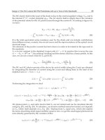
4%) and rebleeding rate (about 6%) [16]. Angiographic
localization has been shown to be more precise than scin-
tigraphic methods. The 1-year rebleeding rate could be
decreased from 42% without angiographic localization to
14% with angiography (fig. 1).
Lower Gastrointestinal Bleeding –
The Role of Endoscopy
Dig Dis 2003;21:19–24
23
Fig. 1.
Management of acute severe LGIB.
Intraoperative diagnostic endoscopy has become most
attractive to examine the small or large bowel with entero-
scopes or colonoscopes after laparotomy, pleating of the
bowel on the instrument, and translumination. Identifica-
tion of bleeding sites has been possible in 83–100% [30].
Preliminary studies report on a theoretical advantage of
this combined approach, especially in the management of
small bowel hemorrhage, which cannot be identified with
usual techniques [31]. New techniques such as wireless
capsule endoscopy may improve the diagnosis in patients
with LGIB as well.
References
1 Zuccaro G Jr: Management of the adult patient
with acute lower gastrointestinal bleeding. Am
J Gastroenterol 1998;93:1202–1208.
2 American Society for Gastrointestinal Endos-
copy: The role of endoscopy in the patient with
lower gastrointestinal bleeding. Gastrointest
Endosc 1998;48:685–688.
3 Zuckerman GR, Prakash C, Askin MP, Lewis
BS: AGA technical review on the evaluation
and management of occult and obscure gas-
trointestinal bleeding. Gastroenterology 2000;
118:201–221.
4 Longstreth GF: Epidemiology and outcome of
patients hospitalized with acute lower gastroin-
testinal hemorrhage: A population-based
study. Am J Gastroenterol 1997;92:419–424.
5 Bramley PN, Masson JW, McKnight G, Herd
K, Fraser A, Park K, Brunt PW, McKinlay A,
Sinclair TS, Mowat NA: The role of an open-
access bleeding unit in the management of
colonic haemorrhage. A 2-year prospective
study. Scand J Gastroenterol 1996;31:764–
769.
6 Jensen DM, Machicado GA: Colonoscopy for
diagnosis and treatment of severe lower gas-
trointestinal bleeding. Routine outcomes and
cost analysis. Gastrointest Endosc Clin North
Am 1997;7:477–498.
7 Peura DA, Lanza FL, Gostout CJ, Foutch PG:
The American College of Gastroenterology
Bleeding Registry: Preliminary findings. Am J
Gastroenterol 1997;92:924–928.
8 Zuckerman GR, Prakash C: Acute lower intes-
tinal bleeding. I. Clinical presentation and
diagnosis. Gastrointest Endosc 1998;48:606–
617.
9 Luk GD, Bynum TE, Hendrix TR: Gastric
aspiration in localization of gastrointestinal
hemorrhage. JAMA 1979;241:576–578.
10 Cuellar RE, Gavaler JS, Alexander JA, Brouil-
lette DE, Chien MC, Yoo YK, Rabinovitz M,
Stone BG, Van Thiel DH: Gastrointestinal
tract hemorrhage. The value of a nasogastric
aspirate. Arch Intern Med 1990;150:1381–
1384.
11 Jensen DM, Machiado GA: Management of
severe lower gastrointestinal bleeding; in Bar-
kin JS, O’Phealn CA (eds): Advanced Thera-
peutic Endoscopy, ed 2. New York, Raven
Press, 1994, pp 201–208.
12 Kok KY, Kum CK, Goh PM: Colonoscopic
evaluation of severe hematochezia in an Orien-
tal population. Endoscopy 1998;30:675–680.
24
Dig Dis 2003;21:19–24
Messmann
13 Costamagna G, Shah SK, Riccioni ME, Fos-
chia F, Mutignani M, Perri V, Vecchioli A, Bri-
zi MG, Picciocchi A, Marano P: A prospective
trial comparing small bowel radiographs and
video capsule for suspected small bowel dis-
ease. Gastroenterology 2002;123:999–1005.
14 Ell C, Remke S, May A, Helou L, Henrich R,
Mayer G: The first prospective controlled trial
comparing wireless capsule endoscopy with
push enteroscopy in chronic gastrointestinal
bleeding. Endoscopy 2002;34:685–689.
15 Laine L: Acute and chronic gastrointestinal
bleeding; in Feldman M, Scharschmidt BF,
Sleisinger MH (eds): Sleisinger’s & Fordtran’s
Gastrointestinal and Liver Disease. Philadel-
phia, Saunders, 1999, pp 198–219.
16 Vernava AM 3rd, Moore BA, Longo WE, John-
son FE: Lower gastrointestinal bleeding. Dis
Colon Rectum 1997;40:846–858.
17 Zuckerman GR, Prakash C: Acute lower intes-
tinal bleeding. II. Etiology, therapy and out-
comes. Gastrointest Endosc 1999;49:228–238.
18 Winkler R: Ursachen und Klinik der peranalen
Blutung; in Häring R (ed): Gastrointestinale
Blutung. Berlin, Blackwell, 1990, pp 313–319.
19 Almy TP, Howell DA: Medical progress. Di-
verticular disease of the colon. N Engl J Med
1980;302:324–331.
20 So JB, Kok K, Ngoi SS: Right-sided colonic
diverticular disease as a source of lower gas-
trointestinal bleeding. Am Surg 1999;65:299–
302.
21 Stollman NH, Raskin JB: Diagnosis and man-
agement of diverticular disease of the colon in
adults. Ad Hoc Practice Parameters Commit-
tee of the American College of Gastroenterol-
ogy. Am J Gastroenterol 1999;94:3110–3121.
22 Gibbs DH, Opelka FG, Beck DE, Hicks TC,
Timmcke AE, Gathright JB Jr: Postpolypecto-
my colonic hemorrhage. Dis Colon Rectum
1996;39:806–810.
23 Shiffman ML, Farrel MT, Yee YS: Risk of
bleeding after endoscopic biopsy or polypecto-
my in patients taking aspirin or other NSAIDS.
Gastrointest Endosc 1994;40:458–462.
24 Mergener K, Baillie J: Complications of endos-
copy. Endoscopy 1998;30:230–243.
25 Foutch PG: Angiodysplasia of the gastrointesti-
nal tract. Am J Gastroenterol 1993;88:807–
818.
26 Weaver GA, Alpern HD, Davis JS, Ramsey
WH, Reichelderfer M: Gastrointestinal angio-
dysplasia associated with aortic valve disease:
Part of a spectrum of angiodysplasia of the gut.
Gastroenterology 1979;77:1–11.
27 Foutch PG: Angiodysplasia of the gastrointesti-
nal tract. Am J Gastroenterol 1993;88:807–
818.
28 Bhutani MS, Gupta SC, Markert RJ, Barde CJ,
Donese R, Gopalswamy N: A prospective con-
trolled evaluation of endoscopic detection of
angiodysplasia and its association with aortic
valve disease. Gastrointest Endosc 1995;42:
398–402.
29 Jensen DM, Machicado GA, Jutabha R, Ko-
vacs TO: Urgent colonoscopy for the diagnosis
and treatment of severe diverticular hemor-
rhage. N Engl J Med 2000;342:78–82.
30 Lewis BS: Small intestinal bleeding. Gastroen-
terol Clin North Am 2000;29:67–95.
31 Ingresso M, Pete F, Pisani A, et al: Laparosco-
pically-assisted total enteroscopy: A new ap-
proach to small intestinal disease. Gastrointest
Endosc 1999;49:651–653.
Review Article
Dig Dis 2003;21:25–29
DOI: 10.1159/000071336
Management of Acute Cholangitis
Dirk J. Gouma
Department of Surgery, Academic Medical Center, Amsterdam, The Netherlands
Prof. Dirk J. Gouma, MD
Department of Surgery, Academic Medical Center
Meibergdreef 9
NL–1105 Amsterdam (The Netherlands)
Fax +31 20 5669 243, E-Mail
ABC
Fax + 41 61 306 12 34
www.karger.com
© 2003 S. Karger AG, Basel
0257–2753/03/0211–0025$19.50/0
Accessible online at:
www.karger.com/ddi
Key Words
Acute cholangitis
W Endoscopic sphincterotomy W
Laparoscopic CBD exploration W Common bile duct
stones
Abstract
Endoscopic sphincterotomy (ES) is the treatment of
choice for patients with (severe) acute cholangitis. For fit
patients without co-morbidity with mild cholangitis and
CBD stones with a gallbladder in situ, the one-stage lapa-
roscopic approach could be considered as an alternative
in centers with sufficient experience. The results of both
procedures are comparable. Open surgery is relatively
safe. It has a high success rate, good/excellent long-term
results, but is not very attractive for the patient and
should not be used routinely nowadays. Therefore, the
indication should be limited for management of severe
complications after ES as perforations of the duodenum,
large CBD stones and patients with Mirizzi’s syndrome or
intrahepatic stones with stenosis of the bile duct. ES as
primary treatment for CBD stones should be followed by
laparoscopic cholecystectomy in ‘fit’ patients. In patients
with malignant disease, particularly after repeated stent
failure and subsequent cholangitis, bypass surgery
should be considered in patients with a life expectancy of
1 3 months.
Copyright © 2003 S. Karger AG, Basel
After the introduction of endoscopic sphincterotomy
(ES) and percutaneous drainage procedures, the indica-
tion for different surgical and non-surgical approaches of
biliary disorders changed radically and is still subject of
controversy. There is however general agreement that
patients with severe cholangitis should preferably be
treated non-surgically by ES instead of (open) CBD explo-
ration after a randomized trial of Lai et al. [1] clearly
showed a reduction in morbidity from 66 to 34% and a
reduction in hospital mortality from 32 to 10%. Recently,
another trial has been published showing that even in the
absence of CBD stones during the attack of cholangitis,
ES decreased the duration of fever in patients with acute
cholangitis and reduced hospital stay from 4.3 to 2.2 days
and 9.1 to 8.1 days, respectively [2]. However, it did not
decrease the incidence of recurrent acute cholangitis dur-
ing follow-up.
The development of high-quality ES in general hospi-
tals has resulted in a decrease of surgical procedures for
acute cholangitis as well as for the initial management of
CBD stones without cholangitis in many European coun-
tries, particularly in The Netherlands and Germany. In
The Netherlands only 20% of patients with CBD stones
underwent a surgical approach during the past decade. A
minority of these patients suffered from severe cholangi-
tis, the others having symptomatic CBD stones.
26
Dig Dis 2003;21:25–29
Gouma
A recent nationwide survey in Germany, reporting the
surgical management of 98,482 patients with symptomat-
ic gallstone disease and 8,433 patients with CBD stones,
showed that surgical CBD exploration decreased from
7.4% in 1991 towards 3.8% in 1996. In 1998, all universi-
ty hospitals used a two-stage management with preopera-
tive ERCP and ES – the so-called ‘therapeutic splitting’
[3]. Again, no doubt exists today that patients with severe
cholangitis will primarily be managed non-surgically.
Therefore, the discussion about the role of surgery should
also focus on whether there is still a role for surgery in the
treatment of patients with CBD stones with mild cholan-
gitis or without cholangitis.
There have been four randomized trials that compared
open surgery versus ES for the treatment of CBD stones
[4–7]. In the Spanish trial [4], high-risk patients with chol-
angitis and mild biliary pancreatitis were also random-
ized. These trials showed a high success rate for both pro-
cedures, around 90–95%, no significant difference in
morbidity and mortality, but a significantly longer hospi-
tal stay after surgery. ES however was associated with sig-
nificantly more recurrent biliary symptoms and a higher
requirement of additional procedures (1 20%) [4–7]. In a
second study by the same group [8], ES was followed by
laparoscopic cholecystectomy and the recurrence of bili-
ary symptoms in that study reduced to 4%.
Summarizing these trials, open surgery is not inferior to
ES, it is safe and effective but is associated with a longer
hospital stay and in particular, after introduction of the
minimal invasive procedures, it is not very attractive for
patients and therefore not generally accepted nowadays.
More recently, laparoscopic CBD exploration has been
introduced for the management of CBD stones including
patients with mild cholangitis. Again it was generally
accepted that ES should be the treatment of choice for poor-
risk patients with severe cholangitis and pancreatitis [9].
There have been two randomized trials that compared
laparoscopic CBD exploration (LCBDE) with ES. In the
first trial, Rhodes et al. [10] compared LCBDE with lapa-
roscopic cholecystectomy and postoperative ES showing
that LCBDE is as effective as ES in overall clearance of
the CBD stones. There was a significantly shorter hospital
stay in patients treated by LCBDE. A second multicenter
trial [9] compared LCBDE with ES and subsequent lapa-
roscopic cholecystectomy and showed an equivalent suc-
cess rate for both procedures, no significant difference in
complications and mortality but a shorter hospital stay
after LCBDE compared with ES. The authors concluded
that laparoscopic CBD exploration should be preferred
for fit patients (ASA I and II). More recent studies also
showed that primary closure of the bile duct after bile
duct exploration without an external drain by a T-tube
drainage is safe and efficient even in patients with acute
cholecystitis, mild cholangitis or pancreatitis provided
that laparoscopic skills are available [11–12].
Laparoscopic CBD exploration without drainage even
reduced biliary complications from 16 to 4% [12]. In a
recent review on management of CBD stones it was con-
cluded that single-stage laparoscopic treatment without
drainage of the CBD (primary closure) should be advo-
cated as the primary treatment in centers with sufficient
experience in laparoscopic exploration [13]. So far in oth-
er hospitals, ES still remains the treatment of choice, how-
ever training issues and experience will also arise concern-
ing gastroenterologists performing ERCP and ES. There is
no doubt that all patients with CBD stones after previous
cholecystectomy should undergo ES.
Despite increased interest in minimal invasive surgery,
there is an enormous difference in Europe about the ac-
ceptance of laparoscopic CBD exploration and still the
majority of patients, around 90%, are treated with ES.
Therefore, the next question arises, i.e. if the gallbladder
should be removed after successful stone clearance after ES.
As shown in previous trials comparing open surgery and
ES, additional procedures were performed in 20–26% of
the patients after ES [4–7]. In a recent trial from The Neth-
erlands comparing a wait-and-see policy versus laparoscop-
ic cholecystectomy after ES and CBD clearance, 47% of the
patients in the wait-and-see group suffered from recurrent
biliary pain and 47% needed an additional procedure (10%
ERCPs and 37% cholecystectomy) within 2 years after ini-
tial ES. It was concluded that laparoscopic cholecystectomy
should be advocated in fit patients after ES [14].
Another indication for surgery is failure after endo-
scopic treatment or the existence of retained stones. In a
series from the area of open surgery for CBD stones, we
showed that a choledochojejunostomy, as the final solu-
tion for complicated CBD stones, was successful in 98%
even after 8 years of follow-up [15]. These procedures can
now also be performed laparoscopically, as mentioned
before. In elderly patients in particular (1 70 years), gas-
troenterologists generally prefer multiple stent exchanges
even in patients with retained stones and recurrent chol-
angitis instead of a relative simple surgical bypass proce-
dure (choledochoduodenostomy). They should realize
that mortality of these procedures these days is nearly
zero for these patients.
Patients with cholangitis due to Mirizzi’s syndrome are
also an indication for (open) surgery or for a laparoscopic
approach with an extremely high conversion rate. These
Management of Acute Cholangitis
Dig Dis 2003;21:25–29
27
Fig. 1.
Patients with obstructive jaundice due to Mirizzi’s syndrome.
A
ERCP showing a stenosis of the CBD.
B
CT
scan showing an inflammatory mass.
C
Control ERCP 6 weeks after surgery and primary repair of the CBD.
Fig. 2.
Patient with intrahepatic bile duct in
the right hepatic duct with a stenosis at the
distal right hepatic duct (
A
) and CT scan (
B
)
showing entrahepatic bile duct dilatation
and stones.
patients generally present with obstructive jaundice or
cholangitis and endoscopic drainage can be performed
easily as the initial treatment because of the relative
smooth stricture by the impacted stone (fig. 1A). After
adequate biliary drainage and resolving of the inflamma-
tion around the hepatoduodenal ligament (fig. 1B), chole-
cystectomy should be performed with closure of the defect
in the CBD and the stent can be removed after a few
weeks (fig. 1C).
Patients with recurrent cholangitis due to multiple
intrahepatic bile duct stones are generally treated by a
combined endoscopic and percutaneous approach. In par-
ticular if only one lobe is affected and after failure of non-
surgical treatments to remove the stones, these patients
are also candidates for surgery and a hemihepatectomy
should be performed (fig. 2A, B). The surgical approach is
well established in South-East Asia for this common prob-
lem and is even performed laparoscopically nowadays
28
Dig Dis 2003;21:25–29
Gouma
Fig. 3.
A patient with a perforation after ERCP
and free air in the retroperitoneum (
A
) and per-
foration of the duodenum during exploration
(
B
).
[16]. Surgery is also sometimes indicated for severe com-
plications after ES (bleeding/perforation) but in particular
after free perforation of the duodenum or a perforation of
the endoscope at the anastomosis (gastroenterostomy)
after a previous BII resection. Early intervention is war-
ranted in these patients.
In a period of 7 years, 27 patients underwent surgery
for complications of ERCP at the AMC Amsterdam. The
majority suffered from perforations of the duodenum (n =
7) (fig. 3) or at the anastomosis after BII resections (n = 7).
In 1 patient a pancreatoduodenectomy was performed.
The other patients underwent cholecystectomy, closure of
the defect, subsequent CBD exploration with or without a
choledochoduodenostomy or choledochojejunostomy. In
patients with perforations during sphincterotomy or even
small retroperitoneal perforations of the duodenum, con-
servative management is nearly always sufficient. If sub-
sequent leakage and abscess formation occurs, percuta-
neous drainage should be performed and finally if not suc-
cessful diversion of the duodenum should be considered.
Endoscopic biliary stenting has generally been ac-
cepted as the treatment of choice for palliative treatment
in patients with obstructive jaundice due to distal bile
duct or pancreatic malignancy with a limited life expec-
tancy. Four randomized trials comparing stenting and
bypass surgery showed that there is no difference in relief
of obstruction by both methods. Surgery was initially
associated with a higher postoperative morbidity, mortal-
ity and a longer hospital stay. Non-operative treatment
with an endoprothesis however led to recurrent jaundice
and cholangitis in up to 40% and gastrointestinal obstruc-
tion in up to 17% during follow-up [17–20]. In two more
recent studies from our center, the mortality after pallia-
tive surgical bypass procedures decreased to 2.5 and 1%
respectively and postoperative complications were 17 and
12% [21, 22]. Other studies showed similar results and in
selective patients with a life expectancy of 1 6 months,
bypass surgery is safe nowadays [23, 24].
In a recent randomized trial comparing stenting and
bypass surgery in patients who proved to have metastasis
during diagnostic laparoscopy, we clearly showed that
patients after stenting had a shorter hospital-free survival
and more readmissions because of stent dysfunction and
cholangitis compared with patients after bypass surgery
[25]. Therefore, we conclude that patients with recurrent
cholangitis after stent treatment for malignant tumors
should of course first undergo stent exchange, or insertion
of metallic stents, but in a selected group of patients a bil-
iary bypass should also be considered, particularly in
patients with a life expectancy of 1 3 months.
Management of Acute Cholangitis
Dig Dis 2003;21:25–29
29
References
1 Lai EC, Mok FP, Tan ES, Lo CM, Fan ST, You
KT, Wong J: Endoscopic biliary drainage for
severe acute cholangitis. N Engl J Med 1992;
326:1582–1586.
2 Hui CK, Lai KC, Wong WM, Yuen MF, Lam
SK, Lai CL: A randomised controlled trial of
endoscopic sphincterotomy in acute cholangi-
tis without common bile duct stones. Gut 2002;
51:245–247.
3 Huttl TP, Hrdina C, Geiger TK, et al: Manage-
ment of common bile duct stones – Results of a
nationwide survey with analysis of 8,433 com-
mon bile duct explorations in Germany. Zen-
tralbl Chir 2002;127:282–289.
4 Targarona EM, Ayuso RM, Bordas JM, et al:
Randomised trial of endoscopic sphincteroto-
my with gallbladder left in situ versus open sur-
gery for common bile duct calculi in high-risk
patients. Lancet 1996;347:926–929.
5 Hammerstrom LE, Holmin T, Stridberg H.
Ihse I: Long-term follow-up of a prospective
randomized study of endoscopic versus surgi-
cal treatment of bile duct calculi in patients
with gallbladder in situ. Br J Surg 1995;82:
1516–1521.
6 Stain SC, Cohen H, Tsuishoysha M, Donovan
AJ: Choledocholithiasis. Endoscopic sphincter-
otomy or common bile duct exploration. Ann
Surg 1991;213:627–633; discussion: 633–634.
7 Suc B, Escat J, Cherqui D, Fourtanier G, Hay
JM, Fingerhut A, et al: Surgery vs. endoscopy
as primary treatment in symptomatic patients
with suspected common bile duct stones: A
multicenter randomized trial. French Associa-
tions for Surgical Research. Arch Surg 1998;
133:702–708.
8 Trias M, Targarona EM, Ros E, Bordas JM,
Perez Ayuso RM, Balague C, Pros I, Teres J:
Prospective evaluation of a minimally invasive
approach for treatment of bile-duct calculi in
risk patient. Surg Endosc 1997;11:632–635.
9 Cuschieri A, Lezoche E, Morino M, Croce E,
Lacy A, Toouli J, et al: EAES multicenter pro-
spective randomized trial comparing two-stage
vs. single-stage management of patients with
gallstone disease and ductal calculi. Surg En-
dosc 1999;13:952–957.
10 Rhodes M. Sussman L, Cohen L, Lewis MP:
Randomised trial of laparoscopic exploration
of common bile duct versus postoperative en-
doscopic retrograde cholangiography for com-
mon bile duct stones. Lancet 1998;351:159–
161.
11 Decker G, Borie F, Millat B, Berthou JC, De-
leuze A, Drouard F, Guillon F, Rodier JG, Fin-
gerhut A: One hundred laparoscopic choledo-
chotomies with primary closure of the common
bile duct. Surg Endosc 2003;17:12–18.
12 Thompson MH, Tranter SE: Laparoscopic ex-
ploration of the common bile duct. The results
of an all-comers policy. Br J Surg 2002;89:
1608–1612.
13 Tranter SE, Thompson MH: Comparison of
endoscopic sphincterotomy and laparoscopic
exploration of the common bile duct. Br J Surg
2002;89:1495–1504.
14 Boerma D, Rauws EA, Keulemans YC, Janssen
IM, Bolwerk CJ, Timmer R, Boerma EJ, Ober-
top H, Huibregtse K, Gouma DJ: Wait-and-see
policy or laparoscopic cholecystectomy after
endoscopic sphincterotomy for bile duct
stones: A randomised trial. Lancet 2002;360:
761–765.
15 Gouma DJ, Konsten J, Soeters PB, et al: Long-
term follow-up after choledochojejunostomy
for bile duct stones with complex clearance of
the bile duct. Br J Surg 1989;76:451–453.
16 Tang CN, Li MK: Hand-assisted laparoscopic
segmentectomy in recurrent pyogenic cholangi-
tis. Surg Endosc 2003;17:324–327.
17 Bornman PC, Harries-Jones EP, Tobias R, et
al: Prospective controlled trial of transhepatic
biliary endoprosthesis versus bypass surgery
for incurable carcinoma of head of pancreas.
Lancet 1986;i:69–71.
18 Shepard HA, Royle G, Ross APR, et al: Endo-
scopic biliary endoprosthesis in the palliation
of malignant obstruction of the distal common
bile duct: A randomized trial. Br J Surg 1988;
75:1166–1168.
19 Andersen JR, Sorensen SM, Kruse A, et al:
Randomized trial of endoscopic endoprosthe-
sis versus operative bypass in malignant ob-
structive jaundice. Gut 1989;30:1132–1135.
20 Smith AC, Dowsett JF, Russell RCG, et al:
Randomised trial of endoscopic stenting versus
surgical bypass in malignant low bile duct ob-
struction. Lancet 1994;344:1655–1660.
21 Van Wagensveld BA, Coene PP, Van Gulik
TM, et al: Outcome of palliative biliary and
gastric bypass surgery for pancreatic head car-
cinoma in 126 patients. Br J Surg 1997;84:
1402–1406.
22 Van Geenen R, Keyzer-Dekker CM, Van Tien-
hoven G, et al: Pain management of patients
with unresectable peripancreatic carcinoma.
World J Surg 2002;26:715–720.
23 Lillemoe KD, Sauter PK, Pitt HA, et al: Cur-
rent status of surgical palliation of periampulla-
ry carcinoma. Surg Gynecol Obstet 1993;176:
1–10.
24 Parks RW, Johnston GW, Rowlands BJ: Surgi-
cal biliary bypass for benign and malignant
extrahepatic biliary tract disease. Br J Surg
1997;84:488–492.
25 Nieveen Van Dijkum EJ, Romijn MG, Terwee
CB, De Wit LT, Van Der Meulen JH, Lameris
HS, Rauws EA, Obertop H, Van Eyck CH, Bos-
suyt PM, Gouma DJ: Laparoscopic staging and
subsequent palliation in patients with peripan-
creatic carcinoma. Ann Surg 2003;237:66–73.