Fatty Liver Disease : Nash and Related Disorders - part 1 ppt
Bạn đang xem bản rút gọn của tài liệu. Xem và tải ngay bản đầy đủ của tài liệu tại đây (317.41 KB, 32 trang )
Fatty Liver Disease:
NASH and Related
Disorders
Fatty Liver Disease:
NASH and Related
Disorders
Edited by
Geoffrey C. Farrell
Director, Storr Liver Unit, Westmead Hospital, Department of Medicine,
University of Sydney, Sydney, NSW 2006, Australia
Jacob George
Director, Clinical Hepatology, Storr Liver Unit, Westmead Hospital,
Department of Medicine, University of Sydney, Sydney, NSW 2006, Australia
Pauline de la M. Hall
University of Cape Town, Department of Anatomical Pathology, Faculty of Medicine,
Observatory, Cape Town 7925, South Africa
Arthur J. McCullough
Division of Gastroenterology, MetroHealth Medical Center and
Schwartz Center for Metabolism and Nutrition, Cleveland, OH 44102-1998, USA
© 2005 Blackwell Publishing Ltd
Blackwell Science, Inc., 350 Main Street, Malden, Massachusetts 02148-5020, USA
Blackwell Publishing Ltd, 9600 Garsington Road, Oxford OX4 2DQ, UK
Blackwell Science Asia Pty Ltd, 550 Swanston Street, Carlton, Victoria 3053, Australia
The right of the Authors to be identified as the Authors of this Work has been
asserted in accordance with the Copyright, Designs and Patents Act 1988.
All rights reserved. No part of this publication may be reproduced, stored in a
retrieval system, or transmitted, in any form or by any means, electronic,
mechanical, photocopying, recording or otherwise, except as permitted by the UK
Copyright, Designs and Patents Act 1988, without the prior permission of the
publisher.
First published 2005
Library of Congress Cataloging-in-Publication Data
Fatty liver disease : NASH and related disorders / edited by Geoffrey C. Farrell . . .
[et al.].
p. ; cm.
Includes bibliographical references and index.
ISBN 1-4051-1292-1 (alk. paper)
1. Fatty liver.
[DNLM: 1. Fatty Liver. WI 700 F252 2004] I. Farrell, Geoffrey C.
RC848.F3F38 2004
616.3′62—dc22
2004009034
ISBN 1–4051–1292–1
A catalogue record for this title is available from the British Library
Set in 9/11.5pt Sabon by Graphicraft Limited, Hong Kong
Printed and bound in Great Britain at CPI Bath, Bath
Commissioning Editor: Alison Brown
Managing Editor: Rupal Malde
Production Editor: Nick Morgan
Project Manager: Sue Hadden
Production Controller: Kate Charman
For further information on Blackwell Publishing, visit our website:
The publisher’s policy is to use permanent paper from mills that operate a sustainable
forestry policy, and which has been manufactured from pulp processed using
acid-free and elementary chlorine-free practices. Furthermore, the publisher ensures
that the text paper and cover board used have met acceptable environmental
accreditation standards.
v
Contents
Contributors, vii
Preface, x
1 Overview: an introduction to NASH and related fatty liver disorders, 1
Geoffrey C. Farrell, Jacob George, Pauline de la M. Hall & Arthur J. McCullough
2 Pathology of hepatic steatosis, NASH and related conditions, 13
Pauline de la M. Hall & Richard Kirsch
3 The epidemiology and risk factors of NASH, 23
Arthur J. McCullough
4 Insulin resistance in NAFLD: potential mechanisms and therapies, 38
Varman T. Samuel & Gerald I. Shulman
5 NASH as part of the metabolic (insulin resistance) syndrome, 55
Giulio Marchesini & Elisabetta Bugianesi
6 NASH is a genetically determined disease, 66
Christopher P. Day & Ann K. Daly
7 The pathogenesis of NASH: human studies, 76
Arun J. Sanyal
8 Animal models of steatohepatitis, 91
Geoffrey C. Farrell
9 Fatty acid metabolism and lipotoxicity in the pathogenesis of
NAFLD/NASH, 109
Nathan M. Bass & Raphael B. Merriman
10 Cytokines and inflammatory recruitment in NASH: experimental and human
studies, 123
Zhiping Li & Anna-Mae Diehl
11 Mitochondrial injury and NASH, 132
Bernard Fromenty & Dominique Pessayre
12 Cell biology of NASH: fibrosis and cell proliferation, 143
Isabelle A. Leclercq & Yves Horsmans
13 Clinical manifestations and diagnosis of NAFLD, 159
Stephen A. Harrison & Brent Neuschwander-Tetri
14 The clinical outcome of NAFLD including cryptogenic cirrhosis, 168
Stephen H. Caldwell & Anita Impagliazzo Hylton
15 Practical approach to the diagnosis and management of people with fatty liver
diseases, 181
Jacob George & Geoffrey C. Farrell
16 Management of NASH: current and future perspectives on treatment, 194
Paul Angulo & Keith D. Lindor
CONTENTS
vi
17 NAFLD, NASH and orthotopic liver transplantation, 208
Anne Burke & Michael R. Lucey
18 NAFLD/NASH is not just a ‘Western’ problem: some perspectives
on NAFLD/NASH from the East, 218
Shivakumar Chitturi & Jacob George
19 NAFLD/NASH in children, 229
Joel E. Lavine & Jeffrey B. Schwimmer
20 Steatohepatitis resulting from intestinal bypass, 241
Christiane Bode & J. Christian Bode
21 Specific disorders associated with NAFLD, 249
Geraldine M. Grant, Vikas Chandhoke & Zobair M. Younossi
22 Hepatocellular carcinoma in NAFLD, 263
Vlad Ratziu & Thierry Poynard
23 Does NASH or NAFLD contribute to comorbidity of other liver diseases?, 276
Andrew D. Clouston & Elizabeth E. Powell
24 Recent advances, 289
Richard Kirsch, Pauline de la M. Hall, Jacob George, Arthur J. McCullough
& Geoffrey C. Farrell
Index, 303
Colour plate section appears after page 22
CONTRIBUTORS
viii
Geraldine M. Grant
Center for Liver Diseases, Inova Fairfax Hospital,
Department of Medicine, 3300 Gallows Road,
Falls Church, VA 22042-3300, USA
Pauline de la M. Hall
Professor of Pathology, University of Cape Town,
Department of Anatomical Pathology, Faculty
of Medicine, Observatory, Cape Town 7925,
South Africa
Stephen A. Harrison
Assistant Professor of Medicine, University of Texas,
Brooke Army Medical Center, 3851 Roger Brooke
Drive, Fort Sam Houston, TX 7823, USA
Yves Horsmans
Service de Gastro-enterologie, Cliniques
Universitaires Saint Luc, Université Catholique de
Louvain, Brussels, Belgium
Anita Impagliazzo Hylton
Division of Gastroenterology and Hepatology,
Box 800708, University of Virginia Health
Sciences Center, Charlottesville, VA 22908, USA
Richard Kirsch
Registrar, University of Cape Town, Department of
Anatomical Pathology, Faculty of Health Sciences,
Observatory, Cape Town 7925, South Africa
Joel E. Lavine
Professor and Vice Chair, Department of Pediatrics,
University of California San Diego Medical Center,
200 West Arbor Drive, MC 8450, San Diego,
CA 92103-8450, USA
Isabelle A. Leclercq
Collaborateur Scientifique du FNRS, Université
Catholique de Louvain, Laboratoire de Gastro-
entérologie, Avenue E. Mounier 53, B 1200
Brussels, Belgium
Zhiping Li
Assistant Professor, Department of Medicine,
Division of Gastroenterology, Johns Hopkins
University, 720 Rutland Avenue, Baltimore,
MD 21205, USA
Keith D. Lindor
Professor of Medicine, Mayo Medical School, and
Head and Consultant, Division of Gastroenterology
and Hepatology, Mayo Clinic Foundation,
200 First Street SW, Rochester,
MN 55905-0002, USA
Michael R. Lucey
Professor of Medicine and Chief, Section of
Gastroenterology and Hepatology, University of
Wisconsin-Madison Medical School,
600 Highland Avenue, Madison,
WI 53792-5124, USA
Giulio Marchesini
Associate Professor of Metabolic Disease, University
of Bologna, Department of Internal Medicine,
Unit for Metabolic Diseases, Policlinico S. Orsola,
Via Massarenti 9, I-40138 Bologna, Italy
Arthur J. McCullough
Director, Division of Gastroenterology, MetroHealth
Medical Center, 2500 Metrohealth Drive, Cleveland,
OH 44102-1998, USA
Raphael B. Merriman
Division of Gastroenterology, University of California,
PO Box 0538, San Francisco, CA 94143-0538, USA
Brent Neuschwander-Tetri
Associate Professor of Internal Medicine,
Saint Louis University School of Medicine,
Division of Gastroenterology and Hepatology,
3635 Vista Avenue, PO Box 15250, St Louis,
MO 63110-0250, USA
Dominique Pessayre
Institut National de la Santé et de la Recherche,
Unit 481, Hôpital Beaujon, 100 Boulevard du
General Leclerc, 92118 Clichy Cedex, France
Elizabeth E. Powell
Department of Gastroenterology and Hepatology,
The Princess Alexandra Hospital, Ipswich Road,
Woolloongabba, Queensland 4102, Australia
CONTRIBUTORS
ix
Thierry Poynard
Service d’hépatogastroentérologie, Hôpital Pitié
Salpêtrière, 47–83 Boulevard de l’Hôpital,
Paris 75013, Fance
Vlad Ratziu
Service d’hépatogastroentérologie, Hôpital Pitié
Salpêtrière, 47–83 Boulevard de l’Hôpital,
Paris 75013, France
Varman T. Samuel
Yale University School of Medicine, S269 TAC,
300 Cedar Street, New Haven, CT 06520, USA
Arun J. Sanyal
Medical College of Virginia, Internal Medicine/
Gastroenterology, MCV Station Box 980711,
Richmond, VA 23298-0711, USA
Jeffrey B. Schwimmer
Division of Gastroenterology, Hepatology and
Nutrition, Department of Pediatrics, University of
California, San Diego, and Children’s Hospital and
Health Center, 200 West Arbor Drive, San Diego,
CA 92103-8450, USA
Gerald I. Shulman
Investigator, Howard Hughes Medical Institute,
295 Congress Avenue, BCMM, New Haven,
CT 06510, USA
Zobair M. Younossi
Director, Center for Liver Diseases, Inova
Fairfax Hospital, Department of Medicine,
3300 Gallows Road, Falls Church, VA 22042-3300,
USA
1
Abstract
This chapter introduces the history, definitional and
semantic issues, spectrum and general importance
of non-alcoholic fatty liver diseases (NAFLD). Non-
alcoholic steatohepatitis (NASH) is a form of meta-
bolic liver disease in which fatty change (steatosis)
is associated with lobular inflammation, hepatocyte
injury and/or hepatic fibrosis. It comprises a pathogenic
link in the chain of NAFLD that extends from bland
steatosis to some cases of ‘cryptogenic cirrhosis’.
NAFLD and NASH are usually hepatic manifesta-
tions of the insulin resistance (or metabolic) syndrome
(syndrome X), but the factors that transform steatosis
to NASH remain unclear. NAFLD/NASH is the most
common type of liver disease in affluent societies,
affecting between 2 and 8% of the population. NASH
typically causes no symptoms. When present, clinical
features such as fatigue, hepatomegaly and aching
hepatic discomfort are non-specific. In 20–25% of
cases, NASH may progress to advanced stages of hep-
atic fibrosis and cirrhosis; liver failure then becomes
the most common cause of death, and hepatocellular
carcinoma (HCC) may occasionally occur. Correc-
tion of insulin resistance by dietary measures and
increased physical activity (lifestyle intervention) is a
logical approach to prevent or reverse early NASH,
and modest weight reduction can normalize liver test
abnormalities. Drug therapy aimed at reversing insulin
resistance, correcting diabetes and lipid disorders, or
Overview: an introduction to NASH
and related fatty liver disorders
Geoffrey C. Farrell, Jacob George, Pauline de la M. Hall &
Arthur J. McCullough
1
Key learning points
1 Non-alcoholic steatohepatitis (NASH) is a form of metabolic liver disease in which fatty change (steato-
sis) is associated with lobular inflammation, hepatocyte injury, polymorphs and/or hepatic fibrosis.
2 NASH comprises a pathogenic link in the chain of non-alcoholic fatty liver diseases (NAFLD) that
extends from bland steatosis to some cases of ‘cryptogenic cirrhosis’.
3 NAFLD and NASH are usually hepatic manifestations of the insulin resistance syndrome, but the factors
that transform steatosis to NASH remain unclear.
4 In 20–25% of cases, NASH may progress to advanced stages of hepatic fibrosis and cirrhosis; liver failure
then becomes the most common cause of death.
5 Clinicians should consider NAFLD/NASH as a primary diagnosis by its metabolic associations with
obesity, insulin resistance and type 2 diabetes, rather than simply as a disease of exclusion.
6 Correction of insulin resistance by lifestyle modification (dietary measures and increased physical activ-
ity) is a logical approach to prevent or reverse NAFLD/NASH.
Fatty Liver Disease: NASH and Related Disorders
Edited by Geoffrey C. Farrell, Jacob George, Pauline de la M. Hall, Arthur J. McCullough
Copyright © 2005 Blackwell Publishing Ltd
CHAPTER 1
2
imately 60/year). These advances have been reviewed
elsewhere [11–19].
What is NASH?
Terminology and definitions
The spectrum of fatty liver disease associated with
metabolic determinants and not resulting from alcohol
(NAFLD) extends from hepatic steatosis through
steatohepatitis to cirrhosis (Table 1.1). As described
in Chapter 2, NASH can be defined pathologically as
significant steatohepatitis not resulting from alcohol,
drugs, toxins, infectious agents or other identifiable
exogenous causes (Table 1.2). However, standardized
definitions are lacking, particularly of what pathology
is encompassed by ‘significant steatohepatitis’ (such
as types 3 or 4 NAFLD; see Table 1.1). Outstanding
challenges confronting pathological definition include
the following.
1 Agreement on the importance, validity and concord-
ance between observers of histological features of hep-
atocellular injury, especially ballooning degeneration.
2 Categorizing the grade and diagnostic reliability of
patterns of hepatic fibrosis.
3 Interpretation of what cases of ‘cryptogenic cirrhosis’
can be attributed to NASH.
This book adopts general recommendations on
nomenclature for what comprises NASH that are
similar to those suggested by Brunt et al. [20] and
providing ‘hepatocellular protection’ has been shown
to improve liver tests in short-term small studies,
but larger randomized controlled trials are needed
to establish whether any of these approaches arrest
progression of hepatic fibrosis and prevent liver
complications, and at what stage interventions are
cost-effective.
History of NASH
In 1980, Ludwig et al. [1] described a series of patients
who lacked a history of ‘significant’ alcohol intake but
in whom the liver histology resembled that of alcoholic
liver disease. They were the first to use the term ‘non-
alcoholic steatohepatitis’ for this condition, the prin-
cipal features of which were hepatic steatosis (fatty
change), inflammation and exclusion of alcohol as an
aetiological factor. Further small case series were pub-
lished during the next 15 years [2–10]. After much
debate, the entity of NASH became accepted, but it is
only in the last 10 years that NASH and other forms of
metabolic (non-alcoholic) fatty liver diseases (NAFLD)
have been widely recognized and diagnosed in clinical
practice. The pace of research into the pathogenesis,
natural history and treatment of NAFLD/NASH has
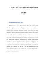
acclerated in the last 5 years (Fig. 1.1). Thus, Marchesini
and Forlani [11] were able to locate only 161 articles
which addressed this topic between 1980 and 1999
(approximately 8/year) but 122 in 2000–01 (approx-
1950 • Cirrhosis noted in diabetics
1970s • Jejuno-ileal bypass liver disease resembles alcoholic hepatitis
1979/80 • Ludwig et al. [1] Coined term NASH for steatohepatitis in non-drinkers
• ~8 papers/year
• Small series
• NASH is benign (Powell et al. 1990 [8])
1994 • Expanded scope of NASH (Bacon et al. 1994 [10])
1996 • CYP2E1 induced in rodent dietary model
• Endotoxin induces inflammation in steatotic liver
1998 • CYP2E1 induced in human NASH
• First NIH conference on NASH
• Pivotal importance of insulin resistance
1999 • Several animal models
• First clinical trials
2002 • ~60 papers/year
• AASLD single topic conference
• First European and Japanese single topic conferences
• NASH established as part of insulin resistance syndrome
2004 • Release of first book on NAFLD/NASH
Fig. 1.1 Chronology of the pace
of research into pathogenesis,
natural history and treatment of
NAFLD/NASH.
INTRODUCTION TO NASH AND RELATED DISORDERS
3
discussed at a single topic conference of the American
Association for Study of Liver Diseases (AASLD),
September 2002, Atlanta, Georgia (see Chapter 2)
[19,20].
When one particular cause of steatohepatitis is evid-
ent, the term steatohepatitis is qualified (e.g. alcoholic
steatohepatitis, drug-induced steatohepatitis, experi-
mental [dietary] steatohepatitis). Such cases are often
referred to as ‘secondary NASH’ (Table 2.2; see Chap-
ters 13, 20 and 21). Because of its strong association
with ‘metabolic’ determinants (obesity, insulin resist-
ance, type 2 diabetes, hyperlipidaemia), the acronym
‘MeSH’ has been been suggested as an alternative for
‘idiopathic’ (or ‘primary’) NASH, but seems unlikely
to gain widespread acceptance.
Non-alcoholic fatty liver diseases
The term NAFLD is gaining acceptance and is use-
ful because it is more comprehensive than NASH
(Table 1.1) [15–17]. NAFLD includes less significant
forms of steatosis either alone (type 1 NAFLD) or with
inflammation but no hepatocyte ballooning or fibrosis
(type 2). The term NAFLD will be used here when
the pathology of metabolic liver disease is not known,
or when specifically referring to the fuller spectrum.
This now includes some cases of cryptogenic cirrhosis
in which steatohepatitis and steatosis are no longer
conspicuous.
Primary and secondary steatohepatitis: the
importance of alcohol
A key definitional issue is potential overlap between
‘primary’ (metabolic) NAFLD/NASH and pathologic-
ally similar fatty liver diseases associated with a single
causative factor (Table 1.2). The most important con-
sideration is the level of alcohol consumption con-
sidered unlikely to have any causal role in liver disease.
Early publications describing ‘alcoholic hepatitis-like
lesions’ were in non-drinkers or those with minimal
intake (less than one drink a week in the Ludwig
series). Since then, reports of NAFLD/NASH have
used a variety of thresholds for alcohol intake. Some
have required rigorous alcohol restriction, particu-
larly for cases of ‘cryptogenic cirrhosis’ attributable to
Table 1.1 Categories of non-alcoholic fatty liver diseases (NAFLD): relationship to NASH. (After Matteoni et al. [15].)
Category Pathology Clinicopathological correlation
Type 1 Simple steatosis Known to be non-progressive
Type 2 Steatosis plus lobular inflammation Probably benign (not regarded as NASH)
Type 3 Steatosis, lobular inflammation and ballooning degeneration NASH without fibrosisamay progress to
cirrhosis
Type 4 Steatosis, ballooning degeneration and Mallory bodies, NASH with fibrosisamay progress to
and/or fibrosis cirrhosis and liver failure
Table 1.2 Causes of secondary steatohepatitis.
Alcohol (alcoholic hepatitis)
Drugs (tamoxifen, amiodarone, methotrexate)
Copper toxicity (Wilson’s disease, Indian childhood cirrhosis)
Jejuno-ileal bypass (see Chapter 20)
Other causes of rapid profound weight loss (massive intestinal resection, cachexia, bulimia, starvation)
Hypernutrition in adults (parenteral nutrition, intravenous glucose)
A-betalipoproteinaemia
Jejunal diverticulosis (contaminated bowel syndrome)
Insulin resistance syndromes (familial and acquired lipodystrophies, polycystic ovary syndrome)
CHAPTER 1
4
for people with NAFLD/NASH are considered in
Chapter 15.
Interaction between steatohepatitis and other
liver disorders
Another challenge is when the metabolic determin-
ants of NASH (Table 1.3) coexist with known causes
of liver disease. The latter include ‘moderate’ levels
of alcohol intake (30–60 g/day in men, 20–40 g/day in
women), hepatitis C and potentially hepatotoxic drugs
(methotrexate, tamoxifen, calcium-channel blockers,
highly active antiretroviral therapy) [28]. The likelihood
that steatosis or the metabolic determinants that result
in NASH contribute to liver injury and fibrotic severity
of other liver diseases is canvassed in Chapter 23.
Importance of NASH
Reasons why NASH is an important form of liver dis-
ease are summarized in Table 1.4.
NASH (e.g. none, or less than 40 g/week) [21,22].
Conversely, other authors have allowed alcohol intake
to be as high as 210 g/week [23].
It is noted that 30 g/day is close to the level of
40 g/day associated with an increased risk of cirrhosis
in women [24]. Safe levels of alcohol intake have also
been difficult to define for other liver diseases, such as
hepatitis C for which less than 10 g/day was recom-
mended by the first National Institutes of Health
(NIH) Consensus Conference in 1997 [25], but up to
30 g/day for men and 20 g/day for women by the sec-
ond NIH Consensus Conference [26]. In this book, the
definition of NASH requires alcohol intake to have
never been greater than 140 g/week (ideally, ≤ 20 g/day
for men and ≤ 10 g/day for women). However, it is
acknowledged that there may be potential for even
these low levels of alcohol intake levels to contribute
to cell injury, fibrogenesis and hepatocarcinogenesis
in steatohepatitis. Conversely, it remains possible that
low levels of alcohol intake confer health benefits in
obese persons with liver disease [27]. The implications
for recommending optimal levels of alcohol intake
Table 1.4 Reasons why NAFLD/NASH is important.
High prevalence of fatty liver disorders in urbanized communities with affluent (‘Western’) economies throughout the world
Most common cause of abnormal liver tests in communitya?2–8% of population have NAFLD
NASH now rivals alcoholic liver disease and chronic hepatitis C as reason for referral to gastroenterologist or liver clinic
NASH is a potential cause of cirrhosis, which may be ‘cryptogenic’, and lead to end-stage liver disease
Liver failure is most common cause of death in patients with cirrhosis resulting from NASH
Standardized mortality of liver disease in type 2 diabetes greatly exceeds vascular disease
NASH recurs after liver transplantation
Hepatic steatosis as a cause of primary graft non-function after liver transplantation
Role of metabolic determinants of NASH in pathogenesis of other liver diseases, particularly hepatitis C and alcoholic cirrhosis
Possible role of NASH/hepatic steatosis in hepatocarcinogenesis
Table 1.3 Metabolic associations of NASH.
Type 2 diabetes mellitus
Family history of type 2 diabetes
Insulin resistance, with or without glucose intolerance
Central obesity (waist : hip ≥ 0.85 in women, ≥ 0.90 in men; waist > 85 cm in women, > 97 cm in men*)
Obesity (BMI ≥ 30 kg/m
2
in white people, ≥ 27 kg/m
2
in Asians)
Hypertriglyceridaemia
Rapid and massive weight loss in overweight subjects
* Values vary between countries; 90 cm for women and 102 cm for men often used in USA.
INTRODUCTION TO NASH AND RELATED DISORDERS
5
The NASH epidemic
In much of the world, abnormal liver tests attributable
to hepatic steatosis or NASH have become the most
common liver disease in the community. Depending
on how an abnormal value for aminotransferase is
defined in studies, such as the Third National Health
and Nutritional Examination Survey (NHANES III),
between 3 and 23% of the adult population may have
NAFLD/NASH [29–31]. In studies that have employed
hepatic imaging, autopsy or biopsy approaches, approx-
imately 70% of obese people have hepatic steatosis
and/or raised alanine aminotransferase (ALT) [12,21,
27,31–37]; NASH is present in approximately 20% of
these [7,27]. In old autopsy studies, ~ 10% of diabetics
had cirrhosis, but other factors (hepatitis B and C)
were possible confounding variables. In more recent
studies, both the prevalence and severity of NASH
appear to be increased considerably in patients with
type 2 diabetes [11,21,36,38–40].
The epidemiology of NAFLD/NASH is discussed in
Chapter 3. Based on the continuing epidemic of obesity
and type 2 diabetes through much of the world, it is
likely that the prevalence of NASH will increase fur-
ther during the next decade. In the USA and Australia,
up to 60% of men and 45% of women are now over-
weight, and about one-third of these are obese [41,
42]. Similar increases have been noted in societies that
until the last one or two generations were particip-
ating in physically active (‘hunter gatherer’) lifestyles
(see Chapter 18). The prevalence of type 2 diabetes has
doubled, trebled or increased 10- to 20-fold (as in
Japanese youth) during the last decade, rates reaching
40% or more of the adult population in some com-
munities [43–45]. Childhood cases of NASH are also
clearly related to obesity and type 2 diabetes (see
Chapter 19) [46,47]. Some possible reasons for high
rates of obesity and type 2 diabetes in contemporary
affluent societies (‘east’ and ‘west’, ‘north’ and ‘south’),
and the implications for prevention and interruption
of NASH are discussed in Chapters 3–5 and 18.
NAFLD/NASH varies in severity and clinical outcome
Steatosis alone has an excellent prognosis. It seems
probable that most cases of steatosis with lobular
inflammation but without conspicuous hepatocyte
injury or fibrosis (NAFLD type 2) behaves in the same
way, with very low rates of fibrotic progression (see
Chapter 3). However, 20–25% of cases with NASH
have or will progress to cirrhosis [15,16,19,21,22,39].
There is mounting evidence that a proportion of
cases of ‘cryptogenic cirrrhosis’ may be attributable to
NASH, in which the histological features of steatohep-
atitis have resolved (see Chapter 14) [15,21,31,35,48].
Rare cases of subacute hepatic failure have also been
attributed to possible NASH [49].
Earlier studies of NAFLD/NASH emphasized the
good overall prognosis [8,10]. More recent studies that
have defined cases according to fibrotic severity indicate
that those with significant fibrosis may progress to liver
failure [15,22,50]. Among cases of cirrhosis, the risk
of death or liver transplantation may be as high as cir-
rhosis resulting from hepatitis C (both ~ 30% at 7 years)
[15,16,22,50]. If this indolent progressive course is con-
firmed in larger prospective studies, NASH will cause a
formidable disease burden in forthcoming decades.
A few well-documented cases of cirrhosis resulting
from NASH have presented with, or less commonly
have terminated in HCC [16,51]. HCC was recently
noted to be a cause of death among obese patients with
cryptogenic cirrhosis [52,53]. However, it is not clear
that all such cases were caused by NASH [22], and sev-
eral were diagnosed within 9 months of presentation.
Others have suggested that steatosis could increase the
risk of HCC associated with other liver diseases [54,55],
but conflicting data have been noted (see Chapter 22).
Metabolic risk factors for NASH may worsen other
liver diseases
As well as providing the setting for NASH, insulin re-
sistance, obesity, type 2 diabetes and hepatic steatosis
are now recognized as factors that favour fibrotic pro-
gression in hepatitis C [56,57]. Obesity is also an inde-
pendent risk factor for alcoholic cirrhosis [58]. Thus,
‘NASH determinants’ may contribute to the overall
burden of cirrhosis directly as the hepatic complication
of obesity, insulin resistance and diabetes, and indirectly
as factors that favour cirrhosis among people with
chronic viral hepatitis or alcoholism (see Chapter 23).
When should the clinician think
of NASH?
Clinicians need to consider that NAFLD/NASH is
the most likely cause of liver test abnormalities in the
CHAPTER 1
6
laboratory tests, such as a raised serum urate, triglyc-
eride, low-density lipoprotein (LDL) cholesterol and
low levels of high-density lipoprotein (HDL) cholesterol
are pointers to insulin resistance. The genetic factors
that could predispose to NASH are considered in
Chapter 6, and the insulin resistance syndrome is dis-
cussed in Chapter 5.
A raised serum ferritin level is a common ‘con-
founder’ in cases of NAFLD/NASH [60–62]. As in
alcoholic liver disease, this most often reflects increased
hepatic release of ferritin as an ‘acute phase reactant’,
reflecting the hepatic inflammatory response and
increased permeability of steatotic and injured hepato-
cytes. If a persistently raised serum transferrin saturation
suggests increased body iron stores, haemochromato-
sis gene testing should be conducted in those with a
northern European or Celtic background. The pro-
posed role of hepatic iron in worsening fibrotic sever-
ity in NASH is controversial (see Chapter 7) [60–62].
Confirming the diagnosis is NASH
Liver biochemical function tests, serum lipids and
other laboratory results
Abnormal biochemical results (liver function tests)
typically comprise minor (1.5- to 5-fold) elevations of
ALT and gamma-glutamyl transpeptidase (GGT). The
following laboratory tests may provide clues to the
presence of cirrhosis: low platelet count, raised aspart-
ate aminotransferase (AST) that is higher than ALT,
and subtle changes in serum albumin or bilirubin that
are not attributable to other causes (see Chapter 14).
presence of metabolic risk factors (Table 1.3), and when
other causes of liver disease have been excluded (see
Chapter 13). The importance of considering NAFLD/
NASH as a primary diagnosis, rather than purely as a
disease of exclusion, is emphasized in this book (see
Chapter 5).
NAFLD/NASH is usually suspected because of ab-
normal liver biochemical tests in an apparently healthy
person with no symptoms (Table 1.5). However,
fatigue, or vague discomfort over the liver with ‘rub-
bery’ hepatomegaly are common. Significant hepatic
pain and tenderness are rare. The presence of a firm
liver edge, or more rarely a palpable spleen, muscle
wasting, ascites, jaundice or hepatic encephalopathy
indicate possible cirrhosis, with or without complica-
tions of portal hypertension and hepatic decompensa-
tion (see Chapters 13 and 14).
In a person with abnormal liver biochemistry tests, a
history of recent weight gain or an expanding waistline
are often clues to the diagnosis of NASH. However,
rapid and extensive weight loss in an obese person can
lead to an initial diagnosis of NASH. Such weight loss
may occur through intercurrent illness, older forms of
obesity surgery (see Chapter 20) or drastic reductions
in energy intake caused by fasting, bulimia or ‘crash’
dieting (Table 1.2). Cycles of rapid weight gain fol-
lowed by precipitant weight loss have led to cirrhosis
or hepatic decompensation [3].
The past medical and family history often provide
clues to metabolic disorders that underlie NASH [59],
particularly type 2 diabetes, and other features and
complications of insulin resistance such as arterial
hypertension and coronary heart disease [11]. Similarly,
Table 1.5 Pointers to NAFLD/NASH in clinical practice.
Unexplained elevation of ALT and GGT, typically minor, in a person with metabolic risk factors (Table 1.3)
‘Rubbery’ hepatomegaly
Recent weight gain and expanding waistline
Lifestyle or medication changes favouring weight gain (marriage, retirement, unemployment, antidepressants)
Family history of type 2 diabetes, NAFLD, vascular disorders or hyperlipidaemia
Raised serum ferritin not attributable to iron storage disorder or alcohol
Abnormalities of hepatic imagingadiffuse echogenicity on ultrasonogram (‘bright liver’), radiolucency on CT
Patient with chronic HCV infection and diabetes and/or obesity, ‘rubbery’ hepatomegaly or steatosis with HCV genotype 1
infections (see Chapter 23)
Patient with chronic HBV infection, raised ALT but non-detectable HBV DNA in presence of metabolic risk factors
ALT, alanine aminotransferase; CT, computerized tomography; GGT, gamma-glutamyl transpeptidase; HBV DNA, hepatitis B
virus DNA; HCV, hepatitis C virus.
INTRODUCTION TO NASH AND RELATED DISORDERS
7
Fasting hypertriglyceridaemia is present in 25–40%
of patients with NASH [8,9,10,16,39]. It may be
associated with hypercholesterolaemia (increased LDL
cholesterol, particularly with low levels of HDL and a
high LDL : HDL ratio). This pattern of lipid disorders
is a feature of the insulin resistance syndrome.
Anthropometric measurements
Because nearly all patients with NASH have central
obesity, anthropometric measurements should be
routinely recorded at liver clinic visits (see Chapter 15).
Height and weight are used to calculate body mass
index (BMI), while girth (circumference at umbilicus),
or waist : hip ratio form simple pointers to central
obesity (see Chapters 5 and 15 for details). Some nutri-
tionists recommend waist circumference as more useful
than body weight for monitoring benefits of lifestyle
change in overweight people.
Determination of insulin resistance
The near universal association of NASH with insulin
resistance means that tests to document this patho-
physiological state should form part of the approach
to diagnosis. Fasting serum insulin and blood glucose
levels can be used to construct the relatively crude (but
practically useful) homoeostasis model assessment of
insulin resistance (HOMA-IR). Values for HOMA-IR
differ between population subgroups. Thus, applica-
tion of this method requires reference to a local group
of normal age-matched controls.
As discussed in Chapter 4, diabetologists prefer an
‘active’ measure of insulin sensitivity as opposed to a
fasting one; the latter will be misleading when there is
secondary failure of insulin secretion by pancreatic β
cells. A simplified 75-g oral glucose tolerance test with
1 and 2 h blood glucose and serum insulin levels can be
very informative. Fasting serum C-peptide level is an
excellent measure of insulin production. It therefore
appears to be a sensitive indicator of insulin resistance
that can be used in hepatological practice.
Hepatic imaging
Hepatic imaging performed as part of investigations
into abdominal pain, abnormal liver tests or suspected
hepatic malignancy may be the first clue to the pres-
ence of steatosis [63]. The sensitivity of hepatic ultra-
sound for steatosis (increased echogenicity, or ‘bright
liver’) appears fairly high, particularly when extensive
steatosis (involving at least 33% hepatocytes) is pres-
ent [63]. CT also appears to be relatively sensitive
for hepatic steatosis, and has the advantage that
nodularity resulting from cirrhosis may sometimes be
appreciated. Careful attention should be given to
features of portal hypertension (portal vein dilatation,
splenomegaly, retroperitoneal varices). Otherwise,
both ultrasonography and computerized tomography
(CT) have low positive predictive value for detecting
features of cirrhosis.
Neither ultrasonography nor CT is able to distinguish
NASH from other forms of NAFLD (see Chapter 13).
Thus, while hepatic imaging is useful for providing
supportive evidence in favour of hepatic steatosis, it
cannot substitute for liver biopsy for elucidating the
fibrotic severity of NASH.
Newer imaging techniques (dual-energy X-ray
absorptiometry [DEXA], magnetic resonance imaging
[MRI]) are also valuable in determining body com-
position. Total body fat can be estimated accurately
with DEXA, but greater interest will come from stud-
ies attempting to discern patterns of adipose tissue
distribution (visceral versus subcutaneous or ectopic);
these patterns are likely to correlate more closely with
insulin resistance (see Chapter 4).
Liver biopsy
Clinical guidelines for when liver biopsy is indic-
ated for suspected NASH are not yet standardized
[16,18], with views ranging from the nihilistic to the
enthusiastic! In considering whether a liver biopsy is
indicated, one approach is to assess risk factors for
fibrotic severity (obesity, diabetes, age over 45 years,
and AST : ALT > 1) and to seek ‘warning signs’ of cir-
rhosis (see Chapter 14) [15,16,18]. One approach is
not to recommend biopsy at first referral (see Chap-
ter 15). If lifestyle intervention aimed at correcting
insulin resistance and central obesity fails to normalize
liver tests, and particularly if there are warning signs
for cirrhosis or the patient expresses a strong desire to
know the severity of their liver disease, the physician
should proceed to liver biopsy (see Chapters 13 and 15).
Liver biopsy interpretation is described in Chapter 2.
In following any paradigm for liver biopsy, it
should be noted that liver test abnormalities in NASH
are poorly related to fibrotic severity. Some patients
CHAPTER 1
8
peroxidation. It is now clear that the steatotic liver is
more susceptible to oxidative stress, as well as to injury
after injection of endotoxin [16,18,64].
The liver normally responds to the chronic presence
of oxidants by increasing synthesis of protective anti-
oxidant pathways, such as those based on reduced
glutathione (GSH). If GSH levels are depleted (as with
fasting, toxins such as alcohol, or consumption by pro-
oxidants), the products of lipid peroxidation create
and amplify oxidative stress. In turn, oxidative stress
can cause liver injury (e.g. by triggering apoptosis
and inciting inflammation). The mechanisms that
may trigger and perpetuate inflammatory recruitment
in NASH, and the importance of cytokines such as
tumour necrosis factor-α (TNF-α) are discussed in
Chapter 10.
Evidence has been deduced from human studies as
well as in experimental models that cytochrome P450
2E1 (CYP2E1) is overexpressed in steatohepatitis [66–
68], most likely because of impaired insulin receptor
signalling. CYP2E1 is a potential source of reduced
(reactive) oxygen species (ROS). In the absence of
CYP2E1, CYP4A takes on the role as an alternative
microsomal lipid oxidase, and it too may generate
ROS [67]. CYP2E1 and CYP4A catalyze the ω and ω-1
hydroxylation of long-chain fatty acids. The products
are dicarboxylic fatty acids, which cannot be subjected
to mitochondrial β-oxidation and are so targeted to
the peroxisome for further oxidation. In turn, this gen-
erates hydrogen peroxide (coupled to catalase) as an
essential by-product [69].
The relative importance of metabolic sites of ROS
generation in hepatocytes (mitochondria, endoplasmic
reticulum, peroxisomes), and products of the inflam-
matory response in contributing to oxidative stress in
steatohepatitis remains unclear; interactive processes
are likely to operate [64]. However, mitochondria
could be a critical source of ROS in fatty liver disorders
(see Chapter 11) [38,70].
Hepatic inflammation and cellular injury to hepato-
cytes can induce and activate transforming growth
factor-β (TGF-β), which has a key role in activating
stellate cells to elaborate extracellular matrix as part
of the wound healing process. It is now apparent
that leptin has a key role in hepatic fibrogenesis, and
leptin also appears to be necessary for appropriate
liver regeneration as part of the ‘wound healing’
response to chronic steatohepatitis and other forms
with NASH cirrhosis may have normal ALT levels.
A nihilistic approach to liver biopsy for NASH
therefore raises the concern that some patients with
advanced hepatic fibrosis and/or cirrhosis would not
be counselled and monitored appropriately. Further,
liver biopsy can sometimes produce unexpected
findings indicative of another liver disease, thereby
changing management.
Why does NASH happen?
The recurrence of NASH after orthotopic liver trans-
plantation (see Chapter 17) is a dramatic demonstra-
tion of the importance of extrahepatic (metabolic)
factors in its pathogenesis. Among these, genetic and
acquired abnormalities of fatty acid turnover and oxida-
tion are likely to be crucial in causing steatohepatitis
[16,17,19,64]; some facilitate accumulation of free fatty
acids (FFA), others favour the operation of oxidative
stress. Factors that facilitate recruitment of an hepatic
inflammatory (or innate immune) response, or deter-
mine the tissue response to liver injury are other poten-
tially relevant variables.
Human and animal studies have started to address
key issues in NASH pathogenesis, such as the nature of
insulin resistanceawhy it occurs, whether it is respons-
ible for inflammation and liver cell injury as well as
FFA accumulation, the mechanisms for inflammatory
recruitment and perpetuation, the biochemical basis
and significance of oxidative stress, the cell biological
basis of hepatocye injury and the pathogenesis of
fibrosis (see Chapters 4, 7, 8 and 10–12). It seems
likely that many such factors are genetically deter-
mined (see Chapter 6). In this way, NASH, like type 2
diabetes, atherosclerosis and some cancers, is the
outcome of an interplay between several genetic and
environmental factors.
Lipid accumulation also favours increased concen-
trations of FFA that may be directly toxic to hepatocytes.
It has recently been proposed that such ‘lipotoxicity’
in NASH results from failure of leptin or other hor-
mones that modulate insulin sensitivity to correct for
insulin resistance [65]. The humoral and dietary mod-
ulation of insulin receptor signalling that underlies this
new concept is discussed in Chapter 4. The fatty liver
also provides an excess of unsaturated FFA, oxidation
of which results in the autopropagative process of lipid
INTRODUCTION TO NASH AND RELATED DISORDERS
9
of liver injury (see Chapter 12). Thus, leptin, origin-
ally characterized as an anti-obesity hormone acting
on the central nervous system to regulate appetite,
could have multiple roles in the pathogenesis of NASH
by modulating fat deposition in hepatocytes (anti-
lipotoxicity), and regulating the hepatic fibrotic and
regenerative response to steatohepatitis. A more de-
tailed account of the cell biology of NASH is presented
in Chapter 12.
Approaches to management of NASH
Lifestyle adjustments
Attempts to correct steatosis and liver injury in NASH
can begin before the diagnostic process is complete
(see Chapter 15). The aim is to correct insulin resist-
ance and central obesity. Rapid and profound weight
loss is potentially dangerous for the person with fatty
liver disease [3]. It is prudent and more realistic to
recommend slow reductions in body weight that are
achievable and sustainable by permanent changes
in lifestyle. It has been shown that such reductions
improve liver tests [71], and there is mounting evid-
ence that this is associated with removal of fat from
the liver, decreased necroinflammatory change and
even resolution of fibrosis [72,73].
In accordance with the results of recent type 2
diabetes intervention studies [74,75], physical activity
should include at least 20 min of exercise each day
(140 min/week), equivalent to rapid walking. The
essentials of dietary modification are the same as for
diabetes: reduce total fat to less than 30% of energy
intake, decrease saturated fats, replace with complex
carbohydrates containing at least 15 g fibre, and rich in
fruit and vegetables. Consideration of low versus high
glycaemic foods (e.g. brown or basmati rice versus
conventional long or short-grain white rice); reduction
of simple sugars and alcohol intake is also likely to be
beneficial.
Some authors have advocated referral to a dietitian
or ‘personal case manager’ to provide education and
closer supervision of dietary regimens and lifestyle
interventions [73–75]. Approaches to lifestyle modifi-
cation and weight reduction are discussed in more
detail in Chapter 15. The effectiveness and cost-
efficacy of such approaches are important aspects that
warrant further study.
Measures to control hyperlipidaemia and
hyperglycaemia
Increased physical activity and low-fat diet improve
insulin sensitivity and can, in some cases, reverse
insulin resistance. The value of exercise in improving
glycaemic control in diabetes is now generally accepted.
In other respects, treatment of diabetes in patients with
NASH should conform to conventional approaches,
although this may change in future if drugs that help
reverse insulin resistance live up to initial promise
against NAFLD/NASH without causing unacceptable
weight gain. These agents include metformin and the
thiolazinediones (see Chapter 16). Drugs that correct
lipid disorders, anti-oxidants (vitamin E, betaine) and
other hepatoprotective agents (ursodeoxycholic acid)
are also under study in NASH (see Chapter 16).
Concluding remarks: can NAFLD/NASH
be prevented or reversed?
Because liver failure does not occur in NAFLD/NASH
unless cirrhosis has developed, reducing or reversing
fibrotic progression must be the ultimate objective of
treatment. While several agents improve liver tests
over the short term in patients with NAFLD/NASH
(see Chapter 16), none have yet (June 2003) been
shown to have long-term efficacy and to impact
on fibrotic progression (but see Chapter 24). In the
absence of evidence of such efficacy, patients should
currently only receive drug therapy directed at NASH
within the context of a clinical trial, particularly as
some of the compounds presently under study carry
toxic potential or other unwanted effects (see Chapters
16 and 24).
There is now compelling evidence that type 2 dia-
betes can be prevented (or at least delayed in onset) by
lifestyle interventions [74,75]. Both the Finnish and US
Diabetes Intervention Projects showed a 58% reduc-
tion in incidence of type 2 diabetes among those at
high risk could be achieved with only modest reduc-
tions in body weight [74,75]. NASH, another con-
sequence of insulin resistance (see Chapter 5), should
also be preventable by changes in diet and physical
activity. There is now evidence that weight reduction
and lifestyle changes nearly always improve liver tests
in NAFLD, and also have potential to improve liver
CHAPTER 1
10
15 Matteoni CA, Younossi ZM, Gramlich T et al. Non-
alcoholic fatty liver disease: a spectrum of clinical and
pathological severity. Gastroenterology 1999; 116: 1413–9.
16 Angulo P. Non-alcoholic fatty liver disease. N Engl J Med
2002; 16: 1221–31.
17 Younossi ZM, Diehl AM, Ong JP. Non-alcoholic fatty
liver disease: an agenda for clinical research. Hepatology
2002; 35: 746–52.
18 Farrell GC. Okuda Lecture. Non-alcoholic steatohepatitis:
what is it, and why is it important in the Asia-Pacific
region. J Gastroenterol Hepatol 2003; 18: 124 –38.
19 Neuschwander-Tetri BA, Caldwell SH. Non-alcoholic
steatohepatitis: summary of an AASLD single topic con-
ference. Hepatology 2003; 37: 1202–19.
20 Brunt EM, Janney CG, Di Bisceglie AM, Neuschwander-
Tetri BA, Bacon BR. Non-alcoholic steatohepatitis: a
proposal for grading and staging the histological lesions.
Am J Gastroenterol 1999; 94: 2467–74.
21 Angulo P, Keach JC, Batts KP, Lindor KD. Independent
predictors of liver fibrosis in patients with non-alcoholic
steatohepatitis. Hepatology 1999; 30: 1356–62.
22 Hui JM, Kench JG, Chitturi S et al. Long-term outcomes
of cirrhosis in NASH compared to hepatitis C: same
mortality, less cancer. Hepatology 2003; 38: 420–7.
23 Mulhall BP, Ong JP, Younossi Z. Non-alcoholic fatty
liver disease: an overview. J Gastroenterol Hepatol 2002;
17: 1136–43.
24 Norton R, Batey R, Dwyer T, MacMahon S. Alcohol
consumption and the risk of alcohol related cirrhosis in
women. Br Med J 1987; 295: 80 –2.
25 Consensus Development Panel. National Institutes of
Health Consensus Development Conference Panel State-
ment. Management of hepatitis C. Hepatology 1997; 26
(Suppl. 1): 2S–10S.
26 National Institutes of Health Consensus Development
Conference Statement. Management of Hepatitis C, June
10–12, 2002. Hepatology 2002; 36 (Suppl. 1): S3–S21.
27 Dixon JB, Bhathal PS, O’Brien PE. Non-alcoholic fatty
liver disease: predictors of non-alcoholic steatohepatitis
and liver fibrosis in the severely obese. Gastroenterology
2001; 121: 91–100.
28 Farrell GC. Drugs and steatohepatitis. Semin Liver Dis
2002; 22: 185–94.
29 Erby JR, Silberman C, Lydick E. Prevalence of abnormal
serum alanine aminotransferase levels in obese patients
and patients with type 2 diabetes. Am J Med 2000; 109:
588–90.
30 Clark JM, Brancati FL, Diehl AM. Non-alcoholic fatty
liver disease. Gastroenterology 2002; 122: 1649–57.
31 Ruhl CE, Everhardt JE. Determinants of the association
of overweight with elevated serum alanine aminotrans-
ferase activity in the United States. Gastroenterology
2003; 124
: 71–9.
histology in obese patients with hepatitis C or fatty
liver disorders [71–73] (Chapter 24). Whether this
approach would be a cost-effective way to reduce the
number of patients progressing to cirrhosis and liver
failure is clearly worthy of study.
References
1 Ludwig J, Viaggiano TR, McGill DB, Oh BJ. Non-
alcoholic steatohepatitis: Mayo Clinic experience with an
hitherto unnamed disease. Mayo Clin Proc 1980; 55:
434–8.
2 Adler M, Schaffner F. Fatty liver hepatitis and cirrhosis in
obese patients. Am J Med 1979; 67: 811–6.
3 Capron J-P, Delamarre J, Dupas J-L et al. Fasting in
obesity: another cause of liver injury with alcoholic
hyaline? Dig Dis Sci 1982; 27: 265– 8.
4 Itoh S, Yougel T, Kawagoe K. Comparison between non-
alcoholic steatohepatitis and alcoholic hepatitis. Am J
Gastroenterol 1987; 82: 650–4.
5 Diehl AM, Goodman Z, Ishak KG. Alcohol-like liver
disease in non-alcoholics: a clinical and histologic com-
parison with alcohol-induced liver disease. Gastroentero-
logy 1988; 95: 1056–62.
6 Lee RG. Non-alcoholic steatohepatitis: a study of 49
patients. Hum Pathol 1989; 20: 594 – 8.
7 Wanless IR, Lentz JS. Fatty liver hepatitis (steatohepatitis)
and obesity: an autopsy study with analysis of risk factors.
Hepatology 1990; 12: 1106–10.
8 Powell EE, Cooksley WGE, Hanson R et al. The natural
history of non-alcoholic steatohepatitis: a follow-up
study of 42 patients for up to 21 years. Hepatology 1990;
11: 74–80.
9 Fiatarone JR, Coverdale SA, Batey RG, Farrell GC.
Non-alcoholic steatohepatitis: impaired antipyrine meta-
bolism and hypertriglyceridaemia may be clues to its
pathogenesis. J Gastroenterol Hepatol 1991; 6: 585–90.
10 Bacon BR, Farahvash MJ, Janney CG, Neuschwander-
Tetri BA. Non-alcoholic steatohepatitis: an expanded
clinical entity. Gastroenterology 1994; 107: 1103–9.
11 Marchesini G, Forlani G. NASH: From liver diseases to
metabolic disorders and back to clinical hepatology.
Hepatology 2002; 35: 497–9.
12 Seth SG, Gordon FD, Chopra S. Non-alcoholic steato-
hepatitis. Ann Intern Med 1997; 126: 137– 45.
13 Ludwig J, McGill DB, Lindor KD. Non-alcoholic
steatohepatitis. J Gastroenterol Hepatol 1997; 12: 398–
403.
14 James OFW, Day CP. Non-alcoholic steatohepatitis
(NASH): a disease of emerging identity and importance.
J Hepatol 1998; 29: 495–501.
INTRODUCTION TO NASH AND RELATED DISORDERS
11
32 Bellentani S, Sacoccio G, Masutti F et al. Prevalence and
risk factors for hepatic steatosis in northern Italy. Ann
Intern Med 2000; 132: 112–7.
33 Hasan I, Gani RA, Machmud R et al. Prevalence and
risk factors for non-alcoholic fatty liver in Indonesia.
J Gastroenterol Hepatol 2002; 17 (Suppl): A154.
34 Caldwell SH, Oelsner DH, Iezzoni JC et al. Cryptogenic
cirrhosis: clinical characterization and risk factors for
underlying disease. Hepatology 1999; 29: 664–9.
35 Ratziu V, Giral P, Charlotte F et al. Liver fibrosis
in overweight patients. Gastroenterology 2000; 118:
1117–23.
36 Marchesini G, Brizi M, Morselli-Labate AM et al. Asso-
ciation of non-alcoholic fatty liver disease with insulin
resistance. Am J Med 1999; 107: 450 –5.
37 Marceau P, Biron S, Hould FS et al. Liver pathology
and the metabolic syndrome X in severe obesity. J Clin
Endocrinol Metab 1999; 84: 1513–7.
38 Sanyal AJ, Campbell-Sargent C, Mirshahi F et al. Non-
alcoholic steatohepatitis: association of insulin resist-
ance and mitochondrial abnormalities. Gastroenterology
2001; 120: 1183–92.
39 Chitturi S, Abeygunasekera S, Farrell GC et al. NASH
and insulin resistance: insulin hypersecretion and spe-
cific association with the insulin resistance syndrome.
Hepatology 2002; 35: 373–8.
40 Pagano G, Pacini G, Musso G et al. Non-alcoholic steato-
hepatitis, insulin resistance and metabolic syndrome:
further evidence for an etiologic association. Hepatology
2002; 35: 367–72.
41 www.cdc.gov/nccdphp/dnpa/obesity/trends/maps/
index.htm
42 Dunstan DW, Zimmet PZ, Welborn TA et al. The rising
prevalence of diabetes and impaired glucose tolerance:
the Australian Diabetes, Obesity and Lifestyle Study.
Diabetes Care 2002; 25: 829–34.
43 Daniel M, Rowley KG, McDermott R, O’Dea K. Diabetes
and impaired glucose tolerance in Aboriginal Australians:
prevalence and risk. Diabetes Res Clin Pract 2002; 57:
23–33.
44 Zimmet P, Alberti KG, Shaw J. Global and societal im-
plications of the diabetes epidemic. Nature 2001; 414:
782–7.
45 Omagari KH, Kadokawa Y, Masuda J et al. Fatty liver in
non-alcoholic non-overweight Japanese adults: incid-
ence and clinical characteristics. J Gastroenterol Hepatol
2002; 17: 1089–105.
46 Rashid M, Roberts E. Non-alcoholic steatohepatitis
in children. J Paediatr Gastroenterol Nutr 2000; 30:
48–53.
47 Manton ND, Lipsett J, Moore DJ et al. Non-alcoholic
steatohepatitis in children and adolescents. Med J Aust
2000; 173: 476–9.
48 Poonawala A, Nair SP, Thuluvath PJ. Prevalence of obes-
ity and diabetes in patients with cryptogenic cirrhosis: a
case study. Hepatology 2000; 32: 689–92.
49 Caldwell SH, Hespenheide EE. Subacute liver failure in
obese women. Am J Gastroenterol 2002; 97: 2058–62.
50 Falck-Ytter Y, Younossi ZM, Marchesini G, McCullough
AJ. Clinical features and natural history of non-alcoholic
steatosis syndromes. Semin Liver Dis 2001; 21: 17–26.
51 Shimada M, Hashimoto E, Taniai M et al. Hepatocellular
carcinoma in patients with non-alcoholic steatohepatitis.
J Hepatol 2002; 37: 154–60.
52 Ratziu V, Bonhay L, Di Martino V et al. Survival, liver
failure, and hepatocellular carcinoma in obesity-related
cryptogenic cirrhosis. Hepatology 2002; 35: 1485–93.
53 Bugianesi E, Leone N, Vanni E et al. Expanding the
natural history of non-alcoholic steatohepatitis: from
cryptogenic cirrhosis to hepatocellular carcinoma. Gastro-
enterology 2002; 123: 134–40.
54 Marrero JA, Fontana RJ, Su GL et al. NAFLD may be a
common underlying liver disease in patients with hepato-
cellular carcinoma in the United States. Hepatology
2002; 36: 1349–54.
55 Garcia-Monzon C, Martin-Perez E, Iacono OL et al.
Characterization of pathogenic and prognostic factors
of non-alcoholic steatohepatitis associated with obesity.
J Hepatol 2000; 33: 716–24.
56 Hourigan LF, Macdonald GA, Purdie D et al. Fibrosis in
chronic hepatitis C correlates with body mass index and
steatosis. Hepatology 1999; 29: 1215–9.
57 Hwang SJ, Luo JC, Chu CW et al. Hepatic steatosis
in chronic hepatitis C virus infection: prevalence and
clinical correlation. J Gastroenterol Hepatol 2001; 16:
190–5.
58 Naveau S, Giraud V, Borotto E et al. Excess weight risk
factor for alcoholic liver disease. Hepatology 1997; 25:
108–11.
59 Struben VMD, Hespenheide EE, Caldwell SH. Non-
alcoholic steatohepatitis and cryptogenic cirrhosis within
kindreds. Am J Med 2000; 108: 9–13.
60 George DK, Goldwurm S, MacDonald GA et al. Increased
hepatic iron concentration in non-alcoholic steatohepatitis
is associated with increased fibrosis. Gastroenterology
1998; 114: 311–8.
61 Younossi ZM, Gramlich T, Bacon BR et al. Hepatic iron
and non-alcoholic fatty liver disease. Hepatology 1999;
30: 847–50.
62 Chitturi C, Weltman M, Farrell GC et al. HFE mutations,
hepatic iron, and fibrosis: ethnic-specific associations
of NASH with C282Y but not with fibrotic severity.
Hepatology 2002; 36: 142–8.
63 Saadeh S, Younossi ZM, Remer EM et al. The utility of
radiological imaging in non-alcoholic fatty liver disease.
Gastroenterology 2002; 123: 745–50.
CHAPTER 1
12
70 Caldwell SH, Swerdlow RH, Khan EM et al. Mito-
chondrial abnormalities in non-alcoholic steatohepatitis.
J Hepatol 1999; 31: 430–4.
71 Palmer M, Schaffner F. Effect of weight reduction on
hepatic abnormalities in overweight patients. Gastro-
enterology 1990; 99: 1408–13.
72 Ueno T, Sugawara H, Sujaku K et al. Therapeutic effects
of restricted diet and exercise in obese patients with fatty
liver. J Hepatol 1997; 27: 103 –7.
73 Hickman IJ, Clouston AD, MacDonald GA et al. Effect
of weight reduction on liver histology and biochemistry in
patients with chronic hepatitis C. Gut 2002; 51: 89–94.
74 Diabetes Prevention Program Research Group. Reduc-
tion in the incidence of type 2 diabetes with lifestyle
intervention or metformin. N Engl J Med 2002; 346:
393–403.
75 Tuomilehto J, Lindstrom J, Eriksson J G et al. Prevention
of type 2 diabetes mellitus by changes in lifestyle among
subjects with impaired glucose tolerance. N Engl J Med
2001; 344: 1343–50.
64 Chitturi S, Farrell GC. Etiopathogenesis of non-alcoholic
steatohepatitis. Semin Liver Dis 2001; 21: 27– 41.
65 Chitturi S, Farrell GC, Frost L et al. Serum leptin in NASH
correlates with hepatic steatosis but not fibrosis: a mani-
festation of lipotoxicity? Hepatology 2002; 36: 403–9.
66 Weltman MD, Farrell GC, Ingelman-Sundberg M, Liddle
C. Hepatic cytochrome P4502E1 is increased in patients
with non-alcoholic steatohepatitis. Hepatology 1998; 27:
128–33.
67 Leclercq IA, Farrell GC, Field J, Robertson G. CYP2E1
and CYP4A as microsomal catalysts of lipid peroxides in
murine non-alcoholic steatohepatitis. J Clin Invest 2000;
105: 1067–75.
68 Chalasani N, Gorski C, Asghar MS et al. Hepatic
cytochrome P450 2E1 activity in non-diabetic patients
with non-alcoholic steatohepatitis. Hepatology 2003; 37:
544–50.
69 Reddy JK. Non-alcoholic steatosis and steatohepatitis III:
peroxisomal β-oxidation, PPARα, and steatohepatitis. Am
J Physiol Gastrointest Liver Physiol 2001; 281: G1333–9.
13
Abstract
This chapter provides general background informa-
tion on the pathology of NAFLD/NASH for non-
pathologists, as well as practical help for anatomical
pathologists who report liver biopsies. The main
emphasis is on the definition and illustration of the
various patterns of liver injury that form the broad
spectrum of injury encompassed by the terms non-
alcoholic fatty liver disease (NAFLD) and non-
alcoholic steatohepatitis (NASH). Difficult concepts,
such as the essential requirements and minimal
requirements for a diagnosis of NASH, are addressed.
Currently, the broader term NAFLD is probably
preferable because it embraces simple steatosis and
non-specific steatohepatitis than does the more narrow
term NASH, in which the pathology is virtually identi-
cal to that seen in alcoholic hepatitis and which is usu-
ally complicated by fibrosis. An approach is suggested
for the diagnosis of cirrhosis associated with NASH
and ‘cryptogenic’ cirrhosis seen in people with clinical
risk factors for NASH. Finally, the relatively new con-
cept that hepatocellular carcinoma (HCC) forms part
of the spectrum of NASH complicated by cirrhosis is
discussed briefly.
Introduction
In a landmark study in 1980, Ludwig et al. [1] described
a series of patients who lacked a history of ‘signific-
ant’ alcohol intake but in whom the liver histology
resembled that of alcoholic liver disease. They coined
the term NASH to describe the principal features of
Pathology of hepatic steatosis,
NASH and related conditions
Pauline de la M. Hall & Richard Kirsch
2
Key learning points
1 Non-alcoholic steatohepatitis (NASH) is the term used to describe liver injury that occurs with little or
no alcohol consumption, but which closely resembles alcoholic hepatitis, and is characterized by steatosis,
hepatocyte injury (ballooning degeneration and/or necrosis), a mixed inflammatory infiltrate that includes
neutrophils, with or without pericellular fibrosis.
2 Non-alcoholic fatty liver disease (NAFLD) is a preferable term because it refers to a spectrum of
liver injury that includes simple steatosis, non-specific steatohepatitis and NASH.
3 Reports of liver biopsies showing NAFLD/NASH should include the grade and stage in words, with
or without a numerical score.
4 Some cases of cryptogenic cirrhosis are likely to be the result of ‘burnt-out’ NASH, which can recur
after liver transplant.
5 Hepatocellular carcinoma is now recognized as a rare complication of cirrhosis likely due to NASH.
Fatty Liver Disease: NASH and Related Disorders
Edited by Geoffrey C. Farrell, Jacob George, Pauline de la M. Hall, Arthur J. McCullough
Copyright © 2005 Blackwell Publishing Ltd
CHAPTER 2
14
this condition; namely, hepatic steatosis and inflam-
mation and an aetiology that was ‘non-alcoholic’.
During the next two decades it became apparent that
the histopathological definition of NASH was subject
to a wide range of interpretations. In many studies,
the presence of mild focal macrovesicular steatosis
and lobular inflammation, mainly or exclusively com-
posed of mononuclear cells, was regarded as sufficient
for the histological diagnosis of NASH, while some
insisted on the presence of ballooning degeneration,
and still others required neutrophils and/or fibrosis.
There is still no international consensus regarding the
histopathological criteria for the diagnosis of NASH.
Some have proposed that in addition to steatosis and
lobular inflammation, either ballooning degeneration
or perivenular or pericellular fibrosis should be present
[2,3] (see also Chapter 24).
In their first paper on grading and staging NASH,
Brunt et al. [4] stated that in grade 1 injury one ‘may
see occasional ballooned zone 3 hepatocytes’, but in a
subsequent review article Brunt et al. [5] required hep-
atocellular ballooning to be present for the diagnosis
of NASH. Burt et al. [6] used the term steatohepatitis
when steatosis, ballooning of hepatocytes and any
degree of centrilobular fibrosis was present, while Diehl
et al. [7] regarded centilobular fat accumulation, and
Mallory bodies or zone 3 perivenular and pericellular
fibrosis as cardinal features of NASH. Some patho-
logists occasionally make a diagnosis of ‘NASH’ even
in the absence of steatosis (B. Brunt, personal commu-
nication, see also Chapter 24). Presumably, this is
when the clinical setting is appropriate for NASH and
the biopsy shows all the features required for a diagno-
sis of NASH apart from steatosis. However, it seems
counter-intuitive to use a diagnostic term that includes
steatosis in cases where there is no steatosis.
Clinicopathological studies have been vexed by
these inconsistencies, leading to considerable confu-
sion amongst pathologists, clinicians and patients. In
an attempt to ‘tighten the screws’, Lee [2] suggested
that the diagnosis of NASH should be reserved for
liver biopsies in which the pathology closely resembles
that of alcoholic steatohepatitis. Sheth et al. [3],
Brunt et al. [4], Brunt [5] and Burt et al. [6], amongst
others, have supported this suggestion. The features
in liver biopsies diagnosed as NASH should fulfil
the criteria for alcoholic hepatitis laid down by the
International Hepatopathology Study Group: hepato-
cyte necrosis and the presence of neutrophils amongst
the inflammatory cells, with or without Mallory bodies
[8]. Although liver injury diagnosed as NASH should
be indistinguishable from alcoholic hepatitis, the liver
injury is generally less severe, with fewer or no Mallory
bodies [6,8–11]. In addition, some of the patterns of
injury (e.g. sclerosing hyaline necrosis) seen in alcoholic
hepatitis are not usually evident in NASH [9].
According to such rigid criteria, milder forms
of steatohepatitis, which bear little resemblance to
alcoholic hepatitis, are effectively excluded from being
designated as NASH, leading to the apparent paradox
that ‘steatosis + inflammation + insignificant alcohol
intake’ do not necessarily equal ‘non-alcoholic steato-
hepatitis’. In addition, many hepatopathologists, who
work with animal models for alcohol-induced liver
injury, point out that alcohol-related liver injury in
humans is also frequently non-specific-without Mallory
bodies and with few or no polymorphs, rather than
‘classic’ steatohepatitis with ballooning, neutrophil
polymorphs and Mallory bodies, to support the valid-
ity of their models [12]. Again, it is parodoxical that
the same non-specific pattern of steatohepatitis in human
non-drinkers, which is identical to that seen experimen-
tally in association with alcohol, should not be desig-
nated by the words ‘non-alcoholic steatohepatitis’.
To overcome some of the problems outlined above,
Matteoni et al. [13] suggested the term ‘non-alcoholic
fatty liver diseases’ (NAFLD), which they divided into
four categories:
• Type 1: steatosis alone
• Type 2: steatosis plus lobular inflammation
• Type 3: steatosis, lobular inflammation and bal-
looning degeneration of hepatocytes
• Type 4: steatosis, ballooning degeneration and
Mallory bodies and/or fibrosis
NAFLD is a useful ‘umbrella’ term that covers a
broad spectrum of liver injury and encompasses steatosis
(type 1), a pattern of non-specific steatohepatitis that
does not resemble alcoholic hepatitis (type 2) and
NASH (types 3 and 4). The finding that NAFLD types
3 and 4 are associated with the worst clinical outcomes
provides support for such a classification [13].
Ludwig et al. [1], in the initial paper on NASH,
used the term ‘insignificant amounts of alcohol’ and
reported that ‘most patients had less than one drink a
week’. However, there is a lack of consensus as to what
constitutes ‘insignificant’ or ‘negligible’ alcohol intake.
A recent review on NASH reports on studies that
have allowed from 40 to 210 g/week ethanol [14]. It is
PATHOLOGY OF HEPATIC STEATOSIS, NASH AND RELATED CONDITIONS
15
possible that the alcohol, particularly the higher doses,
is contributing to liver injury, in at least some if not
all of these patients.
Liver pathology
Light microscopy
Steatosis
Steatosis (fatty liver) is characterized by the accumula-
tion of fat droplets in hepatocytes. In NAFLD the fat is
seen mainly as large single macrovesicular droplets
that displace the nucleus to the periphery of the cell
(Plate 1, facing p. 22); a lesser amount of microvesicular
fat may be seen as large numbers of smaller droplets
surrounding a central nucleus (Plate 2). In early or mild
NAFLD, the fat is seen in zone 3 hepatocytes. Simple
steatosis is reversible in a matter of days to weeks.
Biochemically, steatosis is defined as an accumula-
tion of lipid in the liver exceeding 5% of the liver
weight [15]. We, and others, consider the presence of
fat droplets in up to 5% of hepatocytes as within
normal limits [16], while others regard the presence
of any steatosis as abnormal and allocate a score of
1 for even the mildest forms (Table 2.1) [4,5]. When
the steatosis is entirely microvesicular in type, other
aetiologies including alcohol and drugs and, where
appropriate, acute fatty liver of pregnancy should
be considered. Steatotic livers may also contain ‘fat
cysts,’ and lipogranulomas that are mainly located in
zone 3, and are composed of aggregates of lipid-laden
macrophages that stain positively with an antibody
to CD 68.
There is uncertainty about the minimum criteria for
the diagnosis of any type of hepatitis in fatty livers.
The presence of one or two focal collections of mono-
nuclear cells in the parenchyma (Plate 1) or occasional
mononuclear cells in the portal tracts is not sufficient
to warrant a diagnosis of NAFLD/NASH types 2–
4. Nor does the existence of one or more clinical risk
factors for NASH justify the designation of simple fatty
liver as NASH in the absence of hepatocyte injury and
a mixed inflammatory infiltrate. However, a diagnosis
of NAFLD type 1 would be appropriate in such livers.
Alcoholic hepatitis
The essential features are steatosis, hepatocyte necro-
sis and a neutrophil polymorph infiltrate. Ballooned
hepatocytes and Mallory bodies are frequently seen
but are not obligatory for the diagnosis of alcoholic
hepatitis [7,17].
Steatohepatitis
This is a term that implies the presence of both fatty
change and hepatocyte injury accompanied by inflam-
mation. Ludwig et al. [1] made the selection criteria for
inclusion in their study ‘moderate to severe macro-
vesicular fatty change and lobular inflammation’. They
further described the features in liver biopsies as focal
necrosis and a mixed inflammatory infiltrate. Most
of their cases contained Mallory bodies and showed
varying degrees of fibrosis. Thus, the originally described
features of NASH (Plates 3–6) clearly resemble those
of alcoholic hepatitis.
Hepatocyte injury can be in the form of ballooning
degeneration that is reversible, or hepatocyte necrosis
or apoptosis that is irreversible. Some [4,5,16], but not
all authors [7,18], consider the presence of ballooning
degeneration as an absolute requirement for a diagnosis
of NASH. Ballooned hepatocytes are enlarged and have
pale cytoplasm as a result of fluid retention (Plates 3,
4). The problem is that small fat droplets can give the
cytoplasm a ‘cobweb-like’ appearance that closely
resembles that of mildly hydropic cells. Further, in end-
stage cirrhosis, bile stasis, particularly in hepatocytes
at the periphery of the regeneration nodules, results
in hydropic change that gives the cells a ballooned
appearance.
Fat stains (oil red O on frozen tissue, or post-
fixation in osmium tetroxide), which are not routinely
performed, are required to reliably distinguish between
fluid and fat. Apoptotic hepatocytes, seen as shrunken
eosinophilic cells with pyknotic nuclei, can be seen in
NASH but are never as prominent as in viral hepatitis.
Necrotic hepatocytes are not usually prominent, but a
mixed inflammatory infiltrate comprising neutrophils,
lymphocytes and ceroid-laden Kupffer cells can be
seen at the sites where necrotic hepatocytes have dis-
appeared. Again, some authors [4,5], but not others
[7,18], require neutrophils for a diagnosis of NASH.
Table 2.1 Grading of steatosis. (After Brunt [5], with
permission of the author.)
Grade 1 Fat droplets in < 33% hepatocytes
Grade 2 Fat droplets in 33–66% hepatocytes
Grade 3 Fat droplets in > 66% hepatocytes
CHAPTER 2
16
Mallory bodies are seen in hepatocytes, particularly
in zone 3 and especially in those cells showing balloon-
ing degeneration. They appear as irregularly shaped,
deeply eosinophilic masses in the cytoplasm (Plate 3).
Mallory bodies are composed of cytokeratin polypep-
tides, which stain with an antibody to ubiquitin [19].
They are often seen in NASH but, as the case in alco-
holic hepatitis [8], the presence of Mallory bodies is
not obligatory for a diagnosis of NASH.
Another unresolved problem is how much necroin-
flammation is required for a diagnosis of NASH; to
some extent this can be overcome by using a grading
system either as words (mild, moderate, marked) or a
numerical grade (1–3) (Table 2.2).
Fibrosis and cirrhosis
In both alcoholic hepatitis and NASH, fibrosis is first
seen in zone 3 (centrilobular region). The fibrosis is
characteristically pericellular in distribution (Plate 4),
but perivenular fibrosis may also be present. Some
authors advocate the presence of early fibrosis as an
essential feature for the diagnosis of NASH [6,18]. In
children with NASH (see Chapter 19 for a more
detailed discussion) the fibrosis tends to be in the por-
tal tracts rather than in zone 3 (Plate 5A, B).
In clinical series, approximately 20% of patients
with NASH progress to cirrhosis [2,7,13]. In the stag-
ing of NASH, the fibrosis can progress, albeit slowly,
to cirrhosis (Plate 6). In established cirrhosis, there is
complete loss of the normal lobular architecture and
replacement by regenerative nodules of hepatocytes
that are completely surrounded by bands of fibrous
tissue [7]. Marked fibrosis can be seen in haematoxylin
and eosin (H&E) stained sections, but special stains
such as Sirius red (Plate 7), van Gieson or Masson
trichrome are needed for a more accurate estimate of
the severity of fibrosis. The Sirius red stain is preferred
for morphometric analysis because, unlike the Masson
trichrome stain, it reacts specifically with collagen and
does not stain other matrix proteins [20].
Brunt [5] has developed and refined a staging system
for fibrosis in NASH (Table 2.3). Cirrhotic livers usu-
ally show active steatohepatitis (personal observation
of the editors). However, in end-stage liver disease
coming to transplantation, the hepatic steatosis and
necroinflammation may no longer be apparent. In this
situation, the cirrhosis is described as ‘inactive’ (cryp-
togenic cirrhosis) and NASH is sometimes designated
as ‘burnt-out’ [21].
Glycogenated nuclei
The presence of pseudo-inclusions of glycogen in hep-
atocyte nuclei is non-specific, but they are frequently
seen in diabetes mellitus [22] and are therefore a
frequent finding in NASH.
Electronmicroscopy
Megamitochondria (giant mitochondria)
Enlarged mitochondria, often containing paracrystal-
line inclusions, are a frequent ultrastructural finding in
Table 2.2 Grading of necroinflammation. (After Brunt [5], with permission of the author.)
Grade Ballooning Lobular inflammation Portal inflammation
Grade 1 Occasional, zone 3 Polymorphs and mononuclear None or mild
Mild hepatocytes cells, mild and scattered
Grade 2 Obvious, present in zone 3 Polymorphs associated with ballooned None, mild or moderate
Moderate hepatocytes, +/− mild mononuclear cells
Grade 3 Marked, predominantly Polymorphs concentrated in areas Mild or moderate,
Severe zone 3 of ballooning not marked
Inflammation more than in grade 2
Table 2.3 Staging of fibrosis*
Stage 1 Zone 3 pericellular fibrosis (focal or extensive)
Stage 2 Zone 3 pericellular fibrosis (focal or extensive)
plus portal fibrosis (focal or extensive)
Stage 3 Bridging fibrosis (focal or extensive)
Stage 4 Cirrhosis, +/– foci of residual pericellular fibrosis
PATHOLOGY OF HEPATIC STEATOSIS, NASH AND RELATED CONDITIONS
17
NASH (Fig. 2.1) (Plate 5(b)) [23]. Megamitochondria
are well recognized in alcoholic liver disease, even in
the early stages, and occasionally they can even be rec-
ognized by light microscopy as eosinophilic intracyto-
plasmic globules in H&E-stained sections of liver.
However, because giant mitochondria are now becom-
ing increasingly recognized in NASH in both humans
[23] and animal models [24], their presence in liver
biopsies showing steatohepatitis does not necessarily
point to an alcoholic aetiology. The causes and effects
of mitochondrial injury in NASH are discussed in
Chapter 11.
Clinicopathological correlation
Histopathologists cannot make a diagnosis of NASH
purely on morphological grounds; clinicopathological
correlation is essential. Problems for the histopatho-
logist include the following:
• Whether or not to use the term NASH when the
steatosis is mild, hepatocyte injury minimal, without
ballooning, and the inflammatory infiltrate is com-
posed purely of mononuclear cells. This is a partic-
ular problem when the patient does not drink alcohol
but has one or more risk factors for NAFLD/
NASH. Currently, many pathologists prefer to term
this pattern of injury NAFLD type 2 because the
features do not meet the strict criteria for a diagnosis
of NASH.
• What terminology to use when the alcohol history
is not known or has not been stated on the request
form. It is unwise to use the term ‘non-alcoholic’ in the
diagnosis under these circumstances; rather, a diagnosis
of ‘steatohepatitis of uncertain aetiology’ is suggested,
along with a recommendation for clinicopathological
correlation.
During the last decade, clinicians and pathologists
have moved from an era of incorrectly diagnosing
alcoholic hepatitis in people who were non-drinkers to
overdiagnosing NASH, sometimes in excessive alcohol
drinkers. At this stage, the significance of this milder
form of non-specific hepatitis, especially in patients
with risk factors for NASH, is uncertain. A small study
of serial liver biopsies from patients with psoriasis
receiving low-dose methotrexate showed a subset with
risk factors for NASH. These patients all had non-
specific steatohepatitis, not ‘classic’ NASH, yet showed
progressive liver fibrosis while on methotrexate unlike
those who had no risk factors for NASH [18].
Pathologists are frequently asked for advice about
the need for liver biopsy in patients in whom NASH is
suspected on clinical grounds. The results of a study by
Angulo et al. [25] enable the pathologist to provide
some guidance to clinicians. The only correlate for
steatosis was the body mass index (BMI), while for
steatohepatitis a high correlation was found between
the severity of fibrosis and the following:
• BMI
• Older age
• Type 2 diabetes mellitus (insulin resistance)
• Aspartate aminotransferase : alanine aminotrans-
ferase (AST : ALT) ratio > 1
• Female gender [26].
Studies are currently in progress to determine
whether female gender is indeed a risk factor for pro-
gressive liver injury (J. George, personal communica-
tion). This type of information has enabled better
selection of patients in whom a liver biopsy is likely to
yield significant pathology (NASH or NAFLD types 3
and 4) (see also Chapter 24).
Fig. 2.1 Electron micrograph of liver from obese patient
with NAFLD showing giant mitochondria with
paracrystalline inclusions. Magnification × 48 000.
Image courtesy of Dr S. Caldwell, University of Virginia,
Charlottesville, USA.