Fecal Incontinence Diagnosis and Treatment - part 5 doc
Bạn đang xem bản rút gọn của tài liệu. Xem và tải ngay bản đầy đủ của tài liệu tại đây (1.45 MB, 35 trang )
Transvaginal Ultrasonography
TVUS involves placing the probe inside the vagina.
For this application, two different types of probes can
be used. To evaluate transaxial projections, a high-
frequency (up to 16 MHz), 360° transducer is used.
The image plane of this transducer is 90° to the lon-
gitudinal axis. For sagittal and conventional trans-
verse imaging of the pelvic floor, including color
Doppler, a biplane, high-frequency transducer with a
long linear and transverse array is used. Both arrays
are placed at 90° to each other and at 90° to the lon-
gitudinal axis. The probe can be placed resting on the
posterior vaginal wall. With the patient lying on her
back on a table or in a gynecological chair, the ante-
rior vaginal wall will softly contact the surface of the
US transducer without disturbing the functional
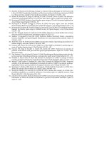
anatomy. TVUS allows evaluation of a complex set of
anatomical structures of the pelvic floor (Fig. 1) [3].
At the external urethral meatus level, the anal canal
will be seen posteriorly in the image, together with
the external anal sphincter (EAS), the internal anal
sphincter (IAS), and often the superficial transverse
perineal muscles within the perineal body in nulli-
para women. Introducing the transducer further in
the cephalad direction (proximal), the ischiopubic
rami, the symphysis pubis, the urethra, the pub-
ourethral ligament, and the pubococcygeus muscle
can be visualized. The puborectalis muscle (PR) will
be seen inferior and lateral to the anal canal, depict-
ing a soft curve upward anterior and lateral to the
vagina, forming almost an ellipsoidal structure
before attaching itself to the inferior side of the sym-
physis pubis. Posteriorly to the anal canal, the
anococcygeal ligament can be identified as a black
triangle in the US image. For transvaginal scanning,
3D US offers significant advantages over convention-
al techniques, in particular if combined with VRM.
Transperineal Ultrasonography
TPUS is a relatively simple technique for assessing
morphologic integrity of both the IAS and EAS [4]. It
is performed with a convex 6-MHz probe placed on
the perineum. Most often, the patient will lie on her
back, with hips flexed and slightly abducted. The left
lateral, sitting, and standing positions are seldom
used. Examination of the anus is made with the
transducer initially applied transversely to the per-
ineal body, identifying the axial view of the anus
using the IAS hypoechoic ring as the landmark in an
image that is similar to that obtained in the mid anal
canal using EAUS. The transducer is then turned 180°
to obtain a longitudinal view of the rectum, with
extension of the hypoechoic IAS appearing above
and below the anal canal in profile. The bright hyper-
echoic elliptical bundle of the PR sling is well demon-
strated.
TPUS offers a dynamic evaluation of the pelvic
130
G.A. Santoro
Fig. 1. Transvaginal ultra-
sonography (TVUS) of
the pelvic floor. Repro-
duced with permission
from [5]
Chapter 11 Imaging of Fecal Incontinence · Invited Commentary
floor [6]. After examination performed at rest, the
patient can be examined during forcible straining
and simulated evacuation so that structures can be
evaluated during action. Observation of the levator
ani (LA) during contraction and on Valsalva may
increase the likelihood of detecting abnormalities of
levator morphology [7–10].
Clinical Application
Anal sphincter defects are a major cause of fecal
incontinence. These defects are often the result of
vaginal delivery [11] or anal surgery (i.e., hemor-
rhoidectomy, sphincterotomy, fistula surgery). Dr.
Maier has provided a comprehensively written and
extensively referenced section on the importance of
EAUS in distinguishing incontinent patients with
intact anal sphincters and those with sphincter
lesions. A limitation of EAUS remains scar identifica-
tion and evaluation of EAS atrophy in patients with
idiopathic fecal incontinence [1].
An advantage of high-resolution 3D EAUS is the
possibility of measuring EAS length, thickness, area,
and volume. The relationship between the radial
angle and longitudinal extent of a sphincter tear can
be assessed and graded. The length of the remaining
intact sphincter muscle can also be evaluated,
improving patient selection for surgical repair of the
anal sphincter complex and helping the surgeon to
judge how far the repair should extend. Volume ren-
dering can be particularly useful in evaluating anal
sphincter lesions [2]. Compared with normal mode,
setting VRM with high opacity, normal thickness,
and high luminance parameters allows better visual-
ization of a rupture of the hyperechoic external
sphincter complex in the anal canal. External sphinc-
ter tear will appear as a low-intensity defect in the
context of the competent, brightest segments of this
striated muscle [2]. To better delineate IAS tears,
VRM should be used with low opacity and normal
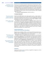
thickness setting. It is also possible to detect EAS
atrophy by using VRM with normal opacity, high
thickness, and high luminance setting to separate
color and intensity data of muscular fibers and fatty
tissue replacement (Fig. 2) [2].
Dr. Maier concentrated most of her chapter on
detecting anal sphincter disruption or atrophy, but it
is increasingly well recognized that many incontinent
women have intact sphincter muscles. In these cases,
LA muscle atrophy or damage is believed to cause the
symptoms [12]. Research demonstrates that the LA is
critically important in supporting the pelvic organs
and maintaining their continence [7–9]. Though
regarded as a single muscle, it is composed of two
functional components: a supportive component
(the iliococcygeus) and a sphincteric component (the
pubococcygeus and the PR). The PR is responsible
for maintaining anorectal junction angulation and
contributes to anal continence. It moves dorsoven-
131
Fig. 2a, b. A 57-year-old woman with a large anterior external anal sphincter (EAS) tear between the 9 and 3 o’clock posi-
tions combined with an internal anal sphincter (IAS) defect between the 7 and 11 o’clock positions as consequence of an
obstetric trauma. Three-dimensional (3D) endoanal ultrasound (EAUS) with normal mode (a). By using volume render
mode (VRM) with normal opacity, high thickness, and high luminance setting, it is also possible to detect EAS atrophy of
the remaining muscular fibers (b). Reproduced with permission from [2]
a
b
trally, narrowing the levator hiatus on straining,
whereas the iliococcygeus moves craniocaudally. LA
damage in women with pelvic floor dysfunction has
been documented using MRI [13–17] or TPUS
[7–10], and the origin of this damage during vaginal
birth has been described [18, 19]. Damage usually
appears in localized regions and more often in the
pubic portion (pubococcygeal and PR) rather than in
the iliococcygeal portion. Lien et al. [20] demonstrat-
ed that the pubococcygeal muscle seen to be injured
is the part of the LA that undergoes the greatest
degree of lengthening during vaginal delivery, sug-
gesting that this injury may be due to rupture of the
muscle from overstretching. Weakness of or damage
to the LA may result in pelvic organ prolapse and uri-
nary or fecal incontinence.
The complex shape and fiber arrangement of the
LA precludes useful measurements of the muscle
being made in standard 2D axial plane. The disad-
vantage of 2D US stems from its inability to easily
disclose the 3D relationships, which may be at the
root of the defects that lead to clinical pelvic floor
pathology. To better understand the specific anatom-
ic defects in women with fecal incontinence, we eval-
uated LA morphology and integrity by using 3D
EAUS and 3D TVUS. Three-dimensional reconstruc-
tion and establishing muscle fascicle direction in 3D
space provides accurate evaluation of LA morpholo-
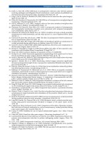
gy. Findings noted in axial sections can be correlated
with findings seen in coronal and longitudinal planes
to confirm the nature and extent of muscle damage
(Fig. 3). In our center, 42 women, 16 with pelvic
organ prolapse and fecal incontinence and 26 asymp-
tomatic volunteers were studied using 3D EAUS and
3D TVUS. Axial, coronal, and longitudinal images
were obtained and the following parameters meas-
ured: levator muscle shape, levator sling arm thick-
ness, levator hiatus width (left-to-right distance), and
length (anterior–posterior distance). Abnormalities
of the pubovisceral portion were determined on each
side and defect severity scored in each muscle from 0
(no defect) to 3 (complete muscle loss). A summed
score for the two sides (0–6) was assigned and
grouped as minor (1–3) or major (4–6) defects. A
summed score of 3 occurring from a unilateral score
of 3 was classified in the major group. In the control
group, bilaterally intact levator sling arms were
observed. In the patient group, ten women (62.5%)
with incontinence and pelvic-organ prolapse showed
PR defects: four had major defects, involving the
right branch in three cases and the left branch in one
case; six presented minor defects of the right branch
(four cases) or left branch (two cases). Lesion site was
more frequently the right branch (seven patients)
than the left branch (three patients). Mean values of
PR right- and left-branch thickness were significant-
ly higher in controls than in patients (9±0.3 mm vs.
7±0.3 mm and 8
±0.6 mm vs. 6±0.2 mm, respec-
tively; P<0.05). Posterior PR thickness was similar in
both groups (7±0.4 mm vs. 7±0.2 mm). Our 3D data
confirm previous reports [13, 14] that levator atro-
phy and structural integrity loss are major cofactors
in female pelvic floor dysfunction.
Conclusions
Ultrasound imaging is becoming the diagnostic stan-
dard in fecal incontinence. Several factors are con-
tributing to its increasing acceptance, the most
132
G.A. Santoro
Fig. 3a, b. Example of a major defect of the right arm of the puborectalis muscle. Axial image (a). Three-dimensional (3D)
reconstruction (b)
a
b
Chapter 11 Imaging of Fecal Incontinence · Invited Commentary
important being the availability of suitable equip-
ment. Recent developments such as high-resolution
3D EAUS with VRM and 3D TVUS and TPUS
enhance the clinical usefulness of the method. It is
hoped that increasing parameter standardization will
make it easier for clinicians and researchers to com-
pare data.
References
1. Santoro GA, Gizzi G (2006) Accuracy and reliability of
endoanal ultrasonography in the evaluation of anal
sphincter injury. In: Santoro GA, Di Falco G. Benign
anorectal diseases. Springer-Verlag Italia, pp 87–98
2. Santoro GA, Fortling B (2007) The advantages of vol-
ume rendering in three-dimensional endosonography
of the anorectum. Dis Colon Rectum 50:359–368
3. Tunn R, Petri E (2003) Introital and transvaginal ultra-
sound as the main tool in the assessment of urogenital
and pelvic floor dysfunction: an imaging panel and
practical approach. Ultrasound Obstet Gynecol
22:205–213
4. Kleinubing H Jr, Jannini JF, Malafaia O et al (2000)
Transperineal ultrasonography: new method to image
the anorectal region. Dis Colon Rectum 43:1572–1574
5. Santoro GA, Di Falco G (2006) Benign Anorectal Dis-
eases. Springer-Verlag Italia
6. Beer-Gabel M, Teshler M, Barzilai N et al (2002)
Dynamic transperineal ultrasound in the diagnosis of
pelvic floor disorders. Pilot study. Dis Colon Rectum
45:239–248
7. Dietz HP (2004) Ultrasound imaging of the pelvic
floor. Part I: two dimensional aspects. Ultrasound
Obstet Gynecol 23:80–92
8. Dietz HP (2004) Ultrasound imaging of the pelvic
floor. Part II: three-dimensional or volume imaging.
Ultrasound Obstet Gynecol 23:615–625
9. Dietz HP, Steensma AB (2005) Posterior compartment
prolapse on two-dimensional and three-dimensional
pelvic floor ultrasound: the distinction between true
rectocele, perineal hypermobility and enterocele.
Ultrasound Obstet Gynecol 26:73–77
10. Dietz HP, Steensma AB, Hastings R (2003) Three-
dimensional ultrasound imaging of the pelvic floor:
the effect of parturition on paravaginal support struc-
tures. Ultrasound Obstet Gynecol 21:589–595
11. Santoro GA, Pellegrini L, Di Falco G (2006) Update in
perineal anatomy and its relevance to obstetric trau-
ma. In: Santoro GA, Di Falco G. Benign anorectal dis-
eases. Springer-Verlag Italia, pp 99–113
12
. DeLancey JOL (2005) The hidden epidemic of pelvic
floor dysfunction: achievable goals for improved pre-
vention and treatment. Am J Obstet Gynecol
192:1488–1495
13. Singh K, Reid WMN, Berger LA (2002) Magnetic reso-
nance imaging of normal levator ani anatomy and
function. Obstet Gynecol 99:433–438
14. Singh K, Jakab M, Reid WMN et al (2003) Three-
dimensional magnetic resonance imaging assessment
of levator ani morphologic features in different grades
of prolapse. Am J Obstet Gynecol 188:910–915
15. Hoyte L, Schierlitz L, Zou K et al (2001) Two and 3-
dimensional MRI comparison of levator ani structure,
volume, and integrity in women with stress inconti-
nence and prolapse. Am J Obstet Gynecol 185:11–19
16. DeLancey JOL, Kearney R, Chou Q et al (2003) The
appearance of levator ani muscle abnormalities in
magnetic resonance images after vaginal delivery.
Obstet Gynecol 101:46–53
17. Chen L, Hsu Y, Ashton-Miller JA, DeLancey JOL
(2006) Measurement of the pubic portion of the leva-
tor ani muscle in women with unilateral defects in 3D
models from MR images. Int J Gynecol Obstet
92:234–241
18. Kearney R, Miller JM, Ashton-Miller JA, DeLancey JOL
(2006) Obstetric factors associated with levator ani
muscle injury after vaginal birth. Obstet Gynecol
107:144–149
19. Kearney R, Sawhney R, DeLancey JOL (2004) Levator
ani muscle anatomy evaluated by origin-insertion
pairs. Obstet Gynecol 104:168–173
20. Lien K-C, Mooney B, DeLancey JOL, Ashton-Miller JA
(2004) Levator ani muscle stretch induced by simulat-
ed vaginal birth. Obstet Gynecol 103:31–40
133
Introduction
Anal continence is assured by the activity of complex
anatomical and physiological structures (anal
sphincters, pelvic floor musculature, rectal curva-
tures, transverse rectal folds, rectal reservoir, rectal
sensation). It is dependent also on numerous other
factors, such as stool consistency, patient’s mental
faculties and mobility, and social convenience. Only
if there is an effective, coordinated integration
between these elements can defecation proceed nor-
mally. On the other hand, fecal incontinence (FI) is
the result of disruption of one or several of these dif-
ferent entities: frequently, it can be due to a multifac-
torial pathogenesis, and in many cases, it is not sec-
ondary to sphincter tears. The disruption could lie in
alterations intrinsic to the anorectal neuromuscular
structures of continence control or be extrinsic to
them, involving extrapelvic control mechanisms. The
primary aim of an effective therapeutic approach
must be the improvement–better, the resolution–of
this distressing condition. Different forms of therapy
are now available so that physicians must select the
best option for each patient. Consequently, the diag-
nostic workup is fundamental to assess, as accurate-
ly as possible, the functional condition of every com-
ponent involved in the continence mechanism and
identify presumed causes of incontinence. In this
regard, some clinicians are very aggressive in using a
variety of tests, whereas others are very minimalist.
This is despite evidence that approximately 20% of
women with FI report a moderate or severe impact
on their quality of life, and 84% of them with poor FI
ask for a physician’s help [1]. Even if there is full
agreement concerning the role played by adequate
data collection of patient history and accurate physi-
cal examination, the importance of each symptom or
sign in the pathophysiologic assessment and in
selecting the appropriate management of each indi-
vidual patient’s FI is still debated. On the other hand,
related to the progressive improvement of knowledge
on continence physiology, several specific instru-
mental tests have been designed for defining the
underlying mechanisms of FI, which are available in
a clinical setting or for investigational purposes.
However, disagreement remains on the choice of
diagnostic procedures and timing.
Clinical Assessment
Investigation of a patient’s history is of utmost
importance. Considering the embarrassment and
reluctance related to FI, it is important to initiate a
positive relationship with the patient. A background
of psychological and emotional suffering is also char-
acteristic of incontinent patients. Moreover, there is
a wide range of personal motivation in searching for
a solution. Some patients have looked for specialists
in this field, perhaps having overcome the lack of
interest or lack of knowledge of general practitioners;
some have become convinced that the problem can-
not be solved. The task of the specialist is to encour-
age patients to undergo clinical assessment and then
to schedule a possible effective treatment.
Maximum efforts must be made to identify symp-
toms of pathogenetic significance and define the type
of FI (urge incontinence, passive incontinence, fecal
soiling, or seepage). However, classification is not
always easy, and an in-depth interview of the patient
is of pivotal importance. It is important to detail
characteristics of normal defecation (occurring with-
out incontinence) and thereafter ascertain the funda-
mental features of the incontinence: timing, dura-
tion, and frequency; type of stool lost; use of pads;
rectoanal sensation during normal defecation and FI
episodes; and influences on health status and quality
of life. These features should be related to possible
events in the patient’s history, including metabolic
and neurological diseases, obstetric and pelvic sur-
gery, neurosurgery, pelvic trauma, chronic inflam-
matory bowel diseases, pelvic irradiation, psychiatric
conditions, and physical and sexual abuse.
The patient interview should effectively address
the physical examination, utilizing all exploratory
and diagnostic techniques necessary to observe phys-
Diagnostic Workup in Incontinent Patients:
An Integrated Approach
Carlo Ratto, Angelo Parello, Lorenza Donisi, Francesco Litta,
Giovanni B. Doglietto
12
ical alterations of the anus, perineum, and pelvis and
to elicit specific reflexes. The checklist shown in
Table 1 could be of help.
Patient’s symptoms and signs should be consid-
ered to classify FI into grades, not only to evaluate
the severity but also to assess the effectiveness of the
therapeutic approach. A number of scales have been
proposed for these purposes, and disagreement
exists on their use; grading systems suggested by the
Cleveland Clinic [2] and Pescatori et al. [3] are some
of the most frequently used.
Another important aspect must be considered: the
patient’s quality of life. This should be considered in
both evaluation of FI severity and treatment assess-
ment. For this parameter also, numerous criteria
have been proposed. Some do not specifically
addressed FI, whereas others do not evaluate the
influence of FI on the general health status of patients
[4–6].
Physiological Investigations
The primary aims of tests used in FI patients are to
better elucidate the pathophysiology and address the
treatment. This is particularly complex, not only due
to the lack of comprehensive knowledge on pelvic
floor morphology and physiology but also because of
the wide variety of tests used, not always as standard
procedures. This assessment must concern both
function [mostly provided by anorectal manometry
(ARM), rectal sensations investigation, and anorectal
electrophysiology (AREP)] and structure [given by
endoanal ultrasound (EAUS) and/or magnetic reso-
nance imaging (MRI)] of all components, pelvic and
extrapelvic, involved in the continence mechanisms.
Due to the multifactorial nature of FI, no one test
alone is sufficient to provide these two types of infor-
mation, and an integration of investigations is need-
ed. When FI occurs with diarrhea, other possible
causes should be explored by endoscopy and stool
tests. As well, when clinical examination suggests
that FI could be secondary to metabolic, neurologi-
cal, or neurosurgical disorders; trauma; bowel
inflammation; irradiation; or psychiatric distur-
bances, specific investigations should be programmed.
Anorectal Manometry and Rectal Sensation
These procedures are usually performed in the same
setting and include the evaluation of rectoanal reflex-
es and rectal compliance. Although they are the most
frequently used diagnostic procedures in proctology,
particularly in FI patients, they are carried out het-
erogeneously because of wide technical variations in
computer software, probes (water perfused or solid
state; uni- or multichannel; difference in number,
location, and shape of openings; difference in loca-
tion and material of balloon), acquisition modality of
pressures (pull through or stationary), and sensa-
tions (inflation of either air or water or using baro-
stat). For these technical differences, it is not possible
to standardize either examination or normal values.
Therefore, it is advisable to establish procedure and
normal values in each laboratory according to age-
and gender-matched healthy subjects [7]. In a study
by Simpson et al. [8], five different manometric pro-
cedures (water-perfused side hole, water-perfused
end hole, microtransducer, microballoon, air-filled
probe) were compared; no significant variations in
anal pressures were found using standard manome-
try techniques, whereas pressures recorded by the
air-filled probe were lower.
In incontinent patients, both resting and squeeze
pressures should be calculated (Fig. 1). The investi-
gator should be very careful to evaluate not only the
136
C. Ratto, A. Parello, L. Donisi, F. Litta, G.B. Doglietto
Table 1. Physical examination of patients with fecal
incontinence (FI)
Examinations Signs
Perianal inspection Skin excoriation/infection
Perianal/perineal scars
Patulous anus
Perineal soiling
Anal ectropion
Hemorrhoidal prolapse
Rectal prolapse
Sphincter deficit
Loss of perineal body
Perineal descent
Fistula
Palpation Pinprick touch
Resting tone
Squeeze tone
Puborectalis at rest, squeezing,
straining
Sphincter deficits
Perianal/perineal scars
Anal/rectal neoplasms
Intussusception
Rectocele
Endoscopy Hemorrhoids
Anal/rectal tumors
Inflammatory bowel disease
Solitary rectal ulcer
Neurological Perianal sensation
Anal reflex
Mental status
Chapter 12 Diagnostic Workup in Incontinent Patients: An Integrated Approach
numeric value (i.e., mean or median) but also to con-
sider pressure profiles, providing information on
asymmetry in the anal canal [due to a limited lesion
of the internal anal sphincter (IAS) or the external
anal sphincter (EAS)] or decreased EAS endurance to
muscle fatigue during prolonged squeeze. Based on a
multichannel acquisition of resting-pressure profile,
it is usually possible to visualize a “vector manome-
try” and identify segments of the anal canal with
increased or decreased pressure (Fig. 2). Following
the routine use of EAUS, clinical utility of vector
manometry has progressively reduced [9], even if,
more recently, an inverted vector manometry has
been suggested, giving good correlations with EAUS
and providing combined functional and anatomic
information [10]. On the other hand, in a number of
incontinent patients, resting and/or squeeze pres-
sures could be normal, related to a nontraumatic
pathophysiology of their incontinence. Although the
rectoanal inhibitory reflex (RAIR) is routinely
evoked (Fig. 3), its meaning in pathophysiological
assessment of FI is not well established. With this
test, the threshold of the reflex and the percentage of
sphincter relaxation, as well as relaxation time and
contraction time, can be calculated. Other reflexes
(coughing) should be elicited to investigate the level
of possible spinal cord lesions. Very important
parameters to be investigated in FI patients are rectal
sensations, commonly studied by inflation of air in a
rectal balloon to elicit threshold and urge sensations,
and maximum tolerated volume. It seems that other
modalities using either electrical or thermal stimula-
tion cannot be standardized at this time [9].
Altered values can be found in FI patients with
metabolic or neurological diseases or following
bowel irradiation, as well as in “idiopathic” FI; how-
ever, in other incontinent patients, rectal sensation
values could be within normal range. Indeed, either a
normosensitive, hypersensitive, or hyposensitive rec-
tum can be found in FI. Despite these different pat-
terns, rectal sensation assessment should be regard-
ed as one of the most useful parameters. In compari-
son with baseline values, variations in rectal sensa-
tion measured under treatment can be of help in the
evaluation of therapeutic effectiveness. Rectal com-
pliance is assessed by progressive inflation (with air
or water, manually or with barostat) of a rectal bal-
loon and registration of rectal pressure; it is defined
137
Fig. 1a, b. Anorectal manometry. a Resting pressure profile
and b squeeze pressure profile in a patient with fecal incon-
tinence (FI) due to a lesion of both internal and external
anal sphincters
a
b
by the ratio of rectal capacity to gradient pressure.
Compliance reduction may cause rectal urgency and
frequent defecation and is usually found in inflamed
rectum (irritable bowel syndrome, ulcerative colitis,
radiation injury), diabetes, or following low spinal
cord lesions. Compliance may be increased in higher
spinal cord lesions.
Endoanal Ultrasound
Specifically designed ultrasound probes and software
are available to investigate the anal canal and rectum
with EAUS. The most useful are those including radi-
al probes with a full 360° field of view and a frequen-
cy range between 5 and 16 MHz. The probe outer
diameter is 1.7 cm or less to minimize any anatomi-
cal distortion. EAUS is usually performed with the
patient in left lateral decubitus position. During the
examination, the probe is inserted into the anal canal
reaching the puborectalis sling showing the U-
shaped aspect. From this level, a manual or mechan-
ical pull-through examination is performed evaluat-
ing the distinct layers and structures of the anal
canal: submucosa, IAS, longitudinal sphincter, EAS,
puborectalis, anococcygeal ligament, puboanalis
muscle, and perineal body (Fig. 4). By convention,
when an axial view is visualized, the anterior edge of
the anal canal should be shown on the screen at 12
o’clock, the left lateral at 3 o’clock, the posterior at 6
o’clock, and the right lateral at 9 o’clock. However, a
more recent EAUS technique allows three-dimen-
sional imaging (3D-EAUS): the 3D structure
138
C. Ratto, A. Parello, L. Donisi, F. Litta, G.B. Doglietto
Fig. 2a, b. Vector manometry in a patient with fecal incontinence (FI) due to lesion of middle-lower internal anal sphinc-
ter, a “standard” vector, b “inverted” vector
Fig. 3. Rectoanal inhibitory reflex (RAIR). R relaxation
time, C contraction time
a
b
Chapter 12 Diagnostic Workup in Incontinent Patients: An Integrated Approach
obtained is the result of numerous axial, rapidly
acquired, two-dimensional (2D) slices. Immediately
after the examination and acquisition of these slices,
the operator is able to navigate inside the 3D struc-
ture observing the anal canal not only in the axial but
also in longitudinal and oblique views (Fig. 5). An
area or volume can be calculated if deemed useful.
Sphincter lesion appears as an hypoechoic area
involving a circumferential segment of the IAS, EAS,
or both (Fig. 6). EAUS is also particularly useful in
differentiating FI patients with and without sphincter
tears. Clinical utility of 3D-EAUS measurement of the
anal sphincter complex in FI patients is under inves-
tigation [11]. Moreover, a “surface render mode”
application is available in the most recently imple-
mented ultrasonographic systems for EAUS (i.e., B-K
Medical Hardware, equipped with 2050 endoprobe).
This image processing allows changing the depth
information of 3D data volume to “see the content
inside a box” and offers accuracy in localizing
sphincter tears.
Anorectal Electrophysiology
AREP includes a few tests directed to patients already
investigated with history and physical assessment
and other procedures (mainly ARM and ultrasound)
in whom pelvic muscular and/or nervous functions
seem to be altered. These tests, used to study the
anorectum, have been derived from myographic and
nerve conduction examinations performed in other
139
Fig. 4a–c. Bidimensional endoanal ultrasound (EAUS): nor-
mal aspect of a upper, b middle, and c lower third of the
anal canal
a
b
c
parts of the body. Since the mid-1980s, an evolution
of instruments, techniques of examination, and indi-
cations has been registered. Electrophysiological
studies are usually carried out with a neuromyograph
system equipped with software dedicated to anorec-
tal physiology to evaluate electrical muscle activity
and nerve functionality. In performing such tests,
either a recording function or an electrostimulating
function or both can be requested. The neuromyo-
graph instrument has to be connected to dedicated
cables and electrodes. A ground electrode soaked in
normal saline is placed around the thigh. The most
preferred patient position is left lateral.
The purpose of electromyography (EMG) is to
investigate the electrical activity of the EAS and the
other striated pelvic floor muscles at rest and during
squeezing and straining. Muscle denervation or rein-
nervation could be found in incontinent patients.
140
C. Ratto, A. Parello, L. Donisi, F. Litta, G.B. Doglietto
Fig. 5. Tridimensional endoanal ultrasound (EAUS): nor-
mal aspect in a longitudinal view
Fig. 6a–c. Endoanal ultrasound (EAUS) in patients with
fecal incontinence (FI) due to a lesion of a internal anal
sphincter, b external anal sphincter, and c both internal and
external anal sphincters
a
b
c
Chapter 12 Diagnostic Workup in Incontinent Patients: An Integrated Approach
Over time, four different types of electrodes have
been developed: concentric needle, monopolar wire,
single fiber, and surface. The concentric needle elec-
trode consists of a thin needle (0.1 mm in diameter)
covered by an insulating resin, which is able to
uptake electrical activity of the small area of the EAS
or puborectalis where it has been inserted under the
guide of digital exploration. This needle is unable to
record single muscle fiber action potentials; record-
ings from the four anal canal quadrants should be
obtained. This procedure is quite uncomfortable for
the patient, and even if multiple recording samples
are taken, the mapping obtained is considered far
from sufficient to delineate accurately the area of
normal and abnormal muscle. The monopolar wire
should reduce discomfort and avoid the electrode
sliding because it is kept in site by a small hook
placed at the electrode tip. The single-fiber electrode
is thinner than the monopolar wire and is able to
record individual motor–unit potentials. An appro-
priate amplification of the signals recorded is neces-
sary. Also, fiber density can be calculated based on 20
different recordings from each anal hemisphere.
Evaluation with single-fiber electrode is more accu-
rate than the two electrodes previously described but
remains uncomfortable. Surface electrodes, mounted
on an endoanal plug or a small external adhesive
plaque, are able to record gross muscle activity but
unable to delimit areas of functional deficit. They are
more useful to study paradoxical contraction of stri-
ated muscles than to evaluate sphincter damage in
incontinent patients. Small polyphasic motor unit
potentials (MUPs) may be identified when myopathic
damage has occurred, whereas large polyphasic MUPs
are found in neurogenic damage; also, a mixed pattern
can be found. This test should be used when a neuro-
genic sphincter weakness is suspected and to distin-
guish selectively disorders of EAS and puborectalis.
Mucosal sensation can be evaluated with electros-
timulation not only in the rectum (as with ARM) but
also in the anal canal using a bipolar ring electrode
(containing two platinum wires 1-cm apart) mount-
ed on a Foley catheter. An appropriate setting of
stimulus duration and rate must be done before
starting the examination. During this test, the elec-
trode is inserted into the anus first. From zero, the
current amplitude is slowly increased until the
patient feels a buzzing or tingling sensation in the
anus. At least three measurements need to be taken,
choosing the lower threshold value for the report. A
similar procedure is used for mucosal sensation
analysis in the rectum. Rectal ampulla must be
reached by the electrode; under slowly increasing
current (parameter setting is different compared
with that used for anal sensation test), three values
should be obtained, taking the lowest as the rectal
threshold sensation to be reported.
Pudendal nerve terminal motor latency (PNTML)
is measured, allowing evaluation of the pelvic floor
neuromuscular integrity (Fig. 7). A disposable St.
141
Fig. 7. Normal pudendal nerve terminal motor latency
(PNTML)
Mark’s pudendal electrode is used, mounted onto
the volar side of the examiner’s gloved index finger.
The index finger is inserted into the rectum, reach-
ing with the fingertip the course of each pudendal
nerve and laying with the proximal finger phalanx
within the anal canal. During this test, both electros-
timulation and recording function have to be acti-
vated. Four cables run within the electrode, convey-
ing stimuli (0.1- or 0.2-ms duration, 1-s. interval, not
exceeding 15 mA) from the machine to the fingertip
(to the anode and cathode) to stimulate the puden-
dal nerve fibers, and from the fingertip to the
machine to record the striated muscle response,
which is visualized on the screen. The latency
(expressed in milliseconds) from the onset of the
stimulus to the first deflection of the response is cal-
culated for each pudendal nerve (n.v.: 2.0±0.2 ms).
Because only the fastest conducting fibers are elicit-
ed during this test, it is possible to find a normal
PNTML value in the presence of pudendal neuropa-
thy, sparing a small amount of conducting fibers.
Imprecise reproducibility and uncertain sensitivity
and specificity are other limits of PNTML.
Evoked potentials can be obtained by stimulating
the cortex or sacral roots to assess the central and
peripheral motor (MEPs) and somatosensory (SEPs)
pathways. Either electrical or magnetic stimulation
can be used, the latter having the advantage of being
painless and able to stimulate deep nervous struc-
tures. Both MEPs and SEPs allow the evaluation of
conduction time of the stimulus (i.e., latency) and
excitability of the intracortical circuit. Sacral MEPs
have been proposed to replace PNTML [12],
although the technical artefacts rate (up to 25%) is
relevant [13–15]. These have been attributed also to
vicinity of recording electrodes to the magnetic field,
and use of an intrarectal ground electrode has been
proposed to minimize artefacts [16]. Evaluation of
SEPs can be performed by application of stimulus to
the rectum, anal canal, anal verge, penis, or clitoris;
this test could be helpful in assessing sensory fiber
lesions, particularly in cases of perineal deficits.
[17–19].
AREP could also include quantification of electri-
cal or thermal sensory thresholds (QSTs) within the
anal canal, sacral anal reflex (SAR) latency measure-
ment in response to pudendal nerve or perianal stim-
ulation, and perianal recording of sympathetic skin
responses (SSRs) [19]. Integration between different
tests can allow a reliable assessment of neuropathy.
Lefaucheur [19] suggests that “needle EMG signs of
sphincter denervation or prolonged TML give evi-
dence for anal motor nerve lesion; SEP/QST or SSR
abnormalities can suggest sensory or autonomic
neuropathy; and in the absence of peripheral nerve
disorder, MEPs, SEPs, SSRs, and SARs can assist in
demonstrating and localizing spinal or supraspinal
disease”.
As mentioned above, indications for AREP are
usually decided on the basis of a patient’s history and
physical assessment if pelvic muscular and/or nerv-
ous disorders are hypothesized; moreover, data from
other diagnostic procedures (mainly ARM and ultra-
sound) should confirm the opportunity to submit the
patient to the AREP.
In patients with sphincter lesion, no electrical
activity may be found in case of wide, complete
replacement of normal muscular tissue with scar, or,
more frequently, polyphasic potentials as signs of a
reinnervation process. Polyphasic potentials do pres-
ent multiple spikes of muscle activity, prolonged in
duration, and an increased fiber density. In evaluat-
ing sphincter injury, EAUS has higher sensitivity and
specificity compared with EMG in mapping the
lesion; however, only EMG can assess neuromuscular
integrity. In this view, these two procedures are com-
plementary to each other.
Evaluation of anal mucosal electrosensitivity
could have a clinical relevance in a few clinical con-
ditions. In neurogenic incontinence, a wide spectrum
of findings can be observed, probably related to the
degree of pudendal neuropathy. Also, rectal sensa-
tion measurements by electrophysiological study are
meaningful. In incontinent patients with sphincter
lesion(s) only, mucosal electrosensitivity could be
normal. In those with neurogenic incontinence, there
could be a wide variability of findings. As concerning
manometric rectal sensation measurement, its mean-
ing has to be intensively interpreted and correlated to
results from other tests.
Alterations of PNTML are identified in relation to
patient’s age, being more frequent in older subjects.
In a large number of patients with FI (with or without
urinary incontinence) and rectal prolapse, the
PNTML is abnormally prolonged. PNTML levels are
thought to have a predicting value in patients under-
going treatment, but this assumption remains con-
troversial.
Defecography and Magnetic Resonance
Defecography is able to assess pelvic floor physiol-
ogy, recording motions at rest and during squeez-
ing, straining, and coughing. The anorectal angle
(ARA) should be calculated. A perineal descent is
frequently found in incontinent patients. More-
over, rectorectal intussusception, rectocele, ente-
rocele, or sigmoidocele may also be diagnosed;
pelvic muscle dyssynergia needs to be adequately
evaluated because it can cause continence distur-
bances [20].
142
C. Ratto, A. Parello, L. Donisi, F. Litta, G.B. Doglietto
Chapter 12 Diagnostic Workup in Incontinent Patients: An Integrated Approach
MRI of anal sphincters has been evaluated using
phased-array coils, but an endoanal coil has been
preferred in studying FI patients [21] because of a
superior accuracy in delimitating the EAS and
sphincter defect; these should be the major advan-
tages of MRI when compared with EAUS. However,
controversy exists about preference toward
endoanal coil [22]. EAS atrophy is more adequately
visualized by MRI than by EAUS, as sphincter thin-
ning occurs due to a decreased amount of muscle
tissue and replacement with fat [23]. However, more
recently, it has been reported that external phased-
array MRI is comparable with endoanal MRI in
depicting EAS atrophy [24]. Endoanal MRI and 3D-
EAUS have a comparable accuracy in detecting atro-
phy and defects of the EAS, even if there is a sub-
stantial difference in grading of external anal
sphincter atrophy [25]. On the other hand, idiopath-
ic IAS degeneration, or IAS atrophy, is better inves-
tigated with EAUS. Terra and Stoker [26], in review-
ing imaging techniques in FI, concluded that both
external phased-array MRI and 3D-EAUS are “valu-
able tools in the diagnostic work up of faecal incon-
tinence. Decisions about the preferred technique
will mainly be determined by availability and local
expertise”.
More recently, use of MRI defecography suggested
[27] to be included in the diagnostic workup of FI
patients to detect previously missed functional alter-
ations of anterior, middle, or posterior pelvic com-
partments. This examination should improve diag-
nosis of rectocele and internal prolapse when com-
pared with clinical evaluation and allow the choice of
a more adequate treatment.
Critical Choice of an Effective Diagnostic Workup
Every kind of examination should contribute to the
diagnosis, offering an interpretation key of the
pathophysiology of a certain disease. Diagnostic
assessment, provided by a panel of clinical and
instrumental tests, should be finalized to plan the
treatment, and those tests should legitimate the ther-
apy chosen. There is evidence concerning the useful-
ness of anorectal testing in the diagnostic workup of
FI: it can add diagnostic information in 19–98% of
patients, influence the management plan in 75–84%
of patients, and alter the management plan in
10–19% of patients compared with clinical assess-
ment alone [28]. Also, a critical evaluation of
cost/effectiveness ratio is of interest. Moreover, post-
treatment reassessment could provide information
on the impact of a particular therapy on the conti-
nence mechanisms. From this perspective, clinical
evaluation and anorectal tests (including those
assessing both structure and function) should be
complementary.
However, correct diagnostic workup is still debat-
ed [29–31]. There is disagreement concerning the
usefulness of instrumental (by ARM) instead of clin-
ical measurement (i.e., digital examination, of anal
resting and squeeze pressures, as well as the primary
role of EAUS in diagnosing sphincter tears), although
there is agreement about the necessity of tests to
assess anorectal sensory functions and possible neu-
ropathy [31].
Use of anorectal tests needs to be performed
scrupulously, and their results must be related to the
entire clinical condition. Of primary importance is
the examiner’s expertise in order to give adequate
indication to a certain test, correct interpretation to
test data, and to visualize imaging of true sphincter
lesions to be distinguished from anatomical asym-
metry of the sphincters. In these conditions, anorec-
tal testing is very well tolerate by most patients with
FI, as demonstrated by Deutekom et al. [32] in a
study evaluating pain, embarrassment, discomfort,
and anxiety in 211 patients with FI undergoing
defecography, MRI, and combined anorectal tests
(including ARM, PNTML, rectal capacity, and sensa-
tion). Those items were classified by Likert scales
(ranging from 1 = none to 5 = extreme). The mean
scores ranged between 1 and 2 for all four items per-
forming all three tests, being MRI, the procedure
with the lowest mean score, and defecography, with
the highest score.
A complete anorectal investigation is justified pri-
marily considering the wide range of possible thera-
peutic options, which include not only regulation of
bowel habits, pelvic floor retraining, and traditional
surgery (i.e., repair of sphincter tears), but also injec-
tion of bulking agents, treatment with sacral nerve
stimulation (SNS), and dynamic graciloplasty or arti-
ficial sphincter. In particular, correct indications to
SNS play a crucial role in obtaining the best results in
this innovative and very effective therapy. Potential
benefits of this therapy seem to be increasing over
the time, covering not only idiopathic neuropathy
but also neuropathy secondary to other diseases or
nervous trauma and, more recently, sphincter lesions,
in the past suitable for sphincteroplasty [33–45].
Hallan et al. [46] found good correlation between
digital basal score and maximum basal pressure and
digital squeeze score and maximum squeeze pres-
sure, even if there were wide ranges of sphincter
function on digital and manometric assessment, with
considerable overlap between patient groups. In that
report, there were similar sensitivities and specifici-
ties of digital scores and ARM in distinguishing con-
tinent and incontinent patients. Agreement exists
about this assumption [29–31]. However, ARM
143
shows higher accuracy in detecting minor abnormal-
ities in anal pressures and increased pressures in
patients with abnormal sphincter relaxation and sub-
sequent fecal seepage [47].
Only minimal attention has been dedicated to
RAIR in FI patients: a possible role of relaxation and
contraction times needs to be elucidated in cases of
mild continence disturbances. Concerning rectal sen-
sations, even if they are frequently found to be altered
(reduced or increased) in FI patients, in other cases,
they can be normal [48, 49]. The assessment of rectal
sensation is preliminary in patients who are possible
candidates for sensory retraining. Indeed, preserva-
tion of rectal sensation before therapy and its
improvement determined by the therapy are suggest-
ed as major determinants of biofeedback success [50].
In some incontinent patients, rectal urgency could be
associated with a hypersensitive rectum and/or
reduced rectal capacity [48, 51] without any sphincter
disruption. In fact, cases with intact sphincters but
presenting a severe/moderate FI need to be investi-
gated for the presence of other possible causes.
EAUS and MRI represent crucial diagnostic tests
in determining which kind of factors plays a major
role in pathophysiology of FI. In particular, both
techniques may detect sphincter defects following
anorectal surgery, even if clinically unsuspected [52].
However, MRI, particularly if dynamic, could give
adjunctive information in selected cases of FI sus-
pected to be associated with perineal descent or rec-
tocele [53]: it is superior to clinical examination and
barium defecography [7, 54].
144
C. Ratto, A. Parello, L. Donisi, F. Litta, G.B. Doglietto
Table 2. A proposed schema of an integrated diagnostic workup in patients with fecal incontinence (FI)
Diagnostic tests: **** mandatory; *** optional; ** on demand; * useless
Type of FI ARM + EAUS AREP MRI Dynamic Barium Others
rectal MRI defeco-
sensory graphy
Obstetric lesions **** **** ***
(PNTML) *** ** **
Iatrogenic lesions **** **** ****
(PNTML) *** * *
Sphincter atrophy **** ****
(only for **** (EMG, **** * *
IAS) sensory, PNTML, EP) (Only for
EAS)
Rectal prolapse **** **** **** (Sensory, *** *** **** Proctoscopy
PNTML)
Rectal resection **** **** **** (Sensory, *** * * Proctoscopy
PNTML)
Pelvic radiotherapy **** **** **** (Sensory, * * * Proctoscopy
PNTML)
Peripheral and **** **** **** (EMG, * * * MRI of CNS
central neuropathies
sensory,
PNTML, EP)
Diabetes and
metabolic diseases **** **** ****
(Sensory, * * * Metabolic
PNTML, EP) assessment
Elderly and **** **** ****
(EMG, Psychiatric
institutionalized
sensory, tests
patients
PNTML, EP) ** *
Congenital **** **** ****
(EMG, * *** **** Urological
PNTML, EP) evaluation
ARM anorectal manometry, EAUS endoanal ultrasound, AREP anorectal electrophysiology, MRI pelvic magnetic resonance imaging,
EMG electromyography, PNTML pudendal nerve terminal motor latency, EP evoked potentials, CNS central nervous system
Chapter 12 Diagnostic Workup in Incontinent Patients: An Integrated Approach
AREP needs to be performed by experts: under
these circumstances, patient compliance is higher
[48] and results more reliable. Clinical utility of EMG
in mapping sphincter lesions has decreased over time
because of the significant reliability of EAUS; howev-
er, it should have a role in cases due to neurogenic
sphincter weakness. PNTML has been a very promis-
ing test, but some peculiar indications in selecting
patients to specific treatments or to predict therapy
outcome have not been supported by recent data [7,
55]. On the contrary, in a retrospective study in FI
patients without sphincter lesions, Ricciardi et al.
[56] found that only a bilateral (but not unilateral)
prolonged PNTML is associated with poorer function
and physiology.
Which Tests in Which Condition?
Because there are now numerous therapeutic
options, it seems justified to intensively evaluate
patients with FI to corroborate the choice. Depend-
ing upon diagnostic tests only could cause inaccurate
pathophysiological assessment and ineffective treat-
ment. The decision process as to which diagnostic
tests should be used in a specific clinical condition is
inevitably related to the specific attitude developed in
a team involved in a patient’s evaluation and cure.
In Table 2, a proposed schema of an integrated
diagnostic workup is presented. From a general point
of view, ARM and rectal sensation assessment should
be considered mandatory in almost every clinical
condition, being widely performed in coloproctolog-
ical laboratories, moderately time consuming, and
allowing considerable useful information. However,
even if ARM could show a pressure pattern of sphinc-
ter asymmetry, it is not enough to diagnose a sphinc-
ter lesion; therefore, integration with other diagnos-
tic tests is mandatory. Rectal sensation assessment
should be useful to eventually identify alterations
due to central or peripheral neuropathy, metabolic
diseases (i.e., diabetes), or radiotherapy given for
pelvic neoplasms (situated at the anus, rectum,
prostate, bladder, or gynecological organs).
Concerning physiological assessment, AREP should
play a crucial role, although its use is rather limited
because specific experience in electrophysiology is
required. EMG performed to map sphincter lesions is
no longer frequently used, but it could be of interest to
visualize denervation or reinnervation patterns in
many clinical conditions (i.e., sphincter atrophy, neu-
ropathies, elderly patients). AREP allows assessment
of both anal and rectal threshold sensations, which
should be mandatory when investigating FI due to rec-
tal prolapse, after rectal resection or irradiation, in
neuropathy and metabolic diseases, and in elderly
patients. PNTML assessment could reveal a pudendal
neuropathy and, then, be useful in a number of FI
cases: in both obstetric and iatrogenic sphincter
lesions, being suggested of importance in choosing
some therapeutic approach (i.e., sphincteroplasty); in
sphincter atrophy; in rectal prolapse or resection; in
irradiated patients; in central/peripheral neuropathies;
in metabolic diseases; and in FI found in either elderly
or pediatric patients. Evoked potentials should com-
plete the AREP evaluation in suspected neuropathies.
In structural assessment of sphincters, there is dis-
cussion concerning the preference toward EAUS
instead of MR, or vice versa, depending on specific
experience in using one test versus the other. In this
debate, it should be considered that EAUS can be
performed by nonradiologists, and it is usually sim-
pler, more available, and less time consuming and
expensive compared with MRI. On the other hand,
MRI needs dedicated personnel with specific experi-
ence. Therefore, even if both EAUS and MRI should
allow similar diagnostic accuracy, in most cases,
EAUS is the preferred mandatory test for imaging,
with MRI being an optional investigation in the more
complex cases. On the contrary, MRI could be used
as a first-line imaging, if chosen. Only for specific
conditions should clinicians prefer one or the other
(i.e., EAUS in suspected IAS atrophy and MRI in sus-
pected EAS atrophy).
Availability of a certain instrumental or diagnostic
procedure is a determinant factor in the diagnostic
process. In some condition, barium defecography
could be the only procedure available to study the
functional imaging in the pelvis, whereas in other
centers, the availability of dynamic MR could allow a
more accurate evaluation. This is the case in FI due to
rectal prolapse or when other pelvic disruptions (i.e.,
rectocele) could have occurred following obstetric
sphincter lesions.
Finally, but not negligibly, other procedures could
be needed to assess specific problems: proctoscopy in
FI due to rectal prolapse (eventual proctitis or soli-
tary ulcer), rectal resection (evaluation of rectal rem-
nant, anastomosis, proctitis), or pelvic irradiation
(assessment of proctitis); central nervous system
MRI in FI cases of suspected central or peripheral
neuropathy; in-depth biochemical assessment in
metabolic diseases; psychiatric and psychometric
tests in FI elderly; and integration of urologic evalu-
ation in any case of double fecal and urologic incon-
tinence, particularly in pediatric patients.
References
1. Bharucha AE, Zinsmeister AR, Locke GR et al (2005)
Prevalence and burden of fecal incontinence: a popu-
145
lation based study in women. Gastroenterology
129:42–49
2. Jorge JM, Wexner SD (1993) Etiology and management
of fecal incontinence. Dis Colon Rectum 36:77–97
3. Pescatori M, Anastasio G, Bottini C (1992) New grad-
ing and scoring for anal incontinence. Evaluation of
335 patients. Dis Colon Rectum 35:482–487
4. Ware JE Jr, Sherbourne CD (1992) The MOS 36-Item
Short-Form Health Survey (SF-36). I. Conceptual
framework and item selection. Med Care 30:473–483
5. Rockwood TH, Church JM, Fleshman JW et al (2000)
Fecal Incontinence Quality of Life Scale: quality of life
instrument for patients with fecal incontinence. Dis
Colon Rectum 43:9–16
6. Donovan J, Bosch R, Gotoh M et al (2005) Symptom
and quality of life assessment. In: Abrams P, Cardozo
L, Khoury S, Wein AJ (eds), Incontinence, vol. 1.
Health Publications, Plymouth, pp 267–316
7. Barnett JL, Hasler WL, Camilleri M (1999) American
Gastroenterological Association medical position
statement on anorectal testing techniques. American
Gastroenterological Association. Gastroenterology
116:732–760
8. Simpson RR, Kennedy ML, Hung Nguyen M et al
(2006) Anal manometry: a comparison of techniques.
Dis Colon Rectum 49:1033–1038
9. Bharucha AE (2006) Update of tests of colon and rectal
structure and function. J Clin Gastroenterol 40:96–103
10. Kaur G, Gardiner A, Duthie GS (2006) A new method
of assessing anal sphincter integrity using inverted
vectormanometry. Dis Colon Rectum 49:1160
–1166
11. West RL, Felt-Bersma RJ, Hansen BE et al (2005) Vol-
ume measurements of the anal sphincter complex in
healthy controls and fecal-incontinent patients with a
three-dimensional reconstruction of endoanal ultra-
sonography images. Dis Colon Rectum 48:540–548
12. Morren GL, Walter S, Lindehammar H et al (2001)
Evaluation of the sacroanal motor pathway by mag-
netic and electric stimulation in patients with fecal
incontinence. Dis Colon Rectum 44:167–172
13. Jost WH, Schimrigk K (1994) A new method to deter-
mine pudendal nerve motor latency and central motor
conduction time to the external anal sphincter. Elec-
troencephalogr Clin Neurophysiol 93:237–239
14. Loening-Baucke V, Read NW, Yamada T, Barker AT
(1994) Evaluation of the motor and sensory compo-
nents of the pudendal nerve. Electroencephalogr Clin
Neurophysiol 93:35–41
15. Sato T, Konishi F, Minakami H et al (2001) Pelvic floor
disturbance after childbirth: vaginal delivery damages
the upper levels of sphincter innervation. Dis Colon
Rectum 44:1155–1161
16. Lefaucheur JP (2005) Intrarectal ground electrode
improves the reliability of motor evoked potentials
recorded in the anal sphincter. Muscle Nerve
32:110–112
17. Jost WH, Loch EG, Muller-Lobeck H (1998) Electro-
physiologic studies of fecal incontinence in the
woman. Zentralbl Gynakol 120:153–159
18. Uher EM, Swash M (1998) Sacral reflexes: physiology
and clinical application. Dis Colon Rectum
41:1165–1177
19. Lefaucheur JP (2006) Neurophysiological testing in
anorectal disorders. Muscle Nerve 33:324–333
20. Rao SS, Ozturk R, Stessman M (2004) Investigation of
the pathophysiology of fecal seepage. Am J Gastroen-
terol 99:2204–2209
21. Hussain SM, Stoker J, Lameris JS (1995) Anal sphinc-
ter complex: endoanal MR imaging of normal anato-
my. Radiology 197:671–677
22. Terra MP, Beets-Tan RG, van Der Hulst VP et al (2005)
Anal sphincter defects in patients with fecal inconti-
nence: endoanal versus external phased-array MR
imaging. Radiology 236:886–895
23. Williams AB, Malouf AJ, Bartram CI et al (2001)
Assessment of external anal sphincter morphology in
idiopathic fecal incontinence with endocoil magnetic
resonance imaging. Dig Dis Sci 46:1466–1471
24. Terra MP, Beets-Tan RG, van der Hulst VP et al (2006)
MRI in evaluating atrophy of the external anal sphinc-
ter in patients with fecal incontinence. AJR Am J
Roentgenol 187:991–999
25. Cazemier M, Terra MP, Stoker J et al (2006) Atrophy
and defects detection of the external anal sphincter:
comparison between three-dimensional anal
endosonography and endoanal magnetic resonance
imaging. Dis Colon Rectum 49:20–27
26. Terra MP, Stoker J (2006) The current role of imaging
techniques in faecal incontinence. Eur Radiol
16:1727–1736
27. Hetzer FH, Andreisek G, Tsagari C et al (2006) MR
defecography in patients with fecal incontinence:
imaging findings and their effect on surgical manage-
ment. Radiology 240:449–457
28. Liberman H, Faria J, Ternent CA et al (2001) A
prospective evaluation of the value of anorectal physi-
ology in the management of fecal incontinence. Dis
Colon Rectum 44:1567–1574
29. Bharucha AE (2006) Pro: Anorectal testing is useful in
fecal incontinence. Am J Gastroenterol 101:2679–2681
30. Wald A (2006) Con: Anorectal manometry and imag-
ing are not necessary in patients with fecal inconti-
nence. Am J Gastroenterol 101:2681–2683
31. Rao SS (
2006) A balancing view: fecal incontinence:
test or treat empirically-which strategy is best? Am J
Gastroenterol 101:2683–2684
32. Deutekom M, Terra MP, Dijkgraaf MG et al (2006)
Patients’ perception of tests in the assessment of faecal
incontinence. Br J Radiol 79:94–100
33. Matzel K, Stadelmaier M, Hohenfellner FP (1995) Elec-
trical stimulation of sacral spinal nerves for treatment
of faecal incontinence. Lancet 346:1124–1127
34. Ganio E, Realis Luc A, Ratto C et al (2005) Sacral nerve
modulation for fecal incontinence: functional results
and assessment of the quality of life. -
orep.it/Rivista%20CEC/sacral_nerve_modulation_for
_feca.htm. Cited 28 Nov 2005
35. Jarrett ME, Varma JS, Duthie GS et al (2004) Sacral
nerve stimulation for faecal incontinence in the UK. Br
J Surg 91:755–761
36. Bernstein AJ, Peters KM (2005) Expanding indications
for neuromodulation. Urol Clin North Am 32:59–63
37. Jarrett ME, Matzel KE, Christiansen J et al (2005)
Sacral nerve stimulation for faecal incontinence in
patients with previous partial spinal injury including
disc prolapse. Br J Surg 92:734–739
38. Kenefick NJ, Vaizey CJ, Nicholls RJ et al (2002) Sacral
nerve stimulation for faecal incontinence due to sys-
146
C. Ratto, A. Parello, L. Donisi, F. Litta, G.B. Doglietto
Chapter 12 Diagnostic Workup in Incontinent Patients: An Integrated Approach
temic sclerosis. Gut 51:881–883
39. Matzel K, Stadelmaier U, Hohenfellner M et al (2001)
Chronic sacral spinal nerve stimulation for faecal
incontinence: long-term results with foramen and cuff
electrodes. Dis Colon Rectum 44:59–66
40. Malouf AJ, Vaizey CJ, Nicholls RJ et al (2000) Perma-
nent sacral nerve stimulation for faecal incontinence.
Ann. Surg 232:143–148
41. Leroi AM, Michot F, Grise P et al (2001) Effect of sacral
nerve stimulation in patients with faecal and urinary
incontinence. Dis Colon Rectum 44:779–789
42. Jarrett ME, Matzel KE, Stosser M et al (2005) Sacral
nerve stimulation for fecal incontinence following sur-
gery for rectal prolapse repair: a multicenter study. Dis
Colon Rectum 48:1243–1248
43. Matzel KE, Stadelmaier U, Bittorf B et al (2002) Bilat-
eral sacral spinal nerve stimulation for fecal inconti-
nence after low anterior rectum resection. Int J Col-
orectal Dis 17:430–434
44. Ratto C, Grillo E, Parello A et al (2005) Sacral neuro-
modulation in treatment of fecal incontinence follow-
ing anterior resection and chemoradiation for rectal
cancer. Dis Colon Rectum 48:1027–1036
45. Jarrett ME, Matzel KE, Stosser M et al (2005) Sacral
nerve stimulation for faecal incontinence following a
rectosigmoid resection for colorectal cancer. Int J Col-
orectal Dis 20:446–451
46. Hallan RI, Marzouk DE, Waldron DJ et al (1989) Com-
parison of digital and manometric assessment of anal
sphincter function. Br J Surg 76:973–975
47. Rao SS, Ozturk R, Stessman M (2004) Investigation of
the pathophysiology of fecal seepage. Am J Gastroen-
terol 99:2204–2209
48. Bharucha AE, Fletcher JG, Harper CM et al (2005
)
Relationship between symptoms and disordered con-
tinence mechanisms in women with idiopathic fecal
incontinence. Gut 54:546–555
49. Sun WM, Donnelly TC, Read NW (1992) Utility of a
combined test of anorectal manometry, electromyog-
raphy, and sensation in determining the mechanism of
‘idiopathic’ faecal incontinence. Gut 33:807–813
50. Chiarioni G, Bassotti G, Stanganini S et al (2002) Sen-
sory retraining is key to biofeedback therapy for
formed stool fecal incontinence. Am J Gastroenterol
97:109–117
51. Siproudhis L, El Abkari M, El Alaoui M et al (2005)
Low rectal volumes in patients suffering from fecal
incontinence: What does it mean? Aliment Pharmacol
Ther 22:989–996
52. Felt-Bersma RJ, van Baren R, Koorevaar M et al (1995)
Unsuspected sphincter defects shown by anal
endosonography after anorectal surgery. A prospec-
tive study. Dis Colon Rectum 38:249–253
53. Stoker J, Halligan S, Bartram CI (2001) Pelvic floor
imaging. Radiology 218:621–641
54. Bharucha AE, Fletcher JG, Seide B et al (2005) Pheno-
typic variation in functional disorders of defecation.
Gastroenterology 128:1199–1210
55. Malouf AJ, Norton CS, Engel AF et al (2000) Long-
term results of overlapping anterior anal-sphincter
repair for obstetric trauma. Lancet 355:260–265
56. Ricciardi R, Mellgren AF, Madoff RD et al (2006) The
utility of pudendal nerve terminal motor latencies in
idiopathic incontinence. Dis Colon Rectum 49:852–857
147
In this era of evidence-based medicine, it is more
important than ever to base clinical decisions on reli-
able information. The dilemma that clinicians face is
how to fully evaluate the patient without performing
unnecessary testing. This decision is particularly dif-
ficult in patients presenting with fecal incontinence
for a number of reasons: the complex nature of con-
tinence, the frequency of concomitant conditions
contributing to the incontinence, and, in general, a
lack of correlation among many of the available tests.
The introduction of multiple new treatment options
with only limited data about their relative roles
makes it even more complicated. The review by Ratto
et al. illustrates some important points regarding the
need to have a focused diagnostic approach to
patients presenting with fecal incontinence.
Normal continence is an intricate process that
involves the coordinated interaction between multi-
ple different neuronal pathways and the pelvic and
perineal musculature [1]. In addition, multiple other
factors, including systemic disease, emotional affect,
bowel motility, stool consistency, evacuation efficien-
cy, pelvic floor stability, and sphincter integrity play a
role in normal regulation [2]. Failure at any level may
result in an impaired ability to control gas or stool.
As pointed out by the authors in Table 1, invalu-
able information can be gathered from physical
examination. At this assessment, it is imperative for
the physician to be able to differentiate normal from
abnormal findings, a skill that can only be garnered
from a complete perineal examination for the many
different types of patients seen by the colorectal sur-
geon, not just those with fecal incontinence. Howev-
er, even a well-performed physical examination has
limitations. Anal inspection and digital rectal exami-
nation are poor for detecting sphincter defects, espe-
cially those less than 90°[3]. Furthermore, specific
questions relating to the overall health and bowel
function of the patient need to be ascertained. How-
ever, even with the most adept history taking and
physical examination, as the authors state, fecal
incontinence often demands further workup with
ancillary studies.
We are in full agreement with the authors in the
value of an integrated approach. The patient present-
ing with fecal incontinence may have a plethora of
concomitant pelvic floor defects that will not only
affect their current function but also impact results
of therapy. Physicians need to have an organized
approach to evaluation to optimally manage these
patients and avoid pitfalls that may ultimately lead to
failed outcomes. In a retrospective review of 21
patients with full-thickness rectal prolapse, 71% were
found to have sphincter defects on ultrasound evalu-
ation, likely contributing to the persistent or recur-
rent fecal incontinence following successful repair of
the rectal prolapse [4]. Similarly, in a study of 28
patients undergoing overlapping sphincteroplasty
who underwent thorough physiologic evaluation, all
patients were found to have associated pelvic floor
disorders, with 57% having more than one abnor-
mality [5]. Further highlighting this point, Hetzer
and colleagues used magnetic resonance (MR)
defecography to demonstrate concomitant pathology
in up to 43% of patients undergoing workup for fecal
incontinence, which resulted in a change in the sur-
gical approach in 27 of 33 patients who underwent
surgery [6]. Therefore, identification of concomitant
pathologies may be a clinically important factor for
optimal treatment outcomes, as pointed out by the
authors of the current study.
Although questions have been raised recently
about the relative contribution of sphincter injuries
to incontinence, sphincter defects are still considered
to play a significant role in the condition. The most
common cause of surgically correctable fecal inconti-
nence is a traumatic injury in the sphincter complex
that can be treated by overlapping sphincteroplasty.
Combined with the increasing success of sacral nerve
stimulation and bulking agents [7–10] for patients
with either a normal pelvic floor or abnormal pelvic
floor anatomy with neuropathy or dyssynergia, it
may be most prudent to first rule out an anatomical
defect. Ancillary tests such as endoanal ultrasound,
MR imaging (MRI), and/or defecography enable
visualization of anatomical abnormalities otherwise
Invited Commentary
Scott R. Steele, Ann C. Lowry, Anders F. Mellgren
Chapter 12 Diagnostic Workup in Incontinent Patients: An Integrate Approach · Invited Commentary
not seen by physical examination alone. The authors
have provided an excellent discussion of the various
modalities of identification of sphincter injuries. We
agree with the authors’ proposed algorithm that
endoanal ultrasound is the most cost-effective, read-
ily available examination for anatomical evaluation.
Despite its frequent use, sphincteroplasty alone
leaves many patients with recurrent symptoms when
reexamined at extended follow-up intervals [11–13].
In contrast to the recent long-term success reported
by Maslekar and colleagues [14] where anterior
sphincteroplasty was deemed successful in 80% of
patients at a median follow-up of 84 months, we pre-
viously reported that only 40% of our 191 patients
undergoing overlapping sphincteroplasty had main-
tenance of continence at long-term follow-up of 10
years [15]. There may be several factors contributing
to a suboptimal outcome after sphincteroplasty,
including weakening of the muscle with time, techni-
cal problems at the original repair, postoperative
infection, failure to achieve a lasting circumferential
sphincter integrity, or unidentified or uncorrected
pelvic floor disorders present at the time of initial
surgery. Atrophy of the external sphincter may also
play a role; if so, MRI may become a more important
part of the preoperative evaluation.
The question remains as to which tests should be
performed in patients with fecal incontinence. Per
the authors’ schema, both anorectal manometry and
anorectal electrophysiology are mandatory in almost
all incontinent patients. Whereas we feel that each of
these procedures provides valuable additional infor-
mation, they do have some drawbacks, as identified
by the authors. Manometry lacks specific correlation
with any anatomical defect as well as the wide varia-
tions in normal pressures with age and gender [16,
17], and manometry has variable efficacy in correlat-
ing with postoperative symptomatic improvement
[18]. Anal electromyography has similarly shown
variable effects on predicting success following
repair, thus limiting its overall use. In a study of 96
patients with fecal incontinence, pudendal neuropa-
thy was only found in 59% [19]. Furthermore, puden-
dal neuropathy, when present, is highly variable for
predicting improvements in continence following
repair [20, 21]. We have previously looked at 83
patients with fecal incontinence with intact sphinc-
ters on ultrasound and no prolapse on defecography
[22]. Only 28% had prolonged pudendal nerve termi-
nal motor latency (PNTML), and unilateral neuropa-
thy did not correlate with manometric or fecal incon-
tinence severity scores, although bilateral neuropa-
thy did correlate with worse scores and decreased
mean resting but not squeeze pressures. Thus, results
of the PNTML are variable. Unfortunately, the pres-
ence of neuropathy may not predict outcome of
repair, and normal PNTML does not exclude prob-
lems with pelvic dysfunction.
In summary, due to the complex nature of the dis-
tal 5–10 cm of the distal alimentary tract and fre-
quency of concomitant conditions, patients with
fecal incontinence cannot be accurately assessed by
one study alone. Availability may be a determining
factor for what studies can be performed. If access is
limited, endoanal ultrasound with the goal of identi-
fying a sphincter injury is the most likely examina-
tion to affect the treatment recommendation. How-
ever, limiting evaluation to one test risks missing
other significant factors that could influence treat-
ment outcome. Although our understanding of
incontinence and appropriate evaluation has
improved, further research is necessary to develop
the most effective and efficient algorithm.
References
1. Bannister JJ, Gibbons C, Read NW (1987) Preservation
of faecal continence during rises in intra-abdominal
pressure: is there a role for the flap-valve? Gut
28:1242–1244
2. Mavrantonis C, Wexner SD (1998) A clinical approach
to fecal incontinence. J Clin Gastrol 27(2):108–121
3. Dobben AC, Terra MP, Deutekom M et al (2006) Anal
inspection and digital rectal examination compared to
anorectal physiology tests and endoanal ultrasonogra-
phy in evaluating fecal incontinence. Int J Colorectal
Dis (Epub ahead of print)
4. Woods R, Voyvodic F, Schloithe AC et al (2003) Anal
sphincter tears in patients with rectal prolapse and
faecal incontinence. Colorectal Dis 5(6):544–548
5. Steele SR, Lee PY, Mullenix PS et al (2006) Is there a
role for concomitant pelvic floor repair in patients
with sphincter defects in the treatment of fecal incon-
tinence? Int J Colorectal Dis 21:508–514
6. Hetzer FH, Andreisek G, Tsagari C et al (2006) MR
defecography in patients with fecal incontinence:
imaging findings and their effect on surgical manage-
ment. Radiology 240(2):449–457
7. Conaghan P, Farouk R (2005) Sacral nerve stimulation
can be successful in patients with ultrasound evidence
of external anal sphincter disruption. Dis Colon Rec-
tum 48(8):1610–1614
8. Rasmussen OO, Buntzen S, Sorensen M et al (2004
Sacral nerve stimulation in fecal incontinence. Dis
Colon Rectum 47(7):1158–1161
9. Tjandra JJ, Lim JF, Hiscock R, Rajendra P (2004)
Injectable silicone biomaterial for fecal incontinence
caused by internal anal sphincter dysfunction is effec-
tive. Dis Colon Rectum 47(12):2138–2146
10. Malouf AJ, Vaizey CJ, Norton CS, Kamm MA (2001)
Internal anal sphincter augmentation for fecal incon-
tinence using injectable silicone biomaterial. Dis
Colon Rectum 44
(4):595–600
11. Barisic GI, Krivokapic ZV, Markovic VA, Popovic MA
(2006) Outcome of overlapping anal sphincter repair
after 3 months and after a mean of 80 months. Int J
149
Colorectal Dis 21(1):52–56
12. Londono-Schimmer EE, Garcia-Guperly R, Nicholls RJ
et al (1994) Overlapping anal sphincter repair for fae-
cal incontinence due to sphincter trauma: five-year
follow-up of functional results. Int J Colorectal Dis
9:100–103
13. Yoshioka K, Keighley MR (1989) Sphincter repair for
fecal incontinence. Dis Colon Rectum 32:39–42
14. Maslekar S, Gardiner AB, Duthie GS (2007) Anterior
anal sphincter repair for fecal incontinence: Good
long-term results are possible. J Am Coll Surg
204(1):40–46
15. Bravo Gutierrez A, Madoff RD, Lowry AC Et al (2004)
Long-term results of anterior sphincteroplasty. Dis
Colon Rectum 47(5):727–731; discussion 731–732
16. Cali RL, Blatchfored GJ, Perry RE et al (1992) Normal
variation in anorectal manometry. Dis Colon Rectum
35:1161–1164
17. Emblem R, Dhaenens G, Stien R et al (1994) The
importance of anal endosonography in the evaluation
of idiopathic fecal incontinence. Dis Colon Rectum
37:42–48
18. Hill K, Fanning S, Fennerty MB, Faigel DO (2006)
Endoanal ultrasound compared to anorectal manome-
try for the evaluation of fecal incontinence: a study of
the effect these tests have on clinical outcome. Dig Dis
Sci 51(2):235–240
19. Roig JV, Villoslada C, Lledo S et al (1995) Prevalence of
pudendal neuropathy in fecal incontinence. Results of
a prospective study. Dis Colon Rectum 38(9):952–958
20. Chen AS, Luchtefeld MA, Senagore AJ et al (
1998)
Pudendal nerve latency. Does it predict outcome of anal
sphincter repair? Dis Colon Rectum 41(8):1005–1009
21. Osterberg A, Graf W, Edebol Eeg-Olofsson K et al
(2000) Results of neurophysiologic evaluation in fecal
incontinence. Dis Colon Rectum 43(9):1256–1261
22. Ricciardi R, Mellgren AF, Madoff RD et al (2006) The
utility of pudendal nerve terminal motor latencies in
idiopathic incontinence. Dis Colon Rectum 49(6)
852–857
150
S.R. Steele, A.C. Lowry, A.F. Mellgren
SECTION III
Treatment of Fecal Incontinence
Introduction
Criteria for patient selection to a certain treatment
are of central importance in the management of fecal
incontinence (FI). Even though the understanding of
continence physiology has improved, there persists a
lack of comprehensive knowledge regarding the very
complex mechanisms by which various structures
contribute to the regulation of continence control. It
is now assumed that a continuous modulation of dif-
ferent stimuli is necessary to effectively maintain the
various functions involved with continence. On the
other hand, the instruments available to measure or
analyze parameters associated with continence,
albeit numerous and sometimes sophisticated, are
not used in a standardized manner, so that data
obtained at one center are not comparable with those
obtained in another. Also, the entire diagnostic
workup is still debatable, being routinely limited to
clinical examination in the opinion of some, whereas
others recommend extensive evaluation. However,
other aspects must be considered in the decision-
making process surrounding treatment choice. These
aspects include the patient’s background and feelings
about his or her FI and the clinician’s attitude toward
proposing a particular treatment. Usually, patients
are very frustrated about their FI, and are sometimes
convinced that it is ineluctable and incurable. Some-
times they are highly motivated to find the right solu-
tion but want to avoid suffering or very complicated
solutions that can significantly alter their lifestyle.
Sometimes they are so depressed about their FI that
they are willing to undergo whichever form of treat-
ment their physician recommends.
Whereas it is necessary for the physician to con-
sider the patient’s perspective when making a treat-
ment decision, it is also necessary to consider the
consequences of treatment, some possibly ineffec-
tive, when deciding upon a first-line approach. For
instance, submitting a patient with very poor conti-
nence to a prolonged period of rehabilitation during
which significant improvement is not seen could
increase the patient’s skepticism to the point of
refusing any other form of therapy. On the other
hand, choosing a complex therapy as the first-line
approach, for example, dynamic graciloplasty or arti-
ficial sphincter, must be based on very strict criteria
in order to avoid submitting the patient to excessive
treatment. Finally, but fundamental in our opinion, it
is unreasonable and irresponsible to offer the patient
a stoma as a unique solution for FI without an appro-
priate diagnosis and a rational therapeutic approach.
Another aspect that influences patient selection to
treatment is the evaluation of results from the appli-
cation of different therapies. These evaluations
should help the identification of specific subgroups
of patients to be recommended for a specific
approach as well as allow assessment of the effective-
ness of a particular therapy. However, there are many
published reports about treatment of patients inho-
mogeneously selected to a therapy. Moreover, the
rapid evolution of new techniques has determined
that in a few years, the same type of patients will be
treated with very different methods. Indeed, treat-
ment selection criteria are rapidly changing parallel
with technical advances. On the other hand, there
remains debate about how to measure the effective-
ness of a treatment for FI: reduction of FI episodes;
positive impact on quality of life; changing of physi-
ological parameters.
Patient Selection
Baseline evaluation of symptoms described by a
patient presenting with FI is fundamental in order to
establish severity of continence dysfunction and its
impact on the patient’s lifestyle. Usually, this can be
derived from a clinical assessment (including clinical
history and physical examination), as well as from
the evaluation of a diary kept by the patient concern-
ing normal bowel movements and episodes of FI,
specifying which kind of material has been lost (gas,
liquid, solid stool). Frequency, circumstances, and
the patient’s sensations and attempts to avoid leak-
age before and during stool passage, are of interest.
Patient Selection and Treatment Evaluation
Carlo Ratto, Angelo Parello, Lorenza Donisi, Francesco Litta,
Giovanni B. Doglietto
13
Clinical features allow the physician to establish an FI
score according to different available scales. There-
after, more information about the pathophysiology
of FI can be gleaned from instrumental examination,
including anorectal manometry (ARM) and sensory
testing, endoanal ultrasound (EAUS), anorectal elec-
trophysiology (AREP), magnetic resonance (MR),
and defecography.
Instrumental assessment is worthwhile because
clinical findings alone are not sufficient to correctly
plan treatment. Ternent et al. [1] found that using
ARM, EAUS, and pudendal nerve terminal motor
latency (PNTML) changes the treatment plan in 20%
of patients. This tendency was confirmed by the same
group of researchers [2] on 90 patients submitted ini-
tially to a clinical evaluation from which a pretest
therapeutic plan was documented (medical for 45
patients, surgical for 45 patients). Thereafter, ARM,
EAUS, and PNTML were performed, and a posttest
management consensus was reached. In 10% (nine
patients), a change of treatment plan was observed
after the physiological tests (from medical to surgical
in five patients; from surgical to medical in three
patients; change of surgical procedure in one
patient). Within patients assigned to a pretest med-
ical treatment, EAUS was the only anorectal test sig-
nificantly responsible of changing the strategy:
among patients indicated for pretest surgical
approach, EAUS and PNTML prompted the varia-
tion. In a similar study performed at our institution
in 2002 involving 63 patients (unpublished data), we
found that clinical features alone were unable to indi-
cate treatment in 25 patients (39.7%), whereas after
ARM, EAUS, and AREP, these “undefined” patients
were assigned to medical treatment in 13 cases and to
surgery in 12 cases. For the entire group of patients,
including those “undefined,” a disagreement
between pretest and posttest treatment plan was
found in 30 cases (47.6%). In five patients (7.9%),
there was a change of pretest approach (from medical
to surgical in two; from surgical to medical in three).
Numerous other studies have supported the integra-
tion of clinical assessment and physiological tests to
improve the understanding of FI and patient treat-
ment selection [3–6].
Medical Treatment
Medical treatment includes diet, drugs, supportive
measures, rehabilitation, and biofeedback. They are
usually chosen for either “elective” reasons or for
patients who cannot be treated by a surgical
approach. Specifically, poor clinical conditions limit-
ing anesthesia and/or surgery could be valid criteria
for a nonoperative approach, whereas a patient’s age
could be only relatively limiting. On the other hand,
psychological problems or disturbances should sug-
gest avoidance of very complex surgical procedures
that require patient compliance. Specific bowel dis-
eases (chronic inflammatory diseases; irritable bowel
syndrome) with uncontrolled symptoms should con-
traindicate a major surgical approach. When life-
threatening clinical conditions are involved (evolv-
ing diseases; chronic diseases; neoplasms not radical-
ly treated), the choice of treatment should consider
the patient’s life expectancy and the possible benefits
in quality of life.
“Elective” indication for medical therapy should
include minor FI without physiologic or morpholog-
ic alterations; in cases with minor abnormalities, a
medical approach could be considered as a first-line
intervention. Also, individuals with continence dys-
functions related to altered feces quality (i.e., diar-
rhea) should be expected to gain benefit from a con-
servative treatment approach. In this area, the
patient must be advised to improve perianal hygiene,
carefully use absorbent cotton diapers and tampons,
and reduce or avoid foods that induce loose stools
and increase gastrointestinal transit and gas produc-
tion (milk derivates; legumes; excess fiber). Diarrhea
needs to be fully investigated and, consequently,
treated with medication when appropriate. Specific
drug treatment has to be initiated in cases of chronic
bowel disease. Also, the pathophysiology of soiling
should be fully elucidated to determine between
operative and nonoperative treatment; when it is
minor, occasional, or without either significant phys-
iologic dysfunction or sphincter lesions, a conserva-
tive approach can be attempted using postevacuation
irrigating water enema or anal plugs as supportive
measures.
Pelvic floor rehabilitation, including biofeedback,
kinesitherapy, sensory retraining, and electrostimu-
lation, is frequently regarded as a first-line treatment
for FI. However, disagreement exists about indica-
tions for rehabilitative techniques. Lack of standard-
ized methods makes it difficult to compare results of
this approach, even in patients accurately selected.
Moreover, in the limited number of well-conducted
studies, there is no agreement concerning outcome
parameters to measure or predict therapy outcome
[7, 8]. A rational modulation of the algorithm for
rehabilitation could play a key role for therapy suc-
cess. Patient compliance and good psychological sta-
tus are preliminary requirements for rehabilitation,
being predictors of therapy success [9, 10]. Selection
criteria cannot be based on anal pressures [11–14],
whereas altered threshold and rectal urgency sensa-
tions have been found to be predictive of a positive
treatment response [7, 14–16]. Sensory retraining
could be used both in individuals with reduced rectal
154
C. Ratto, A. Parello, L. Donisi, F. Litta, G.B. Doglietto
Chapter 13 Patient Selection and Treatment Evaluation
sensation and in patients with very high sensory lev-
els [16–18]. Although controversies exist about the
outcome predictive value of PNTML in individuals
undergoing rehabilitation [7, 8, 13], its alteration
seems to be regarded as a predictor of negative
response [7, 8]. However, an external anal sphincter
defect is not an absolute negative predictor of success
[7, 8, 19]. Biofeedback, electrostimulation, and kine-
sitherapy could be scheduled in patients with such a
defect.
Surgical Treatment
Until the recent past, in cases of intractable severe FI,
criteria for selecting patients to surgical treatment
concerned sphincter lesions or pudendal neuropathy
with perineal descent and altered anorectal angle. In
the former condition, a sphincteroplasty was indicat-
ed in cases of limited lesion without PNTML alter-
ation, whereas a sphincter replacement operation
(dynamic graciloplasty, artificial sphincter, gluteo-
plasty) was indicated when a wide lesion, fragmented
sphincters, or failure of previous sphincteroplasty
occurred. In the latter condition, a postanal repair
was indicated. Recently, other therapies have been
more widely used, such as injectable bulking agents
or the recently introduced radiofrequency. Since
1995, sacral nerve stimulation (SNS) has been intro-
duced into the panorama of treatment options, deter-
mining a significant rearrangement of selection crite-
ria.
Sphincteroplasty
Sphincter lesions due to obstetric trauma (third- and
fourth-degree tears) have traditionally been submit-
ted electively to sphincteroplasty. This technique can
be performed by edge-to-edge approximation or
overlapping of the external anal sphincter (Fig. 1).
Immediate repair, at the time of delivery or delayed
to 24 h, has been suggested to obtain best results.
However, sphincteroplasty can frequently be per-
formed a few decades after childbirth, when the
patient presents clinically with FI. Manometric
parameters (squeeze pressure; resting pressure; anal
canal length) seem not to be useful for patient selec-
tion to sphincteroplasty, whereas a pudendal neu-
ropathy, measured by a prolonged PNTML (particu-
larly if bilateral), should be considered as a predictor
of poor outcome [20–26]. However, conflicting
results are also reported [27–31], attributable to cor-
rect definition of PNTML normality, adequate evalu-
155
Fig. 1a–f. Sphincteroplasty. a Perineal incision. b The external anal sphincter is isolated at the level of a scar. c The external
anal sphincter is incised at the level of the scar. d The overlapping sphincteroplasty is prepared. e Multiple stitches are
placed. f The overlapping sphincteroplasty is completed
a
d
e
f
b
c
ation of pudendal neuropathy when assessed by stan-
dard PNTML measurement with St. Mark’s electrode,
and the role of symmetric pudendal innervation [31].
Although EAUS is determinant today in diagnosing a
sphincter tear, ultrasonographic aspects are not con-
sidered valid criteria to select patients to this proce-
dure. To improve the long-term results displayed by
sphincteroplasty alone, which are sometimes limited
[32–34], this operation has been performed within a
total pelvic floor repair [35] or with anterior levator-
plasty [36]. However, again, anorectal physiological
parameters were not predictive of symptom
improvement.
Postanal Repair
Neuropathic FI associated with perineal descent and
without sphincter lesions seems, theoretically, to be
the best indication to postanal repair. Unfortunately,
no physiological parameters have been found to be
indicative for this approach [37–40]. Considering the
limited long-term effectiveness of this treatment,
patients with these indications could be more effec-
tively approached by other procedures. Indeed, indi-
cations for postanal repair have been significantly
reduced over time. The procedure has been advocat-
ed as part of a total pelvic floor repair in conjunction
with anterior levatorplasty.
Dynamic Graciloplasty, Artificial Bowel Sphincter,
Gluteoplasty
These procedures must be regarded as major sphinc-
ter replacement operations, dedicated only to
patients with very severe FI due to a wide sphincter
lesion (more than half the circumference) or frag-
mented sphincters not amenable to neither sphinc-
teroplasty or other surgical approaches (i.e., SNS). In
case of failure of previous sphincteroplasty (when
there is no indication to redo it), which is not suitable
for SNS, these techniques can also be indicated.
Moreover, if severe FI is consequent to neuropathy or
anorectal malformations, one of these operations
could be performed (specifically, in cases of neu-
ropathy when SNS has failed). Usually, patients pres-
ent a very low or absent squeeze pressure, which is
associated with a decreased or absent resting pres-
sure if an internal sphincter lesion/alteration coex-
ists. When pudendal neuropathy occurs, PNTML
could be altered. Dysfunctions of rectal sensations
should be regarded as negative predictors of success,
as reported in different experiences [41–43]. The
156
C. Ratto, A. Parello, L. Donisi, F. Litta, G.B. Doglietto
Fig. 2a–f. Dynamic graciloplasty, a skin incision, b gracilis muscle is exposed and c isolated, d perianal tunnel is prepared,
e gracilis muscle has been transposed in perianal space and a “gamma” loop is prepared, f the electrostimulator (connect-
ed to the electrodes implanted close to the nerve pedicle of the gracilis muscle) is placed in a subfascial pocket at the level
of the rectum abdominis muscle
abc
d
e
f