Spinal Disorders: Fundamentals of Diagnosis and Treatment Part 5 doc
Bạn đang xem bản rút gọn của tài liệu. Xem và tải ngay bản đầy đủ của tài liệu tại đây (533.48 KB, 10 trang )
bench”or“scamnum” (the Latin expression for traction table) with which
patients were stretched, both horizontally and with underarm and leg distraction
in suspension. In later times, only little progress was made regarding the etiology
and treatment of spinal deformities.
Spinal deformities were
thought to result from
spinal luxation
Even at the end of the Middle Ages, the common belief was that a spinal defor-
mitywascausedbyaspinalluxation.Therefore,suchdeformitieswerecalled
“spina luxata” and the term included every kind of scoliosis and kyphosis. In
1544, the famous Italian surgeon Guido Guidi (1508–1569) proposed treating
such spinal deformities by using the techniques of a traction table as introduced
by Hippocrates and elaborated by Oribasius (325–405
A.D.) [91]. The surgical
textbook Chirurgia `eGraecoinLatinumConuersa, written by Guido Guidi (alias
Vidus Vidius) contains many illustrations depicting different types of extension
machines also known as traction tables [42].
Par ´e (1510– 1590)
introduced a brace
for scoliosis treatment
A less cruel method of treating spinal deformities was developed by Ambroise
Par´e (1510–1590). The father of French surgery also reintroduced the ligature of
vessels. He suggested treating scoliosis by an iron plate brace (
Fig. 4d) [79], which
had to be changed in size during the acceleration phase of child growth at least
every 3 months.
Blount introduced
theMilwaukeebrace
A revolutionary step forward in scoliosis bracing was made by the American
orthopedic surgeon Wa lter Put nam Blount (1900–1992), who was devoted to
scoliosis and its treatment. In 1945, Blount introduced the so-called “Milwaukee
brace”, which is still in use today [7].
Glisson developed
a swing suspension
by the head and armpits
The English physician Francis Glisson (1616–1691), professor of medicine for
over 41 years at Cambridge, wrote extensively on rickets in his pioneering book
On Rickets (De Rachitide, Sive Morbo Puerili, qu i Vulgo The Rickets Dicitur Trac-
tatus) in 1650. He assumed that scoliosis was caused by rickets and that the
pathomechanism was based on the unequal and asymmetric bone growth of the
spine [39]. Therefore, he developed a swing suspension by head and armpits
known as the “English swing”or“Glisson swing”(
Fig. 4e) [39].
Heister’s iron cross
served as a prototype
for later scoliosis braces
Since then, many spinal extension machines have been developed and prop-
agated, for example, the extension chair introduced by the French surgeon
Pierre Dionis (birth date unknown – 1718) in 1707 [30]. In his Cours d’Op´era-
tion de Chirurgie, Pierre Dionis also mentioned for the first time the use of an
iron cross for correcting spinal scoliosis. The cross became well known as Heis-
ter’s cross, because the German surgeon Lorenz Heister (1683–1758) first
depicted the iron cross in his textbook of surgery [49, 50]. Heister’s cross was
used as a kind of scoliosis brace and served as a prototype for later scoliosis
braces (
Fig. 4f
).
The book “Orthopedia”
made Nicholas Andry
the father of modern
orthopedics
In 1741, the French pediatrician Nicholas Andry (1658–1742) published his
epoch-making and pioneering textbook “Orthop´edie” and became the father
ofmodernorthopedics[3].Agreatpartofhisbookdealtwiththedescription
of scoliosis prevention, giving especial attention to sitting and postural habits
and recommending for example physical exercises and a specially designed
chair.
Venel invented a spinal
extension machine
(orthopedic bed)
Influenced by the Enlightenment, the Swiss orthopedic and former obstetri-
cian Jean-Andr´e Venel (1740–1791) founded the world’s first orthopedic hospital
in the small Swiss town of Orbe in 1780. He developed a new treatment regime for
spinal deformities in 1785 [113]. Venel believed that two kinds of procedures
were suitable: first axial extension along the spine and second application of
forces in transverse planes at the region of deviation. Furthermore, he was con-
vinced that the treatment of scoliosis does not tolerate any interruption. Based on
such ideas, he developed a brace for daily activities called an “appareil du jour”
and an orthopedic bed, an extension machine, for the night called an “appareil
de la nuit”(
Fig. 4g, h). Venel’s invention resulted in a hype boom during the fol-
12 Section History of Spinal Disorders
lowing half century and all sorts of different orthopedic beds were developed. In
1829, Johann Friedrich Diefenbach (1792–1847), one of the most important
orthopedic surgeons of the 19th century in Germany, catalogued the various
extension beds and chairs, filling 70 pages [61].
Scoliosis Surgery
Tenotomy and myotomy
was the early but
unsuccessful treatment
for severe scoliosis
In the first half of the 19th century, tenotomy and myotomy were used for severe
scoliosis both because of the prominent paraspinal muscles and the muscle dys-
function theory as outlined above. A very prominent advocate of tenotomy was
the French surgeon Jules Ren´eGu´erin (1801–1886), who developed this tech-
nique in 1835 and treated 1349 patients [41].
After the initial enthusiasm, some terrible outcomes were experienced by
patients and the method was abandoned. It may be of interest that the contro-
versy over this technique was one of the first incidences of doctors criticizing and
attackingeachotherinprintandincourt.
Hibbs performed the first
spinal fusion for scoliosis
In 1911, the American surgeon Russel A. Hibbs (1869–1933) fused the spine
for tuberculosis and suggested extending this method also to scoliosis, as
explained in more detail below [46]. He first performed an in situ fusion in 1914
and later corrected the curve with a cast until fusion had occurred. He gave sev-
eral reports of his technique and advocated a long fusion before the deformity
became severe [53, 54].
After the first successful instrumentations of the spine performed by W. F. Wi l -
kins (1845–1935) [122] and a little bit later by Berthold Ernst Hadra
(1842–1903) [45], many efforts were made to stabilize the spine with instrumen-
tation,e.g.bytheGermanorthopedicsurgeonFritz Lange (1864–1952) [69].
Harrington developed
a milestone spinal
instrumentation system
Finally, however, it was the American orthopedic surgeon Paul Randall Har-
rington (1911–1980) who succeeded in developing an appropriate system for sco-
liosis instrumentation (
Fig. 4i
) [37]. This spinal instrumentation system known as
“Harrington instrumentation” consisted of stainless steel hooks and rods, which
allows the correction of the spinal curvature by distraction (
Fig. 4j
). Harrington
invented this spinal instrumentation system after a severe poliomyelitis epidemic
in the late 1950s. He popularized spinal instrumentation in his milestone paper
Treatment of Sco liosis: Correction and Internal Fixa tion by S pine Instrumentation
published in 1962 [47]. The early technique consisted only of instrumentation.
Fusion was later added because of the initial poor outcome.
Dwyer developed
the first anterior spinal
instrumentation system
Luque introduced segmen-
talspinalcorrection
In 1969, the Australian surgeon Alan Fr ederick Dwyer (1920–1975) intro-
duced the first anterior spinal compression system for scoliosis correction [31].
More than a decade later the Mexican surgeon Eduardo Luque developed a poste-
rior segmental fixation system, which allowed segmental stabilization without
the need for a postoperative cast [74]. In 1984, the French surgeons Yves Cot rel
and Jean Dubousset introduced their posterior derotation system, a system con-
sisting of stainless steel pedicle screws, rods, hooks and transverse traction
Cotrel and Dubousset
introduced the concept
of spinal derotation
devices[22].Bymeansofthissystem,itwaspossiblenotonlytoaddresslateral
deviation of the spine but also apical rotation and thereby improve the sagittal
profile of the spine. Cotrel-Dubousset instrumentation started a new area in spi-
nal surgery.
Juvenile Kyphosis
Scheuermann first described
juvenile kyphosis
The Danish radiologist Holger Werfel Scheuermann (1877–1960), head radiolo-
gist at the Cripple’s Hospital in Denmark, first described juvenile kyphosis in his
thesis which he presented to the University of Copenhagen in 1921. Scheuermann
History of Spinal Disorders Chapter 1 13
reported on a series of 105 adolescent patients (80% males) suffering from a sag-
ittal curvature but with only a minimal coronal deviation [105]. Thus, he postu-
lated a new group of spinal disorder, which begins during puberty and is associ-
ated with a genuine thoracic kyphosis. Initially,his thesis was rejected by the uni-
versity committee. In 1957, he was finally awarded an honorary doctorate in rec-
ognition of his work. Nevertheless the entity became known as Scheuermann’s
disease.
The German pathologist Christian George Schmorl (1891–1932) performed
pathoanatomical studies on more than 5000 spinal specimens which he later
published in his famous book The Human Spine. Schmorl first described the
intercorporal disc prolapses known nowadays as Schmorl’s node [106], which
are frequently seen in juvenile kyphosis.
Spondylolisthesis
An Obstetrical Problem
Herbiniaux described the first
case of spondylolisthesis
Spondylolisthesis must have been observed in ancient times but was probably
first mentioned in 1782 by the Belgian surgeon and obstetrician G. Herbiniaux
(1740 – end of the 18th century). He claimed that it interfered with childbearing
andresultedinthedeathofbothmotherandchild[52].
Kilian coined the term
“spondylolisthesis”
In 1854, Herman Friedrich Kilian (1800–1863) coined the term “spondylolis-
thesis”, which means the “downward gliding of the spine” [64].
In 1882, Franz Ludwig Neugebauer (1856–1914), an obstetrician in Warsaw,
published a monograph on spondylolisthesis in which he described exactly the
clinical features of spondylolisthesis also in relation to obstetrical problems of a
narrowing birth canal in patients with severe spondylolisthesis [89]. In 1976,
Wiltse, Newmann and Macnab were the first to classify spondylolisthesis into
five categories: dysplastic, isthmic, degenerative, traumatic and pathological
types [124].
Surgery
In 1893, Sir William Arbuthnot Lane (1856–1938), who became famous for
introducing the “no touch” or fully instrumental technique of surgery, per-
formed a decompressive laminectomy on a 34-year-old woman who suffered
from progressive gait disturbance, leg weakness and loss of sensation in the lower
limbs. During the operation, he found a forward slipping of the body and neural
arch of L5 on the sacrum without any defect [67].
The first anterior interbody
fusion was performed
by Burns
In this context, the history of the anterior interbody fusion technique should
briefly be reviewed because this surgical technique was first successfully per-
formed in a 14-year-old boy with spondylolisthesis by the English surgeon Burns
in 1933 [14]. Burns’ technique consisted of driving an autologenous tibia dowel
through the fifth lumbar vertebra into the sacrum (
Fig. 5).
Lane and Moore published the first routine series of anterior interbody fusion
in 1948 and shortly after Harmon brought his series to the public in 1950 and
1960 [46, 68]. Since then, many modifications have been made. In the late 1950s,
the American surgeon Humphries and his team first introduced the plate system
for anterior interbody fusion, which consisted of an especially designed com-
pression plate primarily for the lumbosacral joint that was fastened onto the
Hodgson developed an
anterior fusion technique
with bone graft insertion
anterior surface of the vertebra by screw [60]. At the same time, the orthopedic
surgeon Arthur Ralph Hodgson (1915–1993), head of the Orthopedic and
TraumaUnitattheUniversityofHongKong,developedananteriorfusionby
using bone grafts for tuberculosis treatment as explained in more detail below
14 Section History of Spinal Disorders
Figure 5. Spondylolisthesis
Anatomical drawing of the first successful
interbody fusion by B.H. Burns in 1933 [14]
(with Permission from Elsevier).
[58]. In 1936, Jenkins tried to reduce the slip with traction and fusion [63]. Three
decades later, Paul Harrington used his spinal instrumentation system to reduce
severe spondylolisthesis [48].
Back Pain and Sciatica
Not back pain but back
related disability has dra-
matically increased in the
last five decades
Back pain has been known since the start of written history. Probably the first
report of back pain and sciatica can be found in an ancient text, the so-called
Edwin Smith Surgical Papyrus presumably written around 1550
B.C. [10]. The Edwin Smith Surgical
Papyrus first described
back pain (1550
B.C.)
In the industrialized countries, back pain today is the second most common
reason for seeking medical care. Back pain accounts for 15% of all sick leaves and
is the most common cause of disability for persons under 45 years of age. How-
ever, in historical textbooks, only little information is available on backache.
Waddell stated: “At first glance, backache appears to be a problem only since
World War II. At second glance, we realize that not back pain but back related dis-
ability became a medical problem at the end of the last century” [118].
A Wrong Mixture of Fluids
Hippocratic texts first
described sciatica
The first descriptions of spinal pain, called sciatica, are also found in the Hippo-
cratic texts Predictions II (Praedictiones II) [57].
The Predictions are a collection of medical texts concerning especially symp-
toms, course, differential diagnosis and prognosis of a selection of different dis-
eases. It is assumed that the famous Greek physician, Hippocrates of Cos
(460–370
B.C.), the father of the Hippocratic oath, and his scholars contributed to
this ancient medical textbook. Of note, Hippocrates did not differentiate between
symptoms caused by spinal and femoral problems. Both entities were called “sci-
atic” at that time.
The outstanding and important Greek physician Galen of Pergamon (130–
200
A.D.), who became physician to the Emperor Marcus Aurelius (121–180),
described low back pain in his Definition of Medicine (Definitiones Medicae)sim-
ilar to the Hippocratics [36]. Both the Hippocratics and Galen assumed a wrong
History of Spinal Disorders Chapter 1 15
Initially “sciatica” described
hip, buttocks, loin as well
as leg pain
mixtureoffluidstobethecauseofsuchsymptomsaccordingtotheso-called
“fluid doctrine” of Hippocrates. Other ancient physicians had more or less the
same explanation for the sciatic pain syndrome. During antiquity and the Middle
Ages, this view persisted and the term “sciatic” served as a description for hip,
buttocks, loins and leg pain.
The Italian physician Domenico Felice Antonio Cotugno (1736–1822) first
differentiated sciatica from hip related pain in his pioneering study De Ischiade
Nervosa Commentarius (Commentary on Nervous Sciatica) (1764). The nervous
sciatica was called “iscias nervosa Co tunni”alsoknownasthe“malum Co tunni”
or “Cotugno syndrome” (
Fig. 6a) [21]. He was such a skilled clinical examiner he
wasabletodividehisCotugnosyndromeintotwoentities:
anterior “iscias nervosa postica ”
posterior “iscias nervosa antica”
Cotugno first differentiated
nervous sciatica from
musculoskeletal leg pain
The anterior “iscias nervosa postica” was described as pain radiating from the
groin along the inside of the thigh and down the lower leg. The posterior “iscias
nervosa antica” corresponded to pain radiating from the greater trochanter
majoralongtheoutsideofthethighanddownintothelowerleg.Cotugno
thereby became the first author to describe the lumboradicular syndrome.
Brown first assumed neural
irritation to be a cause
of back pain
However, the true cause of the nervous sciatica still remained unknown. He was
still very close to the antique fluid doctrine. Cotugno is also known for his dis-
covery of cerebrospinal fluid as outlined above, his discovery of aqueductus of
the inner ear and his description of the typhoid ulcers. It was finally the English
physician Brown of Glasgow in 1828 who first suggested that irritation of the ner-
vous system could be responsible for back pain [13].
a
b
Figure 6. Back pain and sciatica
a Domenico Felice Antonio Cotugno (1736– 1822). b The Half
Joints of the Human Body published in 1858 by the German
pathologist Hubert von Luschka (1820–1875).
16 Section History of Spinal Disorders
cd
e
Figure 6. (Cont.)
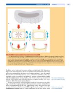
c The illustration depicted in TheHalfJointsoftheHumanBodyshows
a nucleus protrusion of the intervertebral disc between the 12th tho-
racic and 1st lumbar vertebra.
d The drawing shows removal of a so-
called “extradural chondroma” depicted in the paper by Fedor Krause
(1857–1937) and Heinrich O. Oppenheim (1858–1919) in 1908.
e This drawing shows the concept of a disc compressing the cauda
equina as seen by Joel E. Goldthwait (1867– 1961).
Disc Herniation
Luschka (1820–1875) first
described a protruded disc
AfterabriefreportofprotrudeddiscwrittenbythegreatpathologistVirchow
in 1858, the German pathologist Hubert von Luschka (1820–1875) publish-
ed a detailed and concise description and illustration of a protruded disc in
his epoch-making monograph The Half Joints of the Human Body (
Fig. 6b
)
[75].
He supposed that these disc protrusions were caused by a tumor like cartilage
outgrowthofthenucleuspulposusandcalledsuchprotrusionsanomaliesof
intervertebral discs (
Fig. 6c). Notwithstanding Luschka’s descriptions of a subli-
gamentary and intraligamentary outgrowth of a cartilage-gelatinous mass from
the nuclear material with a consecutive transligamentary burst, the effective ori-
gin of these disc protrusions and the clinical link to the sciatica were still unex-
plained for another 70 years. Luschka’s scientific publications and anatomic text-
books became the gold standard of the time because of their clear presentation
and excellent drawings.
Christian George Schmorl (1862–1932), Director of the Pathological Institute
in Dresden, studied more than 5000 spine specimens. In 1928, he published two
History of Spinal Disorders Chapter 1 17
cases of disc protrusion, which he interpreted as supplementary nuclei pulposi,
remnants of the primitive chorda, respectively.
Andrea first proposed
a degenerative origin
of disc protrusion
Finally, in 1929, it was a disciple of Schmorl, Rudolf Andrae, who gave the
accurate explanation for the disc protrusion. In his work On Cartilage Node in the
Posterior End o f Intervertebral Disc Near by the Spinal Canal,Andraeconfirmed
Schmorl’s observations by describing 56 similar cases in 365 examined spines.
Furthermore, he proposed that disc protrusion is based on a degenerative dis-
ruption of annular fibers which permits extrusion or sequestration of nuclear
material. In addition he could exclude the theory of a neoplastic process as cause
for disc protrusion [2]. Even though the pathophysiological mechanism was elu-
cidated, there was no link to the clinical symptom of sciatica.
Krause and Oppenheim
(1958–1919) first performed
a discectomy
With the advent of neurotopic diagnosis using dermatomes at the end of the
19th century, specific operative intervention for the spine and spinal cord
became possible. On 23 December 1908, the German surgeon Fedor Krause
(1857–1937), who worked at the Augusta Hospital in Berlin together with the
German neurologist Heinrich O. Oppenheim (1858–1919), was the first to oper-
ate on a disc prolapse in a patient who had suffered from severe sciatic pain for
several years and had developed an acute cauda equina syndrome [90]. The
operation (
Fig. 6d)consistedof:
laminectomy L2–L4
splitting the dura
mobilizing the cauda equina by a retractor
exploring the operation field
removing a small tumor mass
After the operation, the patient felt much better and the neurological problems
disappeared. Following the theory of Luschka, Krause and Oppenheim supposed
that this fibrocartilage mass was an enchondroma.
Goldthwait first proposed
that sciatica is caused
by a disc prolapse
In 1911, the American physician Joel E. Goldthwait (1866–1961) reported on
a 39-year-old patient who initially suffered from an affection of the sacroiliac
joint. The patient underwent inadequate manipulations and subsequently
developed a cauda equina syndrome. Based on this case, he proposed that a
prolapse of the intervertebral disc could be an explanation for many cases of
lumbago, sciatica and paraplegia (
Fig. 6e
) [40]. At the same time, the physicians
George S. Middleton (1853–1928) and John H. Teacher (1869–1930) reported a
case of a laborer who had sustained a disabling injury during work while lifting
a heavy object [74, 85]. The patient suffered from sciatica and paraplegia. The
authors suggested that a disc rupture caused the severe clinical condition of that
patient.
Disc Surgery
In 1929, the famous Walter E. Dandy (1886–1946), professor of neurosurgery at
JohnsHopkins,discoveredthatnodulesofdiscalorigincouldproducesciatica
by compression and that their removal would cure pain. He published this
hypothesisintheArchives of Surgery [25], but unfortunately little attention
waspaidtothisarticle,becausehecalledtheprotrusionsandprolapsestu-
mors. However, it was not until 1934 that the American neurosurgeon William
Jason Mixter (1880–1958) and the orthopedic surgeon Joseph Seaton Barr
(1901–1963), working at the Massachusetts General Hospital, established that
the supposed neoplastic process was just a prolapse of the disc (
Historical Case
Study
).
Mixter and Barr established
the link between disc
prolapse and sciatica
They also discovered the long missing link between sciatica and disc protru-
sion [86].
18 Section History of Spinal Disorders
a
Historical Case Study
The following text represents a short extract of the milestone article “Rupture of the intervertebral disc with involvement
of the spine canal” (
a) (Massachusetts Medical Society, with permission): written by William Jason Mixter (b) and Joseph
Seaton Barr (
c) in 1934 [86]:
“The symptoms and signs of these so-called chondromata, which we believe in most instances represent rupture of the
intervertebral disc, have been discussed at length by Elsberg and Stookey. The symptoms depend entirely on the loca-
tion and size of the lesion. There is often a history of trauma not immediately related to the present condition. Numbness
and tingling, anaesthesia, partial or complete loss of power of locomotion, are usually present. Bladder and rectal sphinc-
ter may be involved. The condition of the reflexes varies with the level of the lesion. If it is compressing the cauda equina
the tendon reflexes may be absent; if higher, compressing the cord, the legs may be spastic and the reflexes exaggerated
with positive Babinski sign. If the lesion is low in the spine, the physical examination may be suggestive of low back strain
or sacro-iliac strain. X-ray examination may be entirely negative, but narrowing of the intervertebral space is often pre-
sent and is of significance, as it ordinarily means escape of the nucleus pulposus, not necessarily but possibly into the spi-
nal canal Therefore we have developed certain ideas as to the operation when we suspect this lesion to be present.
History of Spinal Disorders Chapter 1 19
bc
Historical Case Study (Cont.)
Exposure of the spine and laminectomy are performed as usual except that the laminectomy is narrow and on the side
where the lesion is suspected, for we believe that a ruptured disc is a weakened disc and the strength of the spine should
be preserved as much as possible. The dura is opened and the spinal canal carefully explored, particular attention being
given to the intervertebral discs in front of the cord and the intervertebral foramina. If the lesion is found in the midline
it is approached by incising the dura over it as suggested by Elsberg. If it is lateral, the dura is closed and the dissection
carried out to the side between the dura and the bone. If lesion is suspected in the intervertebral foramen it may be nec-
essary to carry the removal of bone well out to the side, even taking in part of the pedicle. After removal the tumor is
exposed. It frequently comes away without any dissection and if not, section across its base or removal with curette is
bloodless. Though we have done it in only two cases, we believe that it may be advisable to slip bone chips in between
the stumps of the laminae before closing the wound, in order to facilitate fusion. After removal of the tor piece of the disc
one frequently finds an opening through which a probe may be passed into the nucleus pulposus We conclude from
this study: a that herniation of the nucleus pulposus into the spinal canal, or as we prefer to call it, rupture of the interver-
tebral disc, is a not uncommon cause of symptoms. That the lesion frequently has been mistaken for cartilaginous neo-
plasm arising from the intervertebral disc That the treatment of this disease is surgical and that the results obtained are
very satisfactory if compression has not been too prolonged.”
This finding rapidly attracted surgeons and basic researchers to the interverte-
bral disc. The enthusiasm to solve back pain and sciatica surgically by disc exci-
sion started as Macnab called it “the dynasty of disc” [77]. The disc was thereaf-
termaderesponsibleforallkindsofbackandlegpainandmanytreatmentfail-
ures were the consequence.
Love developed the
interlaminar “key hole”
approach for discectomy
In the early days, the disc prolapse was removed by a full transdural approach
with laminectomy. In 1939, Grafton Lo ve, a surgeon at the Mayo Clinic, published
a new method which he called “key hole” laminectomy, an intralaminar approach
for disc prolapse removal, which preserved spinal stability. Therefore, his ap-
proach served also as a precursor to the microscopically assisted approach [73].
Lyman Smith introduced
chemonucleolysis
for disc prolapses
The American physician Lyman Smith developed a less invasive method for
disc protrusions and reported his results in 1964 [109]. He injected chymopapain
into the disc to shrink the disc protrusion. Although chemonucleolysis was effec-
tive, this method went out of fashion because of some cases of anaphylactic reac-
tion and transverse myelitis.
Caspar and Williams
introduced microdiscectomy
In 1975, Hijkata of Japan first reported on a percutaneous lumbar nucleotomy
technique by a posterolateral approach [35]. In the late 1970s, the German neuro-
surgeon Caspar and the American neurosurgeon Williams introduced the use of
the microscope for minimally invasive discectomy, which today has become the
standard technique in many centers [17, 123].
In 1986, P.W. A s c h er performed the first percutaneous laser decompression of
intervertebral discs [14], but this technique never demonstrated clinical efficacy.
20 Section History of Spinal Disorders
U. Fernström implanted
the first lumbar disc
prothesis
A further milestone in the treatment of degenerative disc disease was the devel-
opment of an artificial disc, which allowed lumbar motion to be preserved. U.
Fernström first implanted a rudimentary lumbar disc replacement consisting of
a single steel ball in the late 1950s [34].
After several less promising developments of different designs, K. Schellnack
and K. Büttner-Janz developed the SB Charite disc prothesis at the Charit´e(Hos-
pital) in Berlin in the early 1980s [15]. Further developments of this prothesis
type resulted in the first FDA approved total disc arthroplasty device.
The Facet Syndrome
It was the Belgian anatomist Andreas Vesalius (1514–1564), professor of anat-
omy at the University of Padua, who first correctly described the facet joint in his
epoch-making anatomical textbook De H u mani Corporis Fabrica Libri Septi in
1543 [116]. The American Joel E. Goldthwait (1867–1961), first surgeon-in-chief
of the Orthopedic Department at the Massachusetts General Hospital, first real-
ized that the facet joints also play an important role in low back pain [40]. Finally,
Ghormley coined the term
“facet syndrome”
in 1933, R.K. Ghormley is credited as having coined the term “facet sy ndrome”
for back pain caused by altered facet joints [38]. This syndrome was re-popular-
ized by Vert Mooney in 1976 [87], but debate continues about the clinical entity.
Spinal Stenosis
Portal made the first
description of spinal
stenosis in 1803
The first evidence of spinal stenosis can be found in Egyptian mummies. The first
report of a spinal stenosis is attributed to the French surgeon Antoine Portal
(1742–1832) in 1803. He observed at autopsy three specimens with narrowing of
the spinal canal [93]. He was also able to relate the pathological findings to the
typical clinical symptoms of spinal stenosis.
Vittorio Putti was the first
to report the relevance
of foraminal stenosis
The Italian orthopedic surgeon Vittori Putti (1880–1940), one of the most
outstanding European orthopedic surgeons of the first half of the 20th century,
emphasized the relevance of anomalies or acquired degenerative alterations of
a b
Figure 7. Spinal stenosis
a Vittorio Putti (1880–1940). b Henk Verbiest (1909 –1997).
History of Spinal Disorders Chapter 1 21


