Primary Care of Musculoskeletal Problems in the Outpatient Setting - part 3 pptx
Bạn đang xem bản rút gọn của tài liệu. Xem và tải ngay bản đầy đủ của tài liệu tại đây (618.2 KB, 35 trang )
ligament. The deformity, not present in grade 1 injury, is present in grade
2 and 3 injury. The deformity may not be obvious initially because of the
edema from the trauma. Palpation usually reveals tenderness over the AC
joint. The crossover test (see Figure 5.12) is positive. This test is performed by
asking the patient to abduct the painful shoulder to 90° and then adduct the
shoulder by attempting to touch his uninjured shoulder with the hand of the
injured side. Once the patient has touched the opposite side the examiner
pushes down on the elbow of the affected side while the patient resists. With
grade 1 injuries, like the patient above, the crossover maneuver is possible and
the patient can resist downward pressure on the elbow but there will be sig-
nificant pain. With grade 2 injuries the patient can do the crossover maneu-
ver but is not be able to actively resist any force you apply. With grade
3 injuries it is very painful to attempt the crossover test and they usually are
unable to complete the task.
4.3. Imaging
Most diagnoses can be made with a thorough history and physical examina-
tion. If a fracture is suspected plain film X-rays of the shoulder are usually
sufficient to make the diagnosis. Magnetic resonance imaging (MRI) would
not be indicated unless other injuries are suspected.
68 E.J. Shahady, J. Buseman, and A. Nordgren
FIGURE 5.20. Grade 3 tear. (Reproduced from Shahady E, Petrizzi M, eds. Sports
Medicine for Coaches and Trainers. Chapel Hill, NC: University of North Carolina
Press; 1991:52, with permission.)
4.4. Treatment
For grade 1 to 3 AC injuries, ice and NSAIDs are the mainstay of initial
treatment. Most AC separations will heal without surgical intervention.
A sling is recommended until the pain subsides. The pain lasts for 1 to 6 weeks
depending on the grade of separation. Within a few days, initiate a rehabili-
tation program in order to restore a full ROM. The patient is asked to remove
the arm from the sling and slowly begin to make circular motions. The circle
of the motion is gradually increased as tolerated. The time needed for com-
plete healing varies from 1 week to up to 12 weeks depending on the grade of
the tear. Resume full activity when the patient has a full ROM and there is no
tenderness in the AC joint region on palpation. Treatment by a physical ther-
apist may be needed in grade 2 and 3 injuries.
It is important to tell the patient that the deformity or lump will not go
away but it does not lead to a decrease in ROM or function. For some
patients the cosmetic result is not acceptable and they wish to have surgery.
Surgery for grade 3 injuries may also be indicated for elite athletes or labor-
ers who are dependent on a more speedy recovery and a more stable AC joint
that can endure significant stress earlier in the recovery process. Consultation
with an orthopedic surgeon will help with this decision.
5. Acromioclavicular Joint Arthritis
In some patients who sustained a grade 1 injury at a younger age the symptoms
of AC joint discomfort may return. The usual time period is 15 to 20 years after
the initial injury. The patient usually has forgotten about the initial injury and
complains of the gradual onset of anterior superior shoulder pain that is made
worse with abduction and adduction of the shoulder. The patient may have
been treated unsuccessfully for other diagnosis before this one is considered.
The examination will be negative for rotator cuff disease. Tenderness is present
over the AC joint and the crossover test is positive similar to a grade 1 injury.
Over 50% of these patients respond to an injection of lidocaine and a steroid
into the joint and the shoulder strengthening exercises described at the end of
the chapter. If there is no response to injections, consultation with an orthope-
dic surgeon for possible surgery should be considered.
6. Fractured Proximal Humeral Head
Humeral head fractures can occur in patients over 55 who fall on their out-
stretched arm. Falling on the outstretched arm is a mechanism of injury for
several fractures and/or soft tissue injuries. Injuries more common in younger
patients include rotator cuff tear, AC joint separation, and fractures of the
scaphoid, radius, and ulna. Fractured radial head in the elbow and fractured
5. Shoulder Problems 69
humeral head in the shoulder are more common in older patients. Be alert for
the possibility of more than one injury occurring with this type of fall. The
primary care clinician can treat many of the proximal humerus fractures.
Once the diagnosis of fracture of the proximal humerus is suspected, a
neurologic and vascular evaluation of the upper extremity should be con-
ducted. Injuries to the axillary nerve and brachial plexus as well as the axil-
lary and brachial artery are rare but possible. A good radial pulse and no
sensory or motor loss of the deltoid region and the lower arm will rule out
these possibilities.
X-rays help not only with the diagnoses but also with decisions about
treatment. Because of the insertion of the rotator cuff tendons, the proximal
head of the humerus generally fractures along four predictable cleavage lines.
Regardless of the number of fragments, proximal humerus fractures are clas-
sified by the displacement and degree of angulation. Neer 1 fractures have no
more than 1-cm displacement of any fragment and no more than 45° of angu-
lation. More than 85% of proximal humerus fractures are nondisplaced Neer 1
and can be treated nonoperatively. The radiologist should help with diagnosing
the degree of displacement and angulation. Any fracture that is open or associ-
ated with neurological or vascular deficit requires referral.
Treatment of Neer 1-part fractures includes a sling for comfort and early
ROM exercises (about 5 to 10 days after the injury). Patients should begin
with pendulum exercises with the injured arm out of the sling. They perform
this movement by bending at the waist, allowing the arm to fall toward the
floor, and rotating it in a circle. With time, the size of the circle is increased
and the sling removed during the exercise (see Fig. 5.30).
Early movement is important to reduce residual stiffness and deformities.
Two weeks following the injury start the following:
1. Abduct the shoulder by progressively walking the fingers up the wall.
2. Internally rotate by placing the hand of the fractured shoulder behind the
back and progressively move up the back.
3. Increase elbow ROM by flexing and extending the elbow when it is out of
the sling.
Discontinue the sling gradually after 4 to 6 weeks. Physical therapy refer-
ral may be helpful if the patient is having difficulty with achieving the
exercises. This is especially true in the elderly.
7. Case
7.1. History
A 16-year-old male football player presents to your office directly from football
practice complaining of left shoulder pain that began after attempting a tackle
with the left arm. His past health is excellent and he has no past shoulder
problems or a family history of shoulder problems. Upon examination, he is in
70 E.J. Shahady, J. Buseman, and A. Nordgren
acute distress with shoulder pain. He is holding his left arm close to his
abdomen to protect it from movement. He thinks he heard his shoulder pop
when he was making the tackle and has been in extreme pain since that time.
On observation of the left shoulder, with his shirt removed, the lateral shoul-
der looks square compared with the roundness of the right shoulder. A bulge
is present below the distal clavicle. There is no deformity or tenderness over the
AC joint or the clavicle. The bulge is tender and there is an empty space under
the acromion laterally. He resists any attempt to move the arm away from the
abdomen, and attempts to externally rotate or abduct the shoulder are very
painful. There is no sensory loss over the deltoid region or any part of the
shoulder and arm. He has good pulses and no loss of color in the extremity.
7.2. Thinking Process
Think of how the injury occurred. Preparing for a tackle the patient inter-
nally rotated both shoulders in order to grab the runner. The runner over-
powered his internal rotation grasp and forced the shoulder into external
rotation. The most likely injury given the mixture of forces would be an ante-
rior dislocation or subluxation of the humeral head outside the glenoid
fossae. Other injuries still need to be considered. This is not the usual mecha-
nism of injury for AC joint separation and clavicle fracture but palpation of
those areas is important to rule out these possibilities. As noted previously,
there was no deformity or tenderness over the AC joint or the clavicle so these
diagnosis are not likely. Subluxation is probably ruled out by the lack of his-
tory of a prior shoulder injury. The physical examination will be the same for
both a subluxation and a dislocation. The remaining parts of the examination
are classical for a dislocation. Refusing to move the arm, a square shoulder, a
tender bulge, and emptiness where the humeral head should be are all charac-
teristic of both dislocation and subluxation. As there is no history of recurrent
subluxation the diagnosis of dislocation is most likely in the presence of this
type of trauma. The diagnosis or dislocation was made and the shoulder was
reduced using an active countertraction force as noted in Figure 5.21. After the
reduction, an X-ray of the shoulder was performed and it revealed no evidence
of fracture. A 6-week rehabilitation program was prescribed and the patient
was able to return to his usual activities following the rehabilitation.
8. Glenohumeral Joint Dislocations
The glenohumeral joint lies between the glenoid fossa and the humeral head.
The flat surface of the glenoid provides no bony stability like that provided
by the acetabulum of the hip for the head of the femur. The stability of the
joint is dependent upon soft tissue structures like the glenoid labrum, gleno-
humeral ligaments, and rotator cuff muscles. Injury to any of these soft tissue
structures makes the joint susceptible to dislocation, instability, and/or
subluxation. The relative lack of stability makes the joint one of the most
5. Shoulder Problems 71
commonly dislocated joints. Ninety percent of shoulder dislocations are
anterior, with the rest being posterior. Inferior dislocations are rare. In
younger patients, most shoulder dislocations are caused by sports injuries
whereas falls are the usual cause in the elderly. For anterior dislocations, the
mechanism of injury is an excessive external rotation or abduction force,
while posterior dislocations usually occur when the humeral head is driven
posteriorly.
The usual symptoms are immediate pain and an unwillingness to move the
affected arm. The patient tends to cradle the affected arm with the other arm.
Inspection reveals a square shoulder, a bulge where the humeral head now
rests, and emptiness beneath the acromion. Before considering reduction, a
neurovascular evaluation should be done. After comparing the radial pulses
assess for axillary nerve deficit. Axillary nerve deficit is the most common neu-
rologic deficit associated with shoulder dislocation. Contraction of the deltoid
is not possible when an axillary nerve deficit is present. To test for deltoid
function, place a hand on the patient’s elbow while the arm is at the patient’s
side. Ask him or her to gently abduct the shoulder while you resist the attempt
to do so. If there is no nerve injury, you will feel the deltoid contract.
8.1. Imaging
Order standard three-view X-rays to rule out humeral fractures. Obtain the
X-rays after the reduction unless there is an open fracture or a neurologic
deficit. Delaying reduction makes relocation more difficult.
72 E.J. Shahady, J. Buseman, and A. Nordgren
FIGURE 5.21. Relocating a shoulder dislocation by countertraction in a chair.
8.2. Treatment
Muscle spasm sets in shortly after dislocation, making reduction more diffi-
cult. The quicker the reduction is performed the easier it is for the patient and
the clinician. Early reduction also requires less force and provides dramatic
relief from pain. Numerous reduction techniques can be used, for example,
the self-reduction technique (Figure 5.22) in which the patient interlaces his
or her fingers and places them around the flexed knee on the same side of the
dislocation. The patient then leans backward, and the reduction occurs. This
technique works well for recurrent subluxations. In the gravity method, the
patient is placed prone with the affected shoulder supported and the arm
hanging over the examination table, bench, or training room table with a
weight attached to the hand. A weight of 5 lb usually is sufficient (Figure
5.23). Gravity stretches the muscle spasm and reduction occurs. The trac-
tion–countertraction method has the patient sitting on a chair or a bench and
leaning forward as an assistant places his arms around the patient’s torso to
provide countertraction (see Figure 5.21). Gentle but steady pressure is then
applied to the affected arm as the countertraction is increased. The pull is
gradually increased until the shoulder relocates. There is an older method
known as the hippocratic method. In this technique, the clinician places a
foot in the axilla to apply countertraction while providing traction on the
5. Shoulder Problems 73
FIGURE 5.22. Self-reduction of a dislocation of the shoulder.
affected arm. Potential neurovascular damage to the axillary area has led to
this method being abandoned.
For all attempted reductions, applying ice to reduce discomfort and reas-
surance to reduce anxiety aid the process. Reduction is more difficult for
patients that are more muscular and when the reduction is delayed for greater
than 20 min. It may be necessary to use an injectable narcotic and/or an anx-
iolytic if the above methods do not initially work. This of course will require
a setting that can provide appropriate monitoring. On rare occasions, general
anesthesia is needed for reduction.
It is essential to advice patients younger than 20 to wait 6 weeks before they
return to any activity that may lead to stressful combined shoulder abduction
and external rotation. Contact sports as well as some work-related activities
may produce this type of stress. These patients may feel capable of resuming
participation after 2 to 3 weeks, and they may seek clearance from their pri-
mary care clinician. It is important that the 6-week rule be adhered to in this
younger age group to decrease the incidence of repeated dislocations. In older
patients, the time required for recovery is less. A rule of thumb is 5 weeks for
20- to 30-year-olds, 4 weeks for 30- to 40-year-olds, and 3 weeks for 40- to
50-year-olds. For patients older than 50 years, the shoulder should be mobi-
lized as soon as symptoms permit (similar to the fractured proximal humerus).
Rehabilitation should be handled by an experienced physical therapist that
74 E.J. Shahady, J. Buseman, and A. Nordgren
FIGURE 5.23. Gravity reduction of the shoulder.
will help motivate the patient to adhere to their exercises. The primary care
clinician also needs to help with this motivation. Surgery can be an option
initially but the majority opinion is to attempt nonsurgical treatment first and
reserve surgery for the patient who fails conservative management because of
recurrent dislocations. If the patient experiences recurrent dislocations, imag-
ing studies should be done to access for indications that the subluxations have
damaged parts of the glenoid or the humerus. Defects in the anterior inferior
aspect of the glenoid rim are referred to as Bankart lesions and those in the
posterior lateral aspect of the humeral head are known as Hill–Sachs lesions.
Both plain films and MRI demonstrate these lesions.
9. Rotator Cuff Tears
Rotator cuff disease represents a spectrum of conditions that begins with
inflammation of the cuff tendons that may progress to impingement of the
cuff and a tear. Tears can also occur acutely with trauma. Rotator cuff tears
are classified as acute, chronic, and chronic with an acute extension. They are
then divided further into full- or partial-thickness tears. Full-thickness tears
are more common in younger patients under age 35 and are usually the result
of a traumatic event like a fall. Partial tears are more characteristic of chronic
tears. The incidence of tears increases with age and many of the chronic tears
are not symptomatic.
Knowledge of the anatomy of the rotator cuff provides an understanding
of cuff function and pathology. The cuff surrounds the anterior, posterior,
and superior portions of the glenohumeral joint. The cuff consists of the ten-
dons from the subscapularis, supraspinatus, infraspinatus, and teres minor
muscles. The subscapularis attaches to the lesser tuberosity of the humerus
and the other three attach to the greater tuberosity. The primary function of
the cuff is to provide a compressive force that keeps the humeral head cen-
tered in the glenoid. The subacromial bursa lies between the coracromial arch
and the rotator cuff. The bursa provides a frictionless surface for movement
and limits contact between the cuff and the acromion. Instability occurs
when cuff muscles are weak. Unopposed movement of the deltoid muscle
now causes the humeral head to move away from the center of the glenoid,
leading to the cascade of inflammation, impingement, subluxation, and in
some cases a tear (impingement cascade).
The patient with a chronic rotator cuff tear may have a history of recre-
ational or work-related overhead motion activities. Overhead activities pre-
dispose to rotator cuff injury by creating repeated microtrauma. The
microtrauma leads to the impingement cascade and eventually to microtears.
The other parts of the history and physical are the same as those listed in the
impingement syndrome that will be discussed in Section 9.1. Some specific
parts of the examination that are more indicative of rotator cuff tear include
atrophy in the infraspinatus and supraspinatus fossae, lift-off test shown in
5. Shoulder Problems 75
Figure 5.24 (inability to lift the internally rotated arm off the back) for sub-
scapularis tear, and the drop arm test (Figure 5.25). During the drop arm test
the patient is asked to abduct the arm to 180° and then gradually lower it to
the side. At 90° the arm will quickly drop to the side. No matter how many
times the motion is tried once 90° is reached the arm drop cannot be con-
trolled. This indicates a rotator cuff tear.
Injecting the subacromial space with 5 to 10 cc of lidocaine helps differen-
tiate rotator cuff tears from other forms of rotator cuff disease. Patients with-
out tears experience dramatic improvement in all provocative tests for
impingement and the above tests for tears. If there is no improvement after
the injection in these tests a tear is more likely.
9.1. Treatment
Patients with acute rupture following trauma usually have full-thickness
tears. These patients are younger and have no prior history of shoulder
problems. They may benefit from surgery and should be evaluated by an
orthopedic surgeon but do not be surprised if the treatment chosen is non-
surgical.
Chronic tears whether they be full or partial thickness may be asympto-
matic or associated with all the symptoms characteristic of the impingement
76 E.J. Shahady, J. Buseman, and A. Nordgren
FIGURE 5.24. Lift-off test for subscapularis weakness of the shoulder.
syndrome. The treatment is nonsurgical and consists of the rehabilitation
program of shoulder exercises described at the end of this chapter.
10. Labrum Tears
As previously discussed, the glenoid labrum is a fibrocartilaginous rim
around the glenoid fossa. It functions to increase the area and depth of the
glenoid cavity and contributes to the stability of the glenohumeral joint.
Before the use of shoulder arthroscopy and MRI, glenoid labrum lesions
were unusual except in association with anterior shoulder subluxation and
dislocation. In the mid 1980s, labrum lesions that involved the long head of
the biceps (LHB) were being noted in throwing athletes who had shoulder
problems. Because the LHB is contiguous with the superior labrum, both the
labrum and the LHB were pulled off the glenoid in these throwing athletes.
This led to the definition of labral injuries as superior labrum anterior pos-
terior (SLAP) lesions. SLAP lesions were categorized into four types. Type
I has minimal degenerative changes with no avulsion of the biceps tendon
and the labrum edges are firmly attached to the glenoid rim. Types II through
5. Shoulder Problems 77
FIGURE 5.25. Drop arm test of the shoulder is positive when the patient is unable to
keep the arm abducted to 90° and the arm drops to the patient’s side.
IV demonstrated progressive labral detachment, bucket handle type tears,
and eventual disruption of the biceps tendon.
Patients with labrum injury will usually complain of pain with overhead
activities and popping, clicking, or catching at the shoulder joint especially
when pronating and supinating the wrist with the arm abducted to 90°. They
may also complain of weakness, stiffness, and pain while lying on the affected
extremity. The examination will reveal positive tests for impingement and sub-
luxation like the empty can test (Figure 5.13), Neer test (Figure 5.14),
Hawkins test (Figure 5.15), and the anterior apprehension test (Figure 5.16).
The O’Brien test is helpful for labrum tears (Figure 5.26). The arm of the
painful shoulder is brought into 90° of adduction across the body and 90° of
forward flexion. Forward flexion is resisted and the patient is asked to pronate
and supinate the wrist. If the pain is worse with pronation (thumbs down) and
relieved by thumbs up (supination), the test is positive for a SLAP lesion.
When a patient is not responding to nonsurgical conservative treatment for
shoulder pain and a history of subluxation and/or painful overhead activities
is present, labrum injury should be considered. An MRI will help make this
diagnosis. Type 1 lesions usually respond to nonsurgical treatment but the
other types of lesions usually require surgery followed by a good rehabilita-
tion program.
78 E.J. Shahady, J. Buseman, and A. Nordgren
FIGURE 5.26. O’Brien test for labrum tears. Forward-flex the arm to 90° and place
downward pressure on the arm. Ask the patient to resist. Thumbs down causes pain
and thumbs up reduces pain in labrum tears.
11. Case
11.1. History
A 48-year-old man presents to your office with a 1-year history of intermit-
tent right shoulder pain that has become worse over the past 6 weeks. The
pain now awakens him at night and he cannot sleep on his right shoulder. He
works as an auto mechanic, is an avid tennis player, and is right hand domi-
nant. The pain is worse when he raises his arm above his shoulder, making it
difficult to work, play tennis, and comb his hair. He has not worked the past
week. He has no history of shoulder trauma and does not experience click-
ing or catching of the shoulder with any movements. The pain is dull, achy,
and not burning in character. The pain is located over the anterior deltoid
area, does not radiate, and neck movements do not intensify the pain.
The left arm is abducted first when removing his shirt and the right arm is
not abducted as he removes his shirt. When observing the shoulders anteriorly
and laterally the humeral head of the right shoulder is more forward than
the left. Observing the shoulders from the rear reveals obvious atrophy over
the right scapula in the area of the supraspinatus and infraspinatus fossa.
Active motion comparing right with left reveals limited abduction to 90° on
the right compared with 180° on the left (Figure 5.3), external rotation is lim-
ited to 15° on the right compared with 45° on the left (Figure 5.7). Internal
rotation is 80° on both sides (Figure 5.6). Both the Neer and Hawkins tests are
positive for impingement (Figures 5.14 and 5.15). Resisted external rotation
(Figure 5.9), resisted abduction (Figure 5.10), and the empty can test (Figure
5.13) reveal normal 4/4 strength on the left and decreased strength of 2/4 on
the right. The apprehension test (Figure 5.16) is positive for significant dis-
comfort. The O’Brien test for a labrum tear is negative.
The crossover test is negative (Figure 5.12). The Spurling maneuver (see
Figure 9.6) with head compression does not reproduce the pain. Sensory eval-
uation of cervical nerves C4 through C8 reveals no sensory loss. Motor func-
tion is difficult to evaluate because of the pain-limiting muscle movement.
11.2. Thinking Process
This is obviously a chronic problem with a long history and no prior trauma.
Acute rotator cuff tear is unlikely but a chronic tear may be a possibility.
Cervical nerve root compression is unlikely given the lack of radiating or
burning pain, negative Spurling maneuver, and lack of sensory nerve loss.
The most obvious issue with this patient is the difficulty with raising the arm
above his head (shoulder abduction). His pain is aggravated by all work and
recreational activities as well as activities of daily living that involve abduc-
tion. Pain with abduction suggests impingement syndrome. The tests for
impingement (Neer and Hawkins) are both positive so impingement is pres-
ent. However, this patient has more than impingement.
5. Shoulder Problems 79
The observation of the humeral head being more forward on the right sug-
gests weakness of the external rotators. This weakness is confirmed by the
atrophy noted over the infraspinatus fossa of the posterior scapula, the loca-
tion of the external rotators (infraspinatus and teres minor), the limitation of
active external rotation to only 15°, and the strength of external rotation
reduced to 2/4. Atrophy of the supraspinous fossa and the reduced strength
of the empty can test (2/4) confirm weakness of the supraspinatus muscle, the
rotator cuff abductor. The other significant positive test is the anterior appre-
hension test. This indicates subluxation of the humeral head on the gleno-
humeral joint.
The most likely diagnosis given the history and examination is inflammation
and impingement of the supraspinatus muscle and tendon accompanied by
atrophy of the external rotators of the cuff and subluxation. Partial-thickness
tears may also be present. Recurrent subluxation can lead to tears of the
labrum as well as defects in the glenoid rim (Bankart lesions) and the humeral
head (Hill–Sachs lesions). The O’Brien test is negative so a SLAP lesion or
labrum tear is not likely. Both plain films and MRI should be performed to
demonstrate the presence of defects in the glenoid rim, humeral head, and
labrum tears.
The plain film was negative and his MRI revealed areas in the supraspina-
tus consistent with microtears. He was treated with NSAIDs for 10 days and
referred to a physical therapist for extensive rehabilitation. He responded
very well to the rehabilitation and after 6 months has regained most of his
strength, is back to full-time work, is playing tennis, and is able to raise his arm
above his head without difficulty.
12. Rotator Cuff Disease
Rotator cuff pathology is the most common cause of shoulder pain. Most of
the time, a spectrum of pathology is present. Early on in the process, one diag-
nosis may be appropriate but as the process continues, multiple diagnoses are
appropriate. Because the treatment is similar for the majority of the diagnoses,
searching for only one diagnosis is nonproductive. However, understanding
the spectrum of pathology helps the clinician perform a focused history and
physical examination and then put the pieces of the puzzle together.
12.1. History and Physical
The most common scenario for the shoulder pain of rotator cuff pathology is
a middle-aged patient with chronic shoulder pain who has the impingement
syndrome. This syndrome involves impingement of the supraspinatus tendon
and the subacromial bursa against the corocoacromial arch when the arm is
abducted. The impingement leads to inflammation, edema, small tears, and for-
mation of scar tissue. These pathological changes lead to decreased movement
80 E.J. Shahady, J. Buseman, and A. Nordgren
and eventual atrophy of the rotator cuff muscles, especially the supraspinatus,
infraspinatus, and teres minor. The weak rotator cuff muscles decrease the sta-
bility of the humeral head and it moves off the glenoid fossae (subluxation)
with abduction and external rotation. Subluxation increases the chances of
injury to the labrum, glenoid fossa, and the humeral head.
The early symptoms may only be periodic achy shoulder pain that is worse
at night. This is the first sign of inflammation caused by the impingement
syndrome. As the process progresses, it becomes more painful to abduct and
rotate the shoulder. Patients will now complain about discomfort with comb-
ing their hair, fastening their bra from the back, and performing occupational
or recreational activities that require placing the arm above the shoulder.
Examination at this time will demonstrate positive impingement tests (Neer
and Hawkins, Figures 5.14 and 5.15) and pain and weakness with the empty
can test (Figure 5.13). The rotator cuff tendon is now more edematous and
microtears may be present. Weakness of the cuff muscles now begins to play
a part in the symptoms. The humeral head is not held as tightly in the glenoid
fossae and the patient notices clicking and catching of the shoulder. The
shoulder may come “out of place” and “pop back” in place with or without
additional effort by the patient (subluxation). Each patient may express the
symptoms of subluxation differently. Some may say it just feels limp and they
cannot use it for a few seconds while others may only say it feels “funny.” The
examination now has additional positive signs that include weakness of
abduction and internal and external rotation (Figures 5.8 to 5.10). The appre-
hension test for subluxation (Figure 5.16) may also become positive. The
problem in some patients may progress to a complete tear of the cuff. If it is
an acute tear in a chronically inflamed cuff there may be noticeable increase
in symptoms and a positive drop arm test (Figure 5.25). If the cuff tear is
gradual, there will not be an abrupt change in symptoms and the only addi-
tional physical signs are the atrophy noted over the infraspinatus fossae and
supraspinatus fossa of the posterior scapula. There, of course, would be
marked weakness of the external rotation and abduction accompanying this
atrophy. Some patients, especially older women with type 2 diabetes, may
progress to a frozen shoulder. This will be discussed in Section 13 (Adhesive
Capsulitis, see p. 83).
The above scenario described the usual story for a patient over the age of
45. If the patient is younger, the process is a little different. Teenagers and
young adults usually start out with subluxation and then proceed on to
impingement. These patients will first complain of the dead or limp arm and
then progress to the symptoms of impingement. So the apprehension tests
may be the first positive examination signs followed by the more traditional
signs of inflammation and impingement. As noted in Section 10 (Labrum
Tears) patients can acutely tear the cuff with a fall. Labrum tears are also
possible with recurrent subluxation. Attempt to elicit a history of pain with
wrist pronation/supination while the arm is abducted and adducted 90° and
perform the O’Brien SLAP test to access for labrum tears (see Fig. 5.26).
5. Shoulder Problems 81
12.2. Imaging
If a fracture, Bankart, or Hill–Sacks lesion is suspected, obtain X-rays.
Magnetic resonance imaging is expensive and is used only in those circum-
stances where rotator cuff tears are possible. An orthopedic consultation is
more cost-effective than an MRI. The history and physical is usually sensi-
tive and specific enough to make a preliminary diagnosis of rotator cuff
tear. Do not rely on the MRI to make a diagnosis. It only confirms the diag-
nosis. The goal of treatment is to reduce discomfort and return the patient
to adequate use of the shoulder to perform activities of daily living.
Waiting for the MRI to make the diagnosis delays treatment and prolongs
disability. It also gives the patient the feeling that the diagnosis cannot be
made without the image.
12.3. Treatment
The mainstay of treatment is effective shoulder exercises as outlined at the
end of this chapter. If properly taught and properly performed, the vast
majority of the time patients will respond very well to strengthening and
stretching exercises. However, both patients and clinicians have difficulty
understanding the importance of performing these exercises correctly and
continuously. Most patients will do them for a few days, not see dramatic
improvement, and quit. Most clinicians do not emphasize the need to do
them correctly and continuously. The ideal is for the clinician to provide a
verbal and written explanation of the exercises, teach the patient how to do
them, and then have the patient demonstrate the exercises to the clinician.
At each follow-up visit, the patient should again demonstrate the exercises
to the clinician. Most patients do not do them correctly initially and need
three to four reminders. Referral to a physical therapist also helps assure
that the exercises are done correctly and continuously. It is still important
to ask the patient to demonstrate what the physical therapist has taught
them and reinforce the importance of continuing to do the exercises. Most
patients with shoulder problems will prevent recurrence if they continue
some of their shoulder exercises for life. The exercises at the end of the
chapter are excellent for rotator cuff problems. Make copies and give them
to your patients.
The patient can also take NSAIDs for 4 to 7 days and use heat before exer-
cise and ice massage after exercise. Chapter 1 discusses proper use of
NSAIDs, ice, and heat. Injections with lidocaine and steroids into the sub-
acromial bursa or the glenohumeral joint may also be of benefit especially if
it is difficult for the patient to do the exercises because of pain with rotation
and abduction. These injections are discussed in Section 13 (Adhesive
Capsulitis).
82 E.J. Shahady, J. Buseman, and A. Nordgren
13. Adhesive Capsulitis
Adhesive capsulitis or frozen shoulder in pathological terms is a contracted,
thickened joint capsule around the humeral head. There is absence of synovial
fluid in the capsule and chronic inflammatory changes within the subsyn-
ovial layer of the capsule. In clinical terms, there is an initial loss of internal
rotation followed by loss of forward flexion, external rotation, and abduc-
tion. The patient has 50% loss of internal and external rotation and shoulder
abduction is limited to 70° to 80° at best.
Several conditions have been associated with AC, including diabetes melli-
tus (up to five times more), cervical disk disease, hyperthyroidism, intratho-
racic neoplasms, post stroke and trauma. It is most common in women over
age 50, and almost all patients experienced a period of immobility preceding
the onset of AC. There are two types of AC: primary and secondary.
Primary AC is divided into three phases. Phase I is characterized by a grad-
ual onset of diffuse shoulder pain over a period of weeks. The pain usually is
worse at night and is increased by lying on the affected side. The patient uses
the arm less and stiffness ensues. During Phase II the patient seeks pain relief
by restricting movement. Sometimes, unfortunately, the clinician advises this
decreased movement. The stiffness phase usually lasts 4 to 10 months. Patients
now describe significant restriction of internal rotation like inability to reach
their wallets in the case of men and fastening brassieres in the case of women.
As the stiffness progresses a dull ache is present most of the time but especially
at night. The patient will also experience sharp pain at the limits of their ROM.
This leads to decreased desire to reach these limits and a continuous decrease
in the limit of their ROM. The old adage “if you don’t use it you lose it” is cer-
tainly true here. Phase III is the “thawing” phase. This phase lasts for weeks or
months, and as motion increases, pain diminishes. Without treatment (other
than benign neglect), motion return is gradual in most but may never return to
normal. Patients may say they feel near normal because of their adjustment to
living with limited ROM. Treatment still has value at this stage.
Secondary AC is different only because of the presence of an acute pre-
cipitating event. The event can be an acute injury like a fall. The patient will
usually not move the shoulder after the trauma (as sometimes suggested by
the clinician). The three phases of a classic frozen shoulder may not all be
present and may not follow the previously described chronology. The stiffness
phase (II) sets in quickly and the time frame may differ depending on the
degree of intervention.
13.1. Examination
The examination will differ depending on the phase of AC. Initially the
patient may be holding the involved arm to the side and have great difficulty
with any movement. The key movements that are very difficult are shoulder
5. Shoulder Problems 83
abduction and internal and external rotation. Two movements, the Apley
scratch tests, help discover the degree of disability. The posterior Apley scratch
(Figure 5.27) is performed by placing the arm behind the back as far up on
the back as possible. One side is compared with the other by measuring how
far up the back the hand can reach to scratch the back. There usually is a lit-
tle difference with the dominating arm lagging behind the other arm. This
tests the limit of internal rotation and in a patient with a frozen shoulder, the
arm usually cannot go back any further than the iliac crest without signifi-
cant discomfort. Perform the anterior Apley scratch (Figure 5.28) by elevat-
ing the arm above the head and attempting to scratch the back on the
opposite side. Compare the symptomatic side with the asymptomatic side.
The patient with AC is usually unable to elevate the arm above the shoulder
(90° of abduction) and cannot reach the back on the opposite side. Injecting
the shoulder joint with 3 cc of lidocaine helps determine how much of the
limitation is secondary to pain versus actual adhesive capsulitis. The patient
with AC will not achieve much improvement with the lidocaine injection.
13.2. Treatment
Treatment for primary and secondary AC is the same. It is mainly conserva-
tive using intra-articular injections, heat, gentle stretching, NSAIDs, and
84 E.J. Shahady, J. Buseman, and A. Nordgren
FIGURE 5.27. Posterior Apley scratch.
physical therapy modalities. Adhesive capsulitis is usually self-limited. Once
the pain has subsided, it is not that disabling. A thorough explanation of how
much time it will take for the condition to improve is mandatory. I usually say
it may take 1 year but find that it is more like 4 to 6 months if they do their
exercises faithfully. Closed manipulation and surgery may be needed in
patients who do not respond to conservative measures. Avoiding excessive
immobilization is the key to prevention of AC.
Injection of lidocaine and steroids into the glenohumeral joint may be of
help. There are no randomized studies that demonstrate that these injections
influence outcome. Many clinicians, including this author, feel positive about
their experience with these injections. The injections do help decrease dis-
comfort for a period of 2 to 3 weeks and in some cases increase the chances
that the patient will be able to be successful with exercises. Some authors
report success with distention of the glenohumeral joint using up to 50 cc of
fluid. The fluid is a mixture of 3 cc of lidocaine and the rest is normal saline.
Injecting the bursa or the joint can be accomplished through an anterior or
posterior approach. In most patients with shoulder pathology, the humeral
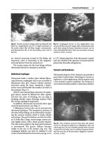
head is moved forward making a posterior approach easier. Landmarks, as
noted in Figure 5.29, help identify the site of the injection. About 1 cm below
the acromion, posteriorly, the humeral head boundaries are identified.
Internal and external rotation of the shoulder with the elbow at the side helps
5. Shoulder Problems 85
FIGURE 5.28. Anterior Apley scratch.
with this identification. There is an obvious sulcus or indentation at the
medial border of the humeral head. As noted in Figure 5.29, the subacromial
bursa is about 1 cm below the acromion and the glenohumeral joint about
3 cm below the acromion. Careful insertion of the needle just barely avoiding
the medial border of the humeral head places it in the correct location. The
depth of the insertion is usually about half the depth of a 1.5-in. 22-gauge
needle. Patient bulk also influences the depth of insertion. With AC, the
injection into the glenohumeral joint will be met with resistance because of
the contracted, thickened joint capsule. If large amounts of fluid are injected
after the first 5 cc the resistance will decrease. Injections can be repeated more
than once. A good rule of thumb is no more than two injections in a month
or three injections a year. This is an anecdotal rule and no good evidence
exists to support it. Also, remember that many of these patients are diabetic
or have the metabolic syndrome and can become diabetic with stress. Steroids
can elevate the blood sugar and patients should be warned so they can adjust
their medications accordingly. This does not mean steroids cannot be used in
diabetics.
86 E.J. Shahady, J. Buseman, and A. Nordgren
Acromion
Subacromial bursa
Glenohumeral joint
FIGURE 5.29. Shoulder injection with landmarks.
14. Arthritis of the Shoulder
The most common inflammatory arthritis of the shoulder joint is rheuma-
toid arthritis. Other systemic disorders like lupus erythematosus, psoriatic
arthritis, ankylosing spondylitis, Reiter’s syndrome, gout, pseudogout, and
scleroderma may cause glenohumeral degeneration but are rare causes. It is
unlikely that shoulder pain would be the presenting symptom for one of these
diseases.
Any patient with known inflammatory arthritis who has shoulder pain
should be evaluated for inflammatory arthritis in that joint. These patients
are still more likely to have the more common shoulder problems but deserve
evaluation to access for signs of the inflammatory arthritis. Treatment is ini-
tially conservative and directed toward controlling pain, inducing a systemic
remission, and maintaining joint motion by physical therapy. The use of
intra-articular steroids may help. Patients with progressive loss of motion or
radiographic destruction should be referred for possible surgical treatment.
Osteoarthritis of the glenohumeral joint is less common because it is a
non-weight-bearing joint. If a patient has prior trauma like a dislocation or
fracture, osteoarthritis should be considered. Osteoarthritis may also be pres-
ent in patients who have engaged in activities like boxing, heavy construction,
or chronic use of a pneumatic hammer. Pain is the usual presentation, but it
is generally not as acute or associated with the spasm seen in inflammatory
conditions. Plain radiographs show narrowing of the glenohumeral joint,
osteophyte formation, sclerosis, and some cyst formation. Patients with
osteoarthritis of the glenohumeral joint do well with conservative therapy.
15. Little Leaguer’s Shoulder
A stress fracture of the growth plate or physis of the proximal humerus is
commonly known as little leaguer’s shoulder. It occurs in high-performance
pitchers between 11 and 13 years of age. In addition to shoulder pain, the
common finding is radiographic evidence of widening of the proximal
humeral physeal plate. Repeated rotational and compressive stress from
throwing produces the stress on the physis. Treatment is usually nonoperative.
Like in the case of little leaguer’s elbow, rest for the remainder of the season
is the key. Encouraging coaches and athletes to develop good pitching skills
can prevent shoulder problems in the skeletally immature athlete. Speed
should be the last skill developed and only after proper technique and con-
trol are established. Many physical therapists and trainers are adept at teach-
ing these techniques. The primary care clinician’s role is early recognition and
prevention. You will be called on to provide advice. Just prescribing cessation
of activity is not enough. Helping coaches and parents understand how to
prevent and rehabilitate is an important additional role. Pitching technique
and number of pitches are associated with injury. Recommendations are to
5. Shoulder Problems 87
avoid throwing breaking pitches between the ages of 9 and 14 years. Pitchers
should focus on fastball and change-up pitches, avoiding a split-finger
change-up. Many authors agree with the USA Baseball News recommenda-
tions for limiting of pitches per game to the following: limits of 52±15 pitches
per game for 8- to 10-year-olds, 68±18 for 11- to 12-year-olds, and 76±16 for
13- to 14-year-olds.
16. Shoulder Exercises
Repeat each of the following exercises two times a day. Rotate from one exer-
cise to the other. Do one set of exercises and then rotate to another exercise and
do a set. Do not exercise past the point of pain. Pain means stop.
A. Pendulum exercises (Figure 5.30): Usually the first shoulder exercise done
once pain has diminished. Bend over and let injured arm hang loose at
your side. Begin to make small circles and gradually increase the circle
size. Pain is the only limiting factor.
B. External and internal rotation 1 (Figure 5.31): Place a hand weight or a can
of soup in your hand and lie on your back in bed. With the elbow flexed to
90° and tucked tightly to or at your side rotate your arm out and then back
88 E.J. Shahady, J. Buseman, and A. Nordgren
FIGURE 5.30. Pendulum exercise for the shoulder.
to your stomach. Remember to keep the elbow tucked to your side at 90°
of flexion. Hold the outer and inner movements for 10 seconds each at
their peak. Repeat 10 to 15 times for one set. Do three sets.
C. External and Internal rotation 2 (Figure 5.32): Place a hand weight or a can
of soup in your hand and lay on the uninjured side in your bed. With the
elbow flexed to 90° and tucked tightly to or at your side rotate your arm
out and then back to your stomach. Remember to keep the elbow tucked
to your side at 90° of flexion. Hold the outer and inner movements for 10 s
each at their peak. Repeat 10 to 15 times for one set. Do three sets.
D. Supraspinatus strengthening (Figure 5.33): Standing with the shoulder
abducted to 90°, elbow straight, arm crossed over about 20°, and thumbs
down, begin to move the arm up and down. Initially use no weight but
within 1 week or so add a small hand weight or a can of soup. Hold each
up and down movement for 5 s and repeat 10 to 15 times. Do three sets.
E. Forward flexion (Figure 5.34): Stand with the injured arm at your side,
elbow straight and a small weight or can of soup in the hand. Forward-
flex the shoulder as high as you can without pain. You may want to start
without a weight and add one as your strength increases. Hold each up
and down movement for 5 s and repeat 10 to 15 times. Do three sets.
F. Backward extension (Figure 5.35): Stand with the injured arm at your
side, elbow straight and a small weight or can of soup in the hand.
5. Shoulder Problems 89
FIGURE 5.31. External and internal rotation 1 exercises.
90 E.J. Shahady, J. Buseman, and A. Nordgren
FIGURE 5.33. Supraspinatus strengthening exercise.
FIGURE 5.32. External and internal rotation 2 exercises.
5. Shoulder Problems 91
FIGURE 5.34. Forward flexion exercise.
FIGURE 5.35. Backward extension exercise.
Backward-extend the shoulder as far back as you can without pain. You
may want to start without a weight and add one as your strength increases.
Hold each up and down movement for 5 s and repeat 10 to 15 times. Do
three sets.
G. Abduction (Figure 5.36): Stand with the injured arm at your side, elbow
straight and a small weight or can of soup in the hand. Abduct the shoul-
der as far as you can without pain. You may want to start without a weight
and add one as your strength increases. Hold each up and down move-
ment for 5 s and repeat 10 to 15 times. Do three sets.
Suggested Readings
Quillen DM, Wuchner M, Hatch RL. Acute shoulder injuries. Am Fam Physician.
2004;70(10):1947–1954.
Mantone JK, Burkhead WZ Jr, Noonan J Jr. Nonoperative treatment of rotator cuff
tears. Orthop Clin North Am. 2000;31:295–311.
92 E.J. Shahady, J. Buseman, and A. Nordgren
FIGURE 5.36. Abduction exercise.