Primary Care of Musculoskeletal Problems in the Outpatient Setting - part 4 pot
Bạn đang xem bản rút gọn của tài liệu. Xem và tải ngay bản đầy đủ của tài liệu tại đây (708.22 KB, 35 trang )
significant problem that will require more extensive measures to treat. A test
that will confirm the diagnosis has been called the chair test. Ask the patient
to try to lift a chair with the elbow extended, hand pronated, and the shoul-
der adducted. Significant pain and inability to lift the chair is diagnostic of
lateral epicondylitis.
Lateral epicondylitis, or tennis elbow, is the most common elbow problem
seen in primary care. The term tennis elbow is a misleading term as only
6. Elbow Problems 103
FIGURE 6.8. Point of maximal tenderness in lateral epicondylitis.
FIGURE 6.9. Resisted wrist extension for lateral epicondylitis.
about 5% of patients with lateral epicondylitis are tennis players. The prob-
lem is seen in association with any sport or occupation that involves repeti-
tive wrist extension. Lateral epicondylitis is caused by degeneration or
tendinosis of the attachment of the musculotendinous tendons of the wrist
extensor muscles to the lateral epicondyle of the distal humerus. The specific
pathophysiology remains to be defined clearly. The primary muscle involved
is the origin of the extensor carpi radialis brevis muscle.
The only other diagnosis that should be entertained is osteoarthritis of the
radiocapitellar joint or the radial head. This is not that common and the his-
tory is different. The key to the diagnosis of radial head pathology is pain
over the radial head, limitation of elbow pronation, and supination (Figure
6.5) and pain with that movement. The radial head is palpated just below the
lateral epicondyle, as noted in Figure 6.6. As the extensor muscle attachments
are in close proximity, it is easy to find discomfort in the vicinity of the radial
head in lateral epicondylitis. But the motions of pronation and supination
will not be limited or produce pain in lateral epicondylitis as they will in
radial head pathology.
The vast majority of the time no additional studies are needed to make the
diagnosis of lateral epicondylitis. If you suspect radiocapitellar osteoarthritis
because of difficulty with elbow pronation and supination, plain film imag-
ing will be needed. Magnetic resonance imaging is rarely indicated.
6.3. Treatment
The initial treatment goals are to decrease pain and inflammation. Therapy
begins with activity modification, ice massage, and NSAIDs. Limit any
recreational or occupational activity that requires repeated wrist extension.
An exercise program should also begin with the initial visit. Start with pas-
sive and active ROM exercises of the wrist and elbow. Pain decreases firing
of the extensor muscles causing weakness, so strengthening of these muscles
should be initiated. Resistance exercises should include wrist flexion–
extension and forearm pronation–supination exercises (see p. 112). These exer-
cises should be continued after successful treatment to prevent recurrence of
symptoms. A physical therapist can be consulted to teach the exercises and use
other modalities. An occupational therapist can help with job-specific exercises.
Steroid injections with 1 cc of local anesthetic and 1 cc of a long-acting
corticosteroid can be of benefit to patients who do not respond to conserva-
tive measures. Use a sterile technique and a 3-cc syringe with a 25-gauge nee-
dle. Identify the area of maximum tenderness for the site of the injection
(Figure 6.8). Repeat injections may be needed. No more than three injections
should be given in a 12-month period.
A trainer or coach may also help in treating athletes who are involved in rac-
quet sports. Some tennis players have symptoms of lateral epicondylitis because
of training errors. Improper body movement, racquet size and position, and
one-hand backhand strokes increase stress on the extensor muscles. Measuring
104 E.J. Shahady
for racquet size and learning proper body movement and types of strokes will
reduce and eliminate lateral elbow pain in many recreational athletes.
Counterforce bracing can also be used. The brace is applied just distal to
the elbow over the extensor muscles origin. The brace provides a constraint
over the extensor musculature and distributes forces to nondiseased portions
of the extensors.
Indication for surgical intervention is a failure to improve after completing
a well-controlled nonoperative treatment program. About 5% of patients
with lateral epicondylitis will require surgery. A minimum of 6 to 12 months
of nonoperative treatment is recommended before considering surgery. Both
open and arthroscopic options can be considered. Good results are expected
in the hands of an experienced surgeon.
7. Medial Epicondylitis
Medial epicondylitis, also known as medial tennis elbow or golfer’s elbow, is
less common than lateral epicondylitis. It is caused by overuse of the flexor
and pronator muscles that attach to the medial epicondyle. Overuse occurs
with any activity that produces a valgus stress on the medial joint line (see
Figure 6.12). Racquet sports, golf, and throwing sports are examples of activ-
ities that are associated with medial epicondylitis. The history is usually one
of a dull ache over the medial elbow and an occupation or sport that requires
repeated elbow pronation–supination or flexion–extension. Examination will
reveal tenderness over the medial epicondyle and medial elbow pain with
resisted wrist flexion (Figure 6.10). This is in contrast to lateral epicondylitis
where the pain is located on the lateral side and is increased with resisted
wrist extension (Figure 6.9). Care should be taken to evaluate the ulnar col-
lateral ligament (UCL) and the ulnar nerve. This will be discussed further in
Section 10 (Ulnar collateral ligament injury) and Section 11 (Ulnar Nerve
Injury (Cubital Tunnel Syndrome)).
Radiographs are usually not needed unless fractures are suspected.
Treatment is similar to lateral epicondylitis and includes activity modifica-
tion, ice massage, NSAIDs, resistance exercises, and corticosteroid injec-
tions. Mechanical analysis of sport or occupation technique may provide
diagnostic and therapeutic information.
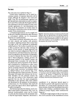
8. Olecrenon Bursitis
The position of the olecrenon (see Figure 6.4) and its overlying bursa makes
it susceptible to injury. Bursitis in this area has also been called student’s
elbow and miner’s elbow. These occupations are noted for a large amount
of time spent leaning on the elbows. The bursitis can be either acute or
chronic.
6. Elbow Problems 105
Acute bursitis can be caused by prolonged pressure or repeated trauma.
The fluid can either be clear or hemorrhagic. Initial therapy includes ice,
NSAIDs, a compression ace wrap, and elimination of the causative factors.
If the bursa is tense and painful it can be aspirated. Elbow pads should be
used in any occupation that requires prolonged pressure or repeated trauma
to the olecrenon area of the elbow.
Chronic bursitis is characterized by thickening of the bursal walls.
Repeated trauma leads to the formation of granulation tissue that grows into
the bursal sac. Examination will reveal a firm rubbery mass that may or may
not be fluid filled. The history is usually one of repeated trauma like falling
on the elbow. There may have been an acute traumatic episode superimposed
on chronic bursal disease. Treatment is similar to acute bursitis except the
occasional patient that might need surgical excision of the bursa.
Suppurative or septic bursitis is not as common as acute and chronic bur-
sitis. Bacteria can be introduced into the bursa from a puncture wound or a
laceration. Erythema, warmth, bursal distention, significant pain, and limi-
tation of motion may be present. Aspiration and identification of the organ-
ism, most often staphylococcus, followed by antibiotics is appropriate
treatment. Suppurative bursitis, on some occasions, has resulted from an
aspiration for acute traumatic bursitis. The blood-filled bursa is an ideal cul-
ture medium for bacteria introduced by the aspiration.
Both acute bursitis and suppurative bursitis can cause warmth but the fluid
is usually bloody or clear in acute bursitis and cloudy in suppurative. Gout
and pseudogout can also present with fluid in the olecrenon bursa. So an
analysis of the fluid for crystals, white blood cell (WBC) type, and bacteria is
appropriate when you are suspicious of one of these entities.
106 E.J. Shahady
FIGURE 6.10. Resisted wrist flexion for medial epicondylitis.
Aspiration of the bursa should be done under sterile conditions. There
should be adequate cleansing of the area and gloves and sterile instruments
should be used. Also use an 18-gauge needle because the fluid may be too
thick for aspiration with a smaller gauge needle.
9. Case
9.1. History
A 13-year-old right-handed boy presents to your office with elbow pain. He
is a pitcher on the baseball team and has noted increased pain as the season
has progressed. Initially the pain was vague and only occurred after a game
that he pitched. The pain now appears after he pitches two to three innings
and he is not able to finish the game. The pain is now localized in the medial
aspect of his right elbow. He also complains of pain with twisting motions
and flexion of the right elbow. Examination reveals a 10° limitation of elbow
extension and an ability to flex only to 100°. Tenderness is noted over the
medial epicondyle and the cubital tunnel (area between the olecrenon and the
medial epicondyle). Tapping over the cubital tunnel produces some tingling,
radiating down to the fifth digit. No other neurological deficit is noted.
Valgus stress (see Figure 6.7) of the elbow in 25° of flexion reveals some lax-
ity compared with the left elbow.
9.2. Thinking Process
The first thing that comes to mind in this case is little leaguer’s elbow. Little
leaguer’s elbow unfortunately has become a term that includes any elbow
problem that occurs in a young throwing athlete. This wastebasket diagnosis
does not help properly diagnose and treat this boy. Three entities—Panner’s
disease, OCD, and medial epicondyle traction apophysitis (META)—need to
be considered in a young athlete with elbow pain. The age of the patient and
the location of the pain help start the process of differentiating the three enti-
ties. Panner’s disease and OCD are problems that result in lateral elbow pain
whereas the epicondyle traction apophysitis results in medial pain. Panner’s
disease is a problem of young children between 7 and 10 years of age and
OCD occurs most commonly in 13- to 16-year-olds (see p. 111). The age of
occurrence of META is dependent on the skeletal maturation of the athlete
and occurs between 11 and 16 years of age. Once the medial epicondyle fuses
the ligamentous structures are more susceptible to injury, and medial (ulnar)
collateral ligament injury is more likely. With the location of the pain on the
medial side and the age of the patient, META is more likely. Osteochondritis
dissecans is still possible and an X-ray will help rule out OCD. The laxity on
valgus stress probably indicates some separation of the medial epicondyle
from the rest of the humerus. The tingling noted when the cubital tunnel is
6. Elbow Problems 107
tapped (Figure 6.11) is associated with pressure on the ulnar nerve. The nerve
is located just below the medial epicondyle under the medial collateral liga-
ment. Swelling in the area places pressure on the nerve and causes ulnar nerve
symptoms.
The stresses placed on the elbow joint by throwing sports provide an expla-
nation for the above-mentioned clinical problems. Throwing motion is not
simple flexion and extension. The elbow goes through significant medial and
lateral stress that can stretch or tear the ulnar (medial) collateral ligament
and radial (lateral) collateral ligament and fracture the distal portions of the
humerus and the proximal portions of the radius and ulna. When the growth
plates are not closed, portions of bone can be separated (avulsed) from the
distal humerus. These stresses can be appreciated by taking your own arm or
a patient’s through the throwing motion while palpating the medial and lat-
eral joints. Figure 6.12 demonstrates the locations of the medial and lateral
joints and their collateral ligaments as well as the capitellum and trochlea of
the humerus and the radial head and ulna. As the arm is brought into the
cocked position the articulation between the capitellum and the radial head
is impacted together (Panner’s disease and OCD) and the medial side UCL
or the medial epicondyle is pulled (META). As the arm is now accelerated,
the object released, and deceleration begins, the lateral side radial collateral
ligament is now stretched and the bony articulation between the ulna and the
humeral trochlea is impacted together. During this phase the radial head
impacts with the capitellum because of pronation. This is especially true
when attempting to throw a curve ball. It is easy then to see how collateral
ligament injury as well as micro- and macrobone fractures can occur.
108 E.J. Shahady
Olecranon
Cubital Tunnel
Medial
Epicondyle
FIGURE 6.11. Medial elbow with landmarks.
Understanding the mechanism helps explain the why and also offers thoughts
on prevention and treatment.
Plain films are a necessity with these types of problems. Avulsion or frag-
mentation of the medial epicondyle will be noted in META. Always request
a film of the opposite side to compare for differences as well as normal vari-
ants in the skeletal immature athlete. Multiple views are recommended to rule
out the presence of loose bodies. Bone scan, CT scans, and MRI may be help-
ful. These more expensive entities should be reserved for those patients who
do not respond to conservative treatment or have more extensive disease on
plain films.
9.3. Treatment
Medial epicondyle traction apophysitis can usually be treated with conserva-
tive measures depending on the amount of separation observed on the X-ray.
If the avulsion is 5 mm or greater, surgery may be indicated. The above
patient had minimal separation and was treated with 3 weeks of no throwing,
ice, and NSAIDs. Steroid injections are never indicated. He was allowed to be
a pinch hitter during the 3 weeks and encouraged to maintain general condi-
tioning so that he remained fit. After the 3 weeks he was gradually allowed to
return to activity. Range of motion and resistance exercises to strengthen the
flexors are indicated. He was first allowed to play outfield and then after
6. Elbow Problems 109
Valgus
stress
Medial
torn
ulnar
collateral
ligament
Radiocapitellar
joint
FIGURE 6.12. Mechanism of injury in throwing sports. (Reproduced from Richmond J,
Shahady E, eds. Sports Medicine for Primary Care. Cambridge, MA: Blackwell
Science; 1996:354, with permission.)
4 weeks allowed to start his pitching routine. The number of innings pitched
was gradually increased and he is now doing well.
Elbow injury in young athletes can be prevented. Pitching technique and
number of pitches are associated with injury. Throwing breaking pitches
increases elbow pain and using change-ups reduces the rate of elbow pain.
Recommendations are to avoid throwing breaking pitches between the ages
of 9 and 14 years. Pitchers should focus on fastball and change-up pitches,
avoiding a split-finger change-up. Many authors agree with the USA Baseball
News recommendations for limiting of pitches per game to the following: lim-
its of 52±15 pitches per game for 8- to 10-year-olds, 68±18 for 11- to 12-year-
olds, and 76±16 for 13- to 14-year-olds.
10. Ulnar Collateral Ligament Injury
The mechanism of injury for UCL stretch or tear is similar to META. If the
growth plate of the medial epicondyle has fused, valgus stress will stretch or
tear the ligament rather than avulse a portion of the epicondyle that occurs
in META. These patients are older, usually young adults, who are engaged in
throwing sports. Symptoms and examination are similar to META. If UCL
rupture occurs the patient may report a sudden event and loss of function.
The hallmark of UCL rupture is valgus instability. Stability of the ligament
can be accessed by applying a valgus force to the medial joint while the shoul-
der is in external rotation and the elbow is at 25° of flexion (see Figure 6.7).
The arm is stabilized by placing the patient’s hand in your armpit, one hand
on the lateral side to exert the valgus force and the other on the medial joint
line to assess the degree of instability. Treatment depends on the expectations
for returning to competitive competition. Surgical management is indicated
if the patient wishes to return to competitive overhead throwing.
Conservative treatment with ice, NSAIDs, and splinting followed by stretch-
ing and strengthening exercises is usually sufficient for treatment if return to
competition is not contemplated. It also can be attempted for recreational
throwing athletes for a 3- to 6-month period.
11. Ulnar Nerve Injury (Cubital Tunnel Syndrome)
The ulnar nerve is susceptible to injury with trauma to the medial side of the
elbow. The ulnar nerve enters the cubital tunnel behind the medial epi-
condyle. The tunnel is made up of the UCL and other lateral ligamentous
structures. The nerve is vulnerable to injury from traction, compression, and
direct trauma that accompanies any problem that involves the medial com-
plex. This includes META, UCL rupture, and medial epicondylitis. Common
symptoms are medial elbow pain that is increased with flexion, numbness and
110 E.J. Shahady
tingling of the fourth and fifth digits, and a positive Tinel’s sign (tingling
induced by tapping over the cubital tunnel, Figure 6.11). Treatment is symp-
tomatic and aimed at the primary condition that has produced the compres-
sion or edema. If this treatment is not successful or ulnar motor nerve
weakness is present surgical correction may be needed.
12. Panner’s Disease
Children of age 7 to 10 are affected and complain of lateral elbow pain with
throwing. The repetitive force of throwing compresses the radial head into
the capitellum. The disorder affects the ossification center. Initially there is
necrosis or degeneration of the ossification center followed by regeneration
and recalcification. Physical examination will reveal pain over the lateral joint
between the capitellum and radial head (Figure 6.12). Range of motion is
usually not limited. The diagnosis is made by X-ray. The capitellar ossifica-
tion center is fragmented and the epiphysis is irregular and smaller compared
with the opposite elbow. This is usually a self-limited problem. Dis-
continuation of throwing, ice, and NSAIDs will produce relief of symptoms.
Follow-up X-rays are needed to document healing. The capitellar epiphysis
usually remodels and returns to a normal appearance. The child can usually
return to throwing within a 6- to 8-week period.
13. Osteochondritis Dissecans
Osteochondritis dissecans (OD) is a more serious problem than Panner’s
disease that affects young adolescents between 13 and 16 years of age. The
cause is thought to be a combination of repetitive stress to the radiocapitel-
lar joint and an interruption of the vascular flow to the capitellum. Both
throwing athletes and gymnasts are susceptible to OD. The story is one of
gradual onset of lateral elbow pain, clicking, locking, and decreased ROM.
The pain increases with throwing or in a gymnast with routines that rely
on the arms to bear all the weight of the body like hanging from a bar by
the hands. Examination will reveal lateral joint line tenderness and limita-
tion of flexion and extension. With the elbow in full extension, pain may be
elicited with attempts to pronate and supinate because of radial head
involvement. Plain X-rays commonly show the characteristic radiolucent
focal defect in the capitellum. If this is noted a loose body may be present.
If the defect is present without a loose body noted, a CT scan with contrast
should be obtained to search for the loose body. Magnetic resonance imaging
can help with early identification of OD. Because of the poorer prognosis
with OD a consultation with an orthopedic surgeon is recommended if you
suspect OD.
6. Elbow Problems 111
14. Medical Problems
14.1. Rheumatoid Arthritis
Elbow involvement occurs in at least half the patients with rheumatoid
arthritis. Soft tissue abnormalities such as joint swelling, olecrenon bursitis
and rheumatoid nodules along the extensor surfaces, warmth, and tenderness
are the earliest findings. Another early finding, often unnoticed by the
patient, is a loss of full extension. Symptoms isolated only to the elbow are a
rare occurrence. The majority of the time, rheumatoid symptoms are also
present in the shoulders, hands, and wrist as well as the elbow. Early recogni-
tion is critical to limiting deformity. Early referral to a physician who can
administer disease-modifying drugs is indicated.
14.2. Osteoarthritis
Osteoarthritis is not common in the elbow as it is not a weight-bearing joint. If
present it is caused by post-traumatic overuse. Loss of motion rather than pain
is what prompts the patient to seek medical attention. Osteophytes and loose
bodies will be noted on plain film radiographs. Treatment is symptomatic.
Surgical decompression is sometimes required to restore functional motion.
14.3. Other Medical Problems
Gout and pseudogout need to be considered when there is a joint effusion.
Gout or pseudogout should be suspected if the effusion is acute and other
joints like the big toe (gout) or the shoulder (pseudogout) are involved.
Examination of aspirated fluid for crystals should be included if either dis-
ease is suspected.
15. Elbow Exercises
Tell patients to perform these exercises two times a day. Rotate from one exer-
cise to the other. Do one set of one exercises and then rotate to another exercise
and do a set. Do not exercise past the point of pain. Pain means stop.
1. Wrist range of motion (Figure 6.13A and 6.13B): Bend your wrist forward
and backward as far as you can. Hold each movement for 5 seconds (s) and
repeat 10 times.
2. Wrist flexion stretch (Figure 6.14): With the injured hand in 30° of flexion
begin to flex the wrist against the resistance of the other hand. Resist the
movement for 15 s and repeat five times.
3. Wrist extension stretch (Figure 6.15): With the injured hand in 30° of exten-
sion, begin to flex the wrist against the resistance of the other hand. Resist
the movement for 15 s and repeat five times.
112 E.J. Shahady
4. Wrist flexion exercise (Figure 6.16): Hold a weight like a can of soup with
your palm facing up. Flex the wrist (bend it upward), hold wrist in maxi-
mum flexion for 15 s, and return to the starting position slowly. Repeat five
times.
5. Wrist extension exercise (Figure 6.17): Hold weight like a can of soup with
your palm facing down. Extend the wrist (bend up) and hold in maximum
extension for 15 s. Return to the starting position slowly and repeat five
times.
6. Wrist radial and ulnar deviation strengthening (Figure 6.18A and 6.18B):
Hold a weight like a can of soup in your hand with your thumb facing up
6. Elbow Problems 113
FIGURE 6.13. (A and B) Wrist range of motion.
and your wrist sideways. Move your wrist up and down through radial and
ulnar deviation. Hold each position for 15 s and repeat five times.
7. Elbow flexion and extension (Figure 6.19A and 6.19B): Place your arm at
your side with your elbow completely straight. Hold a weight like a can of
soup with your palm face up. Flex (bend) your elbow slowly toward your
shoulder as far as it will go. Slowly lower the arm until the elbow is again
completely straight. Hold each position for 15 s and repeat five times.
114 E.J. Shahady
FIGURE 6.14. Wrist flexion stretch.
F
IGURE 6.15. Wrist extension stretch.
8. Pronation and supination of the forearm (Figure 6.20A and 6.20B): Place
your elbow at your side and bend the elbow 90°. Rotate the hand from a
palm-upward to palm-downward position. Hold each position for 15 s and
repeat five times. Add a weight like a soup can once the movements can be
performed easily without a weight.
6. Elbow Problems 115
FIGURE 6.16. Wrist flexion exercise.
FIGURE 6.17. Wrist extension exercise.
116 E.J. Shahady
Redial
deviation
Ulnar Deviation
FIGURE 6.18. Wrist radial and ulnar deviation strengthening.
FIGURE 6.19. (A) Elbow flexion and (B) extension.
Suggested Readings
1. Lyman S, Fleisig G, Andrews J, Osinski E. Effect of pitch type, pitch count, and
pitching mechanics on risk of elbow and shoulder pain in youth baseball pitchers.
Am J Sports Med. 2002;30(4):463–468.
2. Andrews J, Fleisig G. How many pitches should I allow my child to throw? USA
Baseball News. 1996;4:5.
6. Elbow Problems 117
FIGURE 6.20. (A) Pronation and (B) supination of the forearm.
7
Wrist Problems
EDWARD J. SHAHADY
Wrist problems and complaints are another group of common and frus-
trating musculoskeletal issues in primary health care. Wrist injuries can be
intimidating because of the complex anatomy and concern for potential
long-term disability. Overcoming the fear and frustration of caring for wrist
injury becomes possible with a few simple organizational steps:
1. Step 1 is to realize that 95% of patients seen in the office with wrist-
complaints can be classified into three categories and six to seven
problems.
2. Step 2 is to take a focused history that segments the categories into acute
trauma, overuse trauma, and medical disease. You now have a manageable
list (Table 7.1) to begin further investigation.
3. Step 3 is to perform a focused thorough wrist examination. With a focused
history and a thorough wrist examination you most likely now have
the diagnosis. Your knowledge of the usual history and examination
associated with the most common problems has facilitated the diagnostic
process.
4. Step 4 is ordering confirmatory studies if needed (many times they are
not).
5. Step 5 is to start treatment. (This may include appropriate consultation.)
Five percent of the time the diagnosis will not be so obvious. However, not
being one of the 95% is usually obvious. That is when additional studies
and/or a consultation becomes necessary.
Rare or not so frequent problems are usually the ones that receive the most
press. How often do you hear the words “I got burned once” mentioned
about a rare problem that was missed in the primary care setting. Having a
good working knowledge of the characteristics of common problems pro-
vides an excellent background to help recognize the uncommon. The uncom-
mon is easy to recognize once you know the common. Be driven by the search
for the common rather than the expensive intimidating search for the rare
birds.
118
1. Focused History
The first question is whether the onset of the problem was acute. Did it just
start or has it been present for a prolonged period of time? The next question
would depend on the answer to the first. If the onset is acute, then ask about
a fall, especially one that involved breaking the fall with the outstretched hand
(a common mechanism in wrist injury). If the onset is not acute, ask about
occupations, hobbies, or sports that involve repeated wrist movement like
computer use, painting, and throwing sports. A recent change in occupation
or intensity of activity is also important. Ask about any chronic disease. Wrist
pain can accompany collagen vascular diseases like rheumatoid arthritis.
2. Focused Physical Examination
Begin by asking the patient to go through four motions without your help.
This examination establishes a marker for severity of the injury and a base-
line to assess degree of recovery. The four wrist motions are ulnar deviation
(Figure 7.1); radial deviation (Figure 7.2); wrist extension (Figure 7.3); and
wrist flexion (Figure 7.4).
It is important to start every examination by defining range of motion
(ROM) and noting any difference between one wrist and another. Use a
goniometer to precisely assess degrees of motion. The remaining examina-
tion will be dictated by the focused history.
3. Case
3.1. History
A 33-year-old female secretary presents to your office with right wrist pain
for the past 6 months. It was relieved by taking a few ibuprofen tablets but
now the pain is persistent and makes it difficult for her to type. She also notes
7. Wrist Problems 119
TABLE 7.1. Classification of common wrist problems.
Overuse trauma
●
Carpal tunnel syndrome
●
De Quervain’s tenosynovitis
●
Other tendonitis
Acute trauma
●
Fractured scaphoid bone of the wrist
●
Scapholunate ligament rupture
●
Wrist sprain
Medical problems
●
Arthritis associated with other diseases like rheumatoid arthritis
120 E.J. Shahady
30
degrees
FIGURE 7.1. Ulnar deviation; 30° is normal.
20
degrees
FIGURE 7.2. Radial deviation; 20° is normal.
7. Wrist Problems 121
FIGURE 7.3. Wrist extension; normal is up to 80°.
F
IGURE 7.4. Wrist flexion; normal is up to 80
°.
some weakness when she grasps objects and occasionally she drops her cof-
fee cup. She is frustrated by her inability to open jars or twist off lids. She
feels like she is an old woman before her time. She reports no falls and she has
no other complaints. Her general health is excellent. She is a power walker
and takes no medications.
3.2. Thinking Process
The lack of a fall or other acute trauma makes the diagnosis listed under
acute trauma less likely. Still perform the examination maneuvers to assess
for trauma as some patients forget the fall that occurred some time ago.
Probably this examination will be negative for acute trauma. Medical causes
are also unlikely, as she has no history of chronic disease that suggests this.
If this is the first sign of a systemic chronic disease, it will soon become obvi-
ous by a lack of response to your treatment and appearance of other symp-
toms of that disease. This makes an overuse syndrome most likely. Additional
questions and the wrist examination that will focus on overuse syndromes
should make the diagnosis.
Carpal tunnel syndrome is a possibility and it has a characteristic history.
These patients usually have occupations that require prolonged or repeated
wrist flexion. They also usually complain that the pain is located on the palm
of the hand in the thenar area (thumb side). The pain may radiate the elbow.
Numbness and tingling in that area of the palm are usually present. The pain
may radiate up to the elbow and it is aggravated by wrist flexion and exten-
sion. These patients may also awaken at night with pain and numbness
because the wrist falls into flexion with sleep. Grasping objects is painful and
they may report dropping cups and glasses.
This patient is a secretary and she spends 6 h a day with computer data
entry. Her pain is in the thenar area and is accompanied by numbness and
tingling. She awakens in the morning feeling like she needs to rub or shake
her hand to “get the circulation back.” The diagnosis of carpal tunnel syn-
drome is most likely. The physical examination should be preformed to con-
firm this diagnosis and to rule out the other possibilities.
Carpal tunnel syndrome is a compression neuropathy of the median nerve.
The physical examination will focus on assessing the sensory and motor func-
tions of that nerve. First observe the palm of the hand and compare the
thenar area of the injured hand to the noninjured hand to look for atrophy.
Next, tap over the median nerve at the wrist (Figure 7.5). A positive test
(Tinel’s sign) is the production of tingling on the palmer surface of the thumb,
index finger, and a portion of the middle finger. Test the strength of the thenar
muscles by assessing the ability of the thumb to move toward the little finger
against resistance (Figure 7.6). Placing the wrists together in maximum flexion
(Phalen’s test) for 45 s or less will cause numbness or aching. (Figure 7.7).
De Quervain’s tenosynovitis is an inflammation of the tendon sheaths of
the abductor pollicis longus and the extensor pollicis brevis at the first dorsal
122 E.J. Shahady
7. Wrist Problems 123
FIGURE 7.5. Median nerve percussion (Tinel’s test).
FIGURE 7.6. Thenar muscle strength test.
compartment over the radial styloid. These tendons form the floor of the
anatomic snuffbox of the wrist (Figure 7.8). Palpate the tendons over the
radial styloid and at the floor of the snuffbox for pain and tenderness. Perform
Finkelstein’s test (Figure 7.9) by asking the patient to ulnar-deviate the wrist
with the thumb flexed and abducted into the palm places tension on the ten-
dons and their sheaths. The examiner may have to provide an additional force
to complete the test. Reproduction of the pain clinches the diagnosis.
Examination of the snuffbox to evaluate the scaphoid (Figure 7.10) is
important to rule out a subtle fracture that has been overlooked from past
trauma. How to do this examination will be explained when scaphoid frac-
tures are discussed in Section 7 (Scaphoid Fractures).
This patient had positive Phalen’s and Tinel’s signs. She had no thenar atro-
phy and thumb opposition was not weak. Her Finkelstein’s test is negative and
there is no tenderness over the radial styloid (Figure 7.8). This history and phys-
ical examination makes the diagnosis of carpal tunnel syndrome most likely.
3.3. Additional Studies
No further studies are needed at this time unless a scaphoid fracture is sus-
pected. Nerve conduction studies are helpful if the diagnosis is not clear or
surgery is a therapeutic consideration.
124 E.J. Shahady
FIGURE 7.7. Phalen’s test.
7. Wrist Problems 125
Extensor Pollicis Brevis
Abductor Pollicis Longus
Snuff
Box
Radial
Styloid
FIGURE 7.8. Anatomic snuffbox area.
FIGURE 7.9. Finkelstein’s test.
3.4. Treatment for Carpal Tunnel Syndrome
Most cases will respond to conservative measures. First, identify and decrease
the offending activity that is causing the problem. Workstation modifications
to decrease wrist flexion can be very beneficial. Examples include adjusting
keyboard height and supports for the wrist and forearm. Intermittent breaks
from typing every few hours decreases fatigue. If none of these measures work,
consider a change in occupation or sport technique. Writing a note to the
patient’s employer, coach, or teacher explaining how these changes will increase
productivity helps patients obtain the cooperation of their superiors. If no
contraindication exists, a short course (7 to 10 days) of nonsteroidal anti-
inflammatory drugs (NSAIDs) or steroids will help. Be sure to provide the
patient instructions to take the medication at the appropriate dose for
7 to 10 days, as described in Chapter 1. Wrist splints to limit flexion and exten-
sion are a mainstay of treatment (Figure 7.11). The splint should be worn at
night and if needed during the day. Patients are sometimes reluctant to use the
splint during the day because it interferes with work performance or they do
not want to call attention to the problem. At times, a short-arm cast can be
employed to assure complete rest of the wrist. Carpal tunnel syndrome some-
times occurs with pregnancy. It usually terminates with the end of pregnancy
and is responsive to conservative measures like splints and steroid injections.
Steroid injections with 1 cc of local anesthetic and 1 cc of a long-acting
corticosteroid can be of benefit to patients who do not respond to conserva-
tive measures. Use a sterile technique and a 3-cc syringe with a 25-gauge nee-
dle. Flex the wrist and identify the wrist flexion crease and the palmaris
longus tendon. The needle will be inserted on the ulnar side of the palmaris
longus about 1 cm proximal to the wrist crease (Figure 7.12). Some patients
do not have a palmaris longus tendon. If this is the case use the ulnar side of
the ring finger as a marker to direct the needle. Ask the patient to fully flex
126 E.J. Shahady
FIGURE 7.10. Examination of the snuffbox for scaphoid fracture.
7. Wrist Problems 127
the fingers as noted in Fig. 7.12. Advance the needle at a 45° angle for about
1 cm until you feel resistance. Appropriate location of the needle can be accessed
by moving the ring finger. This should produce movement of the needle. Ask the
patient to now extend the fingers to bring the needle into the carpal tunnel and
slowly inject 1 to 2 cc of the steroid–anesthetic solution. Advice the patient that
there will be some mild soreness and it may require 24 h to feel the full effect.
FIGURE 7.11. Wrist splints.
FIGURE 7.12. Injection for carpal tunnel syndrome.