Primary Care of Musculoskeletal Problems in the Outpatient Setting - part 7 ppt
Bạn đang xem bản rút gọn của tài liệu. Xem và tải ngay bản đầy đủ của tài liệu tại đây (823.74 KB, 35 trang )
Further testing is not indicated if the symptoms are classical and the
patient responds to conservative measures listed below. If the patient does
not respond to conservative measures, nerve conduction studies of the lat-
eral femoral cutaneous nerve can be performed to access for nerve com-
pression. Magnetic resonance imaging of the hip and pelvis are useful to rule
out intra-articular derangement or intrapelvic causes of compression on
the nerve.
The mainstay of treatment for entrapped lateral femoral cutaneous nerve
is nonoperative. Weight reduction, decreased use of constrictive clothing,
nonsteroidal anti-inflammatory drugs (NSAIDs), and local steroid injections
succeed 90% of the time. If symptoms are persistent and disabling, surgical
intervention is warranted. Local nerve block is a useful diagnostic tool and
predictor of benefit from surgical decompression. If injection completely
relieves the patients’ complaints, surgery will usually help.
6. Trochanteric Bursitis
The trochanteric bursa lies over the greater trochanter of femur. Overuse is
the usual cause of the bursitis. It is commonly associated with OA of the hip.
Other factors that contribute to the etiology of trochanteric bursitis include
irritation of the bursa by the overlying iliotibial band (ITB) and biomechan-
ical factors like a broad pelvis in females, leg length discrepancy, and exces-
sive pronation of the foot (see Chapter 15) that change the mechanics of
the ITB.
The patient usually presents with an aching pain over the lateral hip that is
made worse by prolonged standing, lying on the side, or stair climbing. The
pain may radiate to the groin or the lateral thigh.
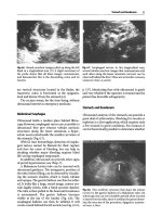
On examination, palpation along the posterior greater trochanter reveals
tenderness (Figure 11.8). The pain is accentuated with external rotation and
abduction and by resisted abduction. Patrick’s (flexion, abduction and exter-
nal rotation (FABER)) test is positive (Figure 11.9) and the hip abductors are
often weak. Test the hip abductor as noted in Figure 11.4. Iliotibial band
tightness may be present. The Ober’s test will be positive if tightness is pres-
ent. See Chapter 12 for a description of this test.
Treatment of trochanteric bursitis consists of rest, ice, ITB stretching,
strengthening of the hip girdle and trunk musculature (especially gluteus
medius), and stretching of the fascia lata and the ITB. The exercises at the
end of this chapter describe how to do this. Leg length discrepancy and
pronation need to be addressed. Inflammation generally responds to non-
steroidal anti-inflammatory medications and local treatment modalities. In
some cases, local corticosteroid injection into the area of tenderness over the
trochanteric bursa may be necessary to achieve symptomatic relief. Rarely, if
the condition is refractory to conservative measures, operative release of the
ITB may be required.
212 E.J. Shahady
FIGURE 11.8. Point of tenderness in trochanteric bursitis.
FIGURE 11.9. Flexion, abduction, external rotation (FABER) test.
Plain X-rays of the hip are helpful in the older population to access for the
presence of OA. Trochanteric bursitis commonly accompanies OA of the hip.
Magnetic resonance imaging is seldom needed unless you suspect tears of the
abductor musculature.
7. Acute Trauma (Case)
7.1. History
A 38-year-old accountant comes to your office complaining of pain in the
back of his leg for the past 3 days. He hurt the leg while playing softball
with his family. He hit the ball and started to run to first base and felt a
catch in the back of his leg. He was unable to continue playing because of
the pain and inability to walk without assistance. The next day the pain
increased and he was unable to work. He noticed a large “black and blue”
area on the back of his leg. He is concerned that he has torn something and
may need surgery. His general health is good. In the last few years, he has
not been as physically active as he was in the past. On examination he walks
with a slight limp and has a 30° loss of knee extension and discomfort with
passive knee extension past 30°. Assess knee extension with the patient sit-
ting and attempting to straighten the leg. This patient was not able to fully
straighten his leg. It remained bent at about 30° of flexion (Figure 11.10).
A 2-by 3-in. ecchymosis is present on his posterior thigh. Flexion of the
214 E.J. Shahady
F
IGURE 11.10. Knee extension limited to 30
° while seated.
knee is painful. Palpation of the posterior thigh with the knee partially
flexed against resistance (Figure 11.11) reveals mild tenderness and a pal-
pable knot at the midthigh.
7.2. Thinking Process
This patient’s history of acute onset of pain after starting to run, difficulty in
walking, and the appearance of an ecchymosis is suggestive of a tear of one
of his muscle groups. Being in the posterior thigh makes a hamstring tear
likely, although adductor tears may produce posterior lateral pain. His phys-
ical examination supports the possibility of hamstring injury. Extension of
the knee stretches the hamstring muscle and hamstring tears produce a spasm
that limits extension. This patient has significant limitation of his knee exten-
sion and ecchymosis of the posterior thigh, so a hamstring muscle tear is
most likely. Judge the severity or extent of injury by the degree of extension
limitation and not by the size of the ecchymosis.
Palpation over the hamstring muscle group with resisted knee flexion may
elicit tenderness, defects, and/or a mass when the hamstring is injured. Both
tenderness and a mass are demonstrated in this patient with resisted flexion
(Figure 11.11). More extensive tears are associated with palpable masses and
11. Hip and Thigh Problems 215
FIGURE 11.11. Knee partially flexed against resistance with muscle belly being
palpated (patient on abdomen).
defects. Other factors responsible for hamstring injuries include inadequate
warm-up, inflexibility, poor conditioning, and muscle strength imbalances
between the hamstring and quadriceps muscles. This patient has decreased
his physical activity in the last few years, is probably not well-conditioned,
and did not stretch or warm up before he started playing.
Hamstring strains are the most common strain-related injuries seen by the
primary care provider. They can be quiet disabling and lead to a loss of work
and recreational time. The site of the tear in patients over age 25 is usually at
the junction of the muscle and the tendon (musculotendinous junction). In
patients under 25 the tear most likely occurs where the tendon attaches to the
pelvis. The apophysis is the ossification center where the tendon attaches to
the bones of the pelvis. These injuries will be discussed in Section 11
(Pediatric Hip Problems) (page 220).
The injury occurs during the stretching or eccentric phase of muscle con-
traction. The force generated during eccentric contraction is greater than the
force generated during the concentric or contracting phase of muscle con-
traction. There are three muscles in the hamstring group: the biceps femoris,
semimembranosus, and semitendinosus. The biceps femoris is the most
commonly injured of the three.
Hamstring strains may be classified into three groups: mild (grade I), mod-
erate (grade II), and severe (grade III). Grade I strains represent a small dis-
ruption of the musculotendinous unit. Grade II strains are partial tears and
grade III have complete rupture. Second-degree strains are associated with
immediate functional loss, a painful palpable mass, marked spasm, and a loss
of knee extension between 20° and 25°. Third-degree tears are associated
with a defect in the muscle, marked spasm, swelling, and an extension loss of
greater than 45°. This patient probably had a grade II hamstring tear. It is
important to classify the injury to give the patient a prognosis. Grade I tears
take days to heal, grade II may take 4 to 6 weeks, and grade III may take up to
6 months. These times are approximations and are modified by response
to treatment.
7.3. Diagnostic Studies
None are needed unless you suspect a fracture or an unusual pathological
entity. Plain X-ray would be a good first step to look at bone and an MRI to
rule out other muscle entities.
7.4. Treatment
Treatment for acute muscle strains is rest, ice, compression, and elevation
(RICE) and NSAIDs. This will reduce the inflammatory process and control
bleeding. Heat and massage in the first week are contraindicated because they
will increase bleeding. After 3 to 5 days, a gradually progressive program of
stretching is started. Range of motion is the key to judging progress. Once
216 E.J. Shahady
full extension is achieved and the pain has subsided, resistance exercises can
be started. This is easier to reach in second-degree tears than in third-degree
tears. For most patients the hamstrings are only 40% to 45% as strong as the
quadriceps and this imbalance increases the risk of hamstring injury. Many
hamstring injuries can be prevented if the hamstring strength in both legs is
60% of the quadriceps strength. The strength can be increased and measured
at any gym that has leg flexion and extension machines. Remember to exer-
cise one leg at a time to assure individual leg strength. A physical therapist
can also be helpful in accessing muscle strength and recommending appro-
priate exercises.
8. Quadriceps Contusion
Direct blows to the quadriceps muscle can occur with certain sports like foot-
ball and soccer or any activity that predisposes to contact with another per-
son or object. The degree of disability depends on the amount of muscular
hemorrhage that occurs. Slow bleeding may occur in the tissues surrounding
the area of impact and the patient may not experience significant symptoms
until the day after the injury. Symptoms include pain over the quadriceps and
difficulty extending the lower leg.
Treatment is similar to that of the injured hamstring muscle. Start with
RICE and NSAIDs. This will reduce the inflammatory process and control
bleeding. Delay heat, massage, and vigorous physical therapy for 48 h or
longer because they may increase bleeding. After 3 to 5 days a gradually pro-
gressive program of stretching is started. Range of motion is the key to judg-
ing progress. Once full extension is reached and the pain has subsided, start
resistance exercises. The disability depends on the amount of muscle involved
in the injury. One measure of severity is the ability to flex the knee. More
severe injuries will have limited flexion. Some authors advise attempting to
aspirate the hematoma but this has met with limited success and increases the
risk of infection. Another popular treatment in the first 24 hours is to keep
the knee flexed at 120 to 140 degrees with an Ace wrap. This treatment may
decrease the flexion loss and enhance recovery. No randomized studies exist
to support this treatment modality and it is difficult for the patient to toler-
ate the flexed position for the full 24 hours.
A major complication of quadriceps contusions is organization of the con-
tusion hematoma into a calcified mass (myositis ossificans). This is a late
occurring phenomena that is felt as a hard mass in the belly of the muscle.
Patients may have forgotten that they had a contusion and may now feel a
mass and become scared. Many of these patients are young so muscle and
bone malignancies are possibilities that come to the mind of the clinician.
Plain films usually reveal a calcified mass but the lateral film demonstrates
that the mass is separate from bone. If the X-ray does not clearly show sepa-
ration from bone, obtain an MRI.
11. Hip and Thigh Problems 217
9. Hip Pointer
This diagnosis includes any contusion or stretch that causes a tear or bleeding
in the muscles that attach to the iliac crest (top of the hipbone). A subpe-
riosteal hematoma and/or a separation of the muscle from the crest can be
quiet disabling. The onset is acute and the degree of disability is determined
by the degree of injury. The symptoms are pain over the iliac crest, point ten-
derness, and pain with stretching of the abdominal muscles. Treat by initiating
RICE and NSAIDs. As the pain decreases start a stretching program. Ability
to exercise and stretch without pain indicates it is okay to return to competi-
tive activity. For adult athletes suffering from a hip pointer due to a contusion,
judicious use of a local corticosteroid injection may help alleviate the pain and
the disability from this condition and hasten the return to activity.
10. Case
10.1. History
A 60-year-old man presents to your office with increasing pain in his left hip.
He has had some hip discomfort off and on for the past 3 years. He is in good
health and takes no chronic medications. He denies smoking and heavy alco-
hol use. He has noted some periodic knee and back pain that responds to
ibuprofen. Previously, the hip pain responded to heat and ibuprofen so he did
not seek medical attention. The pain no longer responds to these measures,
radiates down the lateral part of his leg, and causes a limp. He also notes a
burning type of pain at night in his hip and lower leg. His work demands that
he be on his feet most of the day and he is less able to do that. His past his-
tory is significant for periodic knee and back pain. He is also being treated
for hypertension and type 2 diabetes. His main concern today is his ability to
continue working. He wants to know if he should apply for disability.
Examination of his hands reveals nonpainful Heberden’s nodules of the dis-
tal interphalangeal (DIP) joints (see Chapter 8) in most fingers. Hip exami-
nation reveals some limitation of ROM. Internal rotation is 20° on the right
but limited to 10° on the left with marked discomfort. Abduction is normal
to 50° right but painful and limited to 20° on the left. Flexion is normal to
120° right but limited by discomfort to 80° left. External rotation, extension,
and adduction are normal in both hips. He has tenderness over the greater
trochanter and his Ober’s test is positive on the left side.
10.2. Thinking Process
Osteoarthritis of the hip is the first thing that comes to mind given the
patient’s age and chronicity of the problem. There is no doubt the problem
is in the hip given the history and the examination but is it OA or another
218 E.J. Shahady
disease process like AVN or a malignancy with bone metastasis. As noted
previously, AVN is associated with excessive alcohol use and steroid use. He
denies both, so AVN is unlikely. Prostate cancer is one of the cancers that can
metastasize to bone so a rectal examination is in order. An X-ray will help
rule out malignance and AVN and confirm OA.
OA is usually present in more than one joint. His history of knee and back
pain and the presence of Heberden’s nodules favors this diagnosis.
Tenderness over the greater trochanter and a positive Ober’s test now raise
the suspicion of trochanteric bursitis and tightness of his ITB. Both of these
entities commonly accompany OA of the hip. Iliotibial band tightness and
OA of the hip can produce a burning pain at night in his hip and lower leg.
Diabetic neuropathy may also cause the burning pain.
10.3. Diagnostic Studies
Unlike in the case of rheumatoid arthritis, blood tests are seldom used to
diagnose OA. Rheumatoid arthritis is not usually a disease of the hip but a
sedimentation rate of less than 20 usually confirms the absence of an inflam-
matory arthritis. Plain film X-rays will help confirm your suspicions. Joint
space narrowing, osteophytes, sclerosis, and cyst formation are common.
Radiographic changes of OA are common with aging and mean nothing
unless the patient is symptomatic. The diagnosis and judgment of severity are
made from the clinical picture and not the X-rays. This patient’s films
revealed loss of joint space and early osteophyte formation.
10.4. Treatment
The first line of treatment is to stretch and strengthen all of the hip muscu-
lature and the ITB. Exercises as described at the end of the chapter help
accomplish this task. If trochanteric bursitis is present, treatment as outlined
previously should be initiated. After the patient clearly understands how
important the stretching and strengthening exercises are and you have
demonstrated the exercises, discussion of oral medications can begin. Tylenol
is very effective if used in a dose of 4000 mg a day for a minimum of 10 days.
Patients may not understand the need to take the medication four times a day
for 10 days continuously. Most patients think the medication is only for pain
and will not take it if they do not have pain. Take a few extra seconds to help
them understand and you will find that Tylenol is an effective drug. NSAIDs
can be used as an adjunct to the exercises and Tylenol but not as first-line
therapy. Try COX-1 agents first before going to the more expensive COX-2
agents. Also remember that many patients like the one above are older and
have other chronic diseases like hypertension and diabetes that may be wors-
ened by the use of NSAIDs. Some evidence suggests that glucosamine is an
option but watch the patient’s blood sugar as this medication may causes it
to rise. Referral to a physical therapist is helpful to reinforce exercise therapy
11. Hip and Thigh Problems 219
and for the use of other modalities like ultrasound. Some but not all patients
may go on to require complete replacement of the hip but this is not the fate
of all patients with hip OA. If all of your conservative measures fail, a con-
sultation with an orthopedist will help answer this question.
This patient was taught stretching and strengthening of his hip muscula-
ture and ITB and prescribed 4000 mg of Tylenol a day. He did well and is
being followed closely.
11. Pediatric Hip Problems
11.1. Avulsion Fractures of the Pelvis
These injuries account for 10% to 13% of pelvic fractures and are seen exclu-
sively in children and young adults between 14 and 25 years of age. They
occur at the apophysis or ossification center where the tendon attaches to
bone. These ossification centers, as noted in Table 11.2, appear at age 11 or
12 and do not all fuse until age 25. The mechanism of injury is usually a sud-
den excessive muscle contraction that causes separation of the cartilaginous
area between the apophysis and the bone. Splits done by young girls or
sprints done by track athletes are two common activities that are associated
with avulsion fractures at the apophysis. These injuries are referred to as an
apophysitis.
Once the ossification center fuses, the same excessive muscle contraction
produces injury in the musculotendinous junction of the muscle. Prior to
ossification, the apophysis is the weakest link but after ossification, the mus-
culotendinous junction is the weakest link, so injury will occur there.
Sprinters, jumpers, soccer, and football players have the most apophyseal
injuries. There is usually no history of direct trauma but a sudden muscle
contraction followed by immediate symptoms is the usual story. The same
mechanism that results in a muscle or tendon strain in an adult will cause
avulsion of an apophysis in an adolescent athlete. A good example is ham-
string injury.
220 E.J. Shahady
T
ABLE 11.2. Age of appearance and fusion of ossification centers in the hip and
pelvis.
Location of ossification Appearance Fusion Muscle(s)
centers (years) (years) attachments
Anterior inferior iliac spine 13 –15 16 –18 Quadriceps
Anterior superior iliac spine 13 –15 21 –25 Sartorious
Lesser trochanter 11 –12 16 –17 Iliopsoas
Greater trochanter 2 –3 16 –17 Gluteal
Ischial tuberosity 13 –15 20 –25 Hamstrings
Iliac crest 13 –15 21 –25 Abdominal obliques,
latissimus dorsi
Avulsion injuries associated with the hamstring, adductor, and sartorious
muscle are the ones most commonly seen by the primary care practitioner.
Knowing where these muscles attach to the pelvis and understanding which
muscles are strained with certain sports or activities helps pinpoint the diag-
nosis. Ischial apophysis avulsion occurs with hamstring and adductor injury
and ASIS avulsion occurs with sartorius injury.
Patients, usually young girls doing splits, sustain anterior ischial apophysis
avulsion from adductor avulsion injuries. They usually feel an immediate pull
or pain in the groin. They will present with a limp and groin pain on the
involved side. Examination will reveal tenderness in the groin and pain with
hip abduction and resisted adduction.
Posterior ischial apophysis avulsion is caused by maximum hamstring
eccentric contraction. Hurdlers are most susceptible as they stretch the leg
over the hurdling bar. Pain is immediate and in the posterior buttocks and
groin. Lower leg extension with and without resistance produces symptoms.
X-rays may reveal avulsions of the ischium. Obtaining a comparison view
of the uninjured side helps evaluate the degree of skeletal maturity and the
status of the normal apophysis. Normal pelvic radiographs in this age group
may look abnormal when they are not. The ischial apophysis appears at the
age of 15 years and is one of the last to unite at about age 25.
Avulsion of the ASIS occurs with maximum pull of the sartorious muscle.
This injury usually happens at the beginning of a race as the runner crouches
with the back and hip extended and knee flexed. Coming up from the crouch-
ing position produces a sudden sartorious pull and avulsion at the ASIS.
Examination will reveal tenderness over the rim of the pelvis at the ASIS and
flexion and abduction of the hip will reproduce the pin. Radiographs com-
paring sides demonstrate displacement of the ASIS on the injured side.
Avulsion of the anterior inferior iliac spine (AIIS) is less common. It ossifies
earlier and has less stress placed on it. Contraction of rectus femoris muscle
causes this avulsion. Kicking sports like football and soccer are usually the
mechanism of injury. Examination reveals pain over the lower pelvic rim
close to the groin. Asking the patient to go through the kicking motion
reproduces the pain. Radiographs show distal displacement of a fragment of
the AIIS. Full pelvis radiographs to include the acetabulum and head of the
femur are important for side-to-side comparison to rule out acetabulum and
femoral head injury. Slipped femoral capital epiphysis occurs in athletes of
the same age group and needs to be ruled out. This is discussed later.
Treatment for avulsion fractures of the pelvis includes activity modification,
NSAIDs, ice, and appropriate resting of the joint. Crutches to limit weight
bearing may be needed to limit pain. Bed rest may be ideal but difficult to
accomplish in this age group. Once the pain has diminished, gentle ROM exer-
cises should begin. Once the ROM is accomplished with no pain, stretching
and strengthening exercises for all the muscles of the hip should follow.
Surgical intervention has been described in isolated cases but in most cases is
not indicated and has no advantages over conservative care. Patients treated
11. Hip and Thigh Problems 221
with positioning, protected weight bearing, and progressive rehabilitation can
be expected to return successfully to full activity after 5 to 6 weeks.
12. Case
12.1. History and Exam
A 6-year-old boy presents to your office with a limp on his right side. He was
playing soccer with his friends and kicking a ball when the limp became
noticeable. The parents think the limp has been there for about 3 weeks.
Initially the limp was present only after he played with his friends. Now the
limp is there all the time and the parents are concerned. He also periodically
complains of right knee and groin pain but no other symptoms. There is no
change in his weight or eating behavior and he is “a normal active boy”
according to the parents. His vital signs are normal and he is in no pain as he
sits on the examination table. Right knee examination reveals no tenderness
or instability. Examination of his hips is normal on the left with abduction to
65° but abduction on the right is limited to 25°.
12.2. Thinking Process
Limping in a child this age that lingers for 3 weeks must be taken seriously.
Children in this age group seldom if ever take this route to gain attention.
Possibilities include transient and bacterial synovitis, inflammatory arthritis,
and Legg–Calvé–Perthes disease (LCPD).
Transient or toxic synovitis, a self-limited condition, is the most common
cause of hip pain in children under 10 years of age. The onset is acute and
may follow a recent upper respiratory infection or a traumatic event. There
may also be a low-grade temperature elevation. The onset of symptoms in
this boy was not acute and he is afebrile, making this diagnosis less likely.
Bacterial or septic synovitis has a rapid onset and the patient is acutely ill,
febrile, and refuses to bear weight. This is not the picture in this boy.
Inflammatory arthritis is usually present in more than one joint but this may
be the first manifestation of the disease so further evaluation and time will be
needed to rule out this cause. Legg–Calvé–Perthes disease is characterized by
gradual onset of a mild painful limp. The discomfort is relieved by rest and
aggravated by weight bearing. This picture is certainly compatible with the
story in this boy.
A radiograph of the hip is mandatory in any child with a limp. The X-ray
in this boy revealed widening of the joint space and denseness of the femoral
head suggesting early LCPD. Legg–Calvé–Perthes disease is an idiopathic
osteonecrosis of the femoral head of unknown etiology. It is four times more
common in boys and occurs between 4 and 10 years of age. Fifteen percent
of cases are bilateral. The condition is self-limited but takes about 2 years to
run its course. The most important part of treatment is early recognition and
222 E.J. Shahady
protecting the joint from further damage. Consultation with an orthopedic
surgeon is recommended. Bracing and, occasionally, surgery is advisable.
13. Slipped Capital Femoral Epiphysis
Slipped capital femoral epiphysis is another problem that must be considered
in an older child who is limping. Weakening of the epiphyseal plate of the
head of the femur occurs and results in upward displacement or slippage of
the femoral neck. It is seen most commonly in boys during their rapid growth
spurt between 11 and 16 years of age. The boys are usually tall and thin or
obese with underdeveloped sexual characteristics. The onset of symptoms is
gradual. The symptoms are groin pain with radiation to the knee and a limp
after activity in boys between 11 and 16. The examination reveals limitation
of abduction and internal rotation. X-ray confirms the diagnosis. It is impor-
tant to have the patient in a frog leg position to demonstrate the slippage of
the upper segment. If diagnosis is suspected, give the patient crutches and
obtain an orthopedic consultation. The treatment is usually surgical.
14. Hip Exercises
Repeat each of the following exercises two times a day. Rotate from one exer-
cise to the other. Do one set of one exercise and then rotate to another
exercise and do a set. Do not exercise past the point of pain. Pain means stop.
1. Abduction (Figure 11.12): Stand facing a wall with both feet together and
support yourself with both hands on the wall. Swing the fully extended leg
away from the midline as far as it will go and hold for 10 s. Use ankle
weights to increase the resistance. Bring the leg back to its prior position
and repeat the process 10 times in each leg.
2. Adduction (Figure 11.13): Stand facing a wall with both feet together and
support yourself with both hands on the wall. Swing the fully extended leg
across the opposite leg as far as it will go and hold for 10 s. Use ankle
weights to increase the resistance. (Ankle weights can be purchased in most
sports stores. They have inserts that allow you to increase the weights by
1/2 to 1 lb up to 5 lb total on each side.) Bring the leg back to its prior posi-
tion and repeat the process 10 times in each leg.
3. Backward extension (Figure 11.14): Stand facing a wall with both feet
together and support yourself with both hands on the wall. Swing the fully
extended leg back as far as it will go and hold for 10 s. Use ankle weights
to increase the resistance. Bring the leg back to its prior position and
repeat the process 10 times in each leg.
4. Flexion (Figure 11.15): Stand facing an object like a counter top and grasp it
for support and balance. Bend the knee up into your abdomen and hold it
for 10 s. Use ankle weights to increase the resistance. Bring the leg back to
its prior position and repeat the process 10 times in each leg.
11. Hip and Thigh Problems 223
224 E.J. Shahady
FIGURE 11.13. Adduction exercise.
FIGURE 11.12. Abduction exercise.
FIGURE 11.14. Backward extension exercise.
FIGURE 11.15. Flexion exercise.
226 E.J. Shahady
F
IGURE 11.16. Iliotibial band stretching (standing).
5. Iliotibial band stretching (standing) (Figure 11.16): Cross the normal leg in
front of the injured or painful leg. Bend down, and touch your toes. You
can move your hands across your body toward the uninjured side and you
will feel more stretch on the outside of your thigh on the injured side. Hold
this position for 10 s. Return to the starting position. Repeat 10 times.
Exercises 1, 2, and 4 can be performed lying on your back. Turn over on your
abdomen to perform exercise 3. This may be the preferred position for an
individual who is weaker or has balance problems.
Reference
1. McKee MD, Waddell JP, Kudo PA, Schemitsch EH, Richards RR. Osteonecrosis
of the femoral head in men following short-course corticosteroid therapy: a report of
15 cases. CMAJ. 2001;164:205–206.
Suggested Readings
Boyd KT, Peirce NS, Batt ME. Common hip injuries in sport. Sports Med. 1997;24:
273–288.
Browning KH. Hip and pelvis injuries in runners, careful evaluation and tailored
management, Physician Sportsmed. 2001;29:23–34.
11. Hip and Thigh Problems 227
12
Knee Problems
JOCELYN R. GRAVLEE AND EDWARD J. SHAHADY
The primary care practitioner will encounter many common knee problems.
The problems range from osteoarthritis (OA) in older patients to overuse
injuries like iliotibial band syndrome (ITBS) and acute tears of the collateral
or cruciate ligaments in the younger, more active patients. As with all muscu-
loskeletal problems, a good working knowledge of the epidemiology,
anatomy, associated symptoms, and examination reduce confusion and
enhance the diagnostic and therapeutic process.
Caring for problems is easier if a few simple organizational steps are
followed:
1. Step 1 is to realize that 95% of patients seen in the office with knee
complaints can be classified into the categories of problems noted in
Table 12.1.
2. Step 2 is to take a focused history that segments the categories into acute
trauma, overuse trauma, medical disease, and pediatric problems. This
process reduces the number to a manageable list to initiate further inves-
tigation.
3. Step 3 is to perform a focused physical examination. With a focused his-
tory and examination, you now most likely have a diagnosis. Your knowl-
edge of the usual history and examination associated with the most
common problems has facilitated this process.
4. Step 4 is ordering confirmatory studies if needed (many times they are not).
5. Step 5 is to start treatment. (This may include appropriate consultation.)
Five percent of the time the diagnosis will not be so obvious. But not being
one of the 95% is usually obvious. That is when additional confirmatory
studies and/or a consultation will be required.
Rare or not so frequent problems are usually the ones that receive the most
press. How often do you hear, “I got burned once,” in reference to a rare
problem that was missed in the primary care setting. Having a good working
knowledge of the characteristics of common problems provides an excellent
background to help recognize the uncommon. The uncommon is easy to
recognize once you know the common. Be driven by the search for the com-
mon rather than the expensive intimidating search for the rare birds.
Bone does not provide stability for the knee. Ligaments, cartilage, muscle,
and the capsule provide the support for the joint. The medial and lateral col-
lateral ligaments, the posterior capsule, and the anterior and posterior cruci-
ate (PC) ligaments are the most important structures for stability. Table 12.2
and Figure 12.1 show the anatomic location of ligaments, muscle, and their
function.
12. Knee Problems 229
TABLE 12.1. Common knee problems.
Acute trauma
Anterior cruciate ligament rupture
Medial collateral ligament sprain
Meniscal tear
Patellar subluxation and dislocation
Loose bodies
Overuse
Patellofemoral pain syndrome (chondromalacia patellae)
Iliotibial band syndrome
Infrapatellar tendonitis (jumper’s knee)
Prepatellar bursitis
Pes anserine bursitis
Medical problems
Degenerative joint disease (osteoarthritis)
Pediatric problems
Osgood–Schlatter disease
Physeal fractures
TABLE 12.2. Anatomy and function of key structures of the knee.
Structure Anatomy Function
Medial collateral Originates below the adductor It protects against medial
ligament tubercle of the femur and attaches collapse and assists with
attaches to the upper medial tibia controlling rotation
Lateral collateral Originates on the lateral femoral It protects against lateral
ligament epicondyle and attaches to collapse
the head of the fibula
Anterior cruciate Intra-articular portions of Prevents anterior
ligament the femur and tibia displacement of the tibia
and helps control rotation
of the tibia
Posterior cruciate Intra-articular portions of Prevents backward
ligament the femur and tibia displacement of the tibia
on the femur
Quadriceps muscles Anterior thigh Controls lower leg extension
and prevents patellar
dislocation
Hamstring muscles Posterior thigh Controls flexion and
provides posterior support
for the knee
1. Focused History
Establish whether the problem is acute or chronic or if other chronic diseases
are present. This will get you started down the right path. The mechanism of
injury will many times pinpoint the anatomy involved in the injury. Questions
like the following help put the pieces of the puzzle together. Was there a direct
blow to the knee? Was the foot planted? Was the patient trying to stop or slow
down? Was there any twisting movement? Was the patient landing from a
jump?
Medial collateral ligament injuries occur after a valgus load (stress going
from the outside to the inside or lateral to medial) to the knee usually from
a blow to the lateral aspect of the knee like clipping in football, or a fall
while snow skiing or falling into an embankment that traps the leg. Quick
stops or sharp cuts cause deceleration forces that can rupture the anterior
cruciate ligament (ACL). Planting the foot and pivoting or twisting move-
ments create shearing forces in the joint, causing meniscal cartilage tears.
The twisting can be as benign as that associated with stepping off a curb
and losing your balance. Overuse injuries are associated with discomfort that
has developed over time. Certain characteristics like intensifying one’s exer-
cise routine, changing the terrain to hills or the beach, or different shoes are
all areas that may be causative in the overuse syndrome. A good working
knowledge of knee anatomy (Figure 12.1 and Table 12.1) will help you under-
stand what structures were involved when the acute or overuse injury
occurred.
Do not forget to ask about other medical problems. Diabetics have a
greater incidence of OA. Patients with knee OA usually have evidence of
other signs of OA in the hands (Heberden’s nodes) and the hip or back. Gout
can cause knee problems so a past history of gout would be an important
piece of history. Other types of arthritis are possible but rarely involve the
knee.
2. Focused Physical Examination
Begin by comparing the injured knee with the uninjured one. Look for ery-
thema, swelling, and atrophy of the musculature. The quadriceps muscle
will atrophy when the knee is injured because pain decreases use of the quadri-
ceps muscles. The medial part of the quadriceps, specifically the vastus medi-
alis obliquus (VMO), is the first portion of the muscle to atrophy. This atrophy
can be appreciated by observation and palpation. Ask the patient to tighten
this muscle by extending the knee and starting to lift the leg as noted in Figure
12.2. Palpate the muscle and compare one side with the other. Palpation
should include all the major landmarks: patella, patellar tendon, tibial tuber-
cle, quadriceps tendon, and medial and lateral joint lines (Figure 12.3). It is
easier to find the joint lines with the knee flexed and the foot resting on the
230 J.R. Gravlee and E.J. Shahady
examination table. Next, evaluate the range of motion (ROM) by flexing and
extending the knee. Normal ROM is 0° to 10° of extension to 135° to 160° of
flexion (Figure 12.4A and 12.4B). In one author’s experience (ES), female and
black patients are usually more flexible.
12. Knee Problems 231
Cruciate
ligament
Iliotibial
band
Lateral
meniscus
Medial
collateral
ligament
Distal femur
Lateral
collateral
ligament
Proximal
tibia
Medial
meniscus
FIGURE 12.1. Anatomy of the knee. (Reproduced from Richmond J, Shahady E, eds.
Sports Medicine for Primary Care. Cambridge, MA: Blackwell Science; 1996:392,
with permission.)
vastus medialis
FIGURE 12.2. Partial leg lift to demonstrate vastus medialis muscle location.
232 J.R. Gravlee and E.J. Shahady
Patella
Patellar
Tendon
Tibial Tuberosity
Joint Line
Vastus
Medialis
FIGURE 12.3. Landmarks around the knee.
F
IGURE 12.4. (A) Knee extension.
Other knee evaluation tests include the following:
●
Patellar compression test (Figure 12.5)
●
Medial collateral ligament test (Figure 12.6A)
●
Full extension test for capsular integrity (Figure 12.6B)
12. Knee Problems 233
FIGURE 12.5. Patellar compression test.
FIGURE 12.4. (B) Knee flexion.
234 J.R. Gravlee and E.J. Shahady
A
B
F
IGURE 12.6. (A) Superficial medial collateral ligament test. (B) Full extension test
for capsular integrity (deep medial collateral ligament).
●
Lateral collateral ligament test (Figure 12.7)
●
Anterior drawer (Figure 12.8)
●
Lachman test (Figure 12.9)
3. Case
3.1. History and Exam
A 38-year-old recreational soccer player comes to see you in your office after
hurting her right knee at last night’s match. She complains of a swollen,
painful right knee. She denies being hit, but remembers trying to stop sud-
denly to kick the ball and her knee giving away. She did not hear or feel a
“pop” in her knee. The knee started to swell immediately and she could not
bear weight because of the pain. She was unable to continue playing. She was
very uncomfortable through the night and the knee is more swollen today.
She has had no previous injuries to her knee and does not have any medical
problems. She works as a legal secretary and wants to go back to work as
soon as possible. Examination reveals a swollen knee held in 15° of flexion
as the patient is lying flat on the examination table. Flexion is only possible
12. Knee Problems 235
F
IGURE 12.7. Lateral collateral ligament test.
236 J.R. Gravlee and E.J. Shahady
FIGURE 12.8. Anterior drawer test.
FIGURE 12.9. Lachman test.