Cardiology Core Curriculum A problem-based approach - part 2 pot
Bạn đang xem bản rút gọn của tài liệu. Xem và tải ngay bản đầy đủ của tài liệu tại đây (365.42 KB, 64 trang )
Cardiac stress tests are designed to quantitate the cardiovascular
responses to controlled incremental increases in metabolic demands
using conventional protocols. Stress tests fall into two categories:
physical exercise and pharmacologic. Irrespective of the type of stress
test protocol used, the measurements routinely obtained include heart
rate and blood pressure, electrocardiograms at each incremental
increase in workload, and a continuous account of symptoms. Stress
testing is frequently performed in combination with imaging
techniques, or various nuclear cardiac imaging techniques, which allow
additional measurements to be made during stress testing, including
ventricular contractile performance (i.e. ejection fraction, regional left
ventricular wall motion, and myocardial perfusion and metabolism).
Exercise stress tests
Exercise stress tests are conducted either on a treadmill or a bicycle
ergometer. Exercise is begun at a low workload that the patient can
easily sustain. The workload is then increased in increments at regular
intervals predefined by the exercise protocol until the patient:
• achieves the maximum exercise workload of the protocol
• achieves 85% of their predicted heart rate for age and sex
• achieves his or her anaerobic threshold
• develops typical symptoms with electrocardiographic evidence of
ischemia
• becomes hypotensive, or
• develops ventricular dysrhythmias.
All of these are reasons to terminate any and every stress test,
whether exercise or pharmacologic.
Pharmacologic stress tests
Pharmacologic stress tests are used in patients who cannot engage
in physical exercise for various reasons. They are conducted by
administering a drug that either increases myocardial workload
(dobutamine) or vasodilates the coronary microvasculature
(dipyridamole or adenosine).
Abnormal findings
Abnormal findings during stress that reflect impaired performance
of the coronary circulation and the myocardium are as follows.
Cardiology Core Curriculum
52
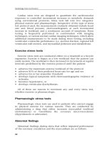
• Characteristic electrocardiographic changes occur during exercise
when the increased myocardial metabolic activity provokes
myocardial ischemia, that is ≥2 mm of planar depression of the ST
segments in the electrocardiographic leads that represent the
distribution of the stenotic coronary artery (Figure 2.12).
• Myocardial ischemia is frequently accompanied by a decline in left
ventricular contractile performance heralded by hypotension,
which denotes proximal severe triple coronary artery stenoses,
stenosis of the left main coronary artery, or ventricular dysfunction.
Decline in left ventricular contractile function can be detected by
imaging studies conducted during or immediately following stress.
• The increased cardiac workload associated with stress may provoke
abnormalities and non-uniformities of myocardial coronary
perfusion, which may be detected by imaging studies conducted
immediately following stress. Alternatively, they may result in
electrical irritability and ventricular arrhythmias that are evident
electrocardiographically.
Cardiac non-invasive imaging and stress testing
53
Baseline ECG
Stress ECG
IVR
VL
VF
VR
VL
VF V
3
V
6
V
1
V
2
V
4
V
5
V
3
V
6
V
2
V
5
V
1
V
4
I
II
II
III
III
Figure 2.12 A pre-exercise (upper) and post-exercise (lower) 12-lead
electrocardiogram (ECG) demonstrating normal ST segments at rest, which
become significantly depressed (below baseline) diffusely in both anterolateral
(leads V
2
–V
6
) and inferior (leads II, III, and aVF) distribution. The diffuse changes
and ST elevation in lead aVR suggest severe left mainstem or proximal left anterior
descending coronary artery stenosis as the anatomic lesion responsible for the
electrocardiographic abnormalities
Cardiac nuclear imaging
Cardiac nuclear imaging provides important information on
cardiac function by employing radiotracer techniques and external
detection equipment. The most frequently used nuclear techniques
are myocardial perfusion imaging, radionuclide angiography, and
metabolic imaging.
Cardiac nuclear imaging is based on the detection of γ rays emitted
by radiopharmaceutical agents administered to patients and
measured by large detectors (i.e. γ cameras) outside the body. The
images created represent various functions of the heart depending on
the type of radiopharmaceutical employed. We briefly discuss
radiopharmaceuticals and detection systems below.
Radiopharmaceuticals
Radiopharmaceuticals are compounds that have two distinct
elements. One is the radioactive material, called a radionuclide, which
is attached to a molecule that distributes in the body according to a
given physiologic function. Radionuclides are unstable elements that
decay to a more stable state by emitting particles or photons from
their nuclei that can be detected. This process is called radioactive
decay. In general, radioactive decay may occur in one of three forms:
α, β, and γ. Both α and β decay involve the emission of particles,
whereas γ decay is characterized by the emission of γ rays
(electromagnetic radiation). Clinical nuclear imaging is based entirely
on γ emitting radionuclides because γ rays pose the least harmful
effect to tissues while having sufficient penetrating power to traverse
the body tissues and be detected externally.
The most widely used radionuclide for clinical testing is
technetium-99m (Tc-99m). This radionuclide is produced by a
generator made of molybdenum-99, which decays to Tc-99m and
emits γ rays of 140 KeV (kiloelectron volts) energy. Tc-99m decays
with a half-life of 6 hours. Another type of radionuclide used in
cardiac imaging is the positron emitting radioisotope. This element
decays by emitting a “positron” from the nuclei, which is a particle of
the same energy (511 KeV) as an electron but is positively charged.
This particle immediately interacts with an electron in the
surrounding matter in a process known as annihilation. Two γ rays of
the same energy and opposite direction are emitted from that process.
An example of a positron emitting radionuclide is fluorine-18, which
has a half-life of 110 min.
The radionuclides above can be attached to other molecules
that have a known distribution in the body and thus form a
Cardiology Core Curriculum
54
radiopharmaceutical agent. An example of a radiopharmaceutical
is Tc-99m sestamibi, which is a myocardial blood flow (MBF) tracer
made of two components: the radionuclide (i.e. Tc-99m) and the
pharmaceutical (i.e. sestamibi). Sestamibi distributes in the myocardium
in proportion to MBF, and its distribution pattern can be detected
because of the presence of technetium in the molecule. In patients
who have suffered heart attacks, abnormal distribution of tracer helps
to define the area of infarction.
12
Detection systems
The overall principle of the detection system is based on the theory
that certain types of crystal emit light when struck by γ rays. One
example of this type of crystal is sodium iodide, which is used in most
clinical scanners. The light output of the crystal is amplified many
times by photomultiplier tubes and by complex electronic circuitry.
This light output can be localized to represent a three-dimensional
map of radionuclide distribution within the myocardium. With the
aid of computers, this information is digitized and images produced
and displayed on computer screens or x ray film.
Large detectors, called γ cameras, are used to image large parts of the
body. The cameras may produce a single image in a given projection
(planar technique) with respect to the organ of interest (for example,
heart or liver), or multiple projections that can be reconstructed into
images known as tomograms. The advantage of the tomographic method
is that the three-dimensional distribution of the radiopharmaceutical
may be determined in detail while avoiding the overlap of structures
that occurs with planar images. This technique is called single photon
emission computed tomography (SPECT) and is designed to image
radionuclides that emit single photons. The other major technique
available is known as positron emission tomography (PET), which is
an imaging method used to detect positron emitting radionuclides.
This is a more complex, but more accurate method and is based on a
principle called coincidence counting. Positron emitting isotopes
decay by giving off two γ rays in exactly opposite directions, each with
the same energy. Using sophisticated electronics, the origin of the
γ rays may be localized within the body more precisely, resulting in a
three-dimensional map of perfusion or metabolism, depending on the
tracer used (see below).
Myocardial perfusion imaging
Myocardial perfusion tracers are used to estimate non-invasively
the relative amounts of blood flow to various regions of the heart.
Cardiac non-invasive imaging and stress testing
55
This test is the most commonly used technique in cardiac nuclear
imaging. In this section we briefly discuss the various radiotracers
available for the assessment of regional myocardial perfusion. We review
a number of tracers based on the mechanism by which these
radiopharmaceuticals measure MBF.
Broadly speaking, there are two major categories of myocardial
perfusion tracers (Table 2.1): those that are retained in the
myocardium and those that are diffusible.
Mechanically retained, or labeled albumin microspheres are not
used clinically because they are large particles (15 µm in diameter),
which if injected intravenously become trapped in the lung capillaries
rather than in the myocardium. In order to be used as myocardial
perfusion tracers, these microspheres must be injected into the left
sided circulation via a catheter placed into the left atrium or ventricle,
which involves a more invasive procedure.
Tracers retained in the myocardium via non-mechanical means are
the most commonly used perfusion agents in clinical practice. These
tracers are retained in the myocardium in proportion to MBF, and
include thallium-201 and Tc-99m sestamibi for SPECT imaging, and
nitrogen-13-ammonia and rubidium-82 for PET imaging.
The retention process takes place during the first few minutes after
tracer injection, and the distribution of activity represents the blood
flow at the time of the injection. This characteristic allows us to image
Cardiology Core Curriculum
56
Table 2.1 Summary of tracers used in myocardial perfusion imaging
Type of tracer Examples
Retained in myocardium Mechanical retention Technetium-99,
*
carbon-11
†
(based on microsphere or gallium-68
†
labeled
size) albumin microspheres
Metabolic retention Thallium,
*
rubidium-82,
†
potassium analogs
(Na/K energy requiring
pump), nitrogen-13
ammonia
†
(retained as
glutamine)
Retained in proportion Technetium-99 sestamibi
to electrical membrane
gradients
Diffusible Hydrophilic Oxygen-15 water
†
Lipophilic Carbon-11 or oxygen-14
butanol
†
Partially diffusible Technetium-99 teboroxime
*
These tracers may be used in
*
single photon emission computed tomography
(SPECT) and
†
positron emission tomography (PET).
patients beginning several minutes after the injection, and permits
longer scan times, which improves image resolution. Further
improvements in resolution are possible using a new acquisition
method synchronized to the cardiac cycle, which also provides
information on left ventricular wall thickening – a measure of
regional function. The ability of tracers to remain in the myocardium
for minutes to hours after administration allows us to perform
interventions such as exercise or pharmacologic stress testing in
patients and then image MBF during flexible time intervals afterward.
The most typical example is the perfusion study performed with
exercise. Exercise is performed in the exercise laboratory, adjacent to
the imaging room, with patients using a treadmill or bicycle
ergometer. The perfusion tracer is administered at peak exercise, and
the patient is allowed to recover for a few minutes to an hour
(depending on the radioisotope), followed by perfusion imaging.
Typically, with thallium agents, a resting scan is performed
approximately 3 hours after the stress imaging, to allow for
comparison with the stress state (Figure 2.13). A smaller dose of
thallium is usually given just before the rest image in order to detect
better ischemic but viable myocardium. Patients with severely
ischemic myocardium may be imaged at 24 hours to allow for further
redistribution of the isotope. Using a tracer with a longer half-life
and higher energy such as technetium sestamibi has an important
practical advantage in that higher quality images are obtained. Thus,
the stress intervention can be performed before the resting
examination and, should the stress perfusion images be normal, this
will eliminate the need for a resting examination.
None of the diffusible tracers are used clinically because of
the complicated scanning techniques required. However, these
techniques are very accurate for quantifying MBF and are used mainly
for clinical research. An example of this group of agents is oxygen-15
water, which is employed with PET scanning.
Imaging protocols
Myocardial perfusion imaging is usually
performed with some form of stress test when used for the diagnosis
of coronary artery disease (CAD) or for evaluation of treatment in
patients with known CAD (for example, angioplasty, bypass surgery,
or medications). Stress testing is necessary to detect regional
differences in myocardial perfusion due to occlusive CAD, because
MBF at rest is not decreased even in the presence of coronary stenoses
with up to 80% reduction in normal vessel diameter. By increasing
MBF with exercise or coronary vasodilators such as dipyridamole or
adenosine, myocardial regions supplied by significantly diseased
coronary vessels may be detected because of their inability to increase
MBF to a degree similar to that in regions supplied by normal vessels.
Cardiac non-invasive imaging and stress testing
57
Given that the radiopharmaceuticals are carried in the blood and
extracted by the myocardium, significantly less tracer is distributed to
areas supplied by diseased vessels, and therefore the total amount of
radiotracer delivered to these regions is less than to normal areas. This
result produces a low intensity segment, or defect, on the scan in
regions subserved by diseased vessels, and permits not only the
detection of the presence of CAD but also assists with localizing the
disease to specific coronary arteries (Figure 2.14).
The most frequently used stress test in clinical practice is the
exercise test. With exercise, there is an increment in heart rate,
blood pressure, and contractility that increases with myocardial
metabolism, and in turn increases MBF in order to increase oxygen
delivery to meet the increased myocardial oxygen demand. An
appropriate increment in MBF in response to the oxygen demand can
be reached in those segments of the myocardium that are supplied
by non-stenotic arteries. This increment in MBF with maximal
exercise or maximal vasodilatation is called the coronary flow reserve,
and is approximately three to four times the normal resting MBF.
Cardiology Core Curriculum
58
Figure 2.13 A normal thallium perfusion scan is shown that illustrates the three
views used for planar clinical cardiac imaging. In the short axis view the right
ventricle is faintly seen to the left of the image, adjacent to the interventricular
septum. The left ventricular (LV) lateral wall is seen to the right of the image. In the
horizontal long axis image, the LV apex is at the top of the image, and the lateral
wall is to the right. In the vertical long axis view, the anteroseptum is seen at the
top of the image, and the LV apex is to the right. Note the homogeneous intensity
pattern in all LV myocardial regions
However, in segments perfused by a stenotic artery there is an
additional resistance in the vessel that prevents an appropriate
increment in MBF. Therefore, patients with CAD will not match their
increased myocardial oxygen demand, resulting in an imbalance
between oxygen demand and supply and producing myocardial
ischemia. This supply/demand mismatch and ischemia may result in a
typical syndrome of retrosternal chest pain associated with sweating,
shortness of breath, and radiation of the pain along the left arm to the
elbow or fingers (angina). In other patients, there may be few or no
symptoms at all, despite electrocardiographic changes demonstrating
myocardial ischemia (silent ischemia). Under these conditions
normally perfused myocardium will demonstrate high MBF and the
region supplied by the stenotic vessel will have lower MBF. If we inject
a myocardial perfusion tracer at this point, the resulting image will
Cardiac non-invasive imaging and stress testing
59
Figure 2.14 Perfusion defects. A transient per fusion defect is seen in the upper
panel that is consistent with exercise-induced ischemia. During stress, the inferior
walls in both the short axis and vertical long axis views exhibit decreased signal
intensity, and therefore decreased perfusion, relative to the remaining walls. The
signal intensity normalizes or reverses in the resting image, demonstrating a
reversible defect. In the lower panel, a fixed defect in a similar location is shown.
A defect noted on the stress images show no reversibility upon rest, which is
consistent with infarction or non-viable tissue. Reinjection of a small amount of
thallium at the time of rest images improves detection of severely ischemic but
viable myocardium
show a regional perfusion imbalance or defect that is not present in a
resting image, when MBF would be more comparable.
There are pharmacologic stress tests that can be used to provoke
these same transient perfusion defects, which involve the use of
potent coronary vasodilators or β-agonists that increase myocardial
oxygen consumption in a similar manner to exercise.
Clinical applications
The major clinical applications of myocardial
perfusion imaging are:
• diagnosis of CAD
• risk stratification in patients with known chronic CAD
• treatment evaluation in patients with known CAD, in particular
following revascularization techniques such as percutaneous
transluminal coronary angioplasty or coronary artery bypass
grafting
• risk stratification after acute myocardial infarction
• evaluation of patients with CAD and left ventricular dysfunction
• evaluation of patients with “silent ischemia”.
Radionuclide angiography
Ventricular function is most commonly assessed with a technique
called multigated image acquisition scanning, which uses a “blood
pool” method approach. Blood labeled with technetium-99 remains
in the intravascular space, or blood pool, and provides a means to
measure the end-diastolic and end-systolic volumes (EDV and ESV,
respectively) of the heart non-invasively. The ejection fraction, or
(EDV – ESV)/EDV, is a common measure of global ventricular
performance. If a stress test is performed after baseline imaging, then
the cardiac “reserve” can be estimated, with a fall in exercise ejection
fraction indicating abnormal reserve.
Metabolic imaging
PET scanning is a technique that can assess myocardial perfusion
and metabolism somewhat more rigorously than thallium scanning.
13
Nitrogen-13-ammonia is a common perfusion isotope, while
18
fluorine deoxyglucose is used as the metabolic tracer that evaluates
the ability of myocytes to use glucose (Figure 2.15). One potential
advantage to PET scanning is that the study may be performed at rest;
however, the use of the above isotopes requires a cyclotron for
production.
Cardiology Core Curriculum
60
Cardiac non-invasive imaging and stress testing
61
Figure 2.15 In this positron emission tomography (PET) image, the perfusion agent
nitrogen-13 ammonia (
13
NH
3
; upper panels) demonstrates decreased resting blood
flow to the lateral wall, as seen in both the short axis and horizontal long axis views.
The metabolic tracer 2-deoxy-2-[
18
F]fluoro-D-glucose (
18
FDG) depicts regions in which
the conversion from free fatty acid substrate use (normal metabolism) to glycolytic
metabolism (ischemic zones) has occurred. High signal intensities in the
18
FDG
images (bottom panels) are seen in segments corresponding to the hypoperfused
regions, which is indicative of ischemia-related changes in metabolism
Case studies
Case 2.1
A 32-year-old male tax accountant presented with a 2 year history
of progressive shortness of breath on exertion such that he could only
walk two blocks on flat ground or climb five stairs. He had never
complained of chest pain or palpitations, and was a non-smoker and
non-drinker.
When aged 15 years, at a school sports medical examination, a
cardiac murmur was detected. In his remote past he had sustained two
unexplained syncopal episodes that were unrelated to exertion or
posture. His father, who had always enjoyed good health as an active
athlete and non-smoker, died suddenly from a “heart attack” at age
37 years. His father’s death prompted an office visit to a cardiologist
who, in addition to eliciting an ejection systolic murmur at the left
sternal edge, recorded a 12-lead electrocardiogram, which revealed
left ventricular hypertrophy and repolarization abnormalities. A
clinical working diagnosis of congenital aortic valve stenosis was
made, and an outpatient two-dimensional echocardiogram was
scheduled, which excluded aortic valve stenosis, but showed left
ventricular hypertrophy with normal systolic function, and no
further recommendations were made. At age 30 years he noticed
reduction in his exercise tolerance and was found to have moderate
mitral regurgitation, and because of the family history of premature
heart disease he underwent cardiac catheterization and coronary
arteriography.
Catheterization demonstrated a cardiac index of 4·1 l/min per m
2
;
ejection fraction 73%; end-diastolic volume index 55 ml/m
2
;
end-systolic volume index 15 ml/m
2
; left ventricular pressure
135/23 mmHg; aortic pressure 102/65 mmHg; a “v” wave in the
pulmonary capillary wedge pressure of 41 mmHg; pulmonary artery
systolic pressure 46 mmHg; and right atrial pressure 9/7 mmHg (mean
6 mmHg). Contrast angiography showed a hyperdynamic left
ventricle with no segmental wall motion abnormality, grade 3+ mitral
regurgitation, an enlarged left atrium, and normal coronary arteries.
In view of the progressive reduction in exercise capacity, the moderate
pulmonary hypertension, and moderately severe mitral regurgitation,
he was referred for mitral valve repair/replacement.
Examination. Physical examination: the patient was comfortable
lying flat. Pulse: 78 beats/min, brisk upstroke, full volume. Blood
pressure: 105/60 mmHg in the right arm. Jugular venous pulse:
normal. Cardiac impulse: forceful, double impulse, regular rhythm.
First heart sound: normal. Second heart sound: reversed splitting.
Fourth heart sound was present. Apical grade 3/6 holosystolic
murmur radiating to axilla, grade 2/6 ejection systolic murmur at
mid-left sternal edge, which increased with Valsalva. Chest
examination: normal air entry, no rales or rhonchi. Abdominal
examination: soft abdomen, no tenderness, and no masses. Normal
liver span. No peripheral edema. Femoral, popliteal, posterior tibial,
and dorsalis pedis pulses: all normal volume and equal. Carotid
pulses: full volume, rapid upstroke, no bruits. Optic fundi: normal.
Investigations. Laboratory findings: normal. Electrocardiogram:
sinus rhythm at 78 beats/min, normal intervals and frontal QRS axis,
severe left ventricular hypertrophy with small Q waves, and 2 mm ST
segment depression and T-wave inversion in leads V
4
through V
6
consistent with strain or lateral ischemia. Chest x ray: mild
cardiomegaly with left atrial enlargement and normal lung fields.
24-Hour ambulatory electrocardiographic monitoring: predominant
cardiac rhythm was sinus, occasional isolated premature ventricular
depolarizations, and three episodes of non-sustained ventricular
tachycardia, with the longest being an 11-beat run at a maximum rate
of 178 beats/min.
Cardiology Core Curriculum
62
Transthoracic two-dimensional echocardiogram. Asymmetric
hypertrophy of the interventricular septum; a small hyperdynamic
left ventricle; systolic anterior motion of the mitral valve; a 30 mmHg
left ventricular outflow tract gradient in systole at rest, which
increased to 64 mmHg with Valsalva in late systole; enlarged left
atrium; and moderately severe mitral regurgitation by color flow
Doppler velocity mapping.
Clinical course. The patient underwent mitral valve surgery, from
which he made an excellent recovery and was discharged from
hospital on postoperative day 7 on β-adrenergic blocking agents. At
3 month follow up his exercise tolerance had increased to 12 blocks
on flat ground.
Questions
1. What are the differential diagnoses of a systolic murmur and
electrocardiographic left ventricular hypertrophy?
2. What is the diagnosis in this patient, and on what clinical and
echocardiographic criteria is the correct diagnosis based?
3. The patient married 6 months after discharge from hospital and
wished to start a family. What is the pattern of genetic inheritance
of his disease and how would you counsel the patient in this
regard?
4. Explain the mechanism of the increase in left ventricular outflow
tract gradient with Valsalva.
5. Why was the patient placed on β-adrenergic receptor blocking
agents?
6. What is the prognostic significance of non-sustained ventricular
tachycardia in this disease?
Answers
Answer to question 1
The differential diagnosis of an ejection systolic
murmur and left ventricular hypertrophy by electrocardiography
includes discrete anatomic lesions causing obstruction to left
ventricular ejection and increased pressure work on the left ventricle;
discrete subaortic stenosis; aortic valve stenosis; supravalvular stenosis;
bicuspid aortic valve with associated coarctation of the aorta;
hypertrophic obstructive cardiomyopathy; and systemic hypertension
with an unrelated innocent systolic murmur.
Answer to question 2
The diagnosis in this patient was hypertrophic
obstructive cardiomyopathy. This diagnosis was based clinically on
the auscultatory findings of an ejection systolic murmur with a
Cardiac non-invasive imaging and stress testing
63
forceful double apical impulse, brisk pulses with rapid upstroke to the
carotid pulses, and augmentation of the cardiac murmur with Valsalva.
The confirmatory two-dimensional echocardiographic findings
comprised asymmetric septal hypertrophy, systolic obliteration of the
left ventricular cavity with supranormal ejection fraction, systolic
anterior motion of the mitral valve, with the left ventricular outflow
tract gradient at rest increasing with Valsalva in late systole.
Answer to question 3
The genetic pattern of inheritance of
hypertrophic cardiomyopathy is dominant, and the patient should be
counseled so that he is cognizant of the likelihood of his progeny
having the same disease. The sudden and unexpected death of his
father, who was otherwise healthy, suggests that he had died from the
same disease.
Answer to question 4
Valsalva increases intra-thoracic pressure and
reduces venous return to the heart so that left ventricular end-
diastolic volume is reduced and systolic anterior motion of the mitral
valve more easily obstructs the left ventricular outflow tract and
thereby augments the systolic gradient.
Answer to question 5
The patient was placed on β-adrenergic
receptor blocking agents to reduce augmentation of the systolic
outflow tract gradients in order to prevent or attenuate the increase in
left ventricular outflow tract gradient with exercise, which is at least
partly mediated by increased catecholamines. Another reason is to
slow the heart rate, which allows longer diastolic filling and greater
left ventricular end-diastolic volume. Furthermore, β-adrenergic
blocking agents may be efficacious in the treatment of non-sustained
ventricular tachycardia.
Answer to question 6
Non-sustained ventricular tachycardia
correlates with sudden death, which accounts for the yearly attrition
rate of approximately 5–8% of patients with familial hypertrophic
cardiomyopathy.
Case 2.2
A 47-year-old female Asian immigrant was brought to the
emergency room with a dominant sided dense hemiplegia and severe
expressive dysphasia. The history obtained from a relative was limited
but included long-term shortness of breath on minimal exercise and
at night, requiring three pillows to sleep, and weight loss over the
previous 6 months.
Cardiology Core Curriculum
64
In early childhood she spent 1 year away from school convalescing
following an acute illness, which consisted of a painful migratory
arthralgia with swelling of both knees and ankles but with no other
stigmata. She was discouraged from playing games and took penicillin
tablets once daily until adulthood. She remained well and next saw a
physician during the last trimester of her second pregnancy, when she
developed an episode of palpitations and became light-headed. When
she was seen by a cardiologist she was in regular rhythm and in no
distress. However, a murmur was detected, which was thought to be
due to increased blood flow velocity associated with the volume
overload state of pregnancy, and no follow up was arranged.
Five years later she noted progressive shortness of breath,
intermittent palpitations provoked by exertion, and was finally
admitted for investigation following hemoptysis. She declined cardiac
catheterization but agreed to have an echocardiogram and was
discharged home on digoxin 0·25 mg/day, furosemide 40 mg/day,
potassium supplements, and warfarin at a dose to maintain an
International Normalized Ratio of 2·5–3·0.
Examination. Physical examination: the patient had a right-sided
neurologic deficit. She was only comfortable at 45°. Pulse: 152
beats/min, irregularly irregular. Blood pressure: 95/70 mmHg. Jugular
venous pulse: 12 cm at 45°. Cardiac impulse: parasternal lift, palpable
P
2
. First heart sound: loud. Second heart sound: split with loud P
2
. At
apex was opening snap close to P
2
. Mid-diastolic murmur at apex.
Chest examination: normal air entry, basal crepitations. Abdominal
examination: enlarged, tender liver. No peripheral edema. Carotid
pulses: normal, no bruits.
Investigations. Laboratory findings: normal hemoglobin, hematocrit,
white blood cell count, platelets, electrolytes, and creatinine. Aspartate
aminotransferase 198 U/l (3·3 µkat/l); alanine aminotransferase 33 U/l
(5·6 µkat/l); International Normalized Ratio 1·3. Electrocardiogram:
atrial fibrillation at 167 beats/min, right axis deviation, right
ventricular hypertrophy, and T-wave and ST-segment depression
throughout the limb and chest leads. Chest x ray: cardiomegaly,
enlargement of the left and right atria and main pulmonary artery,
prominence of the left atrial appendage, and elevation in the left main
bronchus at the carina. Calcification of the mitral annulus, small left
pleural effusion, septal (Kerley B) lines, and cephalization of the upper
lobes of the lung were also noted.
Echocardiogram. Echocardiography demonstrated a heavily calcified,
severely stenotic mitral valve with shortened chordae, and
calcification extending from the valve leaflets to the tips of the
papillary muscles, but with no mitral regurgitation. Left ventricular
size, wall thickness, and function were normal. The left atrium was
dilated with laminated mural thrombus on the left atrial wall behind
Cardiac non-invasive imaging and stress testing
65
the posterior aortic root. The aortic valve was trileaflet and normal,
the right atrium and right ventricle were both dilated with moderate
tricuspid regurgitation through a normal tricuspid valve. A small
hemodynamically unimportant pericardial effusion was present.
Doppler assessment revealed a peak gradient across the mitral valve of
28 mmHg (mean 14 mmHg) and a valve orifice area of 0·7 cm
2
calculated from the pressure half-time, and a pulmonary artery systolic
pressure of 68 mmHg calculated from the tricuspid regurgitant jet.
Hospital course. The patient was not considered a candidate for
mitral balloon valvuloplasty because of the extensive calcification of
the valve leaflets and subvalve apparatus, and the presence of left
atrial thrombus. She underwent mitral valve replacement with a
mechanical prosthesis 3 months later when the risk of exacerbating
her neurologic deficit by cardiopulmonary bypass was considered to
be less likely, and she was discharged on warfarin with an
International Normalized Ratio of 2·9.
Questions
1. What are the clinical diagnoses? What was her childhood illness
and the likely rhythm disturbance during pregnancy?
2. What factors contributed to the neurologic event?
3. Explain the auscultatory findings of a loud first heart sound, loud
P
2
, and the clinical significance of the closeness of the opening
snap to P
2
.
4. Why was the liver enlarged and the liver function tests deranged?
5. Explain the abnormalities on the electrocardiogram.
6. Describe the chest x ray findings that support the clinical
diagnosis.
7. What additional hemodynamic information would be obtainable
at cardiac catheterization?
8. Why was a mechanical rather than a biologic mitral prosthesis
selected for mitral valve replacement in so young a woman?
Answers
Answer to question 1
The clinical diagnoses are mitral stenosis and
cerebral thromboembolism. The disease in childhood was rheumatic
fever, which caused her polyarthralgia even though during the acute
phase there was no rheumatic carditis or cardiac murmur detected.
The rhythm disturbance with light-headedness during the last
trimester of pregnancy was probably paroxysmal rapid atrial
fibrillation, which spontaneously reverted to normal sinus rhythm.
Cardiology Core Curriculum
66
Answer to question 2
The factors that contributed to her neurologic
event include mitral valve stenosis with resultant slow flow in the
left atrium, left atrial enlargement, atrial fibrillation, and poor
anticoagulant status with an International Normalized Ratio of 1·3.
Answer to question 3
The loud first heart sound is due to the closure
of rheumatically thickened mitral valve leaflets; the loud P
2
is due to
the presence of pulmonary hypertension. The closeness of the
opening snap to P
2
relates to the amplitude of left atrial pressure and
the severity of the mitral stenosis, so that the closer the opening snap
is to P
2
, the more severe the mitral stenosis.
Answer to question 4
The liver is enlarged because of systemic
venous hypertension from tricuspid regurgitation, and the deranged
liver function tests indicate acute distension of the liver from
congestive heart failure or from chronic elevation of systemic venous
pressure and “cardiac cirrhosis”.
Answer to question 5
Atrial fibrillation occurs from atrial dilatation
and is part of the natural history of chronic rheumatic heart disease.
The right axis deviation and right ventricular hypertrophy are due to
pulmonary hypertension, and the T and ST abnormalities are digitalis
effects.
Answer to question 6
Left atrial enlargement, prominence of the left
atrial appendage and main pulmonary artery, elevated left main
bronchus, intracardiac calcification of the mitral valve apparatus, and
cephalization of the upper lobes of the lungs.
Answer to question 7
None.
Answer to question 8
The patient is postmenopausal and so
problems with subsequent pregnancy are not an issue. She is in
established atrial fibrillation with a dilated left atrium, and will
therefore require anticoagulation. Importantly, the primary failure
rate of bioprostheses at 10 years is approximately 20%, so she would
need to undergo at least two additional valve replacements.
Therefore, the use of a durable prosthesis over the long term is
desirable, and thus a mechanical prosthesis is the treatment of choice.
Case 2.3
A 59-year-old male business executive was brought to the
emergency room complaining of sudden onset of severe central chest
Cardiac non-invasive imaging and stress testing
67
pain (which he graded 10/10) radiating through to his back,
associated with diaphoresis and nausea. He had never had chest pain
previously and played golf once weekly without any exercise
intolerance or shortness of breath. His past medical history included
an arthroscopy for meniscectomy at age 37 years, and hypertension
treated for 11 years initially with β-adrenergic blocking agents but for
the past 4 years with once daily angiotensin-converting enzyme
inhibitor therapy. Risk factors for coronary artery disease included a
family history (his father had sustained a myocardial infarction at age
61 years and underwent coronary artery bypass vein grafting; his
mother and younger brother had hypertension), he was not diabetic
or a smoker, and had a cholesterol of 230 mg% (5·9 mmol/l).
Examination. Physical examination: the patient was in acute
distress, and was cold, clammy, and complaining of pain in his
mid-back. Pulse: 110 beats/min, normal character. Blood pressure:
160/100 mmHg in right arm. Jugular venous pulse: 8 cm. Cardiac
impulse: prominent, displaced to anterior axillary line. First heart
sound: normal. Second heart sound: split normally on inspiration. No
added sounds. Decrescendo murmur at upper left sternal border.
Chest examination: normal air entry, no rales or rhonchi. Abdominal
examination: soft abdomen, no tenderness, and no masses. Normal
liver span. No peripheral edema. Pulses absent below femoral on the
right, and his right foot was colder than his left. Carotid pulses:
normal, no bruits. Optic fundi: normal.
Investigations. Laboratory findings: normal electrolytes and
creatinine. Two sets of cardiac enzymes normal. Electrocardiogram:
sinus tachycardia, left axis deviation (–32°), minor QRS widening,
left ventricular hypertrophy with non-specific T wave abnormalities
throughout. Chest x ray (anteroposterior): widening of the
mediastinum and an “unfolded aorta”, with cardiomegaly but clear
lung fields. Two-dimensional echocardiogram: mildly dilated left
ventricle with concentric hypertrophy; normal systolic function; no
segmental wall motion abnormalities; mild aortic regurgitation by
color flow Doppler; a dilated aortic root with an intimal flap in the
ascending aorta, which could be identified as extending to the
abdominal aorta from the subxiphoid images; and no pericardial
effusion. Magnetic resonance imaging confirmed the diagnosis and
delineated the extent of the disease and the complications.
Clinical course. Following his echocardiogram, the patient
complained of a further episode of severe interscapular pain only
partly relieved by intravenous morphine, after which his blood
pressure dropped to a systolic pressure of 70 mmHg. Examination
demonstrated that he could no longer move his legs, his right leg
remained cold and pulseless, he had a sensory level at his mid-thorax
(T9), and had not passed urine since admission, although he was still
Cardiology Core Curriculum
68
mentating normally. Heart sounds and aortic regurgitant murmur
were unchanged. On his way to the operating room he became
profoundly hypotensive and developed sinus bradycardia, which was
followed quickly by a cardiac arrest from which he could not be
resuscitated. A postmortem was conducted.
Questions
1. What was the differential diagnosis of the patient’s chest pain?
2. What in the clinical history and physical examination made you
select your working diagnosis?
3. Explain the possible mechanisms for aortic regurgitation. Did any
of these potential etiologies elucidate the seriousness or emergent
nature of the patient’s management? Does any classification of the
disease in question spring to mind?
4. What was the significance of the cold right leg?
5. Why was the patient unable to move his legs, and what was the
significance of the sensory level at T9?
6. The presence of the intimal flap seen by echocardiography and
magnetic resonance imaging was indicative of what?
7. What additional information was provided by magnetic
resonance imaging that was unavailable by transthoracic two-
dimensional echocardiography?
8. What findings would you anticipate at postmortem examination?
Answers
Answer to question 1
Acute myocardial infarction and acute aortic
dissection.
Answer to question 2
A history of hypertension associated with chest
pain radiating to the back does not distinguish between myocardial
infarction and aortic dissection, and the presence of aortic
regurgitation is a common feature of type A aortic dissection, but
aortic regurgitation may also occur in patients with hypertension.
However, the diagnosis of aortic dissection is strongly suggested by
the loss of pulses in the right leg.
Answer to question 3
The probable mechanisms for the aortic
regurgitation include acute dissection of the ascending aorta,
involving the aortic root with prolapse of the valve leaflets, and
dilatation of the aortic root due to longstanding systemic
hypertension. Aortic dissections are classified as type A if they involve
the ascending aorta or aortic arch, and as type B if they are limited to
the descending thoracic aorta, usually beginning at the origin of the
Cardiac non-invasive imaging and stress testing
69
left subclavian artery. The treatment of acute type A dissection is
urgent surgical repair, whereas type B dissections without rupture or
compromise of an organ or limb have a similar outcome with medical
or surgical repair. The presence of aortic regurgitation and the two-
dimensional echocardiographic confirmation of type A dissection
necessitated emergent surgical repair (transesophageal echocardiography
would have been a better choice than transthoracic echocardiography).
Answer to question 4
The cold pulseless right leg was caused by
dissection and subsequent occlusion of the right common iliac artery,
resulting in an ischemic right leg.
Answer to question 5
The type A dissection had occluded the arteria
magna, which has a mid-thoracic origin and supplies the anterior
spinal arteries to the mid-thoracic cord. The anterior spinal artery
supplies the corticospinal (motor), and anterior and lateral
spinothalamic tracts (sensory), interruption of which resulted in
motor paralysis of the legs and the sensory level at T9.
Answer to question 6
The intimal flap is the dissection between the
intimal and medial layers of the aortic wall. The free intimal flap is
identified by both two-dimensional echocardiography and magnetic
resonance imaging.
Answer to question 7
The magnetic resonance imaging scan
demonstrated the extent of the aortic dissection, the sites of the
entrance and exit of the dissection, as well as the occlusion of the
right common iliac artery, which were unavailable by
echocardiography. Because the transesophageal echocardiography
probe cannot be passed beyond the stomach, the proximal abdominal
aorta is the limit of echocardiographic evaluation.
Answer to question 8
Postmortem examination demonstrated
similar anatomic findings in terms of the dissection, but in addition
exsanguination caused by rupture of the descending thoracic aorta
into the left pleural cavity.
Case 2.4
A 48-year-old male physician saw his local physician complaining
of a sensation of heaviness in the chest associated with weakness and
light-headedness that had progressed over the prior 3 weeks. The pain
did not radiate, but was provoked by anxiety and by decreasing
amounts of exertion over the 3 weeks following its onset and was
relieved quickly by rest. He had brought forward his medical
Cardiology Core Curriculum
70
appointment because of an episode the day before in which his chest
discomfort occurred while at rest reading the newspaper. His past
medical history was unremarkable, with no previous illness or
admissions to hospital. His risk factors included a remote smoking
history (he had quit 15 years previously), no hypertension or diabetes,
unknown cholesterol, and a positive family history for early coronary
disease (his father had had two myocardial infarctions at ages 50 and
57 years, one brother had coronary artery bypass graft surgery at age
53 years, and his eldest brother had a myocardial infarction at age
56 years but is now symptom free on medical therapy).
Examination. Physical examination: the patient appeared normal.
Pulse: 78 beats/min, normal character. Blood pressure: 135/80 mmHg.
Jugular venous pulse: normal. Cardiac impulse: normal. First heart
sound: normal. Second heart sound: split normally on inspiration. No
added sounds or murmurs. Chest examination: normal air entry, no
rales or rhonchi. Abdominal examination: soft abdomen, no tenderness,
and no masses. Normal liver span. No peripheral edema. Femoral,
popliteal, posterior tibial, and dorsalis pedis pulses: all normal volume
and equal. Carotid pulses: normal, no bruits. Optic fundi: normal.
Investigations. Laboratory findings: normal levels of sodium,
potassium, blood urea nitrogen, creatinine, creatine phosphokinase,
and creatine kinase-MB. Total cholesterol: 285 mg/dl (7·4 mmol/l).
Electrocardiogram: sinus rhythm, normal QRS axis and intervals,
T-wave flattening in leads V
1
–V
3
. Chest x ray: normal heart size and
clear lung fields. Stress thallium was performed using a standard (Bruce)
protocol. The patient developed his typical chest heaviness, became
hypotensive and presyncopal after 3 min exercise, and had 3 mm
planar depression of the ST segments in leads V
1
through V
4
with
multiple ventricular extrasystoles. Thallium scan revealed a large
reversible defect involving the whole of the anterior left ventricular wall
and anterior septum. Emergent cardiac catheterization: left ventricular
end-diastolic pressure 19 mmHg; no left ventricular angiogram was
performed, and during intubation of the left coronary artery arterial
systolic pressure dropped to 70 mmHg, so that only limited views of the
left coronary artery were obtained; the right coronary artery was
normal. The patient was referred for urgent cardiac surgery.
Clinical course. The patient underwent surgery without any
complications and was discharged home on postoperative day 6.
Questions
1. What was the underlying reason for the hypotension and
presyncope at such a low workload, the electrocardiographic
changes and electrical instability?
2. What were the likely diagnoses?
Cardiac non-invasive imaging and stress testing
71
3. Explain the significance of the “reversible” defect on the thallium
scan and the urgency for the cardiac catheterization.
4. Why did hypotension supervene during left coronary
angiography and not during injection of the right coronary
artery?
5. What were the likely findings of the cardiac catheterization?
6. Why was the patient referred for surgery rather than undergoing
angioplasty?
7. What medical therapy, if any, would you institute following
hospital discharge?
Answers
Answer to question 1
Hypotension during stress testing indicates
that the myocardium cannot meet the increased demands of exercise
and fails to generate a normal blood pressure response. This is usually
due to severe proximal triple vessel disease (left anterior descending,
left circumflex, and right coronary artery) or left mainstem coronary
artery stenosis, or severe ventricular dysfunction. The impressive
anterior electrocardiographic ST-segment depression demonstrates
extensive anterior ischemia at a low workload in the territory of the
left anterior descending coronary artery. The electrical instability and
ventricular ectopy are probably induced by myocardial ischemia.
Answer to question 2
Left mainstem coronary artery stenosis or
severe proximal triple coronary artery disease.
Answer to question 3
The reversible defect on the thallium scan
indicates a large region of left ventricular myocardium that has
severely reduced perfusion, even during mild exertion, which is
completely viable and is normally perfused at rest. This suggests that
if occlusion of the vessel occurred, then the area at risk for infarction
would be large. The reason for the urgent cardiac catheterization was
the probable diagnosis of left main coronary stenosis.
Answer to question 4
Hypotension supervened during left coronary
artery injection because when the catheter engaged the severely
diseased left coronary artery it completely obstructed flow, resulting
in myocardial ischemia. The right coronary artery was normal and the
catheter did not occlude antegrade flow.
Answer to question 5
Coronary arteriography demonstrated a 95%
stenosis of the left main coronary artery and an 80% tubular stenosis
in the mid left anterior descending artery, with normal left circumflex
and right coronary arteries.
Cardiology Core Curriculum
72
Answer to question 6
The treatment of left main coronary stenosis is
left internal mammary artery graft to the left anterior descending
artery and saphenous venous graft to the left circumflex artery.
Angioplasty is contraindicated for left main disease because of risk for
severe acute global ischemia and for acute dissection and closure
should complications occur.
Answer to question 7
Long-term cholesterol lowering therapy should
be instituted because this reduces the incidence of late cardiovascular
events in patients with coronary artery disease.
References
1 Henry WL, DeMaria A, Gramiak R, et al. Report of the American Society of
Echocardiography Committee on Nomenclature and Standards in Two-
dimensional Echocardiography. Circulation 1980;62 :212–7.
2 St John Sutton M, Kotler M, Oldershaw P, eds. Textbook of adult and pediatric
echocardiography and Doppler. London: Blackwell Science, 1996.
3 Skjaerpe T, Hegrenaes L, Hatle L. Noninvasive estimation of valve area in patients
with aortic stenosis by Doppler ultrasound and two-dimensional
echocardiography. Circulation 1985;72:810–8.
4 Currie P, Seward J, Reeder G, et al. Continuous wave Doppler echocardiographic
assessment of the severity of calcific aortic stenosis: a simultaneous Doppler-
catheter correlative study in 100 adult patients. Circulation 1985;71:1162–9.
5 Bonow R, Lakatos E, Maron B, Epstein S. Serial long-term assessment of the natural
history of asymptomatic patients with chronic aortic regurgitation and normal left
ventricular systolic function. Circulation 1991;84:1625–35.
6 Chan K, Currie P, Seward J, Hagler D, Mair D, Tajik A. Comparison of three Doppler
ultrasound methods in the prediction of pulmonary artery pressure. J Am Coll
Cardiol 1987;9:549–54.
7 Klein A, Cohn G. Clinical applications of doppler echocardiography in the
assessment of diastolic function. In: St John Sutton M, Kotler M, Oldershaw P, eds.
Textbook of adult and pediatric echocardiography and Doppler. London: Blackwell
Science, 1996:83–96.
8 Schiller N, Shah P, Crawford M, et al. Recommendations for quantitation of the left
ventricle by two-dimensional echocardiography. J Am Soc Echocardgr 1989;2:
358–67.
9 Marvick T, Nemec J, Stewart W, Salcedo E. Diagnosis of coronary artery disease
using exercise echocardiograph and positron emission tomography: comparison
and analysis of discrepant results. J Am Soc Echocardgr 1992;5:231–9.
10 Marcus M, Schelbert H, Skorton D, Wolf G, eds. Cardiac imaging: a companion to
Braunwald's heart disease. Philadelphia: WB Saunders Co, 1991.
11 Nienaber C, von Kodolitsch Y, Nicholas V, et al. The diagnosis of thoracic aortic
dissection by noninvasive imaging procedures. N Engl J Med 1993;328:1–9.
12 Zaret B, Beller G, eds. Nuclear cardiology: state of the art and future directions. St Louis:
Mosby, 1993.
13 Marshall R, Tillisch H, Phelps M, et al. Identification and differentiation of resting
myocardial ischemia and infarction in man with positron emission tomography,
18
F-labeled fluorodeoxyglucose and N-13 ammonia. Circulation 1983;67:766.
Cardiac non-invasive imaging and stress testing
73
74
3: Cardiac catheterization
CHARLES LANDAU
The advent of improved equipment and techniques plus an
increasing number of non-surgical options for the treatment of
cardiovascular disorders has increased the role of the cardiac
catheterization laboratory in the management of patients with a
variety of diseases that affect the vascular and valvular structures of
the heart.
Diagnostic cardiac catheterization
Definitions and overview
The term “cardiac catheterization” refers to the placement of
hollow plastic tubes (catheters) into the chambers of the heart or into
the epicardial coronary arteries with fluoroscopic guidance for the
purpose of acquiring pressure measurements, collecting blood
samples, and injecting radiographic contrast material in order to
opacify coronary arterial and ventricular anatomy. The latter
procedures are frequently referred to as arteriography or angiography.
A catheterization procedure usually consists, at a minimum, of
acquiring arterial pressures and coronary arteriography.
Right heart catheterization involves the passage of a catheter from
the venous access site via the vena cava to the right atrium, right
ventricle, and pulmonary artery. Pulmonary capillary wedge pressure
(PCWP), a reflection of left atrial pressure as transmitted retrograde
through the pulmonary vasculature, is determined by advancing the
catheter to a terminal segment of the pulmonary arterial tree.
Pressures are measured in each of the chambers, and blood samples
are drawn for the determination of oxygen saturations when
clinically indicated. Determinations of cardiac output are also a
routine aspect of this protocol in most catheterization laboratories.
When clinically indicated, an endomyocardial biopsy can be taken
from the right ventricle using a flexible biopsy forceps after
hemodynamic measurements have been acquired.
Left heart catheterization denotes the passage of a catheter from the
arterial access site retrograde to the left ventricle through the aortic
valve. In order to avoid the consequences of systemic emboli, patients
undergoing a left heart catheterization are usually systemically
anticoagulated with heparin. Pressure is recorded sequentially in the
left ventricle and then, after pull-back of the catheter, in the ascending
aorta. This series of measurements is performed to exclude a pressure
gradient across the aortic valve, a finding indicative of aortic stenosis.
Following these measurements a ventriculogram is performed with a
contrast injection into the left ventricle. The images of the opacified
left ventricle are used to assess regional wall motion, determine
ventricular volumes, calculate global left ventricular function as an
ejection fraction, and assess the presence and severity of mitral
regurgitation, which manifests as the appearance of contrast in the left
atrium during ventricular systole as a result of an incompetent mitral
valve. Selective coronary angiography is routinely performed after
ventriculography, during which the left main and right coronary
arteries are selectively cannulated, followed by contrast injections to
define coronary anatomy, including stenoses and vascular distribution.
Right and left heart catheterization combines the components
described above. In addition to the protocols outlined, simultaneous
measurements of the PCWP and left ventricular pressures are made in
order to evaluate the presence of a pressure gradient across the mitral
valve. This is a pathologic finding that is usually indicative of mitral
stenosis.
Indications
In clinical practice the majority of diagnostic cardiac
catheterizations are undertaken primarily for three reasons
(Table 3.1).
• To obtain anatomic information regarding the coronary arteries
and left ventricular function.
• To quantify hemodynamic abnormalities, including valvular
lesions.
• To determine the site and magnitude of an intracardiac shunt.
Cardiac catheterization should not be undertaken in any patient
who is unable or unwilling to provide informed consent. In addition,
the procedure is contraindicated in those circumstances that increase
the risks associated with the procedure, such as bleeding diatheses,
decompensated congestive heart failure, worsening renal function,
systemic infections and untreated hyperthyroidism, and in those
unwilling to provide informed consent. Because catheterization
involves radiation exposure, pregnancy should be excluded in women
of childbearing age.
Cardiac catheterization
75
Procedural techniques
Cardiac catheterization begins with vascular access. Because this
portion of the procedure involves some patient discomfort, it is
Cardiology Core Curriculum
76
Table 3.1 Indications for cardiac catheterization
Indication Reasons
Definition of cardiac
anatomy
Hemodynamic
assessment
Intracardiac shunt
assessment
To facilitate the diagnosis
of coronary artery
disease
Patients refractory to
medical therapy for
ischemia
Patients with suspected
high-risk anatomy (i.e.
left main or severe
three-vessel disease)
To determine the severity
of valvular abnormalities
To assess the severity
of hemodynamic
derangements in
myocardial or pericardial
disease
Equivocal non-invasive
evaluation
Persistent chest pain
despite negative
non-invasive tests
Dilated cardiomyopathy
Sudden cardiac death
Intolerant of medications
Limiting angina despite
adequate drug therapy
Unstable angina
Postmyocardial infarction
angina
Positive predischarge
exercise test following
myocardial infarction
Markedly positive exercise
test
Diffuse ECG changes
(ST depression) during
spontaneous episodes
of ischemia
Rapid onset pulmonary
edema, presumably due
to ischemia
Aortic stenosis
Aortic regurgitation
Mitral stenosis
Mitral regurgitation
Pulmonic stenosis
Dilated cardiomyopathy*
Hypertrophic obstructive
cardiomyopathy*
Restrictive cardiomyopathy
Constrictive pericarditis
Left ventricular diastolic
dysfunction
Atrial septal defect
Ventricular septal defect
Aorto-pulmonary window
Patent ductus arteriosus
*Can also be utilized to assess the efficacy of treatment.