CATHETER-RELATED INFECTIONS IN THE CRITICALLY ILL - PART 2 docx
Bạn đang xem bản rút gọn của tài liệu. Xem và tải ngay bản đầy đủ của tài liệu tại đây (724.52 KB, 19 trang )
6
Catheter-Related Infections in the Critically Ill
PATHOGENESIS
There are two major sources of IVD-related BSI: 1) colonization of the
IVD, catheter-related infection, and 2) contamination of the fluid
administered through the device, infusate-related infection Contaminated
infusate is the cause of most epidemic intravascular device-related BSIs; in
contrast, catheter-related infections are responsible for most endemic device-
related BSIs.
Understanding the pathogenesis of IVD-related BSIs is essential to
devising strategies for prevention of these infections; however few published
studies have determined the mechanism of IVD-related colonization and
infection using sophisticated molecular techniques to prove or disprove
potential routes of infection.
In order for microorganisms to cause catheter-related infection they must
first gain access to the extraluminal or intraluminal surface of the device
where they can adhere and become incorporated into a biofilm that allows
sustained colonization and, ultimately hematogenous dissemination
Microorganisms gain access to the implanted IVD by one of three
mechanisms: skin organisms invade the percutaneous tract, probably
facilitated by capillary action, at the time of insertion or in the days
following; microorganisms contaminate the catheter hub (and lumen) when
the catheter is inserted over a percutaneous guidewire or later manipulated;
or organisms are carried hematogenously to the implanted IVD from remote
sources of local infection, such as a pneumonia (Figure 1).
With short-term IVDs (in place <10 days) peripheral IV catheters,
arterial catheters and non-cuffed, non-tunneled CVCs most catheter-related
BSIs are of cutaneous origin, from the insertion site, and gain access
extraluminally, occasionally intraluminally For long-term catheters
tunneled, cuffed CVCs, totally implantable ports and PICCs luminal
colonization has been shown to be the major mechanism leading to BSI
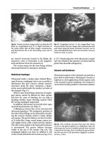
(17,18). A characteristic pulsed-field gel electrophoresis image obtained
from a short-term noncuffed CVC causing BSI is shown in Figure 2 and
from a long-term cathter (PICC), in Figure 3.
MICROBIOLOGY
The distribution of microorganisms that cause IVD-related BSIs vary by
the type of device used (Table 3) (19). For example, microorganisms found
Nasia Safdar, Leonard A. Mermel, and Dennis G. Maki
7
on patient’s skin, which gain access to the IVD extraluminally, occasionally,
intraluminally—coagulase-negative staphylococci (39%), Staphylococcus
aureus (26%), and Candida spp. (11%)—account for 76% of IVD-related
BSIs with short-term noncuffed devices of all types; only 14% are caused by
gram-negative bacilli. In contrast, with long-term surgically implanted
devices, such as cuffed and tunneled catheters, PICCs, and subcutaneous
central venous ports, gram-negative bacilli, which gain access intraluminally
and grow rapidly within the infusate in the device, account for nearly half of
IVD-related BSIs; only 2% are caused by Candida spp.
Figure 1. Potential sources of infection of a percutaneous IVD: the contiguous skin
flora, contamination of the catheter hub and lumen, contamination of infusate, and
hematogenous colonization of the IVD from distant, unrelated sites of infection
(from C.J Crnich and D.G Maki (4)).
8
Catheter-Related Infections in the Critically Ill
Figure 2. Pulsed-field gel electrophoresis image showing the probable pathogenesis
of a central venous catheter-related bacteremia with coagulase-negative
Staphylococcus. The isolates from the catheter tip, blood, and skin of the insertion
sit
e
were all concordant, indicating an extraluminal route of infection (29).
Nasia Safdar, Leonard A. Mermel, and Dennis G. Maki
9
Figure 3. Pulsed-field gel electrophoresis image showing the probable pathogenesis of
a PICC-related bacteremia with Serratia marcescens. The isolates from the catheter
tip, blood, hub and fluid were all concordant, indicating an intraluminal route of
7infection.
10
Catheter-Related Infections in the Critically Ill
RISK FACTORS FOR INFECTION AND IMPLICATIONS FOR
PREVENTION
IVDR-BSIs are largely preventable. Strategies for prevention can be
successful only if based upon a sound understanding of the risk factors and
pathogenesis of IVD-related BSI. A growing body of literature in recent
years has greatly enhanced our understanding of the risk factors for IVD-
related BSI; a recent review summarizes the major risk factors with short-
term noncuffed CVCs (Tables 4,5, and 6) (20).
Nasia Safdar, Leonard A. Mermel, and Dennis G. Maki
11
12
Catheter-Related Infections in the Critically Ill
Nasia Safdar, Leonard A. Mermel, and Dennis G. Maki
13
Training and Experience of the Inserter
CVCs are associated with significant potential for life-threatening
iatrogenic complications besides catheter-related BSI, including
pneumothorax, vascular injury, arrhythmias and thromboembolism.
Armstrong et al. identified inserter experience as an important risk factor for
CVC-related BSI in a prospective study of 169 catheters (21). Moreover, a
survey of U.S. academic medical centers has shown that up to one-half of
clinicians who use PA catheters have major gaps in their understanding of
when to use the catheter and how to interpret the data derived from it (22).
Only in recent years are U.S. institutions requiring formal training of house
officers in the techniques of vascular access. Intensified training and
educational programs can greatly reduce the baseline risk of CVC-related
BSI in a center.
Intravenous Therapy Teams
Good technique is essential. Studies have shown that the use of special IV
therapy teams, consisting of trained nurses or technicians who can assure a
consistent and high level of aseptic technique during catheter insertion and in
14
Catheter-Related Infections in the Critically Ill
follow-up care of the catheter, have been associated with substantially lower
rates of catheter-related BSI and are cost-effective.
Institutional IV teams should be encouraged, but even if an institution
does not have an IV team, it can greatly reduce its rate of IVDR BSI by
formal education of nurses and physicians and strict adherence to IVD care
protocols (23).
Sterile Barrier Precautions
Mermel et al. (24) found in a prospective study of 302 pulmonary-artery
catheters that failure to use maximal sterile barriers at the time of catheter
insertion increased the risk of catheter-related infection more than two-fold
(RR, 2.1). Whereas the issue has not been studied extensively, in one well-
controlled randomized trial it was found that the use of maximal sterile
barriers when inserting a CVC in a patient with cancer greatly reduced the
risk of CVC-related BSI (RR, 0.20) (25).
It seems clear that physicians inserting a CVC should wear a long-sleeved
sterile surgical gown and sterile gloves and, to be in compliance with
universal precautions, a mask and eye cover; the potential insertion site
should be draped with a large sterile sheet (23). Maximal sterile barrier
precautions are not necesssary for peripheral arterial catheters used for
hemodynamic monitoring, where sterile gloves and a sterile fenestrated
drape will suffice based on a a prospective study showing no difference in
colonization but the study was underpowered to show a difference in
catheter-related BSI (26).
Site of Insertion
At least six studies, including one randomized clinical trial, have found
that percutaneous insertion of a CVC in an internal jugular or femoral vein is
associated with a substantially higher risk of catheter-related BSI than
insertion in a subclavian vein (RR, 1-3.3) (24,27-31). Femoral line insertion
also dramatically and independently increases the risk of the life-threatening
complications deep venous thrombosis (30). Whereas placement in an
internal jugular or femoral vein is associated with less risk of pneumothorax
an
d
permits control of local hemorrhage by the application of pressure, the
risk of mechanical complications with central venous cannulation, such as
pneumothorax or hemorrhage, has greatly declined in recent years (59),
Nasia Safdar, Leonard A. Mermel, and Dennis G. Maki
15
reflecting better training in the techniques of percutaneous catheter insertion
and greater experience. It should be possible to place a CVC percutaneously
in the subclavian vein with a very low risk of barotrauma, in the range of 1%
or less.
We believe these data indicate that training programs should strive to
encourage use of the subclavian vein as the preferred site of access for CVCs
(23) (other than catheters needed for long-term hemodialysis), and should
assure that house officers are trained in establishing central access in the
subclavian vein. Catheterization of the femoral vein should be kept to a
strict minimum and if accessed during a code situation, the catheter should
be changed to an alternative site as soon as it’s safe to do so. Tunneling a
CVC appears to reduce the risk of catheter-related BSI, both with catheters
placed in the internal jugular or femoral veins, and might be considered if
circumstances mandate cannulation of an internal jugular or femoral vein
rather than a subclavian vein (e.g., severe coagulopathy or a hemodialysis
catheter).
Catheter Exchange Over a Guidewire
The Seldinger technique for catheter insertion has been a major advance,
permitting the great central veins to be cannulated with considerably less risk
of pneumothorax and vascular injury. To avoid iatrogenic mechanical
complications associated with percutaneous insertion of another CVC, new
catheters are commonly inserted over a guidewire in the site of an old
catheter. Numerous studies have examined the impact of this practice on the
risk of infection (32-43), most did not utilize multivariable techniques. Eight
randomized trials to address this issue have had conflicting results (33-37,42-
44). The best prospective randomized trial, which included pulmonary-
artery catheters, found a nearly two-fold increased risk of catheter-related
BSI with CVCs replaced on a periodic basis in old sites over a guidewire (9
vs 5 cases per 1000 catheter-days); 75 percent of all catheter-related BSIs in
the study population occurred within 72 hours of catheter exchange over a
guidewire (35). However, a systematic review of the effect of guidewire
exchange and new site replacement strategies for central venous catheters in
critically ill patients did not find a statistically significant reduction in
catheter-related BSI with routine guidewire exchange (RR 1.72, 95% CI
0.12-1.91) (45).
16
Catheter-Related Infections in the Critically Ill
If a CVC is replaced because of suspected infection without signs of
sepsis, or the catheter has malfunctioned (e.g it is cracked), it is reasonable to
replace a catheter in the same site over a guidewire if the patient has limited
sites for new access or would be at a very high risk for percutaneous central
venous cannulation in a new site (e.g., coagulopathy or morbid obesity) (23).
However, it is imperative that the same meticulous aseptic technique and use
of full sterile barriers that are mandatory during the insertion of any new
CVC be employed. After vigorously cleansing the site with the antiseptic
solution, inserting the guidewire, removing the old catheter and cleansing the
site once more with the antiseptic solution, the operator should reglove and
ideally redrape the site, as the original gloves and drapes are likely to have
become contaminated from manipulation of the old catheter.
It is also essential to routinely culture the old catheter and, if the patient is
febrile or shows other signs of sepsis, to obtain blood cultures (23). If these
cultures demonstrate that the old catheter was infected, the new catheter
placed in an old site should ideally be immediately removed to prevent
progression to catheter-related BSI or perpetuation of ongoing BSI, as a new
catheter has been inserted into an infected tract; need for continued access
would mandate placement of a new catheter in a new site. If culture of the
old catheter shows that it is not colonized, it has been possible to preserve
access and exclude it as the cause of fever and sepsis without subjecting the
patient to the hazards associated with percutaneous insertion of a new
catheter.
In general, if an old insertion site is inflamed, especially if there is
purulence, the patient shows signs of sepsis that might be originating from
the catheter or the patient has cryptogenic bacteremia or candidemia, it is
strongly recommended that a new catheter not be inserted over a guidewire
into an old, potentially-infected site (23).
HEAVY COLONIZATION OF THE INSERTION SITE AND
CUTANEOUS ANTISEPSIS
Colonization of the insertion site will be greatly influenced by the choice
of the site for insertion. In a prospective study, it was found that the density
of the transient cutaneous microflora was highest at the base of the neck, the
site of insertion of an internal jugular vein catheter, as contrasted with over
the upper chest, the site for insertion of a subclavian vein catheter. In
Nasia Safdar, Leonard A. Mermel, and Dennis G. Maki
17
neonates, there is a significantly greater density of microbes in the combined
jugular and femoral sites than either the umbilical or subclavian site.
Given the powerful evidence for the importance of cutaneous micro-
organisms and particularly the density of the microflora at the potential
insertion site in the pathogenesis of CVC-related infection, measures to
reduce cutaneous colonization of the insertion site would seem of the highest
priority, particularly the choice of the chemical antiseptic used for
disinfecting the site. In the United States, iodine-based disinfectants,
particularly iodophors such as 10% povidone-iodine, are used most widely.
Chlorhexidine, a biguanide with potent and broad-spectrum activity, exhibits
prolonged antimicrobial activity on the skin surface after a single application,
in contrast to alcohol or iodine-based antiseptics. To date, seven prospective
randomized clinical trials have compared the efficacy of 10% povidone-
iodine and chlorhexidine antisepsis for vascular access. The largest, a
prospective randomized trial with 750 CVCs and arterial catheters used in
patients in an ICU, showed that 2% chlorhexidine was superior to 10%
povidone-iodine or 70% alcohol for prevention of CVC-related BSI (RR,
0.16). In six of the seven trials to date, chlorhexidine was superior to
povidone-iodine for preventing catheter colonization, and in two, CVC-
related sepsis was reduced significantly.
These studies in aggregate indicate that a 0.5 - 2% chlorhexidine-alcohol
tincture or a 1-2% aqueous solution is more effective than iodophors or 70%
alcohol for prevention of CVC-related colonization and BSI. Two recent
meta-analyses of randomized trials comparing chlorhexidine to 10%
povidone-iodine for cutaneous antisepsis found a 50% reduction in the risk
of CVC-related BSI with the use of chlorhexidine.
Disinfection of skin should be done with an appropriate antiseptic prior to
catheter insertion and at the time of dressing changes. A 2% chlorhexidine-
based preparation is preferred. Alternatively, tincture of iodine, an iodophor,
or alcohol could be used. Allow the antiseptic to remain on the insertion site
and to dry before inserting the catheter. Allow povidone-iodine to remain on
the skin for at least 2 minutes, or longer if it is not yet dry before inserting
the catheter.
18
Catheter-Related Infections in the Critically Ill
Site Dressings
The importance of the cutaneous microflora in the pathogenesis of CVC-
related infection suggests that the dressing applied to the insertion site could
have considerable influence on the incidence of catheter-related infection. In
recent years, transparent polyurethane film dressings have become available.
They secure the device more reliably, permit continuous inspection of the
site, and are generally more comfortable than gauze and tape; moreover, they
permit patients to bathe and shower without saturating the dressing. Studies
of polyurethane dressings on short-term non-cuffed CVCs have yielded
conflicting results; however, a meta-analysis of the largest and most
rigorously controlled randomized trials has shown that these dressings do not
materially increase the risk of CVC-related BSI (RR 0.99, 95% CI 0.90-
1.09) (46).
Either sterile gauze or sterile, transparent, semipermeable dressing may
be used to cover the catheter site. If the patient is diaphoretic, or if the site is
bleeding or oozing, a gauze dressing is preferable to a transparent, semi-
permeable dressing (23).
Manipulations of the System
Contamination of infusate, stopcocks or catheter hubs, the cause of many
CVC-related BSIs, has been the cause of most outbreaks of infusion-related
bacteremia or candidemia.
In general, running infusions should be manipulated as little as possible,
and persons handling or entering the system should first wash their hands or
don clean gloves (23). Efforts should be made to limit entry into the
monitoring circuit for the purpose of drawing blood or other tests (23). The
number of stopcocks in the system should also be kept to a minimum. It is
unknown whether wiping a stopcock which has been opened with an anti-
infective agent might be of value.
Prolonged Catheter Placement
Exactly how long non-cuffed short-term CVCs can be left in place safely,
particularly in critically ill patients in an ICU, has not been adequately
assessed. In general, however, most studies that have examined duration of
placement as a risk factor have shown that prolonged placement significantly
Nasia Safdar, Leonard A. Mermel, and Dennis G. Maki
19
increases the cumulative risk of infection, particularly insertions longer than
5-7 days (27,37) A simple but elegant mathematical model has also been
derived demonstrating the increased risk of catheter-related bloodstream
infection for each day of catheterization (47). The need for continued use of
an intravascular catheter should be frequently reassesed and the device
should be removed as soon as the intended use is over (48).
Finally, what has not been conclusively established is whether routine
replacement of a non-cuffed CVC to a new site at periodic intervals, such as
every 4-5 days, significantly reduces the risk of CVC-related BSI in patients
requiring prolonged central access. While some studies report no decline in
the incidence of CVC-related BSI with routine replacement (35,37,49), most
that have examined this issue have not had sufficient statistical power to
answer the question (37,44,50). In the absence of conclusive data affirming
benefit, central venous or arterial catheters should not be routinely replaced
solely for the purpose of reducing the risk of catheter-related infection (23).
The question thus remains unanswered; however, the availability of novel
technology may obviate this concern. The studies of anti-infective-coated
CVCs show a sufficiently reduced risk of CVC-related BSI that it would
appear that with the use of such technology in patients requiring prolonged
central access, it should be safe to leave a CVC in place for 10-20 days, if
necessary, perhaps even longer if the device is dedicated to total parenteral
nutrition or anti-infective therapy. Moreover, the use of chlorhexidine-
impregnated dressings or engineered contamination-resistant catheter hubs
can also reduce risk and permit prolonged cannulation with a very low risk of
infection.
REFERENCES
Maki DG, Mermel LA. Infections due to infusion therapy. In: Bennet JV, Brachman
PS, eds. Hospital Infections. Philadelphia: Lippincott-Raven Publishers; 1998:689-
724.
Raad I. Intravascular catheter-related infections. Lancet. 1998;351:893-898.
Mermel LA. Prevention of intravascular catheter-related infections. Ann Intern Med.
2000;132:391-402.
Crnich CJ, Maki DG. The promise of novel technology for the prevention of
intravascular device-related bloodstream infection. I. Pathogenesis and short-term
devices. Clin Infect Dis. 2002;34:1232-1242.
1.
2.
3.
4.
20
Catheter-Related Infections in the Critically Ill
Mermel LA, Farr BM, Sherertz RJ, et al. Guidelines for the management of
intravascular catheter-related infections. J Intraven Nurs. 2001;24:180-205.
Smith RL, Meixler SM, Simberkoff MS. Excess mortality in critically ill patients with
nosocomial bloodstream infections. Chest. 1991;100:164-167.
Pittet D, Tarara D, Wenzel RP. Nosocomial bloodstream infection in critically ill
patients:excess length of stay, extra costs and attributable mortality. JAMA.
1994;271:1598-1601.
Arnow PM, Quimosing EM, Beach M. Consequences of intravascular catheter sepsis.
Clin Infect Dis. 1993;16:778-784.
Collignon PJ. Intravascular catheter associated sepsis: a common problem. The
Australian Study on Intravascular Catheter Associated Sepsis. Med J Aust.
1994;161:374-378.
Soufir L, Timsit JF, Mahe C, Carlet J, Regnier B, Chevret S. Attributable morbidity
and mortality of catheter-related septicemia in critically ill patients: a matched, risk-
adjusted, cohort study. Infect Control Hosp Epidemiol. 1999;20:396-401.
Digiovine B, Chenoweth C, Watts C, Higgins M. The attributable mortality and costs
of primary nosocomial bloodstream infections in the intensive care unit. Am J Respir
Crit Care Med. 1999;160:976-981.
Pelletier SJ, Crabtree TD, Gleason TG, Pruett TL, Sawyer RG. Bacteremia associated
with central venous catheter infection is not an independent predictor of outcomes. J
Am Coll Surg. 2000;190:671-680; discussion 680-671.
Renaud B, Brun-Buisson C. Outcomes of primary and catheter-related bacteremia. A
cohort and case-control study in critically ill patients. Am J Respir Crit Care Med.
2001;163:1584-1590.
Rello J, Ochagavia A, Sabanes E, et al. Evaluation of outcome of intravenous catheter-
related infections in critically ill patients. Am J Respir Crit Care Med. 2000;162:1027-
1030.
Crnich CJ, Maki DG. The role of intravascular devices in sepsis. Curr Infect Dis Rep.
2001;3:497-506.
Kluger D, Maki D. The relative risk of intravascular device-related bloodstream
infections with different types of intravascular devices in adults. A meta-analysis of
206 published studies. Paper presented at: Programs and Proceedings of the Fourth
Decennial International Conference on Nosocomial and Healthcare-Associated
Infections, 2000; Atlanta, GA, 2000.
Sitges-Serra A, Puig P, Linares J, et al. Hub colonization as the initial step in an
outbreak of catheter-related sepsis due to coagulase negative staphylococci during
parenteral nutrition. JPEN J Parenter Enteral Nutr. 1984;8:668-672.
Raad I, Costerton W, Sabharwal U, Sacilowski M, Anaissie E, Bodey GP.
Ultrastructural analysis of indwelling vascular catheters: a quantitative relationship
between luminal colonization and duration of placement. J Infect Dis. 1993;168:400-
407.
Maki DG, Kluger DM, Cmich CJ. The microbiology of intravascular device-related
(IVDR) infection in adults: an analysis of 159 prospective studies; implications for
prevention. Paper presented at: 40th Annual Meeting of the Infectious Diseases
Society of America, 2002; Chicago, IL.
5.
6.
7.
8.
9.
10.
11.
12.
13.
14.
15.
16.
17.
18.
19.
Nasia Safdar, Leonard A. Mermel, and Dennis G. Maki
21
20.
21.
22.
23.
24.
25.
26.
27.
28.
29.
30.
31.
32.
33.
34.
Safdar N, Kluger DM, Maki DG. A review of risk factors for catheter-related
bloodstream infection caused by percutaneously inserted, noncuffed central venous
catheters: implications for preventive strategies. Medicine (Baltimore). 2002;81:466-
479.
Armstrong CW, Mayhall CG, Miller KB, et al. Prospective study of catheter
replacement and other risk factors for infection of hyperalimentation catheters. Journal
Infect Dis. 1986;154:808-816.
Iberti TJ, Fischer EP, Leibowitz AB, et al. A multicenter study of physicians’
knowledge of the pulmonary artery catheter. JAMA. 1990;264:2928-2932.
O’Grady NP, Alexander M, Dellinger EP, et al. Guidelines for the prevention of
intravascular catheter-related infections. Centers for Disease Control and Prevention.
MMWR Recomm Rep. 2002;51:1-29.
Mermel LA, McCormick RD, Springman SR, Maki DG. The pathogenesis and
epidemiology of catheter-related infection with pulmonary artery Swan-Ganz
catheters: a prospective study utilizing molecular subtyping. Am J Med.
1991;91(Suppl 3B):1897-1205
Raad, II, Hohn DC, Gilbreath BJ, et al. Prevention of central venous catheter-related
infections by using maximal sterile barrier precautions during insertion. Infect Control
Hosp Epidemiol. 1994;15:231-238.
Rijnders BJ, Van Wijngaerden E, Peetermans WA. Use of full sterile barrier
precautions during insertion of arterial catheters: a randomized trial. Clin Infect Dis.
2003;36:743-748.
Richet H, Hubert B, Nitemberg G, et al. Prospective multicenter study of vascular-
catheter-related complications and risk factors for positive central-catheter cultures in
intensive care unit patients. J Clin Microbiol. 1990;28:2520-2525.
Pittet D. Intravenous catheter-related infections:current understanding. Paper presented
at: Programs and Abstracts of the Thirty-second Interscience Conference on
Antimicrobial Agents and Chemotherapy, 1992; Anaheim, CA.
Heard SO, Wagle M, Vijayakumar E, et al. Influence of triple-lumen central venous
catheters coated with chlorhexidine and silver sulfadiazine on the incidence of
catheter-related bacteremia. Arch Intern Med. 1998;158:81-87.
Merrer J, De Jonghe B, Golliot F, et al. Complications of femoral and subclavian
venous catheterization in critically ill patients: a randomized controlled trial. JAMA.
2001;286:700-707.
Goetz AM, Wagener MM, Miller JM, Muder RR. Risk of infection due to central
venous catheters: effect of site of placement and catheter type. Infect Control Hosp
Epidemiol. 1998;19:842-845.
Tacconelli E, Tumbarello M, Pittiruti M, et al. Central venous catheter-related sepsis
in a cohort of 366 hospitalised patients. Eur J Clin Microbiol Infect Dis. 1997;16:203-
209.
Kealey GP, Chang P, Heinle J, Rosenquist MD, Lewis RW, 2nd. Prospective
comparison of two management strategies of central venous catheters in burn patients.
J Trauma. 1995;38:344-349.
Snyder RH, Archer FJ, Endy T, et al. Catheter infection. A comparison of two catheter
maintenance techniques. Ann Surg. 1988;208:651-653.
22
Catheter-Related Infections in the Critically Ill
35.
36.
37.
38.
39.
40.
41.
42.
43.
44.
45.
46.
47.
48.
49.
50.
Cobb DK, High KP, Sawyer RG, et al. A controlled trial of scheduled replacement of
central venous and pulmonary-artery catheters. N Engl J Med. 1992;327:1062-1068.
Michel LA, Bradpiece HA, Randour P, Pouthier F. Safety of central venous catheter
change over guidewire for suspected catheter-related sepsis. A prospective randomized
trial. Int Surg. 1988;73:180-186.
Eyer S, Brummitt C, Crossley K, Siegel R, Cerra F. Catheter-related sepsis:
prospective, randomized study of three methods of long-term catheter maintenance.
Crit Care Med. 1990; 18:1073-1079.
Oliver MJ, Callery SM, Thorpe KE, Schwab SJ, Churchill DN. Risk of bacteremia
from temporary hemodialysis catheters by site of insertion and duration of use: a
prospective study. Kidney Int. 2000;58:2543-2545.
Pettigrew RA, Lang SDR, Haydock DA, Parry BR, Bremner DA, Hill GL. Catheter-
related sepsis in patients on intravenous nutrition: a prospective study of quantitative
catheter cultures and guidewire changes for suspected sepsis. Br J Surg. 1985;72:52-
55.
Savage AP, Picard M, Hopkins CC, Malt RA. Complications and survival of
multilumen central venous catheters used for total parenteral nutrition. Br J Surg.
1993;80:1287-1290.
Bach A, Stubbig K, Geiss HK. Infectious risk of replacing venous catheters by the
guide-wire technique. Zentralbl Hyg Umweltmed. 1992;193:150-159.
Senagore A, Waller JD, Bonnell BW, Bursch LR, Scholten DJ. Pulmonary artery
catheterization: a prospective study of internal jugular and subclavian approaches. Crit
Care Med. 1987; 15:35-37.
Powell C, Fabri PJ, Kudsk KA. Risk of Infection accompanying the use of single-
lumen vs double-lumen subclavian catheters: a prospective randomised study. Journal
of Parenteral and Enteral Nutrition. 1988; 12:127-129.
Bach A, Bohrer H, Geiss HK. Safety of a guidewire technique for replacement of
pulmonary artery catheters. J Cardiothorac Vasc Anesth. 1992;6:711-714.
Cook D, Randolph A, Kernerman P, et al. Central venous catheter replacement
strategies: a systematic review of the literature. Crit Care Med. 1997;25:1417-1424.
Hoffmann KK, Weber DJ, Samsa GP, Rutala WA. Transparent polyurethane film as
an intravenous catheter dressing. A meta-analysis of the infection risks. JAMA.
1992;267:2072-2076.
Widmer AF. Intravenous Catheter-related Infections. In: Wenzel RP, ed. Prevention
and Control of Nosocomial Infections. 3rd edition ed. Baltimore, MD: Williams and
Wilkins; 1997.
Lederle FA, Parenti CM, Berskow LC, Ellingson KJ. The idle intravenous catheter.
Ann Intern Med. 1992;116:737-738.
Bregenzer T, Conen D, Sakmann P, Widmer AF. Is routine replacement of peripheral
intravenous catheters necessary? Arch Intern Med. 1998;158:151-156.
Uldall PR, Merchant N, Woods F, Yarworski U, Vas S. Changing subclavian
haemodialysis cannulas to reduce infection. Lancet. 1981;1:1373.
Chapter 2
EPIDEMIOLOGY AND PATHOGENESIS OF
CATHETER-RELATED BLOODSTREAM
INFECTIONS
Antonio Sitges-Serra, F.R.C.S (Ed.)
Department of Surgery, Hospital Universitari del Mar, Barcelona, Spain
Introduction
As the only surgeon contributing to this major work on catheter-related
bloodstream infections (CRBSI), this perhaps requires some explanation. In
the mid- seventies, early during my residency training in general surgery, I
was charged to take care of patients receiving parenteral nutrition at the
Hospital de Bellvitge, my home institution, which pioneered this modality
treatment in Spain (1). Our general surgery service was a reference one for
the care of patients with complex postoperative abdominal complications
such as fistulas and short bowel syndrome. Being a young trainee, I took this
responsibility with some fear but with much enthusiasm because it gave me a
unique opportunity both for challenging patient care and for first-line
research. Of the many interesting aspects of parenteral nutrition delivery,
subclavian catheter infections readily attracted my attention. They were
common, they forced us to stop treatment and carried significant morbidity
and even mortality. Initial attempts at controlling this complication by
meticulous skin care and full barrier precautions at catheter insertion were
24
Catheter-Related Infections in the Critically Ill
unsuccessful and, at a given point, catheter sepsis due to coagulase-negative
staphylococci attained almost an endemic proportion. This prompted us to
start a series of investigations that led, in the early 1980s, to the recognition
of the catheter hub as a relevant portal of entry for microorganisms
contaminating central venous catheters (CVCs). The paper reporting this
seminal observation could have appeared some years earlier but,
unfortunately, it was rejected in three medical journals before being accepted
in a journal specialised in artificial nutrition (2).
Our findings had a somehow cold reception in the expert entourage of the
time probably because they came from an unknown unit and because they
challenged established knowledge. In fact, intellectual challenge was
welcomed by our group and represented a potent stimulus for us to
hypothesize that the new paradigm of endoluminal catheter contamination
would have a major impact on issues closely related to pathogenesis of
CRBSI, namely, diagnosis, prevention, treatment and industrial design of
future catheters (3). These thoughts have been largely confirmed by many
research groups and have been the focus of our work spanning over twenty
years (1976-1996) during which we made some contributions to the field and
had the privilege to meet and discuss the issue with experts on both sides of
the Atlantic.
DEFINITIONS
There is a growing consensus on the definitions to be used when dealing
with infections due to intravascular devices. In the present chapter, we have
adhered to a set of definitions recently proposed to improve communication
between researchers and increase scientific accuracy of articles dealing with
catheter infections (4).
1) The term intravascular catheter-related bloodstream infections
(CRBSI) denotes bacteremia or fungemia in a patient who has an
intravascular device, >1 positive blood culture from a peripheral vein,
clinical manifestations of infection and confirming appropriate
microbiological cultures. CRBSI is to be preferred to the term “catheter-
related sepsis” since the concept “sepsis” does not imply bacteremia and is
used to define the systemic inflammatory response syndrome associated with
an infectious focus. “Catheter-related bacteremia” is not accurate since blood
cultures may grow fungal species (fungemia).