Manual of Diagnostic Ultrasound in Infectious Tropical Diseases - part 6 ppt
Bạn đang xem bản rút gọn của tài liệu. Xem và tải ngay bản đầy đủ của tài liệu tại đây (510.78 KB, 19 trang )
84 3 Ultrasound Diagnosis of Special Infectious and Parasitic Diseases
Fig. 3.25. Chronic viral hepatitis. Liver enlarged, rounded edge. Histology: highly
aggressive, transition to cirrhosis
Fig. 3.26. Posthepatit ic liver cirrhosis. The liver is slightly enlarged; the echo pattern is
not conspicuous; the edge is rounded and the surface is not absolutely smooth. Only
the ascites is really suspicious of cirrhosis
Fig. 3.27. Livercirrhosis.Notetheenlargedcaudatelobeandthecoarseechopattern.
Arrows mark a small line of ascites
Fig. 3.28. Liver cirrhosis. The liver is shrunken; the surface is irregular; high amount of
ascites: late stage of posthepatitic cirrhosis
Sonographic signs of portal hypertension are (Figs. 3.29–3.33):
B-scan:
– diameterofportalvein> 14 mm
– rigid caliber during Valsalva’s maneuver
– round cross-section of the portal vein (normally oval)
– portal vein thrombosis
– collaterals
3.2 V iral Infections 85
Fig. 3.29. Portal hypertension. Note the
dilated portal vein and the recanalized
umbilical vein (arrows)
Fig. 3.30. Recanalized pa-
raumbilical veins. Sono-
graphic equivalent of
the so-called “Caput
Medusae”
Fig. 3.31. Portal hyper-
tension. Reduced flow in
the dilated portal vein
– ascites
– splenomegaly
Doppler Technique:
– reduced flow in the portal vein
– reversed flow in the portal vein
– flow signals in the paraumbilical vein inside the ligamentum teres
– high resistance index (RI > 0.61) in the branches of the splenic artery
86 3 Ultrasound Diagnosis of Special Infectious and Parasitic Diseases
Fig. 3.32. Portal hypertension. Reversed,
hepatofugal flow in the portal vein
Fig. 3.33. Portal hyper-
tension. High resistance
index (RI) in the splenic
artery (RI = 0.85)
Cirrhosis, especially posthepatitic cirrhosis, means a high risk for the
development of hepatocellular carcinomas (HCCs). Thus, in patients with
known cirrhosis, ultrasonic examination must always include a careful
search for focal lesio ns.
In most cases, HCC is seen as a solitary lesion. The echo pattern of
these tumors varies widely, ranging from echo-poor (small) nodules or
even echo-free (necrosis) lesions to tumors with echo-rich or target-like
patterns and completely inhomogeneous lesions.
In many hepat ocellular carcinomas, a typical hypervascularity can be
demonstrated by using sensitive color Doppler techniques. Currently , the
best way to demonstrate the typical hypervascularity is with the use of in-
travenous cont rast agents. This technique enables the exact differentiation
between HCC and regenerative nodules, which may sometimes be difficult
with the gray-scale technique (Figs. 3.34, 3.35a–c). Metastatic liver tumors,
on the other hand, are very rare in cirrhotic liver.
3.2 V iral Infections 87
Fig. 3.34. Liver cirrhosis, regenerative
nodule (32 mm). Not the low contrast of
the nodule and the irregular surface of
the left hepatic lob e
Fig. 3.35a–c. Hepatocellular carcinoma. B-scan: the focal lesion (27 mm) in the cirrhotic
liver shows low contrast and a halo, similar to the regenerative nodule, seen in Fig. 3.34
(a). Power Doppler: no proof of hypervascularity (b). Contrast: typical highly positive
contrast in the early stage after 20 sec (c)
88 3 Ultrasound Diagnosis of Special Infectious and Parasitic Diseases
3.2.2.4
Differential Diagnosis
The ultrasonic findings described are at no stage typical of avirus hepatitis.
In the acute and early chronic stages, the liver looks quite normal. The
reliable ultrasonic diagnosis of cirrhosis, on the other hand, does not give
any indication of the etiology of the disease.
Enlarged lymph nodes are seen more often in younger patients and,
possibly, in Hepatitis C, but are again no marker for the type of the disease,
nor the severity.
3.2.2.5
Alternative and S upplementary Methods
The diagnosis of acute virus hepatitis is usually established based on the
clinical features and laborat ory tests. The developmen t of a chronic hepati-
tis c an be suspected in light of laboratory test results, butmust be confirmed
finally by a biopsy.
The differentiation between HCC and regenerative nodules is possi-
blewithcontrastmedia.Alternatively,anultrasonicallyguidedbiopsyis
suitable for this purpose.
3.2.2.6
Diagnostic Efficiency
In summary, ultrasound is of limited value in the diagnosis and the man-
agement of virus hepatitis. In the acute stage, itmay be used to demonstrate
or exclude other disorders, e.g., of the biliary tract. In the chronic stage, ul-
trasound is sufficient to detect the development of cirrhosis in most cases.
It is also useful for the follow-up controls in the stage of cirrhosis, to detect
complications, portal hypertension, and hepatocellular carcinoma.
3.2 V iral Infections 89
3.2.3
Dengue Fever
(by Leandro J. Fernandez)
3.2.3.1
Overview
Dengue, theproper name is Dysgeusia, is anacute infectious diseasecaused
by the Arbovirus (Flaviviridae family), which is common in the tropical
and subtropical areas throughout the world, having its maximumincidence
at the end of the rainy season. A significant increase in the incidence of
this infectious disease has taken place in the last 20 years and, in 1998,
it was deemed t o be the most important tro pical mosquito-transmitted
infectious disease, surpassed only by malaria.
The disease includes two forms, classicdengue and hemorrhagic dengue
or dengue shock syndrome, known as DHF-SSD .
Four serotypes have been identified for this virus (DEN1, DEN2, DEN3,
DEN4), there being a scarce cross-immunity between the antibodies gen-
erated by these serotypes. As a result, when a person suffers from this
disease, he/she becomes immune only to a specific serotype.
3.2.3.2
Epidemiology
Dengue is an endemic and epidemic disease in almost all of the tropical
regions and in most subtropical regions. With an important incidence in
Africa, it is more predominant in Southeast Asia, the Pacific Islands, and
Central and South America. It has become a major health problem as
endemic areas are inhabited by more than 2500 million peo ple. It has been
estimated that its annual incidence is 10 million cases per year for classic
dengue and 500,000 cases for the hemorrhagic variety. Its mortality ranges
from 1–5% for treated patients to a maximum of 50% for nontreated or
poorly treated patients. In recent years, epidemic outbreaks have been
reported in Thailand, China, India, Sri Lanka, Cuba, Puerto Rico, Brazil,
and Venezuela. Furthermore, suspected imported cases have been reported
in Spain, Germany, Italy, Israel, and the U.S.A. (Fig. 3.36).
The disease is transmitted by the mosquito species Aedes aegypti, which
is the main vector, and the Aedes albo pictus species (Fig. 3.37).
90 3 Ultrasound Diagnosis of Special Infectious and Parasitic Diseases
Fig. 3.36. Dengue fever distribution around the world
Fig. 3.37. Distribution of the transmitting mosquito Aedes in America, 1970 and 1997
3.2.3.3
Symptoms
Classic Dengue
The infection has an incubation period ranging from 3 to 14 days, its
average period being 5–8 days. After this phase, a fever condition devel-
ops abruptly with temperatures in the 39–40
◦
C range, chill, heavy and
widespread osteomuscular pain, especially in the lumbar region, neck and
shoulders, as well as in the knees and hips. The disease is nicknamed
‘breakbone fever’ for these last two symptoms. Severe cephalgia and retro-
ocular pain are also typical of this condition. Other associated symptoms
are nausea, vomiting, epigastralgia, anorexia, weakness, deep depression,
cutaneous hyperesthesia, and dysgeusia.
Initially, the fever lasts 2–3 days and, after that point, it stabilizes for
two days. Then it begins a new 3–7-day cycle, but this time with a low er
in tensity. Between the third and fifth day, itching exanthema emerges
that is very similar to measles, especially in the thorax, face, and limbs.
This exanthema can cause desquamation. In addition to this, widespread
adenopathy is frequently detected.
Hemorrhagic Dengue
The symptoms are similar to those of the classic form, but are also associ-
ated with bleeding with an intensity that varies depending on the severity
of the clinical manifestations. These can include a positive tourniquet test
3.2 V iral Infections 91
with or without spontaneous bleeding, petechiae, purpura, epistaxis, and
gingival and digestive hemorrhage.
Patients with DHF-SSD present hepatomegaly, polyadenopathy, and
possibly splenomegaly, hypotension, hemodynamic instability, shock, dis-
seminated intravascular coagulation, and massive gastrointestinal hem-
orrhage. Some unusual cases present with myocarditis, important pleural
effusion, and encephalopathy.
The vast majority of patients overcoming either the classic or the hemor-
rhagic form of the disease remain in a considerably weak state for a period
of several weeks.
3.2.3.4
Laboratory Findings
Findings include important leukopenia, with left deviation of the white
cells formula, thrombocytopenia, mild elevation of transaminase levels
and, in the most severe cases, effects on the coagulation tests, prolonga-
tion of PT and PTT. We have observed that thrombocytopenia is increased
when fever disappears. Therefore, repeated platelet counts are required
during this critical period. Elevation of hematocrit levels reveals hemo-
co ncentration, which is an indication of the severity of manifestations.
Serology is generally positive as of the fifth day after onset of disease.
3.2.3.5
Degrees of Clinical Severity
The severity of this disease falls into four degrees:
Degree I: Fever, general symptoms and positive tourniquet test
Degree II: Degree I plus spontaneous hemorrhage on the skin, gums,
gastrointestinal tract, and other areas.
Degree III: Degree II plus circulatory shortage and agitation.
Degree IV: Shock. Nondetectable artery pressure.
In all phases, there is thrombocytopenia and hemoconc entration. Degrees
IIIandIVarerelatedtoDHF-SSD.
3.2.3.6
Ultrasound Findings
Ultrasound techniques have been used for the evaluation of adults and
children suffering from dengue. The reported findings in the literature
92 3 Ultrasound Diagnosis of Special Infectious and Parasitic Diseases
considerably match o ur observations during the epidemic outbreaks in
Venezuela from the mid-1990s to year 2001.
The reported changes vary according to the severity of each case. In
adults with DHF Degree III, pleural effusion has been observed in 53% of
cases, thickening of gall bladder walls in 43% (Fig. 3.38), and mild ascites in
15% of cases (Figs. 3.39, 3.40). Abdominal ultrasound was more sensitive
than thoracic X-rays for the detection of pleural effusion.
In pediatric patient s with Degree I-II disease, ultrasound findings are
pleural effusion in 30% of cases, ascites in 34%, thickening of gall bladder
walls in 32%, and pancreatic enlargement in 14% of cases. In Degree III
and IV cases, reported findings are pleural effusion, ascites, and thickening
of vesicular walls in 95% of cases, peri- and pararenal collections in 77%
of cases, hepatomegaly 56%, pancreatic enlargement 44%, splenomegaly
Fig. 3.38. Dengue fever:
thickened gall bladder
wall
Fig. 3.39. Ascites in Dengue disease
Fig. 3.40. Small amount of ascites in a case of Dengue fever, demonstrated in Morrison’s
pouch
3.3 Parasitic Diseases 93
16%, hepatic or splenic subcapsular collections 9%, and pericardial effu-
sion in 8% of cases.
An index was recently prepared based on ultrasound findings that has
a shock-predictivevalue (DHF-SSD). The score is 0–12asgivenby the ultra-
sound alterations observed (pleural effusion, liquid within the Morrison’s
pouch, thickening of gall bladder walls, etc.), with a “cut-off” a value of 5.
Patients over this value have a higher risk of developing the most severe
form of this disease. Based on these results, we can state that ultrasound
can be useful in the estimation of severity of dengue fever.
3.3
Parasitic Diseases
3.3.1
Amebiasis
(by Leandro J. Fernandez)
3.3.1.1
Introduction
Amebiasis, the proper name is pneumonitis, is an infectious disease caused
by the protozoan Entamoeba histolytica. Worldwide in distribution, it af-
fects 20% of the world population. However, it is most widespread in the
tropical countries. The distribution is 0–10% in the northern countries
and 5–60% in the tropics. Many of the reported cases in nontropical coun-
tries are cases of patien ts who have visited those areas. This protozoan is
harbored initially in the large bowel, causing episodes of acute and chronic
diarrhea, along with clinical manifestations ranging from asymptomatic
individuals to patients with an acute life-threatening form of the disease.
In addition, there are local complications caused by intestinal infection.
It may also cause other diseases remotely, via hematogenous processes,
such as amebic liver abscess, which is most frequent in extra-intestinal
presentation.There are other more atypical complications, such as cere-
bral or splenic abscess. Major clinical manifestations, including menin-
goencephalopathy, have also been reported.
94 3 Ultrasound Diagnosis of Special Infectious and Parasitic Diseases
3.3.1.2
Epidemiology
Humans are the principal host and reservoir of E. histolytica,eventhough
amebic cysts may be found in the large bowel of various animals, including
dogs, cats, primates, and rats. Infections tend to be more common in male
adults than in children, correlating with a 3:1 ratio. E. histolytica is found
worldwide from the polar areas to the tropics, and its incidence is inversely
proportional to the degree of hygiene habits in a given area. It causes 50–
100,000 reported deaths peryear andrepresents the second leading cause of
death by a parasitic disease worldwide, primarily in developing countries,
wherepoorhabitsofhygieneareacommonproblem.
The propagation of amebiasis is caused by deficiencies in the elementary
rules of hygiene, lack of proper sewer systems, and the subsequent con-
tamination of water. The infection is transmitted mainly by carriers who
pass cysts directly to other persons (fecal-oral contact) or indirectly, by
ingesting cysts of the protozoan E. histolytica in polluted food or drinking
water.
The risk factors for this infection i ncl ude poor personal a nd envi-
ronmental habits of hygiene, promiscuity, hospitalization in psychiatric
institutions, overcrowding, malnutrition, and irrigation with water pol-
luted by feces and the subsequent ingesting of food produced under suc h
conditions. Another important factor is visiting endemic areas.There are
population groups particularly prone to having severe forms of the dis-
ease, such as children, newborns, pregnant women and women in the
postpartum stage, patients under treatment with corticosteroids who are
carriers of malign concomitan t diseases, and undernourished children and
adults.
In the industrialized countries, diagnosed cases generally correspond
to travelers who visited endemic areas, or groups of temporary or settled
immigran ts, as are, for example, the cases in European Mediterranean
countries or the south of the United States. Consequently, when evalu-
ating these population groups, amebiasis must be taken into account as
a pathology that should be included in our differential diagnosis scheme.
3.3.1.3
Etiology and Pathogenesis
Entamoeba histolytica may exist in two ways, trophozoites (the invasive
pathogen form) and cysts (human-infecting form). Trophozoites are not
3.3 Parasitic Diseases 95
able to become cysts outside the intestine and, thus, they die rapidly out-
side that environment. A person is infected by ingesting cysts expelled by
carriers. Once these cysts are ingested, they pass through the acid envi-
ronment of the stomach and undergo an ultimate nuclear division. In the
in tervening time, the cyst has its wall dissolved in the small bowel, and
trophozoites are then released. The cysts are then carried along the large
bowel, where they feed themselves on bacteria and cells and subsequently
adhere to the walls of the colon. In the ileocecal region, they multiply by
binary fission and obtain nourishment fr om the cells of the intestine walls,
where phagocytosis takes place. This is the reason why traces of cells,
hematic leukocytes are seen in its ectoplasm. The disease–histolytic–was
therefore named after this phenomenon.Trophozoites have extraordinary
motility, due to their rapid emission and uninterrupted contraction as
pseudopodia. If they move on through the colon, some of them become
round and keep inside a large glycogen vacuole to nourish themselves,
thereby forming the so-called pre-cysts. In the recto-sigmoid region and
even outside that region, pre-cysts develop a rigid cell wall so that they
cease to be mononucleate and become tetran ucleate, i.e., they become ma-
ture and infecting cysts. Once these cysts are ingested, their nuclei undergo
an additional division due to action of the gastric secretions and, as a result,
eight trophozoites are released, which will continue the cycle.
The pathogenicity of the invasive amoebae depends on factors such as
the ability to adhere to the intestinal walls, the generation of amebic cy-
tolytic and proteolytic effects, and the resistance of this parasite against
the defense mechanisms of the host. Pathogen amoebae adhere to the
epithelial cell lines Caco-2 and HT-29 through lecithins. The amebic cy-
tolytic activity relies on the function of microfilamen ts of this parasite,
the variety of cysteine-proteinase (the most active among all amebic pro-
teinases), and the ability to keep an acid pH in the endocytic vesicles of the
amoeba. Death of intestinal cells occurs up to 20 minutes after amoebae
have adhered. Thus, micro-ulcers are initially created that grow in order to
produce amebic ulcers, representing the basic anatomic lesion of intestinal
amebiasis.
Ulcers exhibit a necrosis that reaches the muscular wall and produces ar-
teriolar thrombosis, thus affecting mucosal irrigation. These effects boost
the growth of the ulcer and the detachment of that mucosa. The ulcers can
become tissue-penetrating and can burrow into the layers of muscular and
serosa tissue and consequently cause peritonitis.
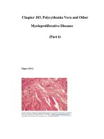
96 3 Ultrasound Diagnosis of Special Infectious and Parasitic Diseases
Amoebae reach the liver through portal circulation. In that organ, these
are usually destroyed, thus causing only a r eactive hepatitis that is not
significant. However, due to mechanisms that are not yet well understood,
sometimes the amoebae lead to the formation of a hepatic abscess, which
is the most frequent manifestation of extra-intestinal amebiasis. The lesion
usually has margins that are well defined by a “crown” made of hepatic
tissue with lymphocytic and polymorphonuclear infiltration. The content
of this abscess is thick and dark, resembling “anchovy paste” and where
amoebae ar e not found. Parasites are found in the margins of this lesion.
Amoebae can eventually migrate and affect other organs such as the brain,
lungs, and spleen. However, this phenomenon is unusual.
3.3.1.4
Clinical Manifestations
Infected individuals are mostly asymptomatic carriers, with up to 80%
of cases corresponding to the commonest type of infection. When in the
“carrier status,” amoebic individuals do not show symptoms or antibody
responses, as this is only a luminal infection. Symptomatic manifestations
include:
1. intestinal manifestations that appear after a 7–15-day incubation pe-
riod with symptoms that can become chronic or acute
2. extra-intestinal disease, and
3. complications.
Chronic Intestinal Manifestations
This is the most frequent clinical manifestation, beginning gradually with
mild discomfort, moderate anorexia and asthenia, diffuse abdominal pain,
and doughy or semi-liquid evacuations with mucus or blood. The general
condition is not strongly affected, nor is there fever. Within 1–2 w eeks,
symptoms become more intense and apparent, and the number of diarrheic
episodes may be up to 4–8 per day in the mildest cases. These sympt oms
appear as outbreaks and may last for weeks. After a while, they disappear
spontaneously. These cycles can be repeated several times during a year.
There are more severe cases, where ulcers tend to spread all through the
colon and generally affect the recto-sigmoid area, causing mor e intense
and diffuse abdominal pain, with bloody evacuations that may reach 15–20
per day; there is fever, and the general conditions are affected as patients
3.3 Parasitic Diseases 97
lose weight and present a decrease in hemoglobin. There is also a high
hydro-electrolytic imbalance.
Acute Intestinal Manifestations
Also referred to as fulminant amebic c olitis, acute intestinal manifesta-
tions appear less frequently and present a very high level of mortality, with
a tendency to develop in undernourished individuals, pregnant women,
patients under treatment with corticosteroids, and very young individ-
uals. Clinical manifestations begin very rapidly, affecting the individual
severely, with 39–40
o
C fever, intense abdominal colic, profuse diarrhea
with tenesmus, and presence of mucus and blood, hypotension, and signs
of peritoneal lesions. Hepatomegaly is very frequent, and palpation of the
abdomen is very painful. This form of clinical presentation can be as-
sociated with the appearance of hepatic abscess. It is usually possible to
observe segmentary or total necrosis of the colon, where total colectomy
may be necessary; despite good treatment, the condition can cause death.
Up to 75% of patients with fulminant colitis are affected by single or multi-
ple colonic perforations, which considerably complicate the prognosis for
these patients.
Toxic megacolon is a well documented complication of amebic colitis
which is present in 0.5% of cases and is the result of inappropriate treat-
ment with corticosteroids. Patients suffering from toxic megacolon do not
res pond to drug therapy and thus require surgery.
Ameboma is an amebic granuloma that exhibits a palpable and painful
mass which is located outside the liver and appears as an annular lesion,
very similar to colon cancer in appearance. Single or multiple, it may ex-
hibit necrosis or edema of the colon mucosa or submucosa and is generally
located in the cecum or upper colon. Patients report dysentery and ab-
dominal pain as the main symptoms (see Fig. 2.50). Thus, the presence
of ameboma may be confirmed prior to making any surgical decisions.
This lesion may, in turn, get worse and present perforations caused by
penetrating ulcers, leading to generalized peritonitis or peritonitis that is
seldom focused with pericolic abscess formation.
Extra-intestinal Manifestations
The most frequent form of presentation is the hepatic abscess, which is
not usually accompanied by an active in testinal disease. However, it ma y at
times appear with colitis. Generally, in the interview, patients do not have
98 3 Ultrasound Diagnosis of Special Infectious and Parasitic Diseases
a history of previous intestinal amebiasis. Some authors, however, report
50%ofcaseswithsuchahistory.
Abscess may be in the acute form (fewer than 10 days) accompanied
by high (39–40
◦
C) fever and a bdominal pain or, in the subacute form, by
a considerable loss of weight, vomiting, chills, anorexia, possible jaundice,
pain in the right hypochondrium (most frequent symptom) and, depend-
ing on the lesion localization, pain in the shoulders, right scapula or, less
frequently, in the epigastrium. Hepat omegaly is a common finding, and
there can be a nonproductive irritating cough.
Hepatic lesions are produced by lysing that is, in turn, caused by the
pr oteolytic enzymes of E. histolytica, primarily hyaluronidase and others,
such as cathepsin B proteinase, collagenase, and neutral pr oteinase. The
abscess begins as a small necrotic focus that is formed by the combination
of neighboring micro-abscesses that grow as a single lesion. The right lobe
is most frequently affected, especially in the posterior segment, due to the
predom inant irrigation coming from the right colon and due t o the larger
amount o f blood coming from the right branch of the portal system.
In an amebic abscess, n ecrotic phenomena take precedence ov er in-
flammatory phenomena. Three main areas have been identified within an
abscess:
1. Central zone, necrotic aspect
2. Middle zone, revealing destruction of parenchyma cells
3. Peripheral zone, where there is still normal hepatic tissue in which
amoebae are found.
Complications
The presence of pericardial friction rub, pleural effusion, bloody sputum,
or peritonitis is an indication of the appearance of complications caused
by draining of the abscess to the pericardium space, pleura, or abdominal
cavity. Pleuro-pulmonary amebiasis is the most frequent com plication of
amebic hepatic abscess. Empyema caused by the rupture and draining of
the abscess presents a 15–35% mortality rate. Hepatobronchial fistulae are
common. Rupture into the pericardium area is unusual, but is co nsidered
a major lesion that becomes apparent in cardiac tamponade and shock,
when the rupture is acute. More frequently, the rupture is subacute and is
accompanied by fever or abdominal pain, developing into intense thoracic
pain and subsequent signs of heart failure.
3.3 Parasitic Diseases 99
Peritoneal rupture may occur in 2–7% of cases and, if perforations are
abrupt, clinical manifestations are characteristically fatal. Most probably,
left lobe abscesses develop to rupture due to their clinical appearance,
which is slower.
There are other extra-intestinal manifestations that are less frequent,
such as cerebral amebiasis initiating cerebral abscesses. The evolution of
this entity is extremely abrupt and, if not treated properly and promptly,
develops to death within 12–72 hours.
Genito urinary amebiasis is unusual. There can be dissemination of
trophozoites in women to the genitourinary tract through recto-vaginal
fistula. Penile amebiasis may occur after anal or vaginal penetration.
Cutaneous amebiasis was previously considered a local complication
of colostomy opening or, a s an e xceptional case, a complication of ab-
scess fistulization into the thoracic wall. Increasing sexual promiscuity
has caused this disease to be included in the list of sexually transmitted
diseases. Muco-cutaneous lesions commonly have an ulcerous appearance
alternating with a condylomatous appearance.
3.3.1.5
Diagnosis
Diagnosis must be supported by an adequate clinical histo ry, ph ysical
examination, and para-clinical examinations, such as laboratory tests, X-
rays, scintigraphy, computerized axial tomography, magnetic resonance,
and ultrasound. Results will depend on localization and presentation of
the disease.
Laboratory tests allow the identification of trophozoites or cysts of E.
histolytica inthefeces,positivecopro-culture,presenceofneutrophilic
leukocytosis, positive serology (up to 96%), and an increase in the rate sedi-
men tation of p acked red cells in intestinal manifestations. Simple thoracic-
abdominal X-rays allow the identification of hepatomegaly and elevation
of the hemidiaphragm in cases of hepatic abscess. Scintigraphy allows the
iden tification of areas with the least concentrations of radioisotopes in
the liver, but is costly and barely specific. Computerized tomography and
magnetic r eso nance imaging are highly sensitive but ha ve low specificity.
Other disadvantages are the use of radiation in the case of tomography, the
need to use contrast materials in many cases, and the high cost of equip-
men t and studies, especially in poor or developing countries. Colonosc opy
100 3 Ultrasound Diagnosis of Special Infectious and Parasitic Diseases
shows the presence of ulcers in the walls of the organ where biopsy samples
can be tak en.
3.3.1.6
Ultrasound
Ultrasound has proved an excellent method for the diagnosis of hepatic
abscess, not only because of its capability to detect lesions, but also because
of its particular characteristics such as portability, precision, accessi bility,
and relative lowcostof studies. U singultrasound, we can calculatethenum-
ber of lesions, their localization, size, and even the degree of liquefaction.
Most hepatic abscesses have a pyogenic origin (88%); those with an amebic
or fungal origin are less frequent, at 10% and 2%, respectively. All types of
abscesses, regardless of their etiology, can be detected using ultrasound.
However, in up to 50% of cases it is not possible to determine the etiologic
origin. This method is also useful in the detection of residual lesions.
3.3.1.7
Hepatic Abscesses
It is worth mentioning that, in the initial phases, amoeb ic hepatic abscesses
may not be detectable and thus ultrasound findings look absolutely n or-
mal, which is not unusual in our clinical practice (Fig. 3.41a). When lesions
become visible to ultrasound, they generally hav e a round or oval shape
in up to 82% of patients, and presen t fine echoes in the core in 56% of
cases (unlike pyogenic abscesses, where these characteristics may reach
60% and 36%, respectively). Amebic abscesses are less echogenic than
hepatic parenchyma. Amebic abscesses are mostly localized in the pe-
riphery of the right lobe and adjacent to the hepatic capsule. Contiguity
with the diaphragm can also be observed. Variable in size, rare cases of
extremely large proportions have been reported, straddling the upper ab-
domen and the pelvis. Amebic hepatic abscesses are generally detected
as a single occurrence, but they can also appear as multiple instances,
usually with poorly defined walls. It has a hypoechoic, cystic (Fi g. 3.41c),
or complicated-cyst image (depending on the degree of liquefaction) and
presents posterior acoustic enhancement with variable intensity in 70–
80% of cases (Figs. 3.41, 3.42). In our experience, Doppler ultrasound
shows peripheral flow with a mild signal int ensity and unspecific in nature
(Fig. 3.43).
3.3 Parasitic Diseases 101
Fig. 3.41a–c. Three phases of amoebic hep-
atic abscess. Not e the low contrast in the
initial phase (a) and the “cystic” appear-
ance in phase 3 (c)
Fig. 3.42. Huge amoebic abscess in the left lobe with an irregular echo pattern
Fig. 3.43. Amoebic liver abscess: Note the CD-signals in the periphery of the abscess
102 3 Ultrasound Diagnosis of Special Infectious and Parasitic Diseases
3.3.1.8
Differential Diagnosis
The above sonographic findings call for differential diagnosis from pyo-
genic abscesses,hemorrhagic cysts, orsubsequently abscessinghematomas
and hepatic tumors with inner necrosis.
Treatmen t of Amoebic He pa tic Abscess
Advanced medical treatment restores to health the majority of individu-
als suffering from amebic hepatic abscess. However, extensive lesions or
refractory cases to anti-amoebic treatment may require draining by percu-
taneous puncture, which, at present, is a safe (and sometimes necessary)
procedure with negligible complications.
Ultrasound is a convenient means for establishing orientation in per-
cutaneous puncture. Giorgi published a study where results from 16,648
3D-assisted biopsies were reviewed, together with 3035 therapeutic pro-
cedures, b oth for pyogenic or amebic abscesses and hydatid cysts and
ablations using radiofrequency. The level of mortality in these cases was
0.6%. There were neither deaths nor major complications after draining of
hepatic abscesses.
Patients requiring percutaneous puncture of their abscesses are those
who do not respond to medical treatment properly within a 48–72-hour
period, present lesions with a diameter greater than or equal to 10 cm, and
with a volume greater than 300 ml. Cases have been reported of patients
in which the rupture of abscesses was resolved by placement of a catheter
for percutaneous drainage. The combination of medical treatment with
metronidazole and percutaneous drainage with or without catheters has
proved to be effective in selected patien ts and can avoid surgical proce-
dures.
Surgery is reserved for patients with important complications such as
empyema, drainage into the pericardium, and major drainage into the
peritoneal cavity.
3.3.1.9
Post-Treatment Evolution
The healing process is evidenced by the progressive reduction of abscess
size. The cavity may last for weeks or months after medical trea tment
has been completed, with an average of seven months. Total recovery may