Mims pathogenesis of infectious disease - part 4 ppsx
Bạn đang xem bản rút gọn của tài liệu. Xem và tải ngay bản đầy đủ của tài liệu tại đây (2.94 MB, 48 trang )
5 The Spread of Microbes through the Body 139
Microorganism
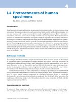
Table 5.2.
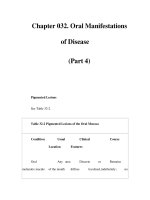
Principal rashes in infectious disease in man
Disease Features
Measles virus
Rubella virus
Parvovirus
Echoviruses 4, 6, 9, 16
Coxsackie viruses A9,
16, 23
Varicella-zoster virus
Coxsackie virus A16
Rickettsia prowazeki
and others
Rickettsia rickettsiae
and others
Streptococcus pyogenes
Measles
German measles 1
Erythema infectiosum
Not distinguishable
Treponema paUidum Syphilis
Treponema pertenue Yaws
Salmonella typhi ~ Enteric fever
Salmonella paratyphi B
J
Neisseria meningitidis Spotted fever
Blastomyces dermatitidis Blastomycosis
Cutaneous
leishmaniasis
Prodromal rashes
Very characteristic
maculopapular rash
Leishmania tropica
Hepatitis B and viral
exanthems
Dermatophytes
(skin fungi)
Maculopapular rashes not
distinguishable clinically
Chickenpox, zoster }
Hand, foot and mouth Vesicular rashes
disease
Typhus }
Spotted fever group Macular or haemorrhagic rash
of diseases
Scarlet fever Erythematous rash caused by
toxin
Disseminated infectious rash
seen in secondary stage,
2-3 months after infection
Sparse rose spots containing
bacteria
Petechial or maculopapular
lesions containing bacteria
Papule or pustule develops into
granuloma; lesions contain
organisms
Papules, usually ulcerating to
form crusted sores; infectious
Dermatophytid or
allergic rash
Streptococcus pyogenes
l
Impetigo a
Staphylococcus pyogenes S
Generalised rash due to
hypersensitivity to fungal or
viral antigens
Vesicles, forming crusts,
especially in children
a
These skin lesions are multiple but like those of erysipelas or warts are formed locally at the sites of
infection, not after spread through the body.
its own immune cells, particularly Langerhans cells (see p. 151), many
mast cells (see p. 161), and recirculatory T-cells are always present in
the dermis.
The skin of man is mostly naked, and is an important thermoregula-
tory organ, under finely balanced nervous control. It is a turbulent,
highly reactive tissue, and local inflammatory events are common-
place. At sites of inflammation, circulating microorganisms readily
localise in small blood vessels and pass across the endothelium. The
skin of most animals, in contrast, is largely covered with fur. Skin
lesions are a feature of many infectious diseases of animals, but these
lesions tend to be on exposed hairless areas where the skin has the
140 Mims' Pathogenesis of Infectious Disease
human properties of thickness, sensitivity and vascular reactivity.
Hence, although virus rashes very occasionally involve the general
body surface of animals, it is udders, scrotums, ears, prepuces, teats,
noses and paws that are more regular sites of lesions. For instance, the
closely related diseases of measles, distemper and rinderpest can be
compared. Cattle with rinderpest may show areas of red moist skin
with occasional vesiculation on the udder, scrotum and inside the
thighs. In dogs with distemper the exanthem often occurs on the
abdomen and inner aspect of the thighs. Yet in human measles there is
one of the most florid and characteristic rashes known, involving the
general body surface. Even in susceptible monkeys, the same virus
produces skin lesions sparingly and irregularly.
Macules and papules are formed when there is inflammation in the
dermis, with or without a significant cellular infiltration, the infection
generally being confined to the vascular bed or its immediate vicinity.
Immunological factors (see Ch. 8) are often important in the production
of inflammation. Measles virus, for instance, localises in skin blood
vessels, but the maculopapular rash does not appear unless there is an
adequate immune response. Virus by itself does little damage to the
blood vessels or the skin, and the interaction of sensitised lymphocytes
or antibodies with viral antigen is needed to generate the inflamma-
tory response that causes the skin lesion. Rickettsia characteristically
localise and grow in the endothelium of small blood vessels, and the
striking rashes seen in typhus and Rocky Mountain Spotted Fever are
a result of endothelial swelling, thrombosis, small infarcts and haem-
orrhages. The immune response adds to the pathological result.
Vascular endothelium is an important site of replication and shedding
of viruses and rickettsias that are transmitted by blood-sucking
arthropods and which must therefore be shed into the blood. After
replication in vascular endothelium, they may be shed not only back
into the vessel lumen, but also from the external surface of the
endothelial cell into extravascular tissues (see also p. 136). Certain
arthropod-borne viruses replicate in muscle or other extravascular
tissues, and can then reach the blood after passage through the
lymphatic system.
Circulating immune complexes consisting of antibody plus microbial
antigen also localise in dermal blood vessels, accounting for the
trichophytid rashes of fungal infections and the prodromal rashes seen
at the end of the incubation period in many exanthematous virus
diseases. Antibodies to soluble viral antigens appear towards the end of
the incubation period in people infected with hepatitis B virus and
form soluble immune complexes. These localise in the skin causing
fleeting rashes and pruritis, and rarely the more severe vascular
lesions of periarteritis nodosa (see Ch. 8).
Certain microbial toxins enter the circulation, localise in skin blood
vessels, and cause damage and inflammation without the need for an
immune response. An erythrogenic toxin is liberated from strains of
5 The Spread of Microbes through the Body
141
Streptococcus pyogenes
carrying the bacteriophage ~, and the toxin
enters the blood, localises in dermal vessels, and gives rise to the
striking rash of scarlet fever.
Vesicles and pustules are formed when the microorganism leaves
dermal blood vessels and is able to spread to the superficial layers of
the skin. Inflammatory fluids accumulate to give vesicles, which are
focal blisters of the superficial skin layers. Virus infections with vesi-
cles include varicella, herpes simplex and certain coxsackie virus infec-
tions. The circulating virus localises in dermal blood vessels, grows
through the endothelium (herpes, varicella) and spreads across dermal
tissues to infect the epidermis and cause focal necrosis. Only viruses
capable of extravascular spread and epidermal infection can cause
vesicles. Inevitably there is an immunopathological contribution to the
lesion, although a primary destructive action on epidermal cells gives
a lesion without the need for the immune response, as with the oral
lesions seen in animals as early as 2 days after infection with foot and
mouth disease virus. A secondary infiltration of leucocytes into the
virus-rich vesicle turns it into a pustule which later bursts, dries, scabs
and heals. Such viruses are shed to the exterior from the skin lesion.
Certain other microorganisms are shed to the exterior after extravasa-
tion from dermal blood vessels. They multiply in extravascular tissues
and form inflammatory swellings in the skin, which then break down
so that infectious material is discharged to the exterior. This occurs
and gives rise to striking skin lesions in the secondary stages of
syphilis and yaws (caused by the closely related bacteria
Treponema
pallidum
and
pertenue)
and is also seen in a systemic fungus infection
(blastomycosis) and a protozoal infection (cutaneous leishmaniasis). In
patients with leprosy,
Mycobacterium leprae
circulating in the blood
localises and multiplies in the skin, and for unknown reasons superfi-
cial peripheral nerves are often involved. The skin lesions do not break
down, although large numbers of bacteria are shed from sites of growth
on the nasal mucosa. Bacterial growth is favoured by the slightly lower
temperature of the skin and nasal mucosa.
Almost all the factors that have been discussed in relation to skin
localisation and skin lesions apply also to the mucosae of the mouth,
throat, bladder, vagina, etc. In these sites the wet surface means that
the vesicles will break down and form ulcers earlier than on the dry
skin. Hence in measles the foci in the mouth break down and form
small visible ulcers (Koplik's spots) a day or so before the skin lesions
have appeared (Fig. 5.3). Similar considerations apply to the localisa-
tion of microorganisms and their antigens on the other surfaces of the
body (see Fig. 2.1). In chickenpox and measles, circulating virus
localises in subepithelial vessels in the respiratory tract, and after
extravasation there is only a single layer of cells to grow through in the
nearby epithelium before the discharge of virus to the exterior. Hence
in these infections the secretions from the respiratory tract are infec-
tious a few days before the skin rash appears and the disease becomes
142
Mims' Pathogenesis of Infectious Disease
recognisable. Much less is known about the localisation of circulating
microorganisms in the intestinal tract. Probably localisation here is
not often of great importance, but this is a difficult surface of the body
to study. In typhoid, secondary intestinal localisation of bacteria takes
place following excretion of bacteria in bile, rather than from blood.
Virus localisation in the intestinal tract is a feature in rinderpest in
cattle but occurs only to a minor extent in measles. When the patient
with measles suffers from protein deficiency, however, it is more
important and helps cause the diarrhoea that makes measles a life-
threatening infection in malnourished children (see p. 378).
The foetus
The blood-foetal junction in the placenta is an important pathway for
infection of the foetus. The number of cells separating maternal from
foetal blood depends not only on the species of animal, there being four
cell sheets for instance in the horse and only one or two in man, but
also on the stage of pregnancy. The junction usually becomes thinner,
often with fewer cell layers, in later pregnancy. There are regular
mechanical leaks in the placenta late in most human pregnancies, and
up to 4.0 ml of blood is transferred across the placenta, but this
appears to be principally in one direction, from foetus to mother. There
is little evidence for the passive carriage of microorganisms across the
placenta, and foetal infection takes place by either of two mechanisms.
If a circulating microorganism, free or cell associated, localises in the
maternal vessels it can multiply, cause damage, locally interrupt the
integrity of the junction and thus infect the foetus.
Treponema
pallidum
and
Toxoplasma gondii
presumably infect the human foetus
in this way. Alternatively, a circulating microorganism can localise and
grow across the placental junction. This occurs with rubella and
cytomegalovirus infections of the human foetus. In both instances, a
placental lesion or focus of infection occurs before foetal invasion. The
microorganisms causing foetal damage are listed in Table 5.3 (see also
p. 334). These, however, are special cases, and special microorganisms.
Nearly always the foetus is protected from microbial as well as from
biochemical and physical insults. The factors that localise micro-
organisms in the placenta are not understood, but blood flow is slow in
placental vessels, as in sinusoids, giving maximal opportunities for
localisation. Once microorganisms are arrested in placental vessels,
their growth may be favoured by particular substances that are
present in the placenta. Erythritol promotes the growth of
Brucella
abortus,
and its presence in the bovine placenta makes this a target
organ in infected cows. Susceptibility of infected cattle to abortion thus
has a biochemical basis. Microorganisms can damage the foetus
without invading foetal tissues. If they localise extensively in placental
vessels and cause primarily vascular damage this of course can lead to
foetal anoxia, death and abortion. Also the toxic products of microbial
5 The Spread of Microbes through the Body
143
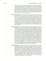
Table
5.3. Principal microorganisms infecting the foetus
Microorganisms Species Effect
Viruses
Rubella virus
Cytomegalovirus
HIV
Hog cholera virus (vaccine strain)
Bluetongue virus (vaccine strain)
Equine rhinopneumonitis
Bovine diarrhoea- mucosal
disease virus
Malignment catarrh virus
Bacteria
Treponema pallidum
Listeria monocytogenes
Vibrio foetus
Man Abortion
Stillbirth
Malformations
Man Malformations
Man About 1 in 5 infants born
to infected mothers
are infected
in utero
Pigs Malformations
Sheep Stillbirths, CNS disease
Horse Abortion
Cow Cerebellar hypoplasia
Wildebeest
Foetus unharmed
Man Stillbirth, malformations
Man Meningoencephalitis
Sheep, cattle Abortion
Protozoa
Toxoplasma gondii
Man
Stillbirth, CNS disease
growth in the placenta or elsewhere and probably cytokines can reach
the foetus and cause damage. High fever and biochemical disturbances
in a pregnant female can adversely affect the foetus.
Miscellaneous sites
There are certain other sites where circulating microorganisms selec-
tively localise. In rats and other animals infected with
Leptospira,
circulating bacteria localise particularly in capillaries in the kidney
and give rise to a chronic local lesion. Infectious bacteria are dis-
charged in large numbers into the urine, which is therefore a source of
human infection. Microorganisms that are discharged in the saliva
(mumps and most herpes-type virus infections in man) must localise
and grow in salivary glands. Those that are discharged in milk must
localise and grow in mammary glands (the mammary tumour virus in
mice and
Brucella,
tubercle bacilli, and Q fever rickettsia in cows). A
few examples, such as
Haemophilus suis
in pigs, Ross River virus in
man (Table A.5), and occasionally rubella virus, localise in joints.
Almost any site in the body, from the feather follicles (Marek's disease)
to testicles or epididymis (mumps in man, the relevant
Brucella
species
in rams, boars, bulls) can at times be infected. Nothing is known of the
mechanism of localisation in these organs.
144
Mims" Pathogenesis of Infectious Disease
Spread via other Pathways
Cerebrospinal fluid (CSF)
Microorganisms in the blood can reach the CSF by traversing the
blood-CSF junction in the meninges or choroid plexus. Capillaries in
the choroid plexus have fenestrated endothelium and are surrounded
by a loose connective tissue stroma (Fig. 3.2). Inert virus-sized particles
and bacteriophages leak into the CSF when very large amounts are
injected into the blood. It is assumed that the viruses causing aseptic
meningitis in man (polio-, echo-, coxsackie, lymphocytic choriomenin-
gitis and mumps viruses) enter the CSF by leakage or growth across
this junction (Fig. 5.6). Once in the CSF microorganisms are passively
carried with the flow of fluid from ventricles to subarachnoid spaces
and throughout the neuraxis within a short time. Invasion of the brain
itself and spinal cord can now take place across the ependymal lining
of the ventricles and spinal canal, or across the pia mater in the
subarachnoid spaces. Nonviral microorganisms entering the CSF
across the blood-CSF junction include the meningococcus, the tubercle
bacillus,
Listeria monocytogenes, Haemophilus influenzae, Strepto-
coccus pneumoniae,
and the fungus
Cryptococcus neoformans.
Pleural and peritoneal cavities
Rapid spread of microorganisms from one visceral organ to another can
take place via the peritoneal or pleural cavity. Entry into the peritoneal
Fig. 5.6 Routes of microbial invasion of the central nervous system. CSF =
cerebrospinal fluid.
5 The Spread of Microbes through the Body
cavity takes place from an injury or focus of infection in an abdominal
organ. The peritoneal cavity, as if in expectation of such events, is lined
by macrophages and contains an antimicrobial armoury, the omentum.
The omentum, originating from fused folds of mesentery, contains mast
cells and lymphocytes, macrophages and their precursors in a fatty
connective tissue matrix. It is movable in the peritoneal cavity and
becomes attached at sites of inflammation.* Microorganisms spread
rapidly in the peritoneal cavity unless they are taken up and destroyed
in macrophages or inflammatory polymorphs. Peritoneal contents
drain into lymphatics opening onto the abdominal surface of the
diaphragm, so that microorganisms or their toxins are delivered to
retrosternal lymph nodes in the thorax, sometimes with slight leakage
into the pleural cavity. Inflammatory responses in the peritoneum
eventually result in fibrinous exudates and the adherence of neigh-
bouring surfaces, which tends to prevent microbial spread.
Microbes entering the pleural cavity from chest wounds or from foci
of infection in the underlying lung have a similar opportunity to spread
rapidly. During pneumonia the overlying pleural surface first becomes
inflamed, causing pleurisy, and later often infected. Pleurisy occurs in
about 25% of cases ofpneumococcal pneumonia. The pleural cavity, like
the peritoneal cavity, is lined by macrophages.
9 145
Nerves
For many years peripheral nerves have been recognised as important
pathways for the spread of certain viruses and toxins from peripheral
parts of the body to the central nervous system (Fig. 5.6). Rabies,
herpes simplex and related viruses travel along nerves at up to
10 mm h -1, but the exact pathway in the nerve was for many years a
matter of doubt and debate. Herpes simplex virus, following primary
infection in the skin or the mouth, enters the sensory nerves and
reaches the trigeminal ganglion (see Ch. 10). Here it remains in latent
form until it is reactivated in later life by fever, emotional or other
factors. The infection then travels down the nerve to reach the region of
the mouth, where the skin is once again infected giving rise to a virus-
rich cold sore. A similar sequence of events explains the occurrence of
zoster long after infection with varicella virus. In cattle or pigs infected
with pseudorabies, another herpes virus, the infection also travels up
peripheral nerves to reach dorsal root ganglia, causing a spontaneous
discharge of nerve impulses from affected sensory neurons, and giving
rise to the signs of'mad itch'. Another herpes virus (B virus) is often
present in the saliva of apparently healthy rhesus monkeys, and people
* Because of its ability to attach to sites of inflammation and infection or to foreign bodies
the omentum has been referred to as the 'abdominal policeman'.
146
Mires' Pathogenesis of Infectious Disease
bitten by infected monkeys develop a frequently fatal encephalitis, the
virus reaching the brain by ascending peripheral nerves from the inoc-
ulation site. Rabies virus slowly reaches the CNS along peripheral
nerves following a bite delivered by an infected fox, jackal, wolf,
raccoon, skunk or vampire bat. It also travels centrifugally from the
brain down peripheral nerves to reach the salivary glands and other
organs. Poliovirus was long thought to reach the CNS via peripheral
nerves, but this was a conclusion from studies with artificially neuro-
adapted strains of virus. In natural infections, poliovirus traverses the
blood-brain junction (Fig. 3.2). Peripheral nerves are affected in
leprosy, the bacteria having a special affinity for Schwann cells, which
are unable to control the multiplying bacteria. The molecular basis for
this targeting of Schwann cells is being unravelled. This causes a very
slow and insidious degeneration of the nerve, but it is certainly not a
pathway for the spread of infection. Peripheral nerves are known to
transport tetanus toxin to the CNS (see Ch. 8), and also prion agents
(scrapie) in experimental infections of mice.
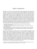
Possible pathways along nerves include sequential infection of
Schwann cells, transit along the tissue spaces between nerve fibres,
and carriage up the axon (Fig. 5.7). The last route is probably an impor-
tant one, although at first sight it might seem less likely. There is a
small but significant movement of marker proteins up normal axons
from the periphery to the CNS, and in experimental herpes simplex
and rabies infections virus particles have been seen in axons by elec-
tron microscopy. In experimental infections, herpes viruses can also
travel in nerves by sequential infection of the Schwann cells associated
with myelin sheaths, but this is not a natural route.
An alternative neural route of spread to the CNS is by the olfactory
nerves. Axons of olfactory neurons terminate on the olfactory mucosa,
the dendrites projecting beyond the mucosal surface giving a direct
anatomical connection between the exterior and the olfactory bulbs in
1. Perineural~
lymphatic
2. Interspaces
in
nerve
Perineureum
3. Endoneural cell elin
(e.g. Schwann ( ;ath
4. Axon
Fig. 5.7 Possible pathways of virus spread in peripheral nerves.
5 The Spread of Microbes through the Body 147
the brain. This route of infection, although at one time a popular postu-
late, is not often important. Aerosol infection with rabies virus (from
the excreta of bats in caves in North America) presumably involves this
route. When administered intranasally in experimental infections of
mice, Semliki Forest virus rapidly enters the olfactory bulbs and
thence into the rest of the brain. Naegleria fowleri, a free-living
amoeba that can lurk in the sludge at the bottom of freshwater pools,
causes a rare but often fatal meningitis in swimmers after infecting by
the olfactory route. The meningococci that live commensally in the
nasopharynx of 5-10% of normal people, and occasionally cause menin-
gitis, were once thought to spread directly upwards from the nasal
mucosa, along the perineural sheaths of the olfactory nerve, and
through the cribriform plate to the CSF. More probably, the bacteria
invade the blood, sometimes causing petechial rashes ('spotted fever'),
and reach the meninges across the blood-CSF junction.
In summary, peripheral nerves are important pathways for the
spread of tetanus toxin and a few viruses to the CNS, and for the
passage of certain herpes viruses between the CNS and the surfaces of
the body. Herpes and rabies viruses can travel both up and down
peripheral nerves. The neural route is not generally used by bacteria or
other microorganisms.
References
de Voe, I. W. (1982). The meningococcus and mechanisms of patho-
genicity. Microbiol. Rev. 46, 162-190.
Drutz, D. J. et al. (1972). The continuous bacteraemia of lepromatous
leprosy. N. Engl. J. Med. 287, 159-163.
Friedman, H. M., Macarek, E. J., MacGregor, R. A. et al. (1981). Virus
infection of endothelial cells. J. Infect. Dis. 143, 266.
Griffin, J. W. and Watson, D. F. (1988). Axonal transport in neurologic
disease. Ann. Neurol. 23, 3-13.
Johnson, R. T. (1982). ~Viral Infections of the Nervous System'. Raven
Press, New York.
Mims, C. A. (1964). Aspects of the pathogenesis of virus diseases. Bact.
Rev. 28, 30.
Mims, C. A. (1966). The pathogenesis of rashes in virus diseases. Bact.
Rev. 30, 739.
Mims, C. A. (1968). The pathogenesis of virus infections of the foetus.
Prog. Med. Virol. 10, 194.
Mims, C. A. (1981). The pathogenetic basis of viral tropism. Am. J.
Pathol. 135,447-455.
Moxon, R. E. and Murphy, P. A. (1978). Haemophilus influenzae
bacteremia and meningitis resulting from survival of a single
organism. Proc. Natl Acad. Sci. U.S.A. 75, 1534-1536.
148
Mims' Pathogenesis of Infectious Disease
Pearce, J. H.
et al.
(1962). The chemical basis of the virulence of
BruceUa abortus
II. Erythritol, a constituent of bovine foetal fluids
which stimulates the growth of
Br. abortus
in bovine phagocytes.
Brit. J. Exp. Pathol.
43, 31-37.
Quagliarello, V. and Schell, W. M. (1992). Bacterial meningitis; patho-
genesis, pathophysiology, and progress. N.
Engl. J. Med.
327,
864-872.
Rambukkana, A. (2000). How does Mycobacterium leprae target the
peripheral nervous system?
Trends Microbiol.
8, 23-28.
Williams, A. E. and Blakemore, W. F. (1990). Pathogenesis of meningitis
caused by
Streptococcus suis
Type 2. J.
Infect. Dis.
162, 474-481.
Williams, A. E. and Blakemore, W. F. (1990). Monocyte-mediated entry
of pathogens into the central nervous system.
Neuropath. Appl.
Neurobiol.
16, 377-392.
6
The Immune Response
to Infection
Antibody response
T-cell-mediated immune response
Natural killer cells
Macrophages, polymorphs and mast cells
Complement and related defence molecules
Conclusions concerning the immune response to
microorganisms
References
156
167
172
173
176
179
181
The immune response is conveniently divided into the antibody and
the cell-mediated component, the latter being transferable from one
individual to another by lymphocytes but not by serum. Antibodies,
since they can be tested and assayed without great difficulty, were the
first to receive attention with the discovery of antibodies to tetanus
and diphtheria toxins in the 1890s. Cell-mediated immunity (CMI) in
the form of delayed hypersensitivity was described more than 50 years
ago, and has received intensive study in the past 30 years. Specific
antibodies and CMI are induced in all infections, but the magnitude
and quality of these responses varies greatly in different infections. It
is not often that the microbial antigens concerned have been individu-
ally defined or identified. More importantly, we have rarely identified
the microbial antigens that induce protective immune responses.
Most antigens are proteins or proteins combined with other sub-
stances, but polysaccharides and other complex molecules also function
as antigens. Substances called haptens, small molecules such as
sugars, cannot by themselves stimulate antibody production, but do so
when coupled to a protein. An antigen stimulates the production of
antibodies that react specifically with that antigen. The reaction can be
thought of as similar to that between lock and key, and it is specific in
the sense that antibody produced against diphtheria toxin does not
react with tetanus toxin. An antibody may, however, have weaker reac-
tivity against antigens closely related to the one that stimulated its
production. For instance, antibodies produced when human serum is
injected into a rabbit will not react with the serum of cows, mice or
chickens, but may give a weak reaction with the serum of the gorilla
149
150 Mires' Pathogenesis of Infectious Disease
and chimpanzee. The antibodies formed against a given antigen will
include representatives from the four main immunoglobulin classes:
IgG, IgA, IgE and IgM. A single antigen molecule may have several
antigenic sites or epitopes, each of which stimulates the formation of a
different antibody. Also, different immunoglobulin molecules vary in
the firmness (avidity) with which they combine with the antigen, but
little is known about antibody avidity in relation to infectious diseases.
The two arms of the immune response are expressed by different
types of immunologically reactive lymphocytes, divided according to
their origin into B (bursa in birds or bone marrow and foetal liver in
mammals) and T (thymus) dependent cells. These two types of cells are
both small to medium-sized lymphocytes, only distinguishable by
specific cell surface molecules identified by immunological techniques.
B cells are concerned with the antibody response and T cells with initi-
ating the cell-mediated immune (CMI) response. B cells bear on their
surface immunoglobulin molecules that act as receptors for antigen.
Different B cells have different antigen-specific receptors (estimated to
be of the order of 109 for each individual). There are about 105 receptors
per cell; they are randomly generated by genetic recombination in the
developing B cell, and when almost any antigen enters the body for the
first time there will be a few B cells that react with it specifically.
Following an encounter with antigen, B cells become activated and
clonally expand to form a pool of memory cells or differentiate to form
plasma cells, the antibody synthesising cells. B cells are located in
various lymphoid tissues, notably spleen and lymph nodes and to a
lesser extent in the blood.
T cells express on their surface the T-cell receptor (TCR), a structure
not dissimilar to the immunoglobulin receptor, but which only recog-
nises antigenic peptides associated with MHC molecules on cell sur-
faces. There are two main types ofTCR, cd~ and y/8,* which are present
on distinct populations of T cells. As with B cells, T cells are clonally
derived, each bearing a unique TCR derived by gene rearrangement
during development. T cells are selected or educated to recognise
foreign antigens in the thymus. A vast repertoire of TCRs are gener-
ated during thymic development, reactive against self-antigens as well
as nonself or foreign antigens. Clearly, the host does not want T cells
capable of damaging its own cells and tissues, and so removes these
cells in the thymus by a process called clonal deletion (apoptotic death),
referred to as negative selection. Equally, the host needs a mechanism
for selecting those T cells destined to recognise foreign antigens and
this is also achieved in the thymus by a process called positive selec-
* ~/~ and y/5 denote the polypeptide chains composing the T cell receptor. Structurally,
the TCR resembles the Fab region of immunoglobulin molecules containing similar
constant and variable regions or domains. These domains form the basis of a diverse
family of important immunological molecules belonging to the immunoglobulin super-
family. Included in this family are MHC molecules, Fc receptors, B7 molecules, CD2,
CD3, CD4, CD8 and ICAM 1-3.
6 The Immune Response to Infection 151
tion. Since all T cells must recognise self-MHC plus 'antigen' during
development, it is still unclear why one population is deleted and the
other selected. A possible explanation involves the avidity of the indi-
vidual TCRs for self-MHC plus antigen: high-avidity interactions lead
to negative signalling and cell death, whereas low-avidity binding
leads to activation and an exit pass to the periphery. Two major classes
of educated T cells leave the thymus, one expressing the CD8 glycopro-
tein (CD8 T cells) and the other expressing the CD4 glycoprotein (CD4
T cells). These cells patrol various lymphoid compartments, waiting for
the opportunity to encounter foreign antigen presented by antigen pre-
senting cells. When this happens, the reactive T cells proliferate and
clonally expand, producing effector and memory cells.
The above is a simplified picture. Things are more complicated
because, nearly always, appropriate responses are produced by cooper-
ation between different types of cell. Dendritic cells and macrophages
play a central role in the induction of immunological responses. Those
in lymphoid tissues are strategically placed to encounter microbes or
their antigens, and at the same time are in close proximity to lymphoid
cells. Microbes and microbial antigens from sites of infection such as
the body surfaces are 'focused' by afferent lymphatics into macro-
phages and dendritic cells in lymph nodes (see pp. 78-79), and when
these materials enter the blood they are taken up by macrophages and
dendritic cells resident in the spleen. These cells serve a vital immuno-
logical function. They act as antigen presenting cells whose function is
to 'process' microbial and other antigens and present them to lympho-
cytes. An example is Langerhans cells* in the epidermis that send
dendritic processes far into the surrounding epithelium. They sample
their environment by endocytosis and macropinocytosis collecting anti-
gens which are then transported into local lymph nodes.
This is separate from the antimicrobial function of macrophages
described in Ch. 4 in which infectious agents are phagocytosed and
killed. The all-embracing word macrophage can be misleading, because
not all of them act as antigen-presenting cells in the induction of an
immune response and it is clear that separate subpopulations of
macrophages carry out the separate functions. For instance, most
Kupffer cells are inefficient inducers of immune responses and there-
fore the uptake of microorganisms by these cells is generally non-
productive from an immunological point of view.
Cell cooperation is an important feature in the induction and expres-
sion of the immune response. Virtually all effector responses are
dependent on T-cell recognition of antigen associated with MHC class
II molecules (see Glossary). These polymorphic membrane glycopro-
* Langerhans cells total 10 9 in man's skin, constituting 2-4% of all epidermal cells. They
belong to the dendritic cell family which are the principal antigen presenting cells
involved in the induction of the adaptive immune response. Dendritic cells are found in
all tissues, with the exception of the brain and cornea.
152 Mims' Pathogenesis of Infectious Disease
teins are located on dendritic cells (including Langerhans cells),
macrophages and B cells, all of which act as the 'professional' antigen
presenting cells of the body, i.e. the main inducers of immunological
responses. These cells function by endocytosing microbial antigens
which become degraded in endosomes by lysosomal enzymes into short
peptides (approximately 15 amino acids in length). These then asso-
ciate with newly formed or recycled MHC class II molecules, which are
presented on the cell membrane. This pathway of antigen presentation
is sometimes referred to as the exogenous pathway (see Fig. 6.1). The
peptide selectively binds to a groove on the MHC molecule, and it is
V-RNA
V ~rotein
I I
Proteosome
9 ~o Degraclati~
Peptides
I
Endogenous pathway Exogenous pathway
e,0n ro e,n
G
\
\
MHC class I MHC class II
r'-'- r-T
MHC class I N ~1 MHC class II
+ peptide + peptide
Fig. 6.1 Simplified scheme for the presentation of antigens via MHC class I
and II molecules. In the endogenous pathway (left), an infecting virus will
express viral RNA (v-RNA) and generate protein. The proteins are subjected to
proteolysis by proteosomes to generate short peptides (Q, A, m) that are trans-
ported into the endoplasmic reticulum (ER) by peptide transporters associated
with antigen presentation (TAP-1 and TAP-2). The peptides interact with
MHC class I to form stable molecular complexes that become directed to the
cell membrane and presented to CD8 T cells. The exogenous pathway (right)
involves the uptake of antigen by endocytosis. Endosomes fuse with lysosomes
leading to proteolysis. These early endosomes fuse with MHC class II-
containing vesicles and selected peptides interact with the MHC molecules.
These structures are displayed on the cell membrane and recognised by CD4 T
cells. In the endoplasmic reticulum MHC class II molecules are protected from
premature contact with other peptides and guided to the Golgi by the invariant
chain (L). This is degraded when the endosome and vesicle fuse, thus allowing
the peptides (O,/~, []) to interact with the class II molecules.
6 The Immune Response to Infection 153
this combination that is recognised by the TCR of CD4 T cells. These
cells function by producing a variety of lymphokines involved in the
activation and differentiation of other cells in the immune response,
hence they are known as T-helper cells. Antibody responses are heavily
dependent on T-helper cells for the generation of memory B cells and
the presence of IgG, IgE and IgA, including high-affinity IgG anti-
bodies in serum. The central role for T-helper cells in the immune
response is summarised in Fig. 6.2.
T-helper cells can be further subdivided according to their function
into two distinct populations of CD4 T cells, Thl and Th2. These cells
are distinguished from each other by the type of cytokines produced.
Thl cells are characterised by the expression of interleukin-2 (IL-2) and
interferon-y (IFN-y) and fail to produce IL-4, IL-5 or IL-10. In contrast,
Th2 cells produce IL-4, IL-5 and IL-10, but not IL-2 or IFN-y. In terms
of their function, Thl cells are associated with delayed-type hypersen-
sitivity (DTH) reactions resulting in the activation of macrophages and
the production in mice of IgG2a antibodies. Th2 cells predominantly
influence B-cell responses to produce IgE, IgA and IgG1 antibodies;
these cells are not involved in DTH reactions. Depending on the nature
of the antigen and the route of infection or immunisation, one particu-
lar Th subset will predominate. For example, microbial infection of skin
will favour Thl cells, where DTH reactions are important, whereas
infections involving parasitic worms will favour Th2 cells, where IgE
antibody is an important effector mechanism. T-cell cytokines are crit-
ical molecules in a number of immunological reactions. A summary of
the cytokines and their actions is shown in Table 6.1.
CD8 T cells, also known as cytotoxic T cells, recognise foreign peptide
in association with MHC class I molecules (found on virtually all cells
of the body). In this instance peptides are generated from proteins
derived within the cell (endogenously), for example, a protein from an
infecting virus, but the pathway of antigen processing and presentation
is different to that of the MHC class II system (see Fig. 6.1). The anti-
genic protein is degraded in the cytoplasm via an enzyme complex called
a proteosome and peptide fragments (approximately nine amino acids
in length) are actively transported into the endoplasmic reticulum
where they encounter newly formed MHC class I molecules. The pep-
tide-MHC complex is then transported to the cell membrane, where it
is recognised by the TCR of CD8 T cells - these cells are often described
as MHC class I restricted. The destruction of an infected cell by these
cytotoxic T cells or the liberation of cytokines with antimicrobial action,
is a major defence mechanism against intracellular microorganisms.*
* It is perhaps useful to attempt a rational explanation for these MHC requirements. A
cytotoxic effector T cell, before releasing its powerful weaponry, needs to know that its
physical contact with foreign antigen (peptide) is actually on the surface of a host cell.
The recognition of antigen plus MHC class I (present on all cells) ensures that this is so.
T-helper cells, on the other hand, need to know that peptide is being offered by a
specialised cell that has been able to carry out suitable processing and presentation, and
the MHC class II requirement for recognition ensures that this is so.
- ~ ~" ,~m ~~
-
- o o u ~ ~ <
,,0~b , , o:
._
~ _ - ~
~ ~ _~
- ~ ~ ~ ~ r ~] ~ T ~ m
oC ~c~eT- o ~ ~ ~oa o >.o' ~C~ ~ ~ ~ m
9 - , ._ .'-" "a b >, ~
r =~o ~=._ "- ~ m
, ~~>, ,-~ , ="d = "= '=
w
:U
,u I i o
.o .~
~ ,.~
~g.~
x
o
o
,- ~ ~'"~~
~,.~_-, m
UJ ~ o~
o "= 9
-a c -a c - ~ '~ ~ "~ ~::"
c ~ c ~ ~ "~ ~ ~
"
~ ~'~.~'
0 O_ 04 0 Cl 9 (D 0 ~ ~ CL~ ~
ra0
X ,
X 4 i._
C~6 ~C
~ ~ .
.~r~ ~ o"
0 ~
0 r Do o
c ~ or,.) -o.r
9 t:~ ~
~= ~
~,,~" ~ 0 ,_ ~
6 The Immune Response to Infection 155
Table 6.1.
Key cytokines produced by T lymphocytes and macrophages in
the immune response to microbial infection
Cytokine Source Target and action
IL-1 M
IL-2 L
IL-3 L
IL-4 L
IL-5 L
IL-6 L,M
IL-10 L,M
IL-12 L,M
IL-13 L
IL-18 M
IFN-y L
TNF-a L,M
TNF-[~ L
TGF-[~ L,M
GM-CSF L,M
Co-stimulator ofT cells. Activates macrophages. Inducer
of fever
Induces proliferation ofT cells and activates natural
killer (NK) cells. Induces antibody synthesis
Growth and differentiation of precursor cells in bone
marrow
B-cell proliferation and differentiation
Induces differentiation of B cells and activates
eosinophils
B- and T-cell growth and differentiation
Activates B cells and inhibits macrophage function
Activates NK cells and directs CD4 T cells to Thl
responses
Induces proliferation of B cells and differentiation of
T cells
Induces proliferation ofT cells
Activates most lymphoid cells
Causes activation of macrophages. Induces
inflammation and fever
Lymphotoxin. Inhibits B and T cells. Causes activation
of macrophages
Inhibits B-cell growth and macrophage activation.
Induces switch to IgA
Induces production of granulocytes and macrophages
L = produced by T lymphocytes; M = produced by macrophages.
When an immune response is initiated, powerful forces are set in
motion, which can be advantageous, but at times disastrous for the
individual (see Ch. 8). So that each response can unfold in a more or
less orderly fashion, it is controlled by a combination of stimulatory
and inhibitory influences. The latter include antigen control and the
activity of regulatory T cells producing immunosuppressive cytokines.
Antigen itself acts as an important regulatory agent. Following its
combination with antibody and uptake by phagocytic cells, it is
catabolised and begins to disappear from the body. Since it is the
driving force for an immune response, this response dies away as
antigen disappears. Immune responses can therefore be regulated by
controlling the concentration and location of antigen. A small amount
of specific antigen or cross-reactive antigens from other sources is
thought to be important for the maintenance of certain types of
immunological memory. As already discussed above, cytokines are
powerful regulators of the immune response (Table 6.1). Whereas some
of these factors activate the immune system, others can exert
inhibitory effects. For example, transforming growth factor-~ (TGF-~)
is a potent inhibitor ofT- and B-cell proliferation. Other cytokines such
as IFN-y inhibit IL-4 activation of B cells, whereas IL-4 and IL-10
156 Mims" Pathogenesis of Infectious Disease
inhibit IFN-y activation of macrophages and hence DTH reactions.
T cells producing these cytokines can therefore be thought of as regu-
lator or suppressor cells. Excessive production of any one of these
cytokines may lead to an inappropriate balance between antibody and
CMI responses, or to a more generalised immunosuppression affecting
the immune response to other microorganisms (see Ch. 7).
In a naturally occurring infection, the infecting dose generally
consists of only a small number of microorganisms, whose content of
antigen is extremely small compared with that used by immunologists,
and quite insufficient on its own to provoke a detectable immune
response. But the microorganism then multiplies, and this leads to a
progressive and extensive increase in antigenic mass. The classical
primary and secondary immune responses merge into one (see Fig.
12.1). Antibodies of various types and reactivities are produced in all
microbial infections, and are directed not only against antigens present
in the microorganism itself but also against the soluble products of
microbial growth, and in the case of viruses against the virus-coded
enzymes and other proteins formed in the infected cell during replica-
tion. Of the antigens present in the microorganism itself, the most
important ones in the encounter between microorganism and host are
those on the surface, directly exposed to the immune responses of the
host. Responses to internal antigenic components are generally less
important, although they are often of great help in detecting past infec-
tion, may appear on infected cells as targets for cytotoxic T cells, and
may play a part in immune complex disease (see Ch. 8).
There are three other important adjuncts to the immune response.
These are complement, phagocytic cells (macrophages and poly-
morphs) and natural killer cells, which are described under separate
headings below. Each is involved in various types of immune reactions.
Antibody Response
Types of immunoglobulin
By the time they reach adult life, all animals, including man, have been
exposed to a wide variety of infectious agents and have produced anti-
bodies (immunoglobulins) to most of them. Serum immunoglobulin
levels reflect this extensive and universal natural process of immuni-
sation. The different classes of immunoglobulin, with some of their
properties, are shown in Table 6.2. All are glycoproteins. The major
circulating type of antibody is immunoglobulin G (IgG). It has the basic
four-chain immunoglobulin structure in the shape of a Y, as illustrated
in Fig. 6.3, and a molecular weight of 150000. The molecule is
composed of two heavy and two light polypeptide chains held together
by disulphide bonds. For a given IgG molecule the two light chains are
~D
O
,.D
O
O
., ~
q~
O
~D
O
O
O
O
O
O
O
O
O
O
O
O~
O
O
O
O
O
O
O
O
L~
r
O
~u r O
rD
O
i;z;
rD
O
i;z;
-I-
O
o
~
=LL~ CD
rD
-I-
~D
O
O
O
O
.~ ~.~
ee
+o
r
C ~ .~
0 ~ "~
~o ~ o ~ c~'~
~ ~ ~
o
c~
c~
c~
.i-i
c~
r
o
o
.r ~
c~
o
,.Q
c~
c~
c~
cD
., 4
c~
r~
o
.4-a
c~ b.O o
158 Mims' Pathogenesis of Infectious Disease
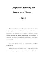
.~ Antigen-binding sites
Fab
k ain
Hinge region / ~ ss~: I "
Fc
J
Heavy
chain
Region with variable amino acid sequence in heavy and light chains, conferring
antigen specificity.
] ]Region with Constant amino acid sequence.
Hinge region enables arms to swing out to 180 ~ and bridge antigenic sites. Papain
digestion of molecule yields two Fab (fragment antigen-binding) portions, and one
Fc (fragment crystallisable) portion which confers biological activity on the molecule
(placental passage, binding to phagocytes, etc.)
Fig. 6.3 Basic Y-shaped (four-chain) structure of immunoglobulin G molecule.
either kappa (~) or lambda (~), and both heavy chains are gamma (y).
The antigen-binding ends of the light and heavy chains have a unique
amino acid sequence for a given antibody molecule and are responsible
for its specificity, while the rest of the chains are identical throughout
a given class of antibody. The molecule can be split into three parts by
papain digestion. Two of these (Fab) represent the arms of the Y and
contain the antigen-binding sites; the third part (Fc) has no antigen-
binding sites, but carries the chemical groupings that activate comple-
ment and combine with receptors on the surface of polymorphs and
macrophages (see below). This last activity of the Fc fragment medi-
ates attachment of antibody-coated microorganisms to the phagocyte,
giving the antibody opsonic activity. The Fc fragment also contains the
groupings responsible for the transport of IgG across the placenta of
some mammals. IgG can pass the placenta in primates, including man,
but not in rodents, cows, sheep, or pigs. Most IgG antibody is in the
blood, but it is also present in smaller concentrations in extravascular
tissues including lymph, peritoneal, synovial and cerebrospinal fluids.
Its concentration in tissue fluids is always increased as soon as there is
inflammation, or when it is being synthesized locally. There are four
6 The Immune Response to Infection 159
subclasses of IgG in man, which differ in heavy chains and in biological
properties such as placental passage, complement fixation and binding
to phagocytes. The amounts present in serum are also different, but
almost nothing is known of their relative importance in infectious
diseases.
Serum IgM is a polymer of five subunits, each with the basic four-
chain structure but with a different heavy chain (p), and has a molec-
ular weight of 900 000. Because it is such a large molecule, it is
confined to the vascular system. Its biological importance is first that,
molecule for molecule, it has five times the number of antigen-reactive
sites as IgG. It therefore has high avidity and is particularly good at
agglutinating microorganisms and their antigens. It also has five times
the number of Fc sites and therefore at least five times the comple-
ment-activating capacity (see below). A mere 30 molecules of IgM
attached to E. coli ensure its destruction by complement, whereas 20
times as many IgG molecules are required. Also, IgM is formed early in
the immune response of the individual. An infectious disease can be
regarded as a race between the replication and spread of the micro-
organisms on the one hand, and the generation of an antimicrobial
immune response on the other. A particularly powerful type of antibody
that is produced a day or two earlier than other antibodies may often
have a determining effect on the course of the infection, favouring
earlier recovery and less severe pathological changes. As each immune
response unfolds, the initially formed IgM antibodies are replaced by
IgG antibodies, and IgM are thus only detectable during infection and
for a short while after recovery. The presence of IgM antibodies to a
microbial antigen therefore indicates either recent infection or persis-
tent infection. A pregnant woman with a recent rubella-like illness
would have rubella IgM antibodies if that illness was indeed rubella.
Measles virus occasionally persists in the brain of children instead of
being eliminated from the body after infection, and the progressive
growth of virus in the brain causes a fatal disease called subacute scle-
rosing panencephalitis. The onset of disease may be 5-10 years after
the original measles infection, but IgM antibodies to measles are still
present because of the continued infection.
IgM antibodies are not only the first to be formed in a given immune
response, but are also the first to be formed in evolution. They are the
only antibodies found in a primitive vertebrate such as the lamprey.
IgM antibodies are also the first to be found during the development of
the individual. After the fifth to sixth month of development, the
human foetus responds to infection by forming almost entirely IgM
antibodies, and the presence of raised IgM antibodies in cord blood
suggests intrauterine infection. The only maternal antibodies that can
pass the placenta to reach the foetus are IgG in type, and thus the pres-
ence of IgM antibodies to rubella virus in a newborn baby's blood shows
that the foetus was infected.
Secretory IgA is the principal immunoglobulin on mucosal surfaces
160
Mims' Pathogenesis of Infectious Disease
and in milk (especially colostrum). It is a dimer, consisting of two
subunits of the basic four-chain structure with heavy chains, and as
the molecule passes across the mucosal epithelium, it acquires an addi-
tional 'secretory piece'. Secretory IgA has a molecular weight of
385000. It does not activate complement (see Ch. 9); although
monomeric IgA-antigen complexes do activate the alternative comple-
ment pathway. It has to function in the alimentary canal, and the
secretory piece gives it a greater resistance to proteolytic enzymes than
other types of antibody. In the submucosal tissues, the IgA molecule
lacks a secretory piece, and enters the blood via lymphatics to give
increased serum IgA levels in mucosal infections.
In the intestine, that seething cauldron of microbial activity, immune
responses are of immense importance but poorly understood. On the
one hand, commensal inhabitants are to be tolerated, but on the other
hand, protection against pathogens is vital. Powerful immunological
forces are present. The submucosa contains nearly 1011 antibody-
producing cells, equivalent to half of the entire lymphoid system, and
in man there are 20-30 IgA cells per IgG cell. Immune responses are
probably generated against most intestinal antigens (see p. 28), and
the sheer number of these antigens is formidable. It is a daunting
prospect to unravel immune events and understand control mecha-
nisms in this dark, mysterious part of the body. It has become clear
that in some species most of the intestinal secretory IgA comes from
bile. Although some of the IgA produced by submucosal plasma cells
attaches to the secretory piece present on local epithelial cells and is
then extruded into the gut lumen, most of it reaches the blood. In the
liver, IgA attaches to the secretory piece which is present on the surface
of hepatic cells, and is transported across these cells (see p. 134) to
appear in bile. This is important in the rat, but perhaps less so in man.
One consequence of the IgA circulation is that, when intestinal anti-
gens reach subepithelial tissues, they can combine with specific IgA
antibody, enter the blood as immune complexes and then be filtered out
and excreted in bile as a result of IgA attachment to liver cells.
There is a separate circulatory system that involves the IgA
producing cells themselves. After responding to intestinal antigens,
some B cells enter lymphatics and the bloodstream, from whence they
localise in salivary glands, lung, mammary glands and elsewhere in
the intestine. Localisation at these sites is achieved by recognition of
particular receptors on vascular endothelial cells called addressins
(see later). In this way, specific immune responses are seeded out to
other mucosal areas, where IgA antibody is produced and further
responses to antigen can be made.
IgA antibodies are important in resistance to infections of the
mucosal surfaces of the body, particularly the respiratory, intestinal
and urinogenital tracts. Infections of these surfaces are likely to be
prevented by vaccines that induce secretory IgA antibodies (see Ch. 12)
rather than IgG or IgM antibodies. However, most patients with selec-
6 The Immune Response to Infection 161
tive IgA deficiencies do not show undue susceptibility to infections of
mucosal surfaces, probably because there are compensatory increases
in the concentration of IgG and IgM antibodies on these surfaces.*
Those that are more susceptible generally have associated deficiencies
in certain IgG subclasses.
IgE is a minor immunoglobulin only accounting for 0.002% of the
total serum immunoglobulins, and it is produced especially by plasma
cells below the respiratory and intestinal epithelia. It has a marked
ability to attach to mast cells, and includes the reagenic antibodies that
are involved in anaphylactic reactions (see Ch. 8). When an antigen
reacts with antibody attached to a mast cell, mediators of inflamma-
tion (serotonin, histamine, etc.) are released. Thus, if a microorganism,
in spite of secretory IgA antibodies, infects an epithelial surface,
plasma components and leucocytes will be focused on to the area as
soon as microbial antigens interact with specific IgE on mast cells. IgE
is considered to be important in immunity to helminths. Larval forms
coated with IgE antibodies are recognised by eosinophils and
destroyed.
In humans, intestinal antibody is measured in duodenal or jejeunal
aspirates, or in faeces ('coproantibody'). Antibody from the entire gut
can be sampled by 'intestinal lavage', when an isotonic salt solution is
drunk until there is a watery diarrhoea, one litre of which is collected,
heat inactivated, filtered and concentrated.
IgD antibodies are for the most part present on the surface of B
lymphocytes. The same cells also carry IgM antibody, and it might be
expected that IgD serves as a receptor for antigen and is involved in
the activation of B cells. However, its main function is not clear.
General features
The antibody response takes place mostly in lymphoid tissues (spleen,
lymph nodes, etc.) and also in the submucosa of the respiratory and
intestinal tracts. Submucosal lymphoid tissues receive microorganisms
and their antigens directly from overlying epithelial cells, and
lymphoid tissues in spleen and lymph nodes receive them via blood or
lymphatics (see Ch. 5). Initial uptake and handling is by macrophages
and dendritic cells, following which antigens are delivered to CD4 T
cells (see above).
On first introduction of an antigen into the body, the antibody
response takes several days to develop. Pre-existing antigen-sensitive
* Also they may show less deficiency in secretory IgA than in the serum IgA which is
usually measured. In any case, the details differ in different species, and in sheep, for
instance, IgG figures as prominently as IgA in the secretory immunoglobulins. Finally,
it must be remembered that in the lower respiratory tract, at least, local CMI responses
can be induced, and may contribute to resistance.
162
Mires' Pathogenesis of Infectious Disease
B lymphocytes encounter antigen via the immunoglobulin receptor.
The antigen is internalised and processed via the exogenous pathway
and presented in association with MHC class II molecules to activated
T-helper cells. T-cell help is provided via CD40 activation and/or
cytokine receptors on B cells, e.g. IL-4 receptor (see Fig. 6.2). The B cells
then:
1. Divide repeatedly, forming a clone of cells with similar reactivity
(clonal expansion), some of which remain after the response is over,
as memory cells.
2. Differentiate, developing an endoplasmic reticulum studded with
ribosomes, in preparation for protein synthesis and export. The
cytoplasm of the cell therefore becomes larger and basophilic.
3. Synthesise specific antibody. The fully differentiated antibody-
producing cell is a mature plasma cell. Each clone of cells forms
immunoglobulin molecules of the same class and the same anti-
genic specificity.
Although the majority of antibody production occurs following T-cell
help, B cells can also become activated directly by polymeric antigens
(antigens with repeating epitopes) which cause cross-linking of specific
immunoglobulin receptors. This is commonly seen with bacteria, but is
also observed with viruses such as polyoma virus, rotavirus and vesic-
ular stomatitis virus. T-cell-independent antibody responses are
largely confined to the IgM isotype and have low affinity and short-
lived memory. However, these responses can be protective and in the
race to stem the dissemination of pathogens in the host such antibody
responses may provide a key defence.*
In a natural infection the initial microbial inoculum is small, and the
immune stimulus increases in magnitude following microbial replica-
tion. Small amounts of specific antibody are formed locally within a few
days, but free antibody is not usually detectable in the serum until
about a week after infection. As the response continues and especially
when only small amounts of antigen are available, B cells producing
high-affinity antibodies are more likely to be triggered, so that the
average binding affinity of the antibody increases as much as 100-fold.
The role of antibody in recovery from infection is discussed in Ch. 9,
the relative importance of antibody and cell-mediated immunity
depending on the microorganism. On re-exposure to microbial antigens
later in life, there is an accelerated response in which larger amounts
of mainly IgG antibodies are formed after only 1 or 2 days. The capacity
to respond in this accelerated manner often persists for life, and
depends on the presence of'memory cells'.
* Remember that every infection is a race between the ability of the invading microbe to
multiply and cause disease, and the ability of the host to mobilise specific and nonspecific
defences - a delay of a day or so on the part of the host can be critical.
6 The Immune Response to Infection
163
Antibodies to a given microbial antigen remain in the serum, often
for many years. Since the half-life of IgG antibody in man is about 25
days, antibody-forming cells are continually active. In some instances
(herpes viruses, tuberculosis) microorganisms remain in the body
after the original infection, and can continuously stimulate the
immune system. In other instances it seems clear that antibody levels
are kept elevated partly by repeated re-exposure to the microbe, which
gives subclinical re-infections and boosts the immune response. This is
known to occur with whooping cough, measles and other infections.
Sometimes, however, antibodies remain present in the serum for very
long periods in the absence of persistent infection or re-exposure. For
instance, five of six individuals who suffered an attack of yellow fever
in an epidemic in Virginia, USA in 1855 were found to have circulating
antibodies to yellow fever 75 years later. There had been no yellow
fever since the time of the original epidemic. Similarly, evidence from
isolated Eskimo communities in Alaska show that antibody to polio-
myelitis virus persists for 40 years in the absence of possible re-
exposure. It is now kaown that antigen can persist on the surface of
follicular dendritic cells (another member of the dendritic cell family
involved specifically with presenting antigen to B cells) in lymphoid
follicles for prolonged periods. This provides a continual source of anti-
genic stimulation to promote B-cell survival and presumably main-
tains B-cell memory. Plasma cells have also been recorded to survive
in the bone marrow for long periods, far in excess of what had previ-
ously been predicted for the half-life of these cells in lymph nodes and
spleen.
As a general rule, the secretory IgA antibody response is short-
lived compared with the serum IgG response.* Accordingly resistance
to respiratory infection tends to be short lived. Repeated infection
with common cold or influenza viruses often means infection with an
antigenically distinct strain of virus, but re-infections with respira-
tory syncytial virus or with the same strain of parainfluenza virus,
for instance, are common. Re-infection of the respiratory tract or
other mucosal surfaces is more likely to lead to signs of disease,
because of the short incubation period of this type of infection. After
re-infection with a respiratory virus there can be clinical disease
within a day or two, before the immune response has been boosted
and can control the infection. This is in contrast to re-infection with
say measles or typhoid; these are generalised infections, and the long
incubation period gives ample opportunity for the immune response
to be boosted and control the infection long before the stage of clini-
cal disease (Fig. 6.4).
* One factor is that, although there are very large numbers of IgA-producing plasma
cells in submucosal tissues, this immunoglobulin is exported to the outside world,
whereas IgG accumulates in the blood as it is produced.