AIRWAY MANAGEMENT IN EMERGENCIES - PART 6 pdf
Bạn đang xem bản rút gọn của tài liệu. Xem và tải ngay bản đầy đủ của tài liệu tại đây (723.04 KB, 32 trang )
position confirmed in the usual fashion. A Shiley
tracheostomy tube will have to have its inner
dilator removed and replaced with the inner
cannula. Once tracheal placement has been
confirmed, the tracheal hook is removed, and
the cannula or ETT is secured.
Early complications of cricothyrotomy
include bleeding, incorrect or unsuccessful
tube placement, cricoid cartilage fracture,
obstruction and subcutaneous emphysema.
Rarely, laryngeal, esophageal, or mediastinal
injury can occur. Pneumothorax, pneumome-
diastinum, and aspiration are also infrequent
complications. After the situation has stabilized,
a cricothyrotomy should be replaced either by
intubation from above, or by conversion to a
formal tracheostomy. This will help minimize
RESCUE OXYGENATION 145
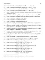
Figure 7–26. With thumb and long finger
stabilizing the thyroid cartilage, the index
finger palpates the cricothyroid membrane.
Figure 7–28. The index finger re-palpates
the cricothyroid membrane within the
wound.
Figure 7–29. A horizontal incision is then
made in the cricothyroid membrane.
Figure 7–27. A 3-cm vertical incision is made
over the cricothyroid membrane.
vocal cord morbidity or the occurrence of sub-
glottic stenosis at the level of the cricoid ring.
᭤ PEDIATRIC OPTIONS FOR RESCUE
OXYGENATION
At the outset, it must be said that a failed oxy-
genation situation is very unusual in the pediatric
population, due in no small measure to the fact
that this population is almost always easy to bag-
mask ventilate. However, as in the adult, if intu-
bation has failed and difficulty is encountered in
maintaining oxygen saturation with BMV, rescue
oxygenation can be achieved with both extra-
glottic devices as well as via transtracheal access.
Extraglottic Device Use in the
Pediatric Patient
Most of the extraglottic devices on the market
are available in pediatric sizes. Some are avail-
able in a full array of sizes while others are
146 CHAPTER 7
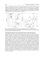
Figure 7–31. A Trousseau dilator is placed in
the cricothyrotomy, and is used to enlarge
the opening, vertically.
Figure 7–30. A tracheal hook picks up
and stabilizes the inferior border of the
thyroid cartilage, and is passed off to an
assistant.
Figure 7–32. A #4 tracheostomy tube is
placed between the arms of the Trousseau
dilator, into the cricothyrotomy opening.
Figure 7–33. The Trousseau dilator and tube
are rotated 90° counter-clockwise, and the
cannula is concurrently advanced down the
trachea.
suitable for use only in larger children (Table 7-1).
As with adults, case reports attest to success-
ful ventilation achieved by EGD use after BMV
had failed.
5
Pediatric Cricothyrotomy
Cricothyrotomy is not performed in children
under the age of eight. In this age group, there
is no developed space between the cricoid ring
and the thyroid cartilage. In addition, signifi-
cant narrowing occurs at the level of the cricoid
ring, which could impede cannula passage in
an emergency. Thirdly, as the cricoid ring is nec-
essary to help maintain patency of an otherwise
substantially membranous trachea, its fracture
during attempted cricothyrotomy could jeopar-
dize subsequent airway patency. For these
reasons, if trans-tracheal access is required in an
emergency in the patient under 8, it should be
obtained below the cricoid ring.
In keeping with the rare nature of the event,
there is very little literature on emergency
cricothyrotomy or tracheotomy in children.
Most clinicians would avoid an open surgical
technique in a pediatric emergency owing to
poor landmarks and the vascularity of the area.
Two other options exist:
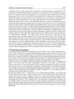
• Needle cricothyrotomy or tracheotomy
with ventilation through an attached pedi-
atric-sized manual resuscitator. A large-bore
IV catheter can be used to access the trachea,
and is connected to the manual resuscitator
in one of two ways: (a) insertion of the con-
nector of a 3.0 mm ID ETT into the IV catheter
hub or (b) attaching the barrel of a 3 cc
syringe, then pushing the connector of a 7.0
mm ID ETT into the end of the syringe barrel
(Figure 7–34). Both options then permit
attachment of a manual resuscitator via the
15-mm ETT connector. Manual ventilation
ensues with 100% oxygen. The chest must
be observed for deflation between ventila-
tions, to avoid the risk of barotrauma.
•A pediatric cricothyrotomy kit (e.g., the
Pedia-Trake Pediatric Emergency Cricothyro-
tomy Kit, Smiths Medical, St. Paul MN) is avail-
able with uncuffed cannulae in sizes of 3, 4,
and 5 mm ID.
᭤ PREDICTING DIFFICULT RESCUE
OXYGENATION
As is the case with predictors of difficult
bag-mask ventilation and difficult laryngo-
scopic intubation, the clinician should evaluate
whether rescue oxygenation via EGD or
cricothyrotomy is predicted to succeed. This
is of particular importance when a rapid-
sequence intubation (RSI) is contemplated in
RESCUE OXYGENATION 147
Figure 7–34. Needle cricothyrotomy set-up
using a large-bore IV, the barrel of a 3-cc
syringe attached to the connector of a 7.0 ETT.
The assembly is attached to a BVM device.
the uncooperative patient with predictors of dif-
ficult bag mask ventilation as well as difficult
laryngoscopy (see Chap. 11).
Predictors of Difficult Extraglottic
Device Use
Simply expressed, EGD use can fail due to an
inability to place the device into or through the
mouth; or even if it has been advanced through
the oral cavity, it can’t be seated in front of the
laryngeal inlet. Thirdly, even if seated well in
front of the laryngeal inlet, adequate ventilation
through the EGD may fail owing to obstructing
pathology at or below the glottis, or poor lung
compliance.
Alternatively, the mnemonic “MOODS” may
be useful to help recall predictors of difficulty in
achieving EGD rescue ventilation:
Mouth Opening limitation. Mouth opening
may be functionally impaired by trismus
and a clenched jaw, or anatomically by TMJ
pathology.
Obstruction at or below the glottic opening.
Glottic edema, foreign body, tumor, or sub-
glottic conditions can all preclude successful
ventilation via an EGD.
Distortion, displacement, or disruption of the
airway. Displacement or distortion of the
laryngeal inlet by pathology such as a
neck hematoma, blunt trauma, or radiation
changes may make it difficult to seat the
EGD directly in the path of the glottic
opening.
Stiff lungs (e.g., bronchospasm) and/or chest
wall. Bronchospasm or chest wall compro-
mise due to conditions such as morbid obe-
sity may cause EGDs to fail, as many (but
not all) have oropharyngeal leak pressures
of 25 cm H
2
O or less.
Predictors of Difficult Cricothyrotomy
The default course of action in a failed oxy-
genation scenario is cricothyrotomy. As with
EGD use, assessment of the patient for pre-
dictors of difficult cricothyrotomy is impor-
tant, particularly if difficulty with laryngoscopy
as well as BMV is predicted. Difficulty can
occur if there are impediments to identifying
the location of the cricothyroid membrane, or
even if its location is evident, if problems are
anticipated in accessing the trachea through it.
The mnemonic “DART” can help recall these
predictors.
Distortion of the anatomy from trauma,
expanding neck hematoma, infection, or
other pathology.
Access problems from obesity or extreme neck
flexion (e.g., ankylosing spondylitis).
Radiation therapy to the neck area in the past.
Tumors.
If RSI is being contemplated in an uncoop-
erative patient with predictors of difficult laryn-
goscopy and difficult bag-mask ventilation,
before proceeding, the clinician should locate
the cricothyroid membrane by palpation. Some
situations will mandate a formal “double setup”,
whereby RSI is undertaken only once the
cricothyroid membrane has already been
marked and prepped, and equipment and per-
sonnel are available for immediate cricothyro-
tomy should failed oxygenation ensue.
᭤ SUMMARY
With application of a consistent approach to
difficult bag-mask ventilation and difficult
laryngoscopy, failed intubation or failed oxy-
genation scenarios will be only infrequently
encountered. However, when the need arises,
extraglottic device use has transformed the air-
way management landscape away from the
old “can’t intubate—cut the neck” directive.
That being said, every clinician with a practice
mandate that includes airway management
should be familiar with indications for, and
knowledge of how to rapidly perform a
cricothyrotomy.
148 CHAPTER 7
REFERENCES
1. Mort TC. Emergency tracheal intubation: complica-
tions associated with repeated laryngoscopic
attempts. Anesth Analg. 2004;99(2):607–613, table
of contents.
2. Davies PR, Tighe SQ, Greenslade GL, Evans GH.
Laryngeal mask airway and tracheal tube insertion
by unskilled personnel. Lancet. 1990;336(8721):
977–979.
3. Levitan RM, Ochroch EA, Stuart S, Hollander JE.
Use of the intubating laryngeal mask airway by med-
ical and nonmedical personnel. Am J Emerg Med.
2000;18(1):12–16.
4. Yardy N, Hancox D, Strang T. A comparison of two
airway aids for emergency use by unskilled per-
sonnel. The Combitube and laryngeal mask.
Anaesthesia. 1999;54(2):181–183.
5. Brimacombe JR. Laryngeal Mask Anesthesia
Principles and Practice. 2nd ed. Philadelphia:
Saunders; 2005.
6. Brimacombe J, Keller C. Insertion of the LMA-
Unique with and without digital intraoral manipu-
lation by inexperienced personnel after manikin-
only training. J Emerg Med. 2004;26(1):1–5.
7. Parmet JL, Colonna-Romano P, Horrow JC, Miller F,
Gonzales J, Rosenberg H. The laryngeal mask airway
reliably provides rescue ventilation in cases of
unanticipated difficult tracheal intubation along
with difficult mask ventilation. Anesth Analg.
1998;87(3):661–665.
8. Brimacombe J, Keller C, Kunzel KH, Gaber O,
Boehler M, Puhringer F. Cervical spine motion
during airway management: a cinefluoroscopic
study of the posteriorly destabilized third cervical
vertebrae in human cadavers. Anesth Analg.
2000;91(5):1274–1278.
9. Keller C, Brimacombe J, Keller K. Pressures exerted
against the cervical vertebrae by the standard and
intubating laryngeal mask airways: a randomized,
controlled, cross-over study in fresh cadavers.
Anesth Analg. 1999;89(5):1296–1300.
10. Levitan RM, Frass M. The Combitube as rescue
device: recommended use of the small adult size
for all patients six feet tall or shorter. Ann Emerg Med.
2004;44(1):92; author reply 92–93.
11. Urtubia RM, Aguila CM, Cumsille MA. Combitube:
a study for proper use. Anesth Analg. 2000;90(4):
958–962.
12. Vezina D, Lessard MR, Bussieres J, Topping C,
Trepanier CA. Complications associated with
the use of the Esophageal-Tracheal Combitube.
Can J Anaesth. 1998;45(1):76–80.
13. Klein H, Williamson M, Sue-Ling HM, Vucevic M,
Quinn AC. Esophageal rupture associated with the
use of the Combitube. Anesth Analg. 1997;85(4):
937–939.
14. Mercer MH. An assessment of protection of the air-
way from aspiration of oropharyngeal contents
using the Combitube airway. Resuscitation.
2001;51(2):135–138.
15. Davis DP, Valentine C, Ochs M, Vilke GM, Hoyt DB.
The Combitube as a salvage airway device for para-
medic rapid sequence intubation. Ann Emerg Med.
2003;42(5):697–704.
16. Calkins TR, Miller K, Langdorf MI. Success and
complication rates with prehospital placement
of an esophageal-tracheal combitube as a
rescue airway. Prehospital Disaster Med.
2006;21(2 Suppl 2):97–100.
17. Staudinger T, Brugger S, Roggla M, et al. [Compar-
ison of the Combitube with the endotracheal
tube in cardiopulmonary resuscitation in the
prehospital phase]. Wien Klin Wochenschr.
1994;106(13):412–415.
18. Blostein PA, Koestner AJ, Hoak S. Failed rapid
sequence intubation in trauma patients: esophageal
tracheal combitube is a useful adjunct. J Trauma.
1998;44(3):534–537.
19. Mercer MH, Gabbott DA. Insertion of the Com-
bitube airway with the cervical spine immobilised
in a rigid cervical collar. Anaesthesia. 1998;53(10):
971–974.
20. Mercer MH, Gabbott DA. The influence of neck
position on ventilation using the Combitube
airway.
Anaesthesia. 1998;53(2):146–150.
21. Brimacombe J, von Goedecke A, Keller C, Brima-
combe L, Brimacombe M. The laryngeal mask
airway Unique versus the Soft Seal laryngeal
mask: a randomized, crossover study in paralyzed,
anesthetized patients. Anesth Analg. 2004;99(5):1
560–1563; table of contents.
22. Hanning SJ, McCulloch TJ, Orr B, Anderson SP.
A comparison of the oropharyngeal leak pressure
between the reusable Classic laryngeal mask airway
and the single-use Soft Seal laryngeal mask airway.
Anaesth Intensive Care. 2006;34(2):237–239.
23. Francksen H, Bein B, Cavus E, et al. Comparison
of LMA Unique, Ambu laryngeal mask and Soft
Seal laryngeal mask during routine surgical proce-
dures. Eur J Anaesthesiol. 2006:1–7.
RESCUE OXYGENATION 149
24. Tan MG, Chin ER, Kong CS, Chan YH, Ip-Yam PC.
Comparison of the re-usable LMA Classic and two
single-use laryngeal masks (LMA Unique and Soft-
Seal) in airway management by novice personnel.
Anaesth Intensive Care. 2005;33(6):739–743.
25. Sudhir G, Redfern D, Hall JE, Wilkes AR, Cann C.
A comparison of the disposable Ambu
AuraOnce(trade mark) Laryngeal Mask with the
reusable LMA Classic(trade mark) laryngeal mask
airway. Anaesthesia. 2007;62(7):719–722.
26. Asai T, Shingu K. The laryngeal tube. Br J Anaesth.
2005;95(6):729–736.
27. Scrase I, Woollard M. Needle vs surgical cricothy-
roidotomy: a short cut to effective ventilation.
Anaesthesia. 2006;61(10):962–974.
28. Frerk C, Frampton C. Cricothyroidotomy; time for
change. Anaesthesia. 2006;61(10):921–923.
29. Sulaiman L, Tighe SQ, Nelson RA. Surgical vs
wire-guided cricothyroidotomy: a randomised
crossover study of cuffed and uncuffed tra-
cheal tube insertion. Anaesthesia. 2006;61(6):
565–570.
30. Wong DT, Prabhu AJ, Coloma M, Imasogie N,
Chung FF. What is the minimum training required
for successful cricothyroidotomy?: a study in man-
nequins. Anesthesiology. 2003;98(2):349–353.
31. Melker JS, Gabrielli A. Melker cricothyrotomy kit:
an alternative to the surgical technique. Ann Otol
Rhinol Laryngol. 2005;114(7):525–528.
150 CHAPTER 7
Chapter 8
How to do Awake Tracheal
Intubations—Oral and Nasal
151
the right hand, will aid in performing awake
direct laryngoscopy.
• Acute-care clinicians should be as compe-
tent in performing an awake intubation as
they are in performing a rapid-sequence
intubation.
᭤ GENERAL CONSIDERATIONS
FOR THE AWAKE TRACHEAL
INTUBATION
Generally, tracheal intubations are performed
in one of three ways:
• Using rapid-sequence intubation (RSI)
• With an “awake” technique, following appli-
cation of topical airway anesthesia
• Facilitated by deep sedation, but without
pharmacologic paralysis
The occasional patient will require a primary
surgical airway. Advantages and disadvantages
of each route appear in Table 8–1 and are dis-
cussed further in Chap. 11.
The American Society of Anesthesiologists’
(ASA) difficult airway algorithm is predicated
upon the clinician first assessing the “likeli-
hood and clinical impact” of encountering dif-
ficulty.
1
If a difficult airway is considered likely
and clinically significant, the algorithm suggests
᭤ KEY POINTS
• If a difficult airway is considered likely
and clinically significant, an “awake”
approach should be considered, if patient
cooperation permits.
• An awake approach describes an intubation
technique facilitated by upper airway anes-
thesia applied topically or with nerve blocks,
with or without light doses of sedation.
• Although commonly used, “deep seda-
tion” should never be counted upon to
“relax” or alleviate clenched teeth, nor
should it be used to compensate for poor
topical airway anesthesia.
• In general, awake intubation should pro-
ceed by the route with which the clinician
has the most comfort and the greatest
experience.
• Local anesthetics can be topically applied
in ointment, jelly, nebulized or atomized
forms through mouth or nose. Nerve
blocks and transtracheal injection are also
options.
• If blood pressure permits, an awake intu-
bation can be performed in the semisitting
or sitting position.
• ‘Precision’ laryngoscopy, whereby the
operator carefully guides a laryngoscope
blade into the mouth using the digits of
Copyright © 2008 by The McGraw-Hill Companies, Inc. Click here for terms of use.
an awake approach. An awake approach to
the airway generally describes an intuba-
tion technique facilitated by upper airway
anesthesia applied topically or with nerve
blocks, in combination with light (e.g., anxi-
olytic) doses of sedation. “Awake” in the con-
text of emergency airway management is
perhaps a misnomer, as the patient requiring
emergency tracheal intubation often has an
impaired level of consciousness (LOC). How-
ever, “awake intubation”, even in the patient
with a depressed LOC, is distinct from tradi-
tional procedural sedation, where the patient’s
LOC might be intentionally altered in an
attempt to overcome resistance to laryn-
goscopy. This latter technique of using deep
sedation without paralysis, although still com-
monly practiced, has none of the benefits of
either awake or rapid sequence (RSI) approaches
to tracheal intubation: indeed, the use of deep
sedation is referred to by some as “tiger” country
in airway management.
2
Currently, RSI is both the most common pri-
mary and secondary rescue approach used to
facilitate tracheal intubation in emergency
departments (EDs) in North America.
3
The lit-
erature supports the use of RSI in the hands of
trained and experienced emergency physicians
(EPs).
4
The decision to use RSI follows an
assessment of the likelihood of encountering
difficulty during the process. In face of pre-
dicted difficulty, awake intubation becomes an
attractive alternative that may provide a wider
margin of safety in many instances. Unfortu-
nately, skillful awake tracheal intubation
receives little attention in the emergency med-
icine (EM) literature or practice. This may relate
to a combination of lack of perceived need,
patient cooperation issues, or deficits in awake
intubation skills teaching and experience.
As with RSI, acute-care clinicians should be
competent and experienced in performing an
awake intubation. This chapter will review the
awake intubation process using either the oral
or nasal route.
152 CHAPTER 8
The Advantages of Awake Tracheal
Intubation
As reviewed in Table 8–1, in a conscious patient,
an awake tracheal intubation delivers the fol-
lowing advantages:
• The patient continues to breathe sponta-
neously.
• The patient continues to maintain a patent
airway.
• The patient continues to protect the airway
against aspiration of gastric contents.
• Light (or omitted) doses of sedative/hyp-
notic agent will generally not present the
same risk of hypotension as those used
for RSI.
Patient Cooperation and Awake
Tracheal Intubation
A degree of patient cooperation is required for
an awake intubation. This may exclude a sig-
nificant proportion of patients requiring emer-
gency tracheal intubation. Indeed, the coopera-
tion issue is one which has made the use of RSI
so widespread in EDs. Patient cooperation fig-
ures prominently in the decision-making
process on how to proceed with tracheal intu-
bation (Fig. 11–3, Chap. 11). However, a blanket
dismissal of a patient’s ability to cooperate with
an awake intubation is also not appropriate:
patients will and can cooperate more often than
commonly perceived. The “actively” uncooper-
ative, physically agitated patient will often not
be rendered cooperative by any means. However,
other patients can be described as “passively”
uncooperative (e.g., the patient in respiratory
failure), and will often permit airway topicaliza-
tion and awake instrumentation. Patients in the
early stages of upper airway obstruction are
usually mentating normally and are ideal can-
didates for an awake approach, as discussed
below.
153
᭤ TABLE 8–1 COMPARISON OF DIFFERENT METHODS OF PROCEEDING WITH TRACHEAL INTUBATION
Intubation Method Advantages Disadvantages
Awake intubation • Patient continues to: • Clinician perception of patient discomfort.
⅙ Breathe spontaneously • Requires an element of patient cooperation.
⅙ Maintain • As with RSI, requires training in indications,
⅙ Protect performing airway anesthesia and direct
. . .his or her airway. laryngoscopic or indirect fiber/videoscopic
• “No bridges burned”. techniques.
• Avoids adverse effects of RSI medications.
• Avoids risk of hypoxemia during transition
from spontaneous respirations to taking
over positive pressure ventilation.
Deep sedation • Perception of a sense of security: • Often gives a false sense of security.
“I haven’t burned any bridges by giving • Retains many of the downsides of RSI while not
a muscle relaxant. . .” delivering the upside of facilitated conditions.
• May help control an uncooperative patient. • Undesirable reflexes intact:
• Perception of a more humane procedure.
⅙ gag/vomiting
⅙ laryngospasm
• No guarantee that deep sedative doses will leave
the patient breathing spontaneously or maintaining
an airway.
• Airway protection ablated in a full stomach patient,
often with no applied cricoid pressure.
• Deep sedative doses of medication can still hemo-
dynamically “crash” the patient.
• Scientific literature clearly documents less optimal
intubating conditions using only deep sedation.
35–37
(Continued)
154
᭤ TABLE 8–1 COMPARISON OF DIFFERENT METHODS OF PROCEEDING WITH TRACHEAL INTUBATION (Continued)
Intubation Method Advantages Disadvantages
RSI • Skeletal muscle relaxation facilitates • Induction drugs may cause profound drop in blood
conditions for direct laryngoscopy. pressure, for example, in shock states.
• Application of cricoid pressure may • Not all physicians are adequately
decrease risk of aspiration. trained in or comfortable using RSI.
• Not dependent on patient cooperation. • “Rescue RSI” not appropriate for all uncooperative
• Drugs may help control undesirable patients, for example, those with obstructing airway
physiologic responses, for example, ICP, HR. pathology.
• High success rates in experienced hands.
4
• Succinylcholine will not always wear off in time to
have patient resume spontaneous ventilation before
life-threatening hypoxemia occurs in “can’t intubate,
can’t oxygenate” situations.
• Fear of “what if I can’t intubate or ventilate?”
• Requires intimate knowledge of all drugs and
contraindications to technique.
Awake tracheotomy or • In the patient presenting with obstructing • Requires requisite surgical skills and equipment.
cricothyrotomy airway pathology, less risk of losing the
airway during application of topical airway
anesthesia or attempted tube passage
from above.
When and Why to do an Awake
Tracheal Intubation
There are three broad reasons to consider an
awake tracheal intubation in emergencies:
A. Predicted difficult airway. An awake
intubation should be considered primarily
if a question exists about whether the
clinician can easily take over what the
patient is presently doing for him- or her-
self. Especially if difficulty is predicted
in both intubating the patient and main-
taining oxygenation with either bag-mask
or a rescue oxygenation technique, then
awake intubation should be considered.
A classic scenario would include the patient
with obstructing pathologic changes in the
airway.
B. Predicted exaggerated hypotensive
response to induction medications used
for RSI. Some patients present with signif-
icant hemodynamic instability and concern
may exist over the effects of RSI induction
agents on the blood pressure. While careful
choice of induction agent and dose, together
with a fluid bolus, will often enable safe
conduct of an RSI in this situation, an
awake intubation is a second option to help
maintain blood pressure during tracheal
intubation.
C. RSI not needed: the arrested, critically
ill, or intrinsically sedated patient: Many
patients requiring intubation in emergen-
cies have a markedly decreased LOC as part
of their presenting condition. Such patients
may be arrested, critically ill, or intrinsically
sedated by their presenting condition, such
as hypercarbia due to respiratory failure.
While not truly “awake” or overtly cooper-
ative, these patients will often not resist a
primary laryngoscopy. This indication is
particularly relevant in the profoundly
hypotensive or arrested patient. In contrast,
the unconscious head-injured patient is still
best intubated with RSI.
Oral or Nasal Route?
In general, awake tracheal intubation should
proceed by the route with which the clinician
has the most comfort and the greatest experi-
ence. For most, this will mean an oral approach.
Blind nasotracheal intubation (BNTI) may be
considered an option when the patient’s
mouth opening is restricted and RSI is con-
traindicated. However, BNTI has relative con-
traindications in certain trauma patients, more
complications, and a lower success rate than
RSI.
4
With either route (oral or nasal), attempts
should be made to topically anesthetize the
airway.
Tools for Awake Tracheal
Intubation
Almost any intubating device can be used for an
awake intubation. Most awake intubations in
the operating room (OR) are performed using
a flexible fiberoptic bronchoscope. However,
direct laryngoscopy, a familiar technique,
can also be used and realistically would be
used for most awake intubations in the emer-
gency, out-of-OR setting. Other tools used for
awake intubations include video-based and
rigid or semi-rigid fiberoptic scopes.
5
A descrip-
tion of fiberoptic stylet use in the awake patient
appears in Chap. 6.
᭤ TOPICAL AIRWAY ANESTHESIA
The very presence of so many different pub-
lished techniques of applying topical airway
anesthesia bears witness to the fact that there
is probably no one best agent or technique.
Local anesthetics can be topically applied in
ointment, jelly, nebulized and atomized forms
through the mouth or nose. Nerve blocks and
transtracheal injection of local anesthetic are
also options.
HOW TO DO AWAKE TRACHEAL INTUBATIONS—ORAL AND NASAL 155
Review of Airway Innervation
The glossopharyngeal nerve innervates the pos-
terior third of the tongue down to and including
the vallecula, as well as the soft palate and
palatoglossal folds (Fig. 3–10, Chap. 3). A “gag”
response will be elicited if the laryngoscope
blade touches or applies pressure to sensitive
structures innervated by this nerve. These struc-
tures can be blocked with topically applied local
anesthetics. The inferior aspect of the epiglottis
and the larynx above the cords are supplied by
the internal branch of the superior laryngeal
nerve (SLN). Touch or pressure to these struc-
tures without anesthesia can stimulate reflex
glottic closure. The SLN can also be blocked top-
ically by application of local anesthetic in the
region of the piriform recesses, located on either
side of the laryngeal inlet. Alternatively, it can be
blocked by injecting a small volume of local
anesthetic (e.g., 2 mL of lidocaine 2%) in the
proximity of the nerve as it pierces the thyrohy-
oid membrane near the lateral edges of the hyoid
bone. Below the cords, sensation is provided by
the recurrent laryngeal branch of the vagus
nerve. Tracheal anesthesia can be attained with
inhalation or application of atomized local anes-
thetic, or a transcricothyroid membrane injec-
tion of local anesthetic.
Topical Airway Anesthesia for
Orotracheal Intubation
Adequate anesthesia for awake oral intubation
using direct laryngoscopy can be achieved with
anesthetic agents applied mainly to the distrib-
ution of the glossopharyngeal nerve (Fig. 3–10,
Chap. 3). Lidocaine can be used as a sole agent:
once applied to the mucosa, it will have maxi-
mal effect in 2–5 minutes, and will act for
about 20 minutes. Lidocaine ointment (in a 5%
concentration) or jelly (2% concentration)
(Fig. 8–1) is applied with a tongue depressor
from the front to back of the tongue, targeting
especially the posterior third. The ointment, if
used, is quite thick and must be applied slowly,
allowing it to “melt” on the tongue surface
(Fig. 8–2). The 2% jelly is easier to apply and will
usually be adequate. The very cooperative
156 CHAPTER 8
Figure 8–1. Lidocaine ointment (in a 5% concentration) or jelly (2% concentration) may be
applied with a tongue depressor.
HOW TO DO AWAKE TRACHEAL INTUBATIONS—ORAL AND NASAL 157
Figure 8–2. Lidocaine ointment once placed
on a tongue depressor is applied to the pos-
terior third of the tongue.
patient can also be coached to “gargle and
swish” liquid 4% lidocaine. Thereafter, other sen-
sitive areas, including the soft palate, posterior
pharynx, tonsillar pillars and hypopharynx
should be targeted, using a “spray as you go”
technique (Fig. 8–3). Lidocaine endotracheal
spray (in a 10% concentration = 10 mg/spray;
not currently available in the USA) can be used,
or 4% lidocaine administered by an atomizing
device. Atomizers include the venerable DeV-
ilbiss atomizer (Fig. 8–4) and the newer Mucosal
Atomization Devices (e.g., MADgic
®
, [Wolfe
Tory Medical Inc., Salt Lake City, UT], Fig. 8–5).
Although the above regimen will generally
allow for awake direct laryngoscopy, if time
permits, additional doses of local anesthetic
can be applied to progressively deeper struc-
tures (e.g., the laryngeal inlet). Gradually
deeper insertion of the laryngoscope blade will
help expose the epiglottis, and then glottic
opening for additional sprays of anesthetic
agent (Fig. 8–6). Oxygen can be readministered
as required in between doses.
Figure 8–3. The soft palate, posterior pharynx, tonsillar pillars, and hypopharynx should be tar-
geted, using a “spray as you go” technique.
Alternatively, 4 mL of 4% lidocaine with or
without neosynephrine 0.5% (1 mL) can be neb-
ulized and delivered either by mask or a mouth
piece (Fig. 8–7). This technique requires some
time (10–15 minutes) and a degree of patient
cooperation.
However applied, care should be taken to
ensure that the maximum recommended dose
of lidocaine (5–7 mg/kg) is not exceeded.
Topical Airway Anesthesia for
Nasotracheal Intubation
A. Vasoconstriction of the nasal mucosa can
be achieved with phenylephrine 0.5% or
oxymetazoline drops. Compared with cocaine
for the prevention of epistaxis, studies sug-
gest that phenylephrine and oxymetazoline
are no less effective (although other studies
have failed to show any advantage over
saline).
6–9
B. The nares can be anesthetized by applying
2% lidocaine jelly to, and inserting a nasopha-
ryngeal airway, or using a cotton pledget
soaked with 2% lidocaine with epinephrine.
Alternatively, one of the previously men-
tioned atomizing devices (e.g., DeVilbiss or
MAD
®
Nasal) can be used.
C. The pharynx is anesthetized with lidocaine
spray, as described in the above section on
“oral” anesthesia.
D. Lidocaine can be simultaneously delivered
to oral and nasal cavities by nebulizer mask.
Although an easy modality to use, results are
usually not as good as those obtained with
more focused application of local anesthetic.
158 CHAPTER 8
Figure 8–4. DeVilbiss atomizer.
Figure 8–5. Mucosal Atomization Device
(MADgic
®
, courtesy of Wolfe Tory Medical Inc.,
Salt Lake City, UT).
HOW TO DO AWAKE TRACHEAL INTUBATIONS—ORAL AND NASAL 159
Figure 8–6. Deeper structures may be targeted with topical airway anesthesia during the
awake laryngoscopy.
Figure 8–7. A mask or mouth-piece may be used to administer aerosolized lidocaine.
᭤ SEDATION FOR THE AWAKE
INTUBATION
Light sedation is the intended state for awake
intubation. It represents a depth of sedation
characterized by anxiolysis, and possibly
decreased pain perception, yet the patient is
readily rousable with verbal or at most, light
physical stimulation. The patient is able to main-
tain protective airway reflexes and a patent
airway, and should be at no risk of becoming
apneic. No sedation is also an option, and may
be most appropriate for the patient presenting
with a tenuous airway due to obstructing airway
pathology.
Deep sedation represents a state of
unconsciousness which may impair the
patient’s respiratory drive and ability to protect
the airway. Deep sedation can be an unintended
complication of light sedation. Consequences of
unintended deep sedation include vomiting and
aspiration with airway instrumentation, laryn-
gospasm, and apnea. It should also be recog-
nized that sedation alone rarely produces patient
cooperation in the actively combative patient.
Although commonly used, deep sedation should
never be counted upon to relax or alleviate
clenched teeth, nor should it be used to com-
pensate for poor topical airway anesthesia.
Sedation Pearls
A. Titrate to effect. Individuals respond dif-
ferently to the same medication dosages.
Small doses should be used initially, for
example, in a 70-kg patient: midazolam
0.25–1 mg/dose and/or fentanyl 25–50 µg/
dose, repeated as needed. Other agents to
consider would include haloperidol (2–5 mg/
dose) or ketamine (20–40 mg/dose). This
latter agent produces a state of dissociative
amnesia and tends to leave protective airway
reflexes intact. However, by sensitizing the
upper airway, ketamine has the theoreti-
cal potential to induce laryngospasm (pri-
marily seen in young children). With this
potential, and its tendency to increase secre-
tions, some clinicians have suggested that
ketamine may not be an ideal sedative agent
for awake intubation. Other sedative agents
with potential application to awake intuba-
tion include remifentanil and dexmedetomi-
dine (Chap. 13).
B. Age differences. The elderly require less
drug to achieve sedation, while children
in general require comparatively more
(in mg/kg).
C. Physiological differences. The patient
with high sympathetic tone (frequently the
case in the emergency intubation popula-
tion) is highly sensitive to low doses of
sedative agents.
D. Pathological differences. The neurologi-
cally impaired patient, for example, has
lower requirements.
E. Reversal agents. Although more often
required in nonairway procedural sedation,
reversal agents (Flumazenil and Naloxone)
should be readily available for benzodi-
azepines and opioids, respectively.
Note that the mainstay of the awake intu-
bation is topical airway anesthesia. Sedatives,
anxiolytics, or narcotics should be used only
as needed. An awake intubation should be
just that! Additional sedation can be adminis-
tered, if needed, as soon as the patient has
been successfully intubated and tube position is
confirmed.
᭤ AWAKE INTUBATION USING
DIRECT LARYNGOSCOPY
If blood pressure permits, an awake intubation
should be performed in the semisitting or sitting
position. This will be mandatory for the patient
in respiratory distress, who will be very reluc-
tant to lie supine. If needed, the clinician can
stand on a stool or a chair. Once the patient has
been prepared, laryngoscopy begins. “Preci-
sion” laryngoscopy, whereby the operator care-
fully guides the laryngoscope blade into the
160 CHAPTER 8
mouth using the digits of the right hand (Fig. 8–8),
will aid in keeping the blade in the lumen of the
oral cavity, avoiding contact with sensitive mucosa
until absolutely necessary. As the blade reaches
the back of the oral cavity, gentle tongue com-
pression will begin, aiming to visualize the
epiglottis. The patient can experience some
“pressure” at this stage. Once the epiglottis is
seen, the blade is positioned, centered, in the
vallecula (Fig. 8–9). At this point, the patient
should be warned that transient discomfort will
be felt during the increased pressure caused by
the laryngoscope “lift” needed to expose the
cords (Figure 8–10). Once seen, the clinician
should maintain visual contact with the cords,
while having a coached assistant place the endo-
tracheal tube (ETT) in his or her hand in the
correct orientation. Expeditious intubation
should then occur while the cords are abducted
during patient inspiration. If the cords are tran-
siently adducted, the clinician should pause with
the ETT poised at the cords until abduction
occurs. As used in the accompanying figures,
curved blade (Macintosh) laryngoscopy is
recommended for awake laryngoscopy, as a
direct lift of the SLN-innervated undersurface of
the epiglottis could otherwise stimulate reflex
glottic closure.
HOW TO DO AWAKE TRACHEAL INTUBATIONS—ORAL AND NASAL 161
Figure 8–8. “Precision” laryngoscopy, whereby the operator carefully guides the laryngoscope
blade into the mouth using the digits of the right hand.
᭤ AWAKE ORAL INTUBATION—A
GENERIC APPROACH
A. Preoxygenation with a nonrebreathing
face mask or manual resuscitator should
occur for 2–5 minutes (as time permits).
Supplemental oxygen should be continued,
as possible, during the application of topical
airway anesthesia.
B. Preparation of monitors, oxygen, BVM
device, suction, ETTs, stylet, laryngoscope
and blades, drugs, alternative intubation
options and rescue devices should be com-
plete. Note that psychological prepara-
tion should be performed with the mentating
patient: the patient should be told the ratio-
nale for the procedure and what to expect,
with reassurance. The patient’s coopera-
tion should be elicited (if possible) with
mouth opening, tongue protrusion, and
avoidance of struggling against the laryngo-
scope blade.
C. Topical airway anesthesia should be
applied, as described above. Sedation can
be titrated to effect, recognizing the need
for ongoing patient cooperation.
D. Awake direct laryngoscopy is performed,
as described above. Unpleasant as this may
sound, it is often well tolerated.
E. The tube location is confirmed. Once
intubated, the ETT cuff is inflated. Often,
the patient will cough, and expiratory flow
can be felt and heard issuing from the prox-
imal tube. The patient should not be able
to vocalize. However, objective confirma-
tion of placement with an ETCO
2
detector
should still be sought. The patient should
be reassured that the procedure has been
successfully completed, that he won’t be
able to talk.
162 CHAPTER 8
Figure 8–9. Once the epiglottis is seen, the blade can be positioned, centered in the vallecula.
F. Additional analgesia and sedation should
be introduced, as blood pressure permits.
Vital signs should be rechecked.
᭤ BLIND NASOTRACHEAL
INTUBATION (BNTI)
BNTI Introduction
BNTI has become increasingly rare in contem-
porary practice. This is appropriate, as it can be
technically challenging, has a higher complica-
tion rate, and compared to RSI, is less frequently
successful.
4
However, it is also a technique that
“occasionally, in cases where laryngoscopy is
difficult, [may permit] a nasal tube [to] enter the
trachea blindly with remarkable ease.”
10
As is
implied in its name, BNTI is performed without
direct visualization of the laryngeal inlet, in a
spontaneously breathing patient. Guided by
breath sounds, a regular endotracheal tube is
placed through the nose and advanced into the
trachea. Corrective maneuvers, if needed, are
suggested by clinical signs. BNTI may be an
option to consider in a patient with predictors
of significant difficulty when RSI is relatively
contraindicated and/or cooperation with an
awake oral intubation may not be expected.
All contraindications to BNTI (including
apnea) are relative, and include upper airway
foreign bodies, bleeding diathesis (including
heparinized, warfarinized, or recently throm-
bolyzed patients), or obstructing airway pathol-
ogy. Midface and/or basal skull fracture
11
has historically been included as a relative
HOW TO DO AWAKE TRACHEAL INTUBATIONS—ORAL AND NASAL 163
Figure 8–10. Once the blade is positioned in the vallecula, an appropriate lift will expose the
cords.
contraindication to BNTI, based on a small num-
ber of case reports of accidental intracranial
passage of nasogastric
12-15
or nasotracheal
tubes.
16–18
However, other published reports
have failed to demonstrate adverse outcomes
following nasal intubation in this same popu-
lation in the prehospital,
19,20
ED,
19
or OR
settings.
19,21
BNTI Technique
As with any other technique, all needed equip-
ment should be assembled. An uncut ETT one
full size smaller than would normally be used
for orotracheal intubation should be selected, for
example, 6.5–7.0-mm internal diameter (ID) for
a female or 7.0–7.5-mm ID for a male. Both
nostrils should be medicated with local anes-
thetic with or without a vasoconstrictor, as detailed
earlier in this chapter. A nasopharyngeal airway
covered in lidocaine jelly or ointment can help
with application of the anesthetic while also
assessing the nasal passage for patency. An
attempt should also be made to apply local anes-
thetic to the pharynx (if feasible) in a manner sim-
ilar to that previously described for the oral
approach.
The head and neck should be placed in the
“sniffing” position.
10
The well-lubricated tube
should be placed in the right nostril (if the option
exists), with the bevel facing the septum. This
tube orientation keeps the leading edge of the
ETT’s bevel away from the vascular Kiessel-
bach’s plexus on the nasal septum. Other clini-
cians advocate preferentially using the most
patent nostril. The tube should be directed infe-
riorly, along the floor of the nasal passage, to
stay within the major nasal airway, beneath the
inferior turbinate. This will also direct the tube
away from the thin bone of the more superiorly
located cribriform plate.
22
A gentle twisting
motion during tube advancement will help
avoid obstruction.
There will be some resistance in the poste-
rior nasopharynx (as is the case when inserting a
nasogastric tube). Gentle pressure and a twisting
motion (e.g., 90° to the left) should allow pas-
sage. As the ETT is advanced, fogging will be
seen and breath sounds should be heard from
the end of the tube. The ETT is further advanced,
with the clinician’s ear near the proximal end of
the ETT, monitoring breath sounds. Maximal
breath sounds will be heard when the ETT is at
the glottic opening. The ETT should then be
quickly advanced through the cords during
inspiration, when the cords are maximally
abducted. The cuff is inflated, and tube position
confirmed in the usual objective fashion. The
ETT should be sited at approximately 28 cm at
the nares in males and 26 cm in females.
BNTI Troubleshooting
Due to the intrinsic shape of the ETT and the
path traveled as it is advanced through the
nasopharynx, the tube is often perfectly directed
up toward the larynx. However, one of four
malpositions can occur.
23
Diagnosis of a mal-
position should be possible by evaluating
(a) breath sounds through the tube; (b) resis-
tance to forward tube passage; and (c) palpation
or observation of tube impingement in the ante-
rior neck. Corrective maneuvers include directing
the tube more anteriorly (by extending the head)
or posteriorly (by lifting then flexing the head);
or by directing it laterally (by twisting the tube to
left or right), as needed. Diagnostic features of,
and corrective maneuvers needed for the four
BNTI malpositions appear in Table 8–2.
Continued difficulty in spite of these correc-
tive maneuvers can occasionally be addressed by
some of the suggestions in the next section,
useful in the patient requiring C-spine precautions.
Performing BNTI with C-Spine
Precautions
For BNTI in the patient requiring manual in-line
neck stabilization, head extension is not an
option if additional anterior direction of the tube
164 CHAPTER 8
is needed. The following three maneuvers can
be used as alternatives:
A. Backward pressure can be applied to the
thyroid cartilage (akin to external laryngeal
manipulation (ELM) performed to improve
the view at direct laryngoscopy), to “bring
the larynx to the tube”.
B. If available, an Endotrol
®
tube
24–26
(Mallinckrodt Inc., St. Louis, MO) can be
used. This tube has a directable tip, con-
trolled by a small loop near its proximal end
(Fig. 8–11). By pulling on the loop, the ETT
tip is flexed, causing it to move anteriorly.
C. With the tube sitting in the oropharynx,
inflation of the ETT cuff with 10–15 mL of
air will elevate the ETT tip up and toward
the laryngeal inlet. The tube is then
advanced until resistance is encountered,
with loud breath sounds. The cuff is deflated
to allow tube passage through the cords,
and is reinflated once tracheal intubation
has been successfully completed.
27, 28
This
maneuver can also be used with Trachlight
guidance, using the device with its inner
stylet removed.
Although the “sniffing” position has long
been thought to be the ideal position for BNTI,
10
the neutral position with cuff inflation appears
to result in similar success rates.
29
BNTI Complications
In addition to failure to intubate, complications
of BNTI may include epistaxis and bacteremia.
Epistaxis severe enough to interfere with intu-
bation or require posterior nasal packing is
unusual, occurring in less than 2.5% of cases.
8,30,31
Moderate bleeding, usually described as blood
visible in, or enough to suction from the poste-
rior pharynx, occurs more often, in up to 14%
of cases.
8,9,30,31
This latter degree of bleeding
would be unlikely to interfere with intubation
attempts, however.
HOW TO DO AWAKE TRACHEAL INTUBATIONS—ORAL AND NASAL 165
᭤ TABLE 8–2 BLIND NASAL INTUBATION: DIAGNOSING AND CORRECTING MALPOSITIONS
Resistance
to Forward Neck Corrective
Malposition Breath Sounds Passage Palpation Maneuver
(None: Correct Present None Nothing None needed
location) palpable
Caught up on Present Present Nothing Flex head slightly (for
adducted cords, palpable anterior commissure
anterior hang-up), rotate tube
commissure, slightly, and readvance
or cricoid
Caught up in Muffled or Present Tube tip in Withdraw, flex head
vallecula absent midline of slightly, do jaw lift and
neck readvance
Piriform sinus Muffled or Present Tube tip felt in Withdraw slightly, rotate
absent lateral neck tube in contralateral
direction, readvance
Esophagus Absent None Nothing Withdraw tube until breath
palpable sounds heard, extend
head, readvance tube
Bacteremia occurred with nasotracheal
intubation
32
in 5.5% of patients in one OR
series. Retropharyngeal perforation, with a risk
of submucosal false passage, has been
described.
30
As previously mentioned, intracra-
nial endotracheal tube passage has also been
reported.
16–18
BNTI Effectiveness
R
OUTINE AND
D
IFFICULT
A
IRWAY
M
ANAGEMENT
In the hands of seasoned clinicians, BNTI suc-
cess rates of up to 92% have been reported in
both routine and difficult airway situa-
tions.
11,30,31
Other series have reported first
attempt success rates of 55%–61% in the hands
of novices “within a reasonable time frame.”
11,33
Both overall and first-attempt success rates
are lower than those reported using other tech-
niques.
4,33
C-
SPINE
P
RECAUTIONS
Historically, BNTI was advocated as the
method of choice for intubation of the patient
with a suspected C-spine injury. In fact, nei-
ther neurologic outcome nor C-spine move-
ment with BNTI in this population has been
shown to differ from that resulting from oral
intubation by direct laryngoscopy.
34
Currently,
known or suspected C-spine injury is not by
itself considered an indication for nasotracheal
intubation, blind or otherwise.
᭤ SUMMARY
Compared with RSI, awake intubation may be
perceived as a more technically challenging pro-
cedure. In contrast to RSI, an element of patient
cooperation is needed to successfully manage a
patient’s airway using an awake approach.
Awake intubation is a necessary skill and should
not become a vanishing art. Success is linked
not only to patient selection but also to the
deliberate acquisition and maintenance of
awake intubation skills. Clinicians should be as
competent and comfortable in performing an
awake tracheal intubation as they are in per-
forming an RSI.
166 CHAPTER 8
Figure 8–11. Endotrol tube
24–26
(Mallinckrodt Inc., St. Louis, MO).
REFERENCES
1. Practice guidelines for management of the difficult
airway: an updated report by the American Society
of Anesthesiologists Task Force on Management
of the Difficult Airway. Anesthesiology. 2003;98(5):
1269–1277.
2. Donati F. Tracheal intubation: unconsciousness,
analgesia and muscle relaxation. Can J Anaesth.
2003;50(2):99–103.
3. Sagarin MJ, Barton ED, Chng YM, et al. Airway man-
agement by US and Canadian emergency medi-
cine residents: a multicenter analysis of more
than 6,000 endotracheal intubation attempts. Ann
Emerg Med. 2005;46(4):328–336.
4. Kovacs G, Law JA, Ross J, et al. Acute airway
management in the emergency department by
non-anesthesiologists. Can J Anaesth. 2004;51(2):
174–180.
5. Kovacs G, Law JA, Petrie D. Awake fiberoptic intu-
bation using an optical stylet in an anticipated dif-
ficult airway. Ann Emerg Med. 2007;49(1):81–83.
6. Gross JB, Hartigan ML, Schaffer DW. A suitable
substitute for 4% cocaine before blind nasotracheal
intubation: 3% lidocaine-0.25% phenylephrine
nasal spray. Anesth Analg. 1984;63(10):915–918.
7. Latorre F, Otter W, Kleemann PP, et al. Cocaine or
phenylephrine/lignocaine for nasal fibreoptic intu-
bation? European Journal of Anaesthesiology.
1996;13:577–581.
8. Katz RI, Hovagim AR, Finkelstein HS, et al. A com-
parison of cocaine, lidocaine with epinephrine, and
oxymetazoline for prevention of epistaxis on naso-
tracheal intubation. J Clin Anesth. 1990;2(1):16–20.
9. Rector FT, DeNuccio DJ, Alden MA. A comparison
of cocaine, oxymetazoline, and saline for nasotra-
cheal intubation. Aana J. 1987;55(1):49–54.
10. Magill IW, Macintosh R, Hewer CL, et al. Lest we
forget. A historic meeting of the Section of Anaes-
thetics of Royal Society of Medicine on 6 Decemeber
1974. Divynyl ether. Anaesthesia. 1975;30(5):
630–632.
11. Iserson KV. Blind nasotracheal intubation.
Ann Emerg Med. 1981;10(9):468–471.
12. Fremstad JD, Martin SH. Lethal complication
from insertion of nasogastric tube after severe
basilar skull fracture. J Trauma. 1978;18(12):
820–822.
13. Fletcher SA, Henderson LT, Miner ME, et al. The
successful surgical removal of intracranial nasogas-
tric tubes. J Trauma. 1987;27(8): 948–952.
14. Gregory JA, Turner PT, Reynolds AF. A complica-
tion of nasogastric intubation: intracranial penetra-
tion. J Trauma. 1978;18(12):823–824.
15. Bouzarth WF. Intracranial nasogastric tube inser-
tion. J Trauma. 1978;18(12):818–819.
16. Horellou MF, Mathe D, Feiss P. A hazard of naso-tra-
cheal intubation. Anaesthesia. 1978;33(1): 73–74.
17. Cameron D, Lupton BA. Inadvertent brain pene-
tration during neonatal nasotracheal intubation.
Arch Dis Child. 1993;69(1 Spec No):79–80.
18. Marlow TJ, Goltra DD, Jr., Schabel SI. Intracranial
placement of a nasotracheal tube after facial frac-
ture: a rare complication. J Emerg Med. 1997;15(2):
187–191.
19. Rosen CL, Wolfe RE, Chew SE, et al. Blind nasotra-
cheal intubation in the presence of facial trauma.
J Emerg Med. 1997;15(2): 141–145.
20. Rhee KJ, Muntz CB, Donald PJ, et al. Does naso-
tracheal intubation increase complications in
patients with skull base fractures? Ann Emerg Med.
1993;22(7):1145–1147.
21. Bahr W, Stoll P. Nasal intubation in the presence
of frontobasal fractures: a retrospective study.
J Oral Maxillofac Surg. 1992;50(5):445–447.
22. Goodisson DW, Shaw GM, Snape L. Intracranial
intubation in patients with maxillofacial injuries
associated with base of skull fractures? J Trauma.
2001;50(2):363–366.
23. Jacoby J. Nasal endotracheal intubation by an
external visual technic. Anesth Analg. 1970;49(5):
731–739.
24. Asai T. Endotrol tube for blind nasotracheal intu-
bation.
Anaesthesia. 1996;51(5):507.
25. Hooker EA, Hagan S, Coleman R, et al. Directional-
tip endotracheal tubes for blind nasotracheal intu-
bation. Acad Emerg Med. 1996;3(6):586–589.
26. O’Connor R E, Megargel RE, Schnyder ME, et al.
Paramedic success rate for blind nasotracheal intu-
bation is improved with the use of an endotracheal
tube with directional tip control. Ann Emerg Med.
2000;36(4):328–332.
27. van Elstraete AC, Pennant JH, Gajraj NM, et al. Tra-
cheal tube cuff inflation as an aid to blind nasotra-
cheal intubation. Br J Anaesth. 1993;70(6): 691–693.
28. Gorback MS. Inflation of the endotracheal tube
cuff as an aid to blind nasal endotracheal intuba-
tion. Anesth Analg. 1987;66(9):916–917.
29. Chung YT, Sun MS, Wu HS. Blind nasotracheal
intubation is facilitated by neutral head position
and endotracheal tube cuff inflation in spontaneously
HOW TO DO AWAKE TRACHEAL INTUBATIONS—ORAL AND NASAL 167
breathing patients. Can J Anaesth. 2003;50(5):
511–513.
30. Tintinalli JE, Claffey J. Complications of nasotracheal
intubation. Ann Emerg Med. 1981;10(3): 142–144.
31. Danzl DF, Thomas DM. Nasotracheal intubations
in the emergency department. Crit Care Med.
1980;8(11):677–682.
32. Dinner M, Tjeuw M, Artusio JF, Jr. Bacteremia
as a complication of nasotracheal intubation.
Anesth Analg. 1987;66(5):460–462.
33. Roppolo LP, Vilke GM, Chan TC, et al. Nasotra-
cheal intubation in the emergency department,
revisited. J Emerg Med. 1999;17(5):791–799.
34. Crosby ET. Airway management in adults after
cervical spine trauma. Anesthesiology. 2006;104(6):
1293–1318.
35. Lieutaud T, Billard V, Khalaf H, et al. Muscle relax-
ation and increasing doses of propofol improve
intubating conditions. Can J Anaesth. 2003;50(2):
121–126.
36. Erhan E, Ugur G, Gunusen I, et al. Propofol—not
thiopental or etomidate—with remifentanil
provides adequate intubating conditions in the
absence of neuromuscular blockade. Can J
Anaesth. 2003;50(2):108–115.
37. Collins L, Prentice J, Vaghadia H. Tracheal intuba-
tion of outpatients with and without muscle relax-
ants. Can J Anaesth. 2000;47(5):427–432.
168 CHAPTER 8
Chapter 9
Rapid Sequence Intubation—
Why and How to do it
169
᭤ INTRODUCTION TO RAPID-
SEQUENCE INTUBATION (RSI)
Historically, the term “RSI” has referred to Rapid
Sequence Induction (of anesthesia), used to
minimize the risk of aspiration in surgical
patients felt to have a “full stomach.” In emer-
gency medicine the term RSI refers to Rapid
Sequence Intubation (RSI). The difference in
semantics refers to a different end-point: in the
operating room (OR), patients are intubated to
provide anesthesia, while in the emergency
department (ED), patients are anesthetized to
facilitate tracheal intubation.
1
Rapid sequence induction was originally
described in the anesthesia literature as a method
of airway management undertaken to minimize
the risk of aspiration in parturients undergoing
emergency Caesarean section.
2
Despite initial
controversy, RSI has now also become part of
everyday emergency medicine (EM) practice. RSI
in emergency care involves the use of a combi-
nation of specific pharmacologic agents to obtain
optimal conditions for tracheal intubation.
RSI use has been shown to increase the like-
lihood of successful tracheal intubation and
decrease complications when compared to
other methods, in most settings.
3
However,
these results assume trained and experienced
clinicians who have learned the process and are
familiar with the drugs used.
᭤ KEY POINTS
• For clinicians with the requisite skills,
rapid-sequence intubation should
become the method of choice for tra-
cheal intubation in emergencies, unless
contraindicated.
• The emergency medicine literature sug-
gests high success rates and low mor-
bidity with the use of RSI in experienced
hands.
• “Successful” tracheal intubation does not
by itself necessarily represent a successful
outcome.
• The evidence favoring pretreatment med-
ications is not compelling. Emphasis
should instead be placed on preventing
hypoxia and hypotension.
• If rapid oxygen desaturation occurs (or is
likely to occur) with onset of apnea
during an RSI, gentle bag-mask ventilation
can and should be performed while
awaiting onset of the muscle relaxant.
• Cricoid pressure has the potential to
impair the view at laryngoscopy, cause
difficulty with bag-mask ventilation (BMV)
and impair extraglottic device (EGD)
placement.
• Laryngoscopy and intubation should pro-
ceed only once the muscle relaxant has
taken effect.
Copyright © 2008 by The McGraw-Hill Companies, Inc. Click here for terms of use.