Clinical Tests for the Musculoskeletal System - part 10 ppt
Bạn đang xem bản rút gọn của tài liệu. Xem và tải ngay bản đầy đủ của tài liệu tại đây (573.92 KB, 24 trang )
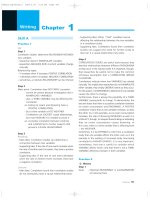
Homans Test
Assesses deep venous thrombosis.
Procedure:
The patient is supine. The examiner lifts the affected leg
and rapidly dorsiflexes the patient’s foot with the knee extended. This
maneuver is repeated with the patient’s knee flexed while the examiner
simultaneously palpates the calf.
Assessment:
Pain occurring upon dorsiflexion of the foot with the knee
extended and flexed indicates thrombosis.
Calf pain with the knee extended can also be caused by intervertebral
disk disease (radicular symptoms) or muscle contractures.
250 Venous Thrombosis
a
b
Fig.
259
Homans test:
a
dorsiflexion of the foot with
the knee extended,
b
dorsiflexion of the foot w ith
the knee flexed
Buckup, Clinical Tests for the Musculoskeletal System © 2004 Thieme
All rights reserved. Usage subject to terms and conditions of license.
Occlusive Arterial Disease
Occlusive arterial disease is often associated with orthopedic disorders.
Notably, nearly 90% of all cases of obliterative arteriosclerosis involve
exclusively the lower extremities. Prior to treating the actual orthopedic
disorder, the physician must take care to exclude or identify any p ossi-
ble arterial ischemic disorders. After obtaining a detailed history, a
diagnosis can usually be made on the b asis of inspection, palpation,
and specific function tests, and usually will not require the use of any
diagnostic technology.
Weakened or absent arterial pulse, cool and pale skin (or cyanotic
skin), patches of erythema, and trophic disturbances are signs of occlu-
sive arterial disease. Ulceration and gangrene are signs of advanced
disease. Where typical symptoms of intermittent claudication (calf
pain after walking short d istances) are present, determining the max-
imum distance the patient can walk without experiencing these symp-
toms can help in estimating the severity of the disorder (Fontaine clas-
sification of the severity of occlusive arterial disease). The d ifferential
diagnosis of intermittent claudication must include spinal claudication
from compression of the cauda equina, the cardinal symptom of lumbar
spinal stenosis. The intermittent claudication in cauda equina pathology
is not a sharply defined clinical syndrome. Radicular symptoms such as
paresthesia, pain, sensory deficits, and weakness can occur in one or both
legs when the patient stands or walks. These symptoms may improve or
disappear when the patient stops moving, as in the vascular form, but
more often will do so only on certain body movements.
Note:
The walking test allows assessment of peripheral circulatory
disruption. The patient is asked to walk up and down a long corridor
for up to thr ee minutes at about 120 paces per minute. The time of
occurrence of symptoms and the site of pain are clinically assessed, as
are gait and any pauses. If the patient pauses after only 60 seconds, this
suggests disruption of vascular supply to the muscles. Symptoms of
moderately severe circulatory disruption will manifest themselves after
1–3 minutes of walking. Symptoms that occur only after three minutes
or more of walking indicate only slight circulatory disruption.
Note that exercise tolerance may be limited by cardiac and pulmo-
nary disorders as well as orthopedic disorders such as osteoarthritis of
the hip or degenerative knee disorders.
Buckup, Clinical Tests for the Musculoskeletal System © 2004 Thieme
All rights reserved. Usage subject to terms and conditions of license.
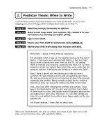
Allen Test
Assesses an arterial ischemic disorder in the upper extremities.
Procedure:
The patient is seated and raises his or her arm above the
horizontal plane. The exam iner grasps the patient’s wrist and applies
finger pressure to block the vascular supply from the radial and ulnar
arteries. The patient then makes a fist so as to force the venous blood out
of the hand via the posterior veins. After one minute, the patient lets the
arm hang down and ope ns the now pale hand. The examiner simulta-
neously releases compression, first from one artery then from the other.
Evaluation:
Rapid, uniform r eddening of the hand in the areas sup-
plied by the respective arteries ind i cates normal arterial supply. If
vascular supply to the hand and fingers is compromised, the ischemic
changes in the hand will only slowly recede.
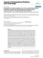
George Vertebral Artery Test (De Klyn Test)
Tests for insuf• ciency of the vertebral artery.
Procedure:
This test requires certain preliminary findings as it is not
entirely without risk. Parameters requiring prior assessment include
blood pressure, arm pulse, and pulses in the common carotid and
subclavian arteries with auscultation to detect any murmurs or bruits.
This test should not be performed if any of these prior examinations
produces significantly abnormal findings. In the absence of any signifi-
cant abnormalities, the seated patient is asked to maximally rotate his
or her head to one side while extending the neck. The test can also be
performed with the patient supine, in which case the patient’s head
projects over the edge of the examining table and rests in t he exam-
iner’s hands. Then with the head hanging dow n (in the De Klyn posi-
tion), the head is maximally rotated and the neck extended. The head
252 Occlusive Arterial Disease
a b
Fig.
260a, b
Allen test:
a
palpation of vessels with
the arm raised,
b
palpation of vessels with
the arm hanging and
evaluation of skin perfu-
sion
Buckup, Clinical Tests for the Musculoskeletal System © 2004 Thieme
All rights reserved. Usage subject to terms and conditions of license.
should remain or be held in maximum rotation and extension for about
20–30 seconds. T he patient is then requested to count out l oud.
Assessment:
Abnormal auscultatory findings in the common carotid
artery, vertigo, visual symptoms, nausea, fatigue, or nystagmus occur-
ring during this maximum rotation and extension indicate stenosis of
the vertebral artery or common carotid artery. The test is especially
important in candidates for treatment (such as traction or manipulative
therapy) of cervical spine symptoms associated with vertigo. The verte-
bral artery provocation test aids in th e differential diagnosis because
nausea, vertigo, and nystagmus initially increase but then rapidly de-
crease in intensity where a vertebral blockade is present. In the pres-
ence of vertebral artery insuf• ciency, the intensity of nausea and vertigo
symptoms will rapidly increase within a few seconds.
Ratschow-Boerger Test
Assessment o f vascular disease in the pelvis and legs.
Procedure:
The supine patient is asked to raise the legs as high as
possible and continuously rotate or plantar flex and dorsiflex the feet.
Occlusive Arterial Disease 253
a b
Fig.
261a, b
George vertebral artery test:
a
starting position,
b
rotation of the head and extension of the cervical spine
Buckup, Clinical Tests for the Musculoskeletal System © 2004 Thieme
All rights reserved. Usage subject to terms and conditions of license.
Assessment:
Patients with normal vascular function will be able to
perform this maneuver witho u t any pain and without the soles of the
feet becoming pale. Patients with compromised vascular function will
experience varying degrees of pain and significant ischemia in the sole
of the foot on the affected side. After about two minutes, the patient is
requested to sit up quickly and let the legs hang over the edge of the
examining table. Reactive hyperemia and refilling of the veins will occur
within 5–7 seconds in patients with normal vascular function. In pa-
tients with compromised vascular function, this reaction will be delayed
in proportion to the severity of vascular stenosis.
˾
Thoracic Outlet Syndrome
Thoracic outlet syndrome is a compression syndrome at the base of the
neck with compromised neurovascular function. Thoracic outlet syn-
drome can be a congenital disorder resulting from factors such as a
cervical rib, a superiorly displaced first rib, atypical ligaments, and the
presence of an atypical small scalene muscle. It may also be acquired as a
result of callus formation, osteophytes on the clavicle and first rib, and
changes in the scalene muscles such as fibrosis or hypertrophy.
254 Occlusive Arterial Disease
a b
Fig.
262a, b
Ratschow-Boerger test:
a
patient supine with the legs raised
b
patient sitting with the legs hanging down over the edge of the examining
table
Buckup, Clinical Tests for the Musculoskeletal System © 2004 Thieme
All rights reserved. Usage subject to terms and conditions of license.
This syndrome may be further differentiated according to the com-
pression site as a cervical rib syndrome, first-rib syndrome, or scalene
muscle syndrome.
Costoclavicular Test
Assesses a neurovascular compression syndrome in the costoclavicular
region.
Procedure:
The patient is seated with the arms hanging relaxed. The
examiner palpates the wrists to take the pulse in both radial arteries,
noting amplitude and pulse rate. Then the patient abducts and exter-
nally both arms and retracts the shoulders. With the patient in this
position, the examiner again palpates the wrists and evaluates the pulse
in both radial arteries.
Assessment:
Unilateral weakness or absence of the pulse in the radial
artery, ischemic skin changes, and paresthesia are cle ar signs of com-
pression of the neurovascular bundle in the costoclavicular region (be-
tween the first rib and clavicle).
Occlusive Arterial Disease 255
a b
Fig.
263a, b
Costoclavicular test:
a
starting position with the examiner palpating the pulse in the radial arteries,
b
palpation of the pulse in the radial arteries in abduction, with arms externally
rotated and shoulders retracted
Buckup, Clinical Tests for the Musculoskeletal System © 2004 Thieme
All rights reserved. Usage subject to terms and conditions of license.
Hyperabduction Test
Indicates a scalene muscle syndrome
.
Procedure:
The stan ding patient abducts both arms past 90° while
retracting the shoulders. Then the patient opens each hand and makes a
fist with each hand for two minutes.
Assessment:
Pain in the shoulder and arm, ischemic skin changes, and
paresthesia are clear signs of compression of the neuro vascular bundle,
which is primarily attributable to changes in the scalene muscles (fib-
rosis, hypertrophy, or presence of a small scalene muscle).
Intermittent Claudication Test
Sign of a costoclavicular compression syndrome
.
Procedure:
The standing patient abducts and externally rotates both
arms. Then the patient is instructed to rapidly flex and extend the
fingers of each hand fo r one minute.
Assessment:
If one arm begins to droop after a few cycles of finger
motion and ischemic skin changes, paresthesia, and pain in the shoulder
and arm occur, this suggests a costoclavicular compression syndrome
affecting neurovascular structures.
Causes include osteophytes, rib changes, and anatomic variations in
the scalene muscles.
256 Occlusive Arterial Disease
a
b
Fig.
264a, b
Hyperabduction test:
a
starting position with both arms ab-
ducted and shoulders retracted,
b
pain elicited in right shoulder
Buckup, Clinical Tests for the Musculoskeletal System © 2004 Thieme
All rights reserved. Usage subject to terms and conditions of license.
Allen Maneuver
Indicates a thoracic outlet syndrome.
Procedure:
The patient is se ated. The affected arm is held in a middle
position alongside the trunk with the elbow flexed 90°. The examiner
stands behind the patient and grasps the patient’s wrist with one han d,
palpating the pulse in the radi al artery. W ith the other hand, the
examiner supports the patient’s upper thoracic spine. The examiner
then draws the patient’s arm backward in to hyperextension and inter-
nal rotation at the shoulder. The patient is asked to rotate his or her head
toward the contralateral side (away from the side being examined).
Assessment:
Weakening or loss of the pulse in the radial artery, pain in
the shoulder and arm, ischemic changes, and paresthesia are signs of a
costoclavicular syndrome (compression of the subclavian artery be-
tween the first rib and the clavicle) or of a scalene muscle syndrome
(compression of the neurovascular bundle between the middle and
anterior scalene muscles due to fibrosis or hypertroph y).
Occlusive Arterial Disease 257
a
b
Fig.
265a, b
Intermittent claudication test:
a
starting position with both arms abducted and externally rotated,
b
pain on the right side with drooping right arm
Buckup, Clinical Tests for the Musculoskeletal System © 2004 Thieme
All rights reserved. Usage subject to terms and conditions of license.
˾
Hemiparesis
Arm-Holding Test
Assessment of latent hemiparesis.
Procedure:
The patient is asked to supinate both arms and raise them
to 90° while keeping his or her eyes closed.
Assessment:
Pronation and a drop in one arm suggest latent central
hemiparesis. Where the arm first drops and then pronates with the
patient’s eyes closed, on e should consider psychogenic influence.
Leg-Holding Test
Assessment of latent central hemiparesis.
Procedure:
The patient is supine and is asked to close his or her eyes
and flex both hips and both knees. The examiner watches the lower legs
to see if they drop down.
Assessment:
The neurologic examination of the lower extremities in a
patient capable of standing and walking begins with inspection of gait.
The patient is asked to stand and walk on tiptoe and then on his or her
heels. This will usually exclude any gross moto r deficits. With the
258 Occlusive Arterial Disease
a b
Fig.
266a, b
Allen maneuver:
a
starting position with the examiner palpating the pulse in the radial arteries,
b
adduction with the arm hyperextended and internally rotated at the shoulder
and the head rotated toward the contralateral side
Buckup, Clinical Tests for the Musculoskeletal System © 2004 Thieme
All rights reserved. Usage subject to terms and conditions of license.
patient supine, the strength of the quadriceps is then tested by having
the patient extend the knee against the examiner’s resistance (L3–L4).
Strength in the extensor digitorum and hallucis longus is tested by
dorsiflexion of the toes (L5) against resistance, and strength in the
triceps surae is tested by plantar flexion of the foot (S1) against resist-
ance. One or both lower legs dropping down during the leg holding test
can be a sign of latent central hemiparesis.
Occlusive Arterial Disease 259
a b
Fig.
267a, b
Arm holding test:
a
Both arms supinated and raised up to 90° with closed eyes,
b
Pronation and drop in one arm
Fig.
268
Leg holding test
Buckup, Clinical Tests for the Musculoskeletal System © 2004 Thieme
All rights reserved. Usage subject to terms and conditions of license.
Buckup, Clinical Tests for the Musculoskeletal System © 2004 Thieme
All rights reserved. Usage subject to terms and conditions of license.
Index
Numbers in
ital ics
indicate figures.
A
Abbott-Saunders test 84–5,
85
abduction
hip 142–3
stress test 46,
46
test 193,
194
hyperabduction 256,
256
valgus stress test 106,
106
abductor pollicis brevis muscle 133 ,
135
abductor pollicis longus muscle 129
abductor pollicis longus tendon
119–20
Achilles tendon 56, 231– 2
acromioclavicular joint 65, 68–9 ,
81–84, 89
arthritis 76, 78, 80–1
acromion 78
Adam forward bend test 22,
22
adduction
correction test 235,
235
hip 142–3
test 193,
194
forced 82–3,
82
varus stress test 105,
106
adductor pollicis muscle 137
Allen
maneuver 257,
257
test 252,
252
anastomoses 2 4 8
Anderson compress i on test 189 ,
190
ankle
joints 225, 235–7
ligaments 199, 204
anvil test 152,
152
ape’s hand deformity 133
Apley
scratch test 76,
76
tests 176–7,
177
apprehension test
anterior 92–3,
93
inferior 101,
101
posterior 97–9,
97
,
99
supine 93–4,
94
arm 75, 131–2, 137, 258–9
adduction 82–3
compression 126–7
forearm 111–12
Arnold crossover test 212,
212
arteries
carotid 253
disease 224, 251
ischemia 252
radial 252, 257–8
sclerosis 251
thrombosis 251–9
ulnar 252
vertebral 252–3
arthritis 167
acromioclavicular joint 76, 78,
80–1
carpometacarp al joint 120–1
glenohumeral joint 76
see also
osteoarthritis, rheumatoid
arthritis, spon dylarthritis
arthrode sis 143
arthroplasty 152–3
asthma 8–9
avascular necrosis 224
Buckup, Clinical Tests for the Musculoskeletal System © 2004 Thieme
All rights reserved. Usage subject to terms and conditions of license.
262 Index
B
Baker cysts 163
Barlow
and Ortolani test 158
test 156–8,
157
belt test 29,
29
Beru sign 90
biceps muscle 89
biceps tendo n 68, 84-90
bicyclists, compression injury 126
Böhler meniscus test 185
Böhler-Krömer test 184 –5,
184
bones
see
individual types of bones
Bonnet sign 50,
50
Bowden test 107,
107
brachialgia 127
Bragard
sign 52
test
meniscus 178–80,
179
spine 52,
53
Brudzinski sign 57,
57
Brunnell-Littler test 122–3,
122
bucket han dle tear 204
bursae 65
bursitis 65–7, 78, 80, 85
C
Cabot test 185–6,
187
calcaneofibular ligament 236
calcaneus 238
calf compression test 231,
231
capsular ligaments 125, 176–7, 197,
236
laxity 97–9
lesions 184
posterior 193
capsule, joint 76
carotid artery 253
carpal tunnel
sign 134,
134
syndrome 127, 134–6
carpometacarpal joint 116, 120–1
cartilage 167, 169–71
cauda equina 251
cervical spine 12–13, 20–21
arthritis 10–11
disks 14
ligaments 14–15, 17
muscles 14–15, 17–19
spondylosis 10–11
chair test 106,
107
chest 7–9
children 154–8
Childress sign 188,
189
chondrocalcinosis 103
Chopart, joint of 236
claudication test 256,
257
clavicle 83,
83
claw hand 115
claw toe 224, 226
Codman sign 63–4,
64
Coleman block test 233–4,
233
collagen 224
collateral ligaments 93, 176, 236
accessory 125
lesions 184
stability 105–6
compression 4 2– 3, 90–1, 189–91
axial 152–3
intervertebral foramina 18–20
nerve root 46–7, 52–3, 158
rib 7–8
spinal cord 158
supinator muscle 114
suprascapular nerve 88–9
syndrome 46–7, 52–3, 112–14
costoclavicular 255–7
median nerve 134 – 6
thoracic outlet syn d rome
254–5, 257–8
compression neuropa thies 115, 224
tibial nerve 240
ulnar nerve 126–7, 139
condyle
femoral 202, 208
osteochondritis dissecans 192
contracture 144–5, 250
gastrocnemius muscle 52
hamstring 143–4, 166
hip 141, 144–6, 148
iliotibial tract 148–9, 206
quadriceps muscle 165, 199
test 218,
218
Buckup, Clinical Tests for the Musculoskeletal System © 2004 Thieme
All rights reserved. Usage subject to terms and conditions of license.
contracture
rectus femoris muscle 144, 165–6
sacroiliac joint 35–6
tenso fasciae latae muscle 148–9
teres major muscle 73–4
coracoclavicular ligame nt 83
costoclavicular compression 255–7
Cozen test 109–10,
110
reverse 110,
111
crepitation
foot 229
patella 170–1
tendons 115
cross–body action 88–9
cruciate ligaments
anterior 95–6, 176, 194–216
posterior 95–6, 187, 193, 208,
216–23
cubital tunnel syn d r ome 103, 112–13,
127, 137–8
cysts
Baker 163
meniscus 176–7
D
dancing patella test 167,
168
Dawbarn test 66–7,
67
De Klyn position 252
de Quervain’s disease 120–1
dead arm sign 92
DeAnquin test 89
deformities
back 22, 244–5
foot 224, 233–4
hand 115, 133
hip 141–3 , 148
knee 22 1
deltoid ligament 236
Derbolowsky sign 41 ,
41
dermatome 47
diabetes mellitus 224
disks 24–6, 54 – 6, 158, 250
extrusion 49 – 5 0
intervertebral 35 –6, 152–3
dislocation
biceps tendon 89
hip 141, 153, 158
humerus 97–9
patella 167, 169, 171
distraction test 17,
17
drawer
signs 100
tests 237,
237
anterior 199–201,
20 0
anterior an d posterior 94–5,
96
posterior 216–17,
216
soft posterolateral 219,
219
Drehmann sign 1 49–50,
151
Dreyer test 175–6,
175
droop test 219,
219
drop arm test 75,
75
Duchenne
antalgic gait 143
sign 54–5,
55
, 154
Dugas sign 90
Dugas test 83–4,
83
dysplasia 160–1
E
elbow 103– 106
compression 112–14
cubital tunnel syndrome 103,
112–13
epicondylitis 106–11
flexion 104–5, 113
emphysema 8–9
epicondylitis 106–11
extension
compression test 2 1 ,
22
hyperextensi o n test 27–8 ,
28
extensor digitorum muscle 259
extensor pollicis brevis muscle 130
extensor pollicis brevis tendon
119–20
extensor po llicis longus mu s cle
118–19, 130
F
fabere test (Patrick test) 34,
35
, 154–5,
155
Index 263
Buckup, Clinical Tests for the Musculoskeletal System © 2004 Thieme
All rights reserved. Usage subject to terms and conditions of license.
facet joint 47, 49, 169–70
femoral nerve 58
femur 141, 143, 150
fibrosis 76
fingers 65, 129, 133, 139–40
Finkelstein test 120–1,
120
Finochietto sign 187,
188
flexion 38–9
compression 2 0– 1
dorsiflexion 225
elbow 104–5, 113
foot 225, 234
hip 141-2, 144–6, 148
hyperflexion 104–5
knee 23 -4
sacroiliac joint 38–9
wrist 140
flexor carpi radialis muscle 140
flexor digitorum profundus muscle
118, 133, 139
flexor digitorum profundus tendon
121–2
flexor digitorum superficialis muscle
118
flexor pollicis brevis muscle 133, 137
flexor pollicis longus muscle 118–19,
139
flexor pollicis long us tendon 121–2
flexor tendo ns, tests 118–25
Fontaine classification 251
foot
calluses 224
deformities 224, 233–4
flexion 225, 23 4
hallux rigidus 229
Köhler disease 224
metatarsalgia 224
Morton neuroma 224
pes adductus 235
pes cavus 226
splay 224, 227 , 230
stress fractures 224
tarsal tunnel syndrome 239–40
warts 224
forward be nd test 29,
29
Fouche sign 178,
178
Fowler test 92
fractures
cervical spine 14
rib 7–9
stress 2 24, 238
Froment sign 137,
138
Fukuda test 100,
100
fulcrum test 102,
102
G
Gaenslen sign 41–2,
42
Galeazzi test 158,
159
Galway and McIntosh 214
gamekeeper’s thumb 1 25
gangrene 251
Gänsslen maneuve r 2 29–30,
229
gastrocnemius muscle 52
genu recurvatum 221
genu va lgum 167
George v ertebral artery test 252–3,
253
Gerber-Ganz drawer tests 96–9,
96
,
98
Gerdy, tubercl e of 149, 206
Gilcrest test 89
glenohumeral joint 64, 76, 99
glide test 167 – 9 ,
168
gluteus medius muscle 153–4
gluteus minimus muscle 153–4
Godfrey test 2 22,
222
golfer’s elbow 103, 110–12
gout 224
gravity sign 220–1,
220
Grifka test 227,
227
grind test 121,
121
grip tests 127–9 ,
128
Thomas 146–8,
147
Guyon’s canal 127
H
hallucis longus muscle 259
hallus rigidus 224
hammer toe 224, 226
hamstring 50, 143–4, 166
hand
ape’s hand 133
claw hand 115
264 Index
Buckup, Clinical Tests for the Musculoskeletal System © 2004 Thieme
All rights reserved. Usage subject to terms and conditions of license.
hand
flexor tendo n 118–25
grip 127–9
ischemic contracture 122
motor function 127–40
pronator quadratus muscle 137
pronator teres muscle 126–7, 137
tenosynovitis 118
Hawkins impingement test 79,
79
head, rotation test 11–12,
11–12
Heberden nodes 118
heel 231–2, 238–9
hemarthrosis 196
hip 142–6 , 153–4
arthrode sis 143
arthroplasty 152–3
deformities 141–3, 148
degenerative joint disease 141
dislocation 141, 153, 158
congen ital 156
disorder 53–4, 149–51
Duchenne, antalgic gait 14 3
dysplasia 160–1
acetabular 141
flexion 141, 14 4 – 6, 148
grip 146–8
inflammation 148, 152–3
instability 156–8
and lumbar rigidity 158–9
osteoarthritis 144, 148, 150, 152–3
tumors 150
Hoffa sign 232,
232
Homans
sign 247
test 250,
250
Hoover
sign 30
test
30
Hueter sign 87–8,
87
Hughston
genu recurvatum test 221,
221
jerk test 215–16,
215
humeral ligament test 88,
88
humerus
dislocation 97–9
epicondylitis 106–11
subluxation 9 2, 97–9
I
iliac compression test 42,
43
iliocostalis lumborum muscle 28
iliotibial tract 148–9, 206
inflammation
arthritis
see
arthritis
bursitis 65–7, 78, 80, 85
epicondylitis 106–11
erythema 115
hip 148, 152–3
osteochondritis 103
pericarditis 8–9
pleuritis 8–9
psoriasis 224
spine 5, 10–12, 56
spondylitis 8–9, 25–6, 35–6
tenosynovitis 118
infraspinatus mu s cle 73–4
instability
elbow 103
hip 156–8
knee 193 – 216
metatarsophalangeal joint
228–9
posterolateral ligament 216–19 ,
221
shoulder 91–102
relocation test 102,
102
Rowe test 94,
94
triquetrolunate joint 124
intercarpal joint 116
interosseous nerve 138–9
interphalangeal joint 119, 122–3, 137,
139, 226
distal 116–18
proximal 116–18, 138
intervertebral foramina 18–20
intrinsic test 139,
139
ischemia 252
J
Jackson comp ression test 19,
20
Jakob
giving way test 213–14 ,
214
maximum drawer test 201,
202
Index 265
Buckup, Clinical Tests for the Musculoskeletal System © 2004 Thieme
All rights reserved. Usage subject to terms and conditions of license.
Jakob
pivot shift test
graded 204–6,
205
reverse 217–18,
217
Jobe
modification 92
supraspinatus test 70–1,
71
joints
arthrode sis 143
arthroplasty 152–3
capsule 76
hemarthrosis 196
inflammation
se e
arthritis;
inflammation
see also
individual type s of joints
K
Kalchschmidt hip dysplasia test
160–1,
161
Kaplan, fibers of 206
Kernig
sign 49
test 56,
56
Keyle 69
Kibler fold 1
test 6,
7
knee 162–3, 171–7
effusion 167
flexion 23–4
genu recurvatum 221
glide 167–9
instability 193–216
ligaments 169, 193–223
motion 163
patella 167–74
reticulum 174
subluxation 104, 204
knock knees
see
genu valgum
Köhler disease 224
Kraus–Web er test 241–2,
243
Krömer test 1 85
kyphosis 244
lumbar 143
test 22,
23
L
Lachman test 194–5,
195
, 200
active 198–9,
199
no-touch 197–8,
198
posterior 216–17,
216
prone 196,
196
stable 196–7,
197
Laguerre test 44 –5,
45
Lasègue
differential test
54
sign 47, 49 – 5 0 ,
49
,
51
, 52
contralateral 51,
52
straight leg drop test 25–6,
26
test
differential 53–4
reverse 58,
58
seated 51
Lasègue-Moutaud-Martin sign 51,
52
Leffert test 94–5,
95
leg 47, 49–52, 258–9
compression 152–3
length 41, 158–9
vascular disease 253–4
Legg-Calvé-Perthes disease 141,
154–5
Lelièvre 226
Lemaire test 214–15
lesions
biceps tendon 86–7, 90
brachial plexus 75, 127
capsular ligament 184
collateral ligament 184
distal nerve 140
median nerve 132, 137, 140
meniscus 176–80, 185–6, 190–1
radial nerve 128, 130
rotator cuff 75
spinal cord 127, 158
ulnar nerve 140
lift–off test 72–3,
73
ligaments 31–3
capsular 125, 176–7, 197, 23 6
laxity 97–9
lesions 184
posterolateral 199, 217–19,
221
posteromedial 193, 199, 204
266 Index
Buckup, Clinical Tests for the Musculoskeletal System © 2004 Thieme
All rights reserved. Usage subject to terms and conditions of license.
ligaments
lateral 199, 204
ankle 25
collateral 193, 237
medial 199
collateral 197, 204
patella 167
pelvic 31–2, 49–50
talofibular 236
see also
individual type s of
ligaments
Linburg test 121–2,
121
Lippman test 89
longissimus thor acis muscle 28
lordosis 141, 145–6
hyperlordosis 143
lumbar 244
Losee test 210– 1 1 ,
211
Lowenberg 246–7,
247
test 247,
247
Ludington
sign 76,
76
test 89
Ludloff-Hohmann test 158
lumbar spine 22–24, 27
disks 152–3
muscles 27–8
nerve root disorder 56
sciatica 30
spondylarthritis 25–6
spondylitis 25–6
stenosis 251
syndrome 27–9
lungs 8–9
Luethy bottle test 1 3 5,
136
M
McConnel l test 171–3,
173
McMurray test 178,
178
Martens test 210,
210
Matthiass postural competence test
241, 244,
245
median ner ve
compression 1 27 , 134–6
palsy 132–3
meningitis 57
meniscus
compression 189–91
cysts 176–7
discoid 176–7
lesions 177–89, 204
Mennell sign 36, 43–4,
43
Merke test 185,
186
metacarpophalangeal joint 116–18,
122, 125, 138
metatarsi 235
hallus rigidus 229
metatarsalgia 227, 230
metatarsophalangeal joint 225, 227–9
Meyer pressure points 247
Mill test 108,
108
Morton neuromas 24–6, 224, 230, 238
Muckard t e st 119–20,
120
Mulder click test 230, 238,
238
muscles 239, 241–4
cervical 83
contractures 165–6, 250
see also
individual type s of
muscles
N
nail sign 135,
135
necrosis
aseptic 154–5, 192
avascular 224
femur 141
Neer 68–9
impingement injection t est 80,
80
impingement sign 78,
78
nerve root
compression 4 6– 7, 52–3, 1 58
disorder 54–5
irritation 49–52, 55–6, 58
nerves
brachial plexus 75
cauda equina 251
distal 140
interossei 138–9
median 1 27 – 9
lesion 132, 137, 140
palsy 115, 13 2–3, 135
paresthesia 134, 136
plantar 238–9
Index 267
Buckup, Clinical Tests for the Musculoskeletal System © 2004 Thieme
All rights reserved. Usage subject to terms and conditions of license.
nerves
radial 114, 131
lesion 128, 130
palsy 115, 129
sciatic 49, 53–4
suprascapular 88–9
tibial 240
ulnar 112–13, 129
compression 126, 139
lesion 140
palsy 115, 138–9
neuralgia 9
neuromas
interdigital 238
Morton 23, 224, 230
neuropathies
compression 1 15 , 126–7, 224, 240
arm 126–7
cauda equina 251
Guyon’s canal 127
median nerve 127, 132
tarsal tunnel syn drome 224
tibial nerve 240
ulnar nerve 126, 139
entrapment, m etatarsalgia 224
medial malleolus 239
neutral–zero method
61
,
104
,
116– 17
,
163
, 225–6
noble compression test 148–9,
148
Noyes test 212–13,
213
O
O test 139,
140
Ober test 149,
150
Ochsner test 133,
133
O’Donoghue test
15
opponens pollicis muscle 132–3, 135
orientation tests 10 4– 6
Ortolani test 156–8,
157
Osgood– S c hlatter disease 162
osteoarthritis 84, 115, 189, 204
carpometacarp al joint 120–1
elbow 103
hallus rigidus 224, 229
hip 144, 148, 150, 15 2 –3
interphalangeal joint 118, 127
metatarsophalangeal joint 229
patella 169–70
sternoclavicular join t 68
osteophytes 16, 68, 204, 256
osteoporosis 238
Ott sign 5,
6
P
painful arc
acromioclavicular joint 81–2,
81
rotator cuff 77–8,
77
palm, adduction 65, 117
palsy 134–6
median nerve 115, 132–3, 135
radial nerve 115, 129– 3 0
serratus muscle 68
ulnar nerve 115, 138–9
paralysis
abductor pollicis brevis muscle
133–5
abductor pollicis longus muscle
129
extensor pollicis brevis muscle
130
extensor po llicis longus mu s cle
118–19
flexor carpi radialis muscle 140
flexor digitorum profundus muscle
118, 133, 139
flexor pollicis brevis muscle 133
flexor pollicis longus muscle
118–19, 139
hemiparesis 258–9
hip 153
interossei nerve 138
opponens pollicis muscle 132–3,
135
supinator muscles 131–2
paresthesia 134–6, 139, 239, 256
nighttime 127
Paessler rotational compression test
190–1,
191
patella 167–74
patellofemoral joint 167
Patrick test (fabere test) 34,
35
, 154–5,
155
268 Index
Buckup, Clinical Tests for the Musculoskeletal System © 2004 Thieme
All rights reserved. Usage subject to terms and conditions of license.
Payr 246
sign
meniscus 180,
180
venous thrombosis 247
test 181,
181
pelvis
ligaments 31–2, 49–50
muscles 242
tendinitis 49–50
percussion test
14
pericarditis 8–9
Perthes test 248–9,
249
pes cavus 226
Phalen tes t 134–5,
134
reverse 136,
136
piriformis sign 50,
50
pivot shift test 202–4,
203
modified 206–7,
207
soft 208–9,
209
plantar muscles 239
pleuritis 9
plexi 75, 127
popliteus sign 185–6,
187
popliteus tendon 193
postdiskectomy sy ndrom e 47
posture 241–5
Matthiass competence test 241,
244,
245
Pratt warn ing 246
pronation test 137,
137
pronator quadratus muscles 137
pronator teres muscle 137
syndrome 126–7
pseudoradicular sign
(pseudo–Lasègue sign) 50
psoas muscle 24–5, 145
psoas sign 25,
25
psoriasis 224,
224
Q
quadriceps muscle 259
atrophy 192
contraction 16 5, 199, 218
quadriceps tendon 175–6
R
radius 47–8, 116, 120
nerve 129 –30
Ratschow-Boerger test 253–4,
254
Reagan test 124,
124
rectus femoris muscle 144–5,
165–6
relocation test 102,
102
rheumatoid arthritis 103, 130, 224
ribs 7–9
Rielander 2 46
rotator cuff
defect 80
impingemen t 67–70, 79
infraspinatus tendon 73–4,
84
lesions 75
painful arc 77– 8
pathology 76
subscapularis tend on 71–2
supraspinatus tendon 70–1, 75,
77–8
tears 67, 70, 76, 78, 82
tendinitis 102
weakness 75
Rowe test 94,
94
S
sacroiliac joint 34–41
abduction 46
disease 42–4
disks 35–6
flexion 38–9
ligaments 31–3, 45
mobility 33–4, 38–41
spondylitis 35– 6
syndrome 46–7
saphenous vein 248–9
scalene muscle 256–7
scaphoid shift test 123,
123
scapholunate ligament 124–5
scapula, displacement 88–9
Schepelmann test
9
Scheuermann disease 241
Schober sig n 5,
6
Index 269
Buckup, Clinical Tests for the Musculoskeletal System © 2004 Thieme
All rights reserved. Usage subject to terms and conditions of license.
Schwartz
and Hackenbruch percussion
method 249
test 249
sciatic nerve 49, 53–4
sciatica 30, 49–51
Bonnet sign (piriformis sign) 50,
50
Kernig sign 49
Lasègue sign 49–50,
49
Turyn sign 49
scoliosis 141–2, 244
serratus muscle 68
sesamoids 224
shift and load test 97–9,
97
,
99
shift test
medial 208,
208
posterior, dynamic 223,
223
scaphoid 123,
123
shoulder 18
bursa 65–7
bursitis 65–7, 78, 80, 85
disorders 59–60
drawer 94–5
frozen 64
impingement 68–9, 78 – 9 , 82–3
instability 92–102
labrum 97
motion 61– 5
osteophytes 6 8
pseudo–stiffening 68
subluxation 9 4, 101
tears 69
Simmond test 231
skier’s thumb 125
skin
dermatatome 47
skin–rolling test
7
slocum test 211,
211
snap test 86,
87
Soto-Hall test
14
speed test 85,
86
spinal canal 47
spinal cord 158
spine 5–7, 23–4, 36–7
disks 20–1, 24–6
facet joint 6, 18–21
syndrome 16
inflammation 56
intercostal joints 6
kyphosis 22–3
ligaments 20
mobility 2–6
muscles 18
nerve root 16, 19
osteophytes 1 6
rheumatoid 152–3
sacroiliac joint
see
sacroiliac joint
Scheuermann disease 241
spondylolisthesis 241
tumors 16, 55
see also
cervical spine, lumbar
spine, thoracic spine
spinous process tap test 24
spondyla rthritis 10–11, 25–6
spondylitis 25–6
ankylosing 8–9, 35–6
spondylolisthesis 47
spondylosis 10–11
sports injuries
bicyclist compression 126
gamekeeper’s thumb 1 25
golfer’s elbow 103, 110–11,
112
skier’s thumb 125
tennis elbow 103, 106–10
springing test
27
, 40,
40
sacroiliac joints 33–4,
33
, 40,
40
Spurling test 16–17,
16
Steinmann
I sign 182,
182
I test 185
II sign 183–4,
183
stenosis 253
sternoclavicular join t 68
sternum compression test 7,
8
stress fractures 224
stress test
motion 109 ,
109
sacroiliac joint 46,
46
Strunsky test 2 27,
228
subacromial bursitis test
66
subluxation
biceps tendon 84–6, 89
glenohumeral joint 99
humerus 92, 97–9
knee 104, 204
patella 169, 173–4
270 Index
Buckup, Clinical Tests for the Musculoskeletal System © 2004 Thieme
All rights reserved. Usage subject to terms and conditions of license.
subluxation
shoulder 94, 101
suppression test 173–4,
174
tibia 201–7, 210, 214–15, 218, 223
subscapularis muscle 67, 70–3,
72
subtalar joint 235–6
sulcus sign 100,
101
supination
stress test 105,
105
test 131,
131–2
supinator muscle 114, 131–2
suppression test 173–4
suprascapular nerve 88–9
supraspinatu s muscle 75–6, 81 , 102
supraspinatus tendon 70–1, 75
T
talocrural joint 225
talofibular ligaments 236
tarsal tunnel syn drome 224
Tinel sign 239,
239
tourniquet sign 240,
240
tarsi, transverse joint 235–6
tears
Achilles tendon 231–2
bucket han d le 204
collateral ligament 125
cruciate ligament 187
meniscus 187, 189, 203
rotator cuff 67, 70, 7 6, 78, 82
subscapularis muscle 71–3
telescope sign 156,
156
tendinitis 67, 86, 103
biceps tendon 86
patella 170
pelvis 49–50
pseudoradicular sign 50
rotator cuff 102
tendons 115
see also
individual types of tendon
tennis elbow 103, 106–10
tenosynovitis 115, 127
abductor pollicis longus muscle
119–20
biceps tendon 85
de Quervain’s disease 119–21
extensor pollicis brevis muscle
119–20
proximal interphalangeal joint 118
thumb 119
tensor fasciae latae m uscle 148–9
teres major muscle 73–5,
74
Thomas grip 146–8
Thompson
compression test 2 3 1,
231
and Kopell horizontal flexion test
88–9,
89
Thomsen sign 55,
55
Thomson test 107–8,
108
thoracic outlet syn drome 127, 254–7
Allen maneuver 257,
257
costoclavicular test 255,
255
thoracic spine 22–30
inflammation 5
kyphosis 22
mobility 5
scoliosis 22
three-phase hyperextension test
34–6,
36
thrombophle bitis 52
thrombosis 52
arterial 251–9
venous 246–50
throwing test 94,
95
thumb 121–2
circumduction 11 7
degenerative joint disease 130
rheumatoid arthritis 130
tendons 119
tenosynovitis 119
tibia
malrotation 167
Osgood-Schlatter disease 162
rotation 204
subluxation 201–7, 210, 214–15,
218, 223
tibiotalocalcaneal joint 235
tilt test 174–5,
175
tinel sign 13 2,
132
, 139, 239,
239
Tinel test 112,
113
tiptoe and heel walking test 56,
57
toes
claw toe 224, 226
displacement test 228–9,
228
Index 271
Buckup, Clinical Tests for the Musculoskeletal System © 2004 Thieme
All rights reserved. Usage subject to terms and conditions of license.
toes
Strunsky test 2 27,
228
Toennis gr adings 15 7–8
tourniquet sign 240,
240
traction test 90,
90
trapezius muscle 68
Trendelenburg
gait 143
sign 153–4,
154
test 248,
248
triceps surae muscle 259
triquetrolunate joint 124,
124
trochlear g roove 167
trunk muscles 242
Tschaklin sign 192
tumors 55
elbow 103
hip 150
nerve root 49–50
neuromas
interdigital 238
Morton 23, 224, 230
pleural processes 8–9
spinal cord 158
spine 16, 55
Turner sign 1 88–9
Turyn sign 49
U
ulcers 251
ulna 116
ulnar ligament 125
ulnar nerve 126–7, 138–9
V
valgus stress test 106,
106
Valsalva test 16,
16
varicose veins 248,
248
varus stress test 105,
106
vastus medialis muscle 192
veins
anastomoses 2 4 8
dorsal 252
saphenous 248–9
thrombophle bitis 52
thrombosis 50, 237, 246–50
varicose 248,
248
vertebrae, blockade 8–9
vertebral artery 252–3
vertigo 10–12
volar hypesthesia 137
W
Wagenhaeuser 241
Watson test 123,
123
Wilson test 192,
192
wrist 123–4, 134–5, 137–8, 140
Y
Yeoman test 47,
47
Yergason test 86,
87
Z
zero-degre e abduction test 70,
76
Zippel 189
Zohlen sign 169,
170
272 Index
Buckup, Clinical Tests for the Musculoskeletal System © 2004 Thieme
All rights reserved. Usage subject to terms and conditions of license.