Textbook of Traumatic Brain Injury - part 2 potx
Bạn đang xem bản rút gọn của tài liệu. Xem và tải ngay bản đầy đủ của tài liệu tại đây (2.02 MB, 66 trang )
60 TEXTBOOK OF TRAUMATIC BRAIN INJURY
(Table 4–2). Because the survivor of a TBI does not
know whether he or she was rendered unconscious by
the trauma, it is important to verify LOC with a witness,
if possible. The survivor may believe that LOC occurred
when, in actuality, he or she was conscious but in a state
of PTA. Introduced by Teasdale and Jennett (1974), the
GCS (see Table 1–2 in Chapter 1, Epidemiology) has
become the standard for measuring the acute severity of
a TBI. Estimating the severity of an acute TBI guides
the physician in quantifying the signs and symptoms as-
sociated with mild, moderate, or severe TBI as well as
the patient’s likely prognosis. According to Asikainen et
al. (1998), the GCS score and duration of LOC and PTA
all have strong predictive value in assessing functional or
occupational outcome for TBI patients. However, Lov-
ell et al. (1999) question the predictive value of LOC
based on the lack of statistical correlation between LOC
and neuropsychological functioning in a large sample of
patients with mild head trauma.
A temporal relationship should be established be-
tween the onset of current signs and symptoms and the
occurrence of the traumatic injury. This information
helps to differentiate the premorbid personality charac-
teristics and psychiatric and behavioral symptoms from
those arising after the brain injury. Any number of emo-
tional and behavioral difficulties that existed in milder
form before the brain injury can be accentuated after it.
Careful consideration of temporal relationships also must
address the phase of recovery and associated behavioral
changes, because improvement after TBI tends to occur
along a continuum, with certain sequelae generally re-
solving before others (e.g., confusion and disorientation
generally resolve before short-term memory impair-
ment). The clinician should also focus attention on the
patient’s psychological reactions and adjustment to injury-
induced cognitive and emotional changes, as well as their
impact on interpersonal relationships, family dynamics,
and employment status.
In the assessment of TBI, it is helpful to categorize
observed signs and symptoms into the broad domains of
cognition, emotion, behavior, and physical symptoms
(Table 4–3). This categorization permits more precise di-
agnosis of the patient’s problems and assists in the formu-
lation of an optimal treatment plan.
Importance of Collateral History
Because insight into disturbances of cognition, behavior,
and emotional state are often compromised in patients
TABLE 4–1. Sample questions for traumatic brain
injury (TBI) assessment
Questions
Rationale
Have you ever hit your head?
Have you ever been in an
accident?
Probe for car/motorcycle/
bicycle/other motor vehicle
accidents, falls, assaults, sports
or recreational injuries
(If so) Did you black out, pass
out, or lose consciousness?
Establish LOC (verify LOC
with witness, if possible)
What is the last thing you
remember before the injury?
Establish extent of retrograde
amnesia
What is the first thing you
recall after the injury?
Estimate duration of LOC and
begin to quantify
posttraumatic amnesia (must
ask further about when
contiguous memory function
returned)
(If no LOC) At the time of the
injury, did you experience
any change in your thinking
or feel “dazed” or
“confused”?
Establish change in mentation
or level of consciousness
What problems did you have
after the injury?
Delineate post-TBI symptoms
(see Table 4–3)
Has anyone told you that
you’re different since the
injury? If so, how have you
changed?
Detect problems outside
survivor’s awareness or those
he/she may be minimizing
Did anyone witness or observe
your injury?
Identify source of collateral
history
Many people who have injured
their head had been drinking
or using drugs; how about
you?
Offer survivor greater
“permission” to admit
substance use
Have you had any other
injuries to your head or
brain?
Identify previous TBIs that may
increase morbidity from
current injury
Note. LOC=loss of consciousness.
TABLE 4–2. Classification of traumatic brain
injury (TBI)
Type
of TBI
Glasgow
Coma
Scale
Loss of
consciousness
Posttraumatic
amnesia
Mild 13–15 30 minutes or less
(or none)
<24 hours
Moderate 9–12 30 minutes to 1
week
>24 hours to <1
week
Severe ≤8 >1 week >1 week
Neuropsychiatric Assessment 61
with brain injury, it is incumbent on the clinician to verify
from collateral sources the accuracy of the patient’s
account of his or her history and symptomatology. In
cases of severe TBI, patients rarely recall the incidents
surrounding the injury. This disturbance in recall of the
incident itself, in conjunction with the patient’s decreased
awareness of his or her deficits, makes accessing collateral
information essential. Collateral history may be obtained
from a variety of sources (Table 4–4), including family and
friends who can describe changes in behavior, cognition,
personality, and general level of functioning since the
brain injury.
Collateral history is also pivotal because survivors of
TBI and their families and friends see the injuries through
different lenses. For example, Sbordone et al. (1998) found
that patients with TBI generally underreported cognitive,
behavioral, and emotional symptoms as compared to those
reported by significant others, regardless of the severity of
injury. For example, 58.8% of significant others in the
study noted emotional lability or mood swings in the pa-
tients with TBI, whereas only 5.9% of the patients re-
ported such difficulties. Circumstantiality was observed by
29.4% of significant others; but none of the patients re-
ported such problems. In those with severe TBI, none of
the patients recognized problems with judgment, whereas
45% of their significant others identified this problem.
Hospital records related to the acute treatment of a
TBI provide invaluable information about the traumatic
event. This information includes the nature of the
trauma (e.g., MVA, fall, or blunt trauma); severity (GCS,
period of unconsciousness, presence of traumatically re-
lated seizures, duration of retrograde amnesia and PTA,
medical complications, and course of recovery); time of
onset and types of neurobehavioral changes that oc-
curred during the acute and postacute phases of recov-
ery; and results of neuroimaging, electrophysiological,
and neuropsychological testing delineating the location
and extent of injury and pattern of cognitive and mem-
ory impairment associated with it. Medical and psychi-
atric records for the period before the trauma are also
helpful in relating current signs and symptoms to past
psychiatric disturbances and premorbid personality, and
can assist in ascertaining the relative contributions of
TABLE 4–3. Traumatic brain injury symptom checklist
Cognitive Emotional Behavorial Physical
Level of consciousness Mood swings/lability Impulsivity Fatigue
Sensorium Depression Disinhibition Weight change
Attention/concentration Hypomania/mania Anger dyscontrol Sleep disturbance
Short-term memory Anxiety Inappropriate sexual behavior Headache
Processing speed Anger/irritability Lack of initiative Visual problems
Executive function (planning, abstract
reasoning, problem-solving,
information processing, ability to
attend to multiple stimuli, insight,
judgment, etc.)
Apathy “Change in personality” Balance difficulties
Dizziness
Coldness
Change in hair/skin
Thought processes Seizures
Spasticity
Loss of urinary control
Arthritic complaints
Source. Adapted from Hibbard MR, Uysal S, Sliwinski M, et al: “Undiagnosed Health Issues in Individuals With Traumatic Brain Injury Living in
the Community.” The Journal of Head Trauma Rehabilitation 13:47–57, 1998.
TABLE 4–4. Sources of collateral history
People Documents
Family Police reports
Friends Emergency medical service reports
Co-workers Medical records
Witnesses to injury Educational history
Medical staff Driving record
Allied health professionals
(occupational, physical,
and speech therapists, etc.)
62 TEXTBOOK OF TRAUMATIC BRAIN INJURY
antecedent variables, the brain injury itself, and current
psychosocial parameters to observed neurobehavioral
changes.
If available, posttrauma psychiatric and/or rehabilita-
tion records help delineate the course of the patient’s re-
covery, including the acute versus chronic nature of pre-
senting psychiatric complaints, and provide a source of
additional behavioral observations. Relevant posttrauma
records also should be reviewed for the emergence of sub-
sequent medical problems, results of neurodiagnostic
studies, and indications of the efficacy and adverse effects
of various treatment interventions the patient may have
received. Additional sources of collateral information that
may prove helpful include police reports and emergency
medical service records (to provide information about the
accident and condition of the patient at the scene), educa-
tional records, and driving record (to provide a history of
prior MVAs).
Current Neuropsychiatric Symptoms
Within days of a mild to moderate TBI, a significant num-
ber of patients experience headaches, fatigue, dizziness,
decreased attention, memory disturbance, slowed speed of
information processing, and distractibility (Levin et al.
1987b; McLean et al. 1983). Other symptoms that fre-
quently occur within the first few days after such an injury
include hypersensitivity to noise and light, irritability, easy
loss of temper, sleep disturbances, and anxiety (Binder
1986). These symptoms, which are often referred to as
“postconcussive” symptoms, are described in more detail
in Chapter 15, Mild Brain Injury and the Postconcussion
Syndrome.
Although there are some discrepancies in the results
of available follow-up outcome studies, it is apparent
that most patients experience substantial resolution of
cognitive, somatic, and emotional symptoms within 1–6
months after a mild brain injury (Barth et al. 1983;
Rimel et al. 1981). However, there is a significant sub-
group of patients who continue to experience difficulties
with reasoning, information processing, memory, vigi-
lance, attention, and depression and anxiety (see Chap-
ter 17, Cognitive Changes).
The symptom profile with moderate TBI is generally
similar to that seen with mild TBI, but the frequency of
symptoms is greater, and they tend to be more severe
(Rimel et al. 1982). Severe TBI is associated with a large
number of chronic neurobehavioral changes, acute as well
as delayed in onset (Table 4–5). Recovery from severe
TBI is typically marked by a number of stages that can be
documented using the Rancho Los Amigos Cognitive
Scale (Table 4–6).
Severe TBI
A common sequence of stages has been identified in the
recovery from severe TBI. It is important to note that not
everyone follows this sequence. For example, one may reach
a particular stage and fail to progress further, or one may
demonstrate features of different stages simultaneously.
The first stage of recovery after a severe TBI is coma,
which is characterized by LOC and unresponsiveness to
the environment. A simple but useful measure of the
depth of coma is the GCS. On emerging from deep coma,
the patient enters the second stage of recovery, a state of
unresponsive vigilance, marked by apparent gross wake-
fulness with eye tracking, but without purposeful respon-
siveness to the environment. The third stage of recovery
is characterized by mute responsiveness, in which there
TABLE 4–5. Neurobehavioral symptoms
associated with severe brain injury
Relative frequencies during
postinjury period (%)
Symptoms 6 months 12 months 2 years
Forgetfulness — — 54
Slowness 69 69 33–65
Tiredness 69 69 28–30
Irritability 69 53–71 38–39
Memory problems 59 69–87 68–80
Decreased initiative — 53 —
Impatience 64 57–71 —
Anxiety 66 58 16–46
Temper outbursts 56 50–67 28
Personality change 58 60 —
Depressed mood 52 57 19–48
Headaches 46 53 23
Childishness — — 60
Emotional lability — — 21–40
Restlessness — — 25
Poor concentration — — 33–73
Lack of interest — — 16–20
Dizziness — — 26–41
Light sensitivity — — 25
Noise sensitivity — — 23
Source. Adapted from Jacobs 1987; Mauss-Clum and Ryan 1981;
McKinlay et al. 1981; Thomsen 1984; and Van Zomeren and Van Den
Berg 1985.
Neuropsychiatric Assessment 63
are no vocalizations, but the patient responds to com-
mands. Identification of this stage depends on demonstrat-
ing the patient’s capacity to carry out simple commands
that will not be confused with reflex activity and do not
depend on intact language function, because the patient
may have an aphasia or apraxia. Requesting that the pa-
tient carry out various eye movements is often the best
task to use, and the movements can range from simple to
complex (Alexander 1982).
The next phase of recovery is characterized by the re-
turn of speech and language function. During this stage,
the patient begins to demonstrate a confusional state akin
to delirium as indicated by fluctuating attention and con-
centration and an incoherent stream of thought (see Chap-
ter 9, Delirium and Posttraumatic Amnesia). The confused
or delirious patient usually displays distractibility, persever-
ation, and a disturbance in the usual sleep/wake cycle. Such
patients may become agitated and demonstrate increased
psychomotor activity. This stage is also frequently associ-
ated with sensory misperceptions, hallucinations, confabu-
lation, and denial of illness (Alexander 1982).
During the stage of confusion, the patient is not able
to form new memories in a normal fashion and is disori-
ented. This stage is the period when posttraumatic anter-
ograde amnesia is prominent. PTA is considered to be
present until the patient is consistently oriented and can
recall particulars of his or her environment in a consis-
tent manner. The duration of PTA can be assessed with
the Galveston Orientation and Amnesia Test (GOAT)
(Levin et al. 1979a, 1979b) (see Figure 8–1 in Chapter 8,
Issues in Neuropsychological Assessment), which moni-
tors both the degree of orientation and recall of newly
learned material. The length of PTA is one of the best in-
dicators of the severity of injury and is a clinically useful
predictor of outcome. Furthermore, the length of PTA
may correlate with the occurrence of psychiatric and be-
havioral sequelae.
When the stage characterized by PTA resolves, atten-
tion and concentration improve, confabulation lessens,
and the sleep/wake cycle normalizes, although problems
often persist with daytime fatigue and insomnia. These
changes mark a major transition from the acute to the
subacute and chronic phases of recovery. This transition
phase is characterized by persistent, though less severe,
disturbances in attention, concentration, memory impair-
ments, and limited awareness of the presence of other dis-
turbances of cognitive function. Some patients also experi-
ence retrograde amnesia, which rapidly shrinks and is
usually relatively short in duration.
As the chronic phase of recovery unfolds, changes in
personality, behavior, and emotions may emerge and be su-
perimposed on the cognitive disturbances. Many patients
with severe TBI complain of forgetfulness, irritability,
slowness, poor concentration, fatigue, and dizziness, in ad-
dition to headache, mood lability, apathy, depressed mood,
and anxiety (Hinkeldey and Corrigan 1990; Thomsen
1984; Van Zomeren and Van Den Burg 1985).
Signs and Symptoms After TBI
The types of signs and symptoms that may occur after a
TBI of any severity are, in part, related to the type of
injury (diffuse or focal) and its anatomical location.
Symptoms that are thought to be associated with DAI
include mental slowness, decreased concentration, and
decreased arousal (Alexander 1982; Gualtieri 1991).
Symptoms after TBI are often linked to lobar or regional
areas of the brain (frontal lobe syndromes or temporal lobe
syndromes). Although such models lend convenience and
TABLE 4–6. Rancho Los Amigos Cognitive Scale
I. No response: Unresponsive to any stimulus
II. Generalized response: Limited, inconsistent, and
nonpurposeful responses—often to pain only
III. Localized response: Purposeful responses; may follow
simple commands; may focus on presented object
IV. Confused, agitated: Heightened state of activity;
confusion, and disorientation; aggressive behavior;
unable to perform self-care; unaware of present events;
agitation appears related to internal confusion
V. Confused, inappropriate: Nonagitated; appears alert;
responds to commands; distractible; does not concentrate
on task; agitated responses to external stimuli; verbally
inappropriate; does not learn new information
VI. Confused, appropriate: Good directed behavior, needs
cuing; can relearn old skills as activities of daily living;
serious memory problems, some awareness of self and
others
VII. Automatic, appropriate: Appears appropriately oriented;
frequently robotlike in daily routine; minimal or absent
confusion; shallow recall; increased awareness of self and
interaction in environment; lacks insight into condition;
decreased judgment and problem solving; lacks realistic
planning for future
VIII. Purposeful, appropriate: Alert and oriented; recalls and
integrates past events; learns new activities and can
continue without supervision; independent in home and
living skills; capable of driving; defects in stress
tolerance, judgment, and abstract reasoning persist; may
function at reduced levels in society
Source. Reprinted with permission from the Adult Brain Injury Service
of the Rancho Los Amigos Medical Center, Downey, California.
64 TEXTBOOK OF TRAUMATIC BRAIN INJURY
order to the understanding of the sequelae of TBI, they may
be too simplistic because individuals often present with
symptoms from several regions. Neuropsychiatric
symptoms may be more closely linked to circuits that
connect a number of lobes and regions involved in sim-
ilar functions. Although it may not be possible to link
structural lesions with symptoms based on anatomical lo-
cation alone, the following syndromes are classic.
Focal lesions involving the convexities of the frontal
lobes (or, more likely, frontal lobe circuitry) are typically
associated with decreased initiation, decreased interper-
sonal interaction, passivity, mental inflexibility, and perse-
veration. Focal lesions involving the orbitofrontal surfaces
are associated with disinhibition of behavior, dysregulation
of mood and anger, impulsivity, and sexually and socially
inappropriate behavior (Cummings 1985; Gualtieri 1991;
Mattson and Levin 1990).
Temporal lobe lesions are often associated with mem-
ory disturbances (left-sided lesions interfering with verbal
memory and right-sided lesions with nonverbal memory),
increased emotional expressiveness, uncontrolled rages,
sudden changes in mood, unprovoked pathological crying
and laughing, manic symptoms, and delusions (Gualtieri
1991). Bilateral temporal lobe injuries may cause a Klüver-
Bucy–like syndrome, characterized by placidity, hyperoral-
ity, increased exploratory behavior, memory disturbance,
and hypersexuality (Cummings 1985; Gualtieri 1991).
Some of the signs and symptoms of TBI result from
the patient’s emotional and psychological responses to
having experienced a TBI and having to deal with its neg-
ative interpersonal and social consequences. Patients with
TBI may experience frustration, anxiety, anger, depres-
sion, irritability, isolation, withdrawal, and denial in re-
sponse to the losses they have experienced. The array of
psychiatric and behavioral symptoms demonstrated by
patients with TBI do not always cluster in a syndromically
defined fashion (with the possible exception of the post-
concussive syndrome in mild TBI), nor do they always al-
low for a specific diagnosis based on DSM-IV-TR criteria
(American Psychiatric Association 2000). Table 4–7
shows common DSM-IV-TR diagnoses used in TBI-re-
lated neuropsychiatric sequelae.
According to a number of studies, TBI appears to be a
risk factor for a number of psychiatric disorders, including
major depression, dysthymia, obsessive-compulsive disor-
der, phobias, panic disorder, alcohol or substance abuse/de-
TABLE 4–7. Traumatic brain injury (TBI)–related DSM-IV-TR disorders
TBI sequelae DSM-IV-TR disorders
PTA Delirium due to TBI (293.0)
Persistent global cognitive impairments in context
of intact sensorium (after resolution of PTA)
Dementia due to TBI, with or without behavioral disturbance (294.11 and 294.10,
respectively)
“Postconcussive” syndrome Cognitive disorder not otherwise specified (294.9) (research criteria specific for
“postconcussional disorder” in Appendix B)
Isolated impairment of memory Amnestic disorder due to head trauma (294.0)
Changes in personality Personality change (apathetic, disinhibited, labile, aggressive, paranoid, other,
combined, unspecified) due to TBI (310.1)
Persistent hallucinations, delusions Psychotic disorder (with delusions or hallucinations) due to TBI (293.81 and
293.82, respectively)
Persistent depression, mania Mood disorder (with depressive, major depressive-like, manic, or mixed features)
due to TBI (293.83)
Persistent anxiety symptoms Anxiety disorder (with generalized anxiety, panic attacks, or obsessive-compulsive
symptoms) due to TBI (293.84)
Impaired libido, arousal, erectile dysfunction,
anorgasmia, etc.
Sexual dysfunction due to TBI: female or male hypoactive sexual desire (625.8
and 608.89, respectively); male erectile disorder (607.84); other female or male
sexual dysfunction (625.8 and 608.89, respectively)
Insomnia, reversal of sleep-wake cycle, daytime
fatigue, etc.
Sleep disorder due to TBI (780.xx): insomnia type (.52); hypersomnia type (.54);
parasomnia type (.59); mixed type (.59)
Note. PTA=posttraumatic amnesia.
Source. Adapted from American Psychiatric Association: Diagnostic and Statistical Manual of Mental Disorders, 4th Edition, Text Revision. Washing-
ton, DC, American Psychiatric Association, 2000.
Neuropsychiatric Assessment 65
pendence, bipolar disorder, and schizophrenia (Hibbard et
al. 1998a; Silver et al. 2001), although the incidence of bipo-
lar disorder and schizophrenia after TBI is much less fre-
quent than depression and select anxiety disorders. Other
psychiatric disorders commonly seen after TBI include
generalized anxiety disorder (Jorge et al. 1993), posttrau-
matic stress disorder (Bryant and Harvey 1999; Hibbard et
al. 1998a), psychosis (Fujii and Ahmed 2001), attention-
deficit/hyperactivity disorder, conduct disorder, and oppo-
sitional defiant disorder (Max et al. 1998). The incidence of
comorbidity is also high, especially for major depression,
anxiety disorders, and substance use disorders, as noted by
Hibbard et al. (1998a) in a study of 100 adults with TBI in
which 44% of patients met criteria for two or more Axis I
disorders. In another study of 100 individuals with TBI fo-
cused on identifying Axis II pathology, Hibbard et al. (2000)
found that 66% of patients met criteria for at least one per-
sonality disorder, most commonly borderline, avoidant,
paranoid, obsessive-compulsive, and narcissistic types.
Given the significant burden of both Axis I and II pathol-
ogy, it is not surprising that those patients with TBI have a
greater lifetime prevalence of suicide attempts (nearly four
times that of individuals without a history of TBI) and
poorer quality of life, according to Silver et al. (2001).
Neurological Symptoms
Brain injuries cause a number of subtle as well as gross neu-
rological disturbances, including visual and sensory distur-
bances, motor dysfunction, ataxias, tremor, aphasias, aprax-
ias, and seizures. Inquiring about neurological symptoms
and a careful neurological examination may shed light on
the nature and extent of brain injury and associated focal
neurological dysfunction. However, it is important to note
that the neurological examination may be entirely normal
despite the presence of a TBI because the examination
focuses primarily on sensorimotor function.
The neurological examination (Table 4–8) should as-
sess various aspects of motor function, such as strength,
tone, gait, cerebellar function (ataxia), fine motor move-
ments (speed and coordination), motor imitation, and re-
flexes. Vision should be tested to identify any field cuts or
diminished acuity. Sensory function, including the sense
of smell, should also be examined. Although infrequently
detected, anosmia (the impairment of the sense of smell)
is a common sequela of TBI often associated with nega-
tive functional outcomes related to orbitofrontal damage
and executive function deficits (Callahan and Hinkebein
1999). Because the olfactory nerves are located in close
proximity to the orbitofrontal cortex, anosmia may serve
as a marker for frontal lobe deficits. Frontal lobe damage
or dysfunction may also be indicated by the presence of
frontal release signs, including the grasp reflex, glabellar
blink reflex (Meyerson’s sign), Hoffmann’s sign, palmo-
mental reflex, and suck, snout, and rooting reflexes.
In addition to focal neurological disturbances after TBI,
there is growing concern that TBI may be a risk factor for
the later development of neurological illnesses, including
Alzheimer’s disease (see Chapter 28, Elderly) and multiple
sclerosis (MS). The association between trauma and MS has
been debated in the literature for many years. Multiple stud-
ies have demonstrated that central nervous system (CNS)
trauma disrupts the blood-brain barrier (BBB), allowing pas-
sage of blood components that deliver the instruments of in-
flammation to the brain (Poser 2000). Lehrer (2000) notes
that cytokines released by TBI disrupt the BBB and precipi-
tate exacerbation in MS. Other investigators disagree and
suggest that brain inflammation may cause a secondary
change in the BBB rather than the opposite (Cook 2000). Al-
though Cook acknowledges the possibility of a slight adverse
effect on the course of MS after trauma, he states that there
is no convincing evidence that physical trauma causes MS. In
addition, the preponderance of evidence reviewed by the
Therapeutics and Technology Assessment Subcommittee of
the American Academy of Neurology reveals no association
between physical trauma and either MS onset or MS exac-
erbation (Goodin et al. 1999).
Patients with severe TBI may experience impairment
in expressive speech and receptive language function (post-
traumatic aphasias), which may be indicated by deficits in
naming, repetition, and word fluency (Levin et al. 1976;
Sarno 1980). Patients with frontal lobe lesions may pro-
duce speech that is simple in structure and poorly orga-
nized. Patients with orbitofrontal damage may demon-
strate confabulation and digressive speech, whereas
patients with left dorsolateral lesions may have linguistic
deficits, marked perseveration, and difficulty initiating
speech (Kaczmarek 1984).
TABLE 4–8. Neurological examination after
traumatic brain injury: key areas of assessment
Sensory Motor Other
Vision (look
for field cuts)
Strength, tone, gait (r/o
ataxia)
Aphasia,
confabulation,
perseveration
Smell (r/o
anosmia)
Fine motor movements,
speed, coordination
(observe for tremor)
Seizures
Frontal release signs
Recognition
(r/o agnosia)
Motor imitation (r/o
apraxia)
Reflexes
Note. r/o=rule out.
66 TEXTBOOK OF TRAUMATIC BRAIN INJURY
Due to the vast array of neuropsychiatric symptoms
that may occur in seizure disorders, it is essential that the
physician carefully evaluate patients with TBI for post-
traumatic seizures (see Chapter 16, Seizures).
Endocrine Symptoms
Endocrine disturbances may be seen subsequent to TBI
(Table 4–9). These tend to appear during the acute phase
of recovery, presumably secondary to DAI and shear-
strain damage to the hypothalamus and pituitary stalk
(Crompton 1971). Abnormalities in thyroid function,
growth hormone release, and adrenal cortical function, as
well as cases of hypopituitarism, hypothalamic hypogo-
nadism, and precocious puberty, all have been described
(Clark et al. 1988; Edwards and Clark 1986; Gottardis et
al. 1990; Klingbeil and Cline 1985; Maxwell et al. 1990;
Shaul et al. 1985; Sockalosky et al. 1987; Woolf et al.
1990). Patients also may experience CNS-mediated
hyperphagia and temperature dysregulation (Glenn
1988). Complaints of feeling cold, without actual alter-
ation in body temperature, may also be seen (Silver and
Anderson 1999). Furthermore, TBI patients in the acute
phase of recovery can develop the syndrome of inappro-
priate antidiuretic hormone, as well as diabetes insipidus
(Bontke and Cobble 1991). In addition, women may
experience menstrual irregularities subsequent to severe
TBI, making inquiry about the menstrual cycle and
reproductive function an important part of the history
(Bontke and Cobble 1991). Patients who have sustained
frontal lobe injuries may manifest behavioral disinhibi-
tion, hypersexuality, and new-onset sexual perversions,
whereas those with temporal lobe injuries may be hypo-
sexual, with decreased libido, and erectile dysfunction
may be seen in men.
Other Physical Symptoms
In a self-reported study involving 338 individuals with
TBI, Hibbard et al. (1998b) identified a high prevalence of
neuroendocrine, neurologic, and arthritic complaints (see
Table 4–3). Physical problems included headaches, sei-
zures, balance difficulties, spasticity, sleep disturbances,
loss of urinary control, and changes in hair/skin texture,
body temperature, and weight. Prevalence of these ongo-
ing health problems was related to duration of LOC.
History Before the Injury
Psychiatric Disorders
Although many neurobehavioral disturbances appear to
result directly from damage to the brain, the contributions
of premorbid personality features, temperament, and ante-
cedent psychiatric disturbances are also important in deter-
mining the nature of post-TBI psychiatric and behavioral
syndromes, particularly in patients with mild to moderate
brain injuries. In a review of mild TBI, Kibby and Long
(1996) note several preinjury factors that influence recov-
ery: alcohol abuse, age, level of education, occupation, per-
sonality, emotional adjustment, and neuropsychiatric his-
tory. Premorbid anxiety, depression, psychosis, personality
disorder, attention deficit hyperactivity disorder, and alco-
hol and/or substance abuse may significantly influence the
recovery from TBI. Individuals with certain personality
disorders (antisocial and obsessive-compulsive) may expe-
rience greater post-TBI adjustment issues (Hibbard et al.
2000). Max et al. (1997) found that preinjury psychiatric
history along with severity of injury and preinjury family
function predicted the development of “novel” psychiatric
disorders in children and adolescents during the second
year postinjury. The presence of mental retardation or
learning disabilities also may influence the presentation of
TBI-associated neurobehavioral disturbances.
Neurobehavioral changes after recovery from TBI result
from the interplay of temperament, underlying personality
traits, premorbid coping mechanisms, TBI-induced alter-
ations in brain function, and injury-related losses and psy-
chosocial stressors. Because all of these factors may influ-
ence outcome, all must be carefully assessed in the
development of a clinical database. Many recent studies of
patients with TBI do not include patients with previous
psychiatric disorders or substance abuse. However, clini-
cal experience indicates that premorbid personality traits,
whether normal or pathological, are often exaggerated af-
ter TBI, possibly due to damage to inhibitory frontal lobe
circuits.
TABLE 4–9. Common endocrine disturbances
after traumatic brain injury
Hypo/hyperthyroidism
Impaired growth hormone release
Impaired adrenal cortical function
Hypopituitarism
Hypothalamic hypogonadism
Precocious puberty
Hyperphagia
Temperature dysregulation
Syndrome of inappropriate antidiuretic hormone
Diabetes insipidus
Menstrual irregularities
Changes in sexual function
Neuropsychiatric Assessment 67
Drug and Alcohol Abuse
Alcohol use is estimated to be a contributing factor in at least
50% of all TBIs (Sparadeo et al. 1990). Among TBI patients
with positive blood alcohol levels at the time of evaluation in
the emergency department, 29%–56% were legally intoxi-
cated (Sparadeo et al. 1990). Alcohol and some substances
may artificially lower the GCS due to their sedative effects
(see Chapter 29, Alcohol and Drug Disorders).
Alcohol use at the time of injury is associated with a
more complicated recovery, as indicated by longer hospi-
talization, longer periods of agitation, and more impaired
cognitive function on discharge (Sparadeo et al. 1990).
Brooks et al. (1989) observed that TBI patients with higher
blood alcohol levels at the time of injury demonstrated
poorer verbal learning and memory function compared to
those with lower blood alcohol levels. A history of excessive
alcohol use before brain injury is associated with an in-
crease in mortality at the time of injury, greater risk of
space-occupying, intracranial lesions acutely, and poorer
overall outcome (Ruff et al. 1990). Continued excessive use
of alcohol in TBI patients may further compromise their
functional capacities, interfere with their rehabilitation,
and place them at greater risk for subsequent TBIs (Strauss
and Sparadeo 1988). Therefore, attention to pre- and
postinjury substance use and abuse is important in assess-
ing current levels of functioning, prognosis for recovery,
and perhaps most important, treatment planning that ad-
dresses the substance abuse problem. Fuller et al. (1994)
found that the CAGE screen and the Brief Michigan Alco-
hol Screening Test are easy to administer and sensitive as
well as specific for substance abuse in this population.
Medical History
A thorough medical history and a careful review of systems
are important parts of the neuropsychiatric evaluation.
Detailed knowledge of prior, as well as current, medical
problems, both related and unrelated to the brain injury,
allows the clinician to assess their impact on the patient’s
overall neurobehavioral status and to take them into account
in making recommendations for safe and appropriate treat-
ments. Any history of early childhood illnesses, particularly
seizure disorders, previous TBIs, and/or attention deficit
hyperactivity disorder, should be sought. A history of prior
TBIs has been associated with a subsequent increased inci-
dence of moderate TBI (Rimel et al. 1982), a longer duration
of postconcussive symptoms (Carlsson et al. 1987), and a
poorer overall outcome (Levin 1989). TBI patients who
eventually develop dementia are more likely to have had
multiple previous brain injuries, alcoholism, and atheroscle-
rosis (Gualtieri 1991). Assessment of developmental mile-
stones and previous levels of cognitive, intellectual, and
attentional functioning also provide the clinician with valu-
able baseline information against which to compare postin-
jury cognitive capabilities and coping strategies.
A detailed history of preinjury, idiopathic, or posttrau-
matic seizure disorders, and associated treatment, is impor-
tant in understanding the impact of seizures and anticonvul-
sants on current cognitive and behavioral functioning.
Detailed knowledge of seizure disorders and their current
treatment is particularly important to the clinician in choos-
ing safe and efficacious psychotropic medications.
Medications
Obtaining a thorough history of past treatment trials with
psychotropic drugs, as well as the current types and doses
of such medications and their efficacy, is important in
establishing the value of previous drug trials, the respon-
siveness of current neurobehavioral symptoms to medica-
tions, and the potential efficacy of pharmacotherapy in
maintaining or enhancing current levels of functioning.
Psychotropic agents, anticonvulsants, and many other
kinds of medication can have important effects on cogni-
tion and behavior, and their contributions to the patient’s
current neurobehavioral status must be ascertained. Ben-
zodiazepines can impair memory and interfere with coor-
dination. Anticholinergic drugs can increase confusion. If a
patient is being treated with anticonvulsants, the clinician
needs to determine whether this is for prophylaxis (and the
patient never had a seizure or had seizures only immedi-
ately after the TBI) or for a continuing seizure disorder.
Patients treated with anticonvulsants for prophylaxis
beyond 1 week may have sedating and cognition-impairing
side effects without any actual seizure prophylaxis. A care-
ful review of the patient’s medication history should also
reveal any drug allergies or drug intolerances.
Family Psychiatric and Medical History
Knowledge of the family psychiatric and medical history
can help in differentiating the increased risk of psychiatric
disturbance due to genetic predisposition from that due
to current psychosocial stressors or the TBI itself. Famil-
iarity with the family history of psychiatric disturbances,
medical illness, deaths, and their causes, can provide a
better understanding of the possible role these factors
may be playing in current abnormalities of emotional and
psychological functioning in a TBI patient.
Social History
Social history encompasses information on 1) family struc-
ture and other support systems; 2) social, school, occupa-
tional, and recreational functioning; and 3) data on legal
68 TEXTBOOK OF TRAUMATIC BRAIN INJURY
problems and personal habits. The social history provides
extremely important data on the patient’s level of current
functioning, the nature and severity of psychosocial stres-
sors, characteristic patterns of adaptation to stress, and the
adequacy of coping mechanisms and social support sys-
tems. Psychopathological reactions may result from severe
stresses associated with the losses and disruptions in an
individual’s life that can be caused by a TBI.
TBI often has an enormous impact on the patient’s fam-
ily (Mauss-Clum and Ryan 1981), as illustrated by the high
frequency of psychiatric symptoms reported by family mem-
bers of patients with TBI (Table 4–10). The clinician must
sensitively assess the level of distress experienced by the fam-
ily and should attempt to understand the quality of the rela-
tionships between the TBI patient and his or her spouse,
children, parents, and siblings. Families are generally more
troubled by behavioral and personality changes that occur in
TBI patients than they are by their physical disabilities
(Brooks 1991). Understanding the nature of the stresses on
the family and the family’s concerns about the TBI patient
enables the clinician to make appropriate referrals for family
and/or couples therapy. In addition to the clinical interview,
a number of self-report instruments, rater-administered
scales, and structured interviews are available to assist in
quantifying and monitoring family functions and adaptation
over time (Bishop and Miller 1988).
It is important to evaluate the patient’s level of social
integration postinjury due to the frequent interruption in
social relationships and subsequent loneliness encoun-
tered by persons with TBI. Patients with severe TBI have
the greatest difficulty establishing new social contacts and
pursuing leisure activities (Morton and Wehman 1995).
School Functioning
Children and adolescents with TBI may experience dis-
turbances in cognition and behavior that interfere with
school functioning. Thus, careful inquiries about learning
difficulties and academic performance, social and inter-
personal interactions with peers, and difficulties with
school authorities or the law are important in understand-
ing the role that the brain injury may be playing in neu-
robehavioral disturbances that are contributing to school
difficulties. This information guides recommendations
for neuropsychological and educational testing, counsel-
ing, behavioral and pharmacologic treatments, and possi-
ble alternative special educational programming.
Formal assessment of cognition and behavior should be
carried out as close to the start of an educational intervention
as possible to establish a baseline against which progress over
time can be measured (Telzrow 1991). Assessment of cogni-
tive function after TBI should be carried out only when a pe-
riod of stability has been achieved—not during the phase of
rapid recovery (Telzrow 1991). Periodic reassessments
thereafter are helpful in adjusting continuing intervention
programs to achieve optimal levels. Any child or adolescent
presenting for evaluation of behavioral problems should be
queried specifically about previous TBI, particularly when
disturbances in attention or memory function, impulsive or
aggressive behavior, mood lability, or impaired social skills
are evident (Obrzut and Hynd 1987).
Occupational Functioning
TBI often has a significant impact on the ability of a
patient to maintain gainful employment. A number of
studies have investigated the percentage of TBI patients
returning to work, and the reported rates vary from 12%
to 96% (Ben Yishay et al. 1987). These authors suggest
that the reasons for this wide degree of variability include
the broad range of severity of the TBI patients sampled,
the absence of uniform criteria for defining return to
work, the lack of verification of actual work performance
and occupational status, and the lack of sufficiently long
follow-up periods to establish reliable data.
According to a review by Kibby and Long (1996), ap-
proximately 90% of patients with mild TBI and 80% with
moderate TBI return to work by 1 year after the injury.
The majority of individuals with mild TBI return to work
by 3 months postinjury. Factors possibly adversely affect-
ing return to work include older age, lower levels of mo-
tivation to work, lower levels of education, poor social
support, or poor coping strategies.
Ben Yishay et al. (1987) cited a study of four comparable
groups of 30–50 TBI patients with moderate to severe brain
TABLE 4–10. Symptoms reported by family
members of patients with severe brain injury
% Reporting
Reported symptom Mother Wife
Frustration 100 84
Irritability 55 74
Annoyance 55 68
Depression 45 79
Decreased social contact 27 77
Anger 45 63
Financial insecurity 18 58
Guilt 18 47
Feeling trapped 45 42
Source. Adapted from Mauss-Clum N, Ryan M: “Brain Injury and the
Family.” Journal of Neurosurgical Nursing 13:165–169, 1981.
Neuropsychiatric Assessment 69
injury who had received extensive rehabilitation and were
considered ready for vocational assessment and placement.
When followed over time, less than 3% of the patients were
able to achieve and maintain competitive employment for
as long as 1 year. The high failure rate was attributed to
cognitive impairments (deficits in attention, memory, and
executive functioning complicated by distractibility and be-
havioral impersistence), problems with apathy and disinhibi-
tion, impaired interpersonal skills, lack of awareness and ap-
preciation of the impact of the injury on functioning, and
unrealistic expectations concerning the suitability of various
types of employment. Clinicians can target these specific ar-
eas in an attempt to facilitate the patient’s return to work by
using a variety of modalities, including psychotropic medica-
tions, supportive psychotherapy, cognitive remediation, and
vocational and occupational rehabilitation.
Physical Examination
Although history is the most critical source of informa-
tion in diagnosing TBI, physical examination is also
important, with particular emphasis on the neurological
examination. Patients with moderate to severe TBI may
have mental status and Mini-Mental State Examination
(MMSE) abnormalities as well as focal neurologic find-
ings that reflect the location and severity of the injury.
However, because the majority of TBIs are mild, the neu-
rological examination is nonfocal and the MMSE normal
in most TBI patients. Frontal release signs may be elicited
in TBI patients who have no focal findings.
Mental Status Examination and
“Bedside" Cognitive Testing
Mental status and MMSE testing should always be car-
ried out as part of a neuropsychiatric evaluation, keeping
in mind that both may be relatively normal, particularly
when deficits due to the TBI are subtle and involve fron-
tal lobe functions. Although neuropsychological testing
provides the most comprehensive “map” of the injury and
its sequelae, the clinician may administer a few simple
tests in the office or at beside to evaluate frontal lobe
functions because the MMSE is inadequate for this pur-
pose. Perhaps the most efficient test is clock drawing.
This exercise provides information not only about the
individual’s executive function, but also attention, visuo-
spatial function, registration of information, and recall.
For a listing of additional tests of frontal lobe functions
that the neuropsychiatrist can easily use, see Table 4–11.
Behavioral Assessment
There are numerous rating scales that can be used to
quantify various aspects of cognition, memory function,
emotion, and behavior (see other chapters for specific
scales for depression, mania, aggression, delirium, agita-
TABLE 4–11. “Bedside” evaluation of frontal lobe function
Test Description Frequent findings
Clock-drawing test Instruct the patient to draw a clock, including all of
the numbers, setting the time at 10 past 11.
Poor planning (numbers inappropriately
positioned; numbers don’t fit inside clock; excess
space inside clock, perseveration, etc.)
Incorrect hand placement: hour and minute hands
inappropriately placed; “stimulus-bound” (hands
connecting 10 and 11), perseveration, etc.
Verbal fluency Number of words that begin with the same letter or
number of animals named in 1 minute
Unable to name 10 or more
Perseveration
Set shifts and sequencing
(verbal and written)
Verbal: 1A–2B–3C (ask the patient to continue the
pattern)
Perseveration
Written (Trails B): ask the patient to connect numbers
and letters in a sequential and alternating manner
(1A–2B–3C, etc.)
Inability to consistently shift sets (1A–2B–3C–4C–
5C–6C, etc., or 1A–2B–3C–3D–3E–3F, etc.)
“Fist-palm-side” Ask the patient to place his or her right fist into left
palm, the right palm into left palm, then right side
of hand into left palm in a sequential manner
Perseveration of movement
“Go–No Go” test Ask the patient to say “two” when one finger is held
up; “one” when two fingers are displayed
Inability to inhibit the visual stimulus (says “one”
when one finger is displayed)
70 TEXTBOOK OF TRAUMATIC BRAIN INJURY
tion, and others). Several rating scales have particular
utility in evaluating behavior and cognition during the
various phases of recovery from TBI.
In the assessment of coma, the GCS described earlier
(see Table 1–2 in Chapter 1, Epidemiology) is one of the
most useful instruments for monitoring changes in levels
of consciousness and the patient’s emergence from coma.
The GCS assesses eye movements, motor coordination,
and verbal responses. The GCS severity index scores
range from 3 to 15, with scores of 3–8 indicating severe,
9–12 moderate, and 13–15 mild injury.
After emergence from coma, the GOAT (see Figure
8–1 in Chapter 8, Issues in Neuropsychological Assess-
ment) can be used to follow the course of improvement in
PTA and establish the end of this period (Levin et al. 1979b).
The GOAT is a 10-item, rater-administered questionnaire,
which assesses orientation to person, place, and time, and re-
call of events before and after the injury. The score is calcu-
lated by subtracting error points from 100. A score of 65 or
less is considered abnormal, whereas borderline abnormal
scores range from 65 to 75 (Levin et al. 1979a, 1979b).
GOAT scores correlate with the severity of injury, and, be-
cause this test provides an assessment of the duration of
PTA, it is helpful in predicting long-term outcome.
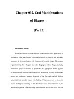
Similar to and highly correlated with the GOAT is the
Orientation Log (O-Log, Figure 4–1)—a scale intro-
duced by Jackson et al. (1998) as a brief measure of orien-
tation for patients undergoing rehabilitation. Health care
providers may use the O-Log to plot a patient’s recovery
curve by assigning a score of 0–3 for each item, adding the
scores, and graphing the sum on the orientation index. In
addition to being brief, this scale has some advantages
over the GOAT, including consistent scoring across items
and the ability to evaluate a patient who is unable to re-
spond (or who responds inaccurately). It can also be ad-
ministered to individuals with speech impairment.
As the period of PTA ends, the patient enters the
chronic phase of recovery, in which assessment of TBI-
related neurobehavioral and neurocognitive changes be-
comes especially important. The previously mentioned
Rancho Los Amigos Scale (see Table 4–6) is a useful tool in
tracking cognitive and behavioral recovery. A more com-
prehensive instrument was developed by Levin et al.
(1987a)—the Neurobehavioral Rating Scale (NRS)—
which measures disturbances in behavior, cognition, emo-
tion, thought content, and language function during the
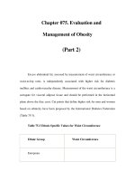
long-term recovery from brain injury. Levin et al. (1990)
enhanced the reliability and content validity of the NRS,
creating the Neurobehavioral Rating Scale—Revised
(NRS-R, Figure 4–2). It consists of a 4-point scale on
which ratings for each item range from absent to severe in
regard to the impact of a particular behavior on the per-
son’s social and occupational functioning. Administration
of the NRS-R requires a 15- to 20-minute structured inter-
view, which includes tests of orientation, attention, con-
centration, memory of recent events, delayed recall, prov-
erb interpretation, and mental flexibility as well as
questions about the emotional state and postconcussional
symptoms. During the administration of the tests the inter-
viewer observes the patient closely for fatigability, signs of
anxiety, disinhibition, agitation, hostility, disturbance of
mood, and difficulties with expressive and receptive com-
munication. Approximately one-third of the item ratings
are solely based on examiner’s observation, whereas the rest
of the items are rated according to the patient’s perfor-
mance on the tasks performed (McCauly et al. 2001). Early
administration after severe TBI followed by serial assess-
ments provide a means of quantifying change in the deficits
over time. Vanier et al. (2000) found the NRS-R to be a
useful tool for predicting psychosocial recovery and assess-
ing neuropsychological factors related to social autonomy.
A thorough clinical neuropsychiatric evaluation requires
careful assessment of cognitive functioning. The Neurobe-
havioral Cognitive Status Examination (NCSE), which can
be completed in 5–20 minutes, is an extremely useful tool for
rapid cognitive screening. Kiernan and colleagues developed
the NCSE to assess attention, orientation, language, visuo-
constructional skills, memory, calculation, abstract reason-
ing, and levels of consciousness (Kiernan et al. 1987;
Schwamm et al. 1987). Most of the NCSE’s assessment cat-
egories begin with a screening item that is a relatively de-
manding test of the skill involved. If the screening item is
successfully completed, no further testing in that domain is
required. This allows for rapid completion when there is lit-
tle cognitive impairment. The NCSE generates a perfor-
mance profile that reflects differentiated functioning and
can be compared to group norms for various neuropsychia-
tric disorders. The NCSE is particularly useful as a screen-
ing tool in identifying patients for whom formal neuropsy-
chological testing is indicated and is a valuable adjunct to
other clinical neurodiagnostic studies when neuropsycho-
logical testing is not readily available. Scales for specific as-
sessment of other psychiatric or behavioral problems are dis-
cussed elsewhere in this text (e.g., the Overt Aggression
Scale [see Chapter 14, Aggressive Disorders] and the Hamil-
ton Rating Scale for Depression).
Additional Assessment Tools
In addition to history, physical, mental status examination,
MMSE, “bedside” cognitive testing, and behavioral assess-
ment, one may incorporate additional evaluation tools to
complete the neuropsychiatric evaluation. These diagnos-
tic tools include neuropsychological testing, structural
Neuropsychiatric Assessment 71
and/or functional neuroimaging, electroencephalogram,
and evoked potentials (see Chapters 5, Structural Imaging;
6, Functional Imaging; and 7, Electrophysiologic Tech-
niques for more information).
Overview of Other Types
of Brain Injuries
In addition to brain injury due to blunt or penetrating
injuries or DAI, brain injury may be due to a number of
other causes. These include metabolic factors such as
hypoxia/anoxia; hypoglycemia, hypothyroidism, and cer-
tain vitamin deficiencies; exposure to CNS toxins such as
heavy metals or other industrial/environmental toxins;
drugs of abuse, including toxic inhalants and carbon mon-
oxide poisoning; and passage of electrical current through
the brain in electrocutions or lightning-related injuries.
Another important and increasingly common kind of
brain injury occurs as a complication of coronary artery
bypass surgery. This kind of diffuse brain injury is
believed to result, in part, from gaseous or particulate
microemboli released into the cerebral circulation as a
result of complications of the bypass procedure itself or
FIGURE 4–1. The Orientation Log.
inappro=inappropriate; incorr=incorrect; MultiChoice=multiple choice; phon=phonetic; Spon=spontaneous.
Source. Adapted from Jackson WT, Novack TA, Dowler RN: “Effective Serial Measurement of Cognitive Orientation in Rehabil-
itation: The Orientation Log.” Archives of Physical Medicine and Rehabilitation 79:718–720, 1998.
72 TEXTBOOK OF TRAUMATIC BRAIN INJURY
FIGURE 4–2. Neurobehavioral Rating Scale––Revised.
F=female; M=male; Mod.=moderate.
Source. Adapted from Vanier M, Mazaux J-M, Lambert J, et al: “Assessment of Neuropsychologic Impairment After Head Injury:
Interrater Reliability and Factorial and Criterion Validity of the Neurobehavioral Rating Scale—Revised.” Archives of Physical Medicine
and Rehabilitation 81:796–806, 2000. Used with permission.
Neuropsychiatric Assessment 73
surgical manipulations that occur during and immediately
after the time the patient is on bypass. The kinds of neu-
rological, cognitive, and behavioral sequelae that occur
with these kinds of brain injury are similar to those seen
with TBI, both with respect to the types and severity of
deficits and the dysfunction and disability they may cause.
As is the case with TBIs, the specific neurocognitive and
behavioral sequelae that occur are dependent on the
regions of the brain that have been damaged.
Anoxia/Hypoxia
Anoxia is defined as inadequate oxygenation of body tis-
sues. Anoxic brain injury owing to a lack of oxygen in the
ambient air is known as anoxic anoxia. Anoxia owing to
acutely decreased blood volume or lowered hemoglobin
concentration in the blood is referred to as anemic anoxia,
and anoxia owing to insufficient cerebral blood flow
because of cerebrovascular accidents, arrhythmias, or car-
diac arrests is called ischemic anoxia. Finally, there is toxic
anoxia, which is because of toxins or metabolites that may
interfere with oxygen utilization.
In general, hypoxia with ischemia is more harmful
than hypoxia alone because potentially toxic metabolic
products such as lactic acid may contribute to tissue dam-
age. The nature of hypoxic ischemic injury is neuropatho-
logically different from traumatic injury, in that the
former affects the neurons themselves, whereas the latter
tends to be an axonal phenomenon. In addition to cardiac
and respiratory arrest, anoxic brain injury occurs in cases
of near drowning, strangulation, and anesthetic accidents
(Wilson 1996).
Although the brain comprises only 2% of the body’s
total weight, it accounts for a disproportionate 20% of the
total oxygen utilization and 65% of the glucose uptake.
Approximately 15% of the cardiac output is directed to
the brain to meet its energy needs (Kuroiwa and Okeda
1994; White et al. 1984). When disruption of the oxygen
delivery system occurs, a series of cerebrovascular ho-
meostatic mechanisms become activated to maintain ade-
quate oxygen supply to the brain (Cohen 1976; Strand-
gaard and Paulson 1984). When there is a sustained
disruption in oxygen supply (for a period of 4–8 minutes
or longer), cerebral infarction and/or disseminated cellu-
lar death may occur (Bigler and Alfonso 1988; Caronna
1979; Cohan et al. 1989; Cohen 1976; Strandgaard and
Paulson 1984; White et al. 1984).
The mechanism of anoxic brain damage comprises a
complex cascade of time-dependent alterations in neuro-
nal function, metabolism, and morphology (Haddad and
Jiang 1993; Pulsinelli et al. 1982). The most important
acute effect of hypoxia on the brain is the release of exci-
tatory neurotransmitters, leading to an influx of sodium,
cellular edema, and consequent cellular injury (Hansen
1985; Kjos et al. 1983; Rothman and Olney 1986).
Longer-term effects are due to an increase in neuronal ex-
citability, which results in calcium influx, formation of
oxygen-free radicals that injure cells, and eventual cell
death (Ascher and Nowak 1987; Choi 1990; Gibson et al.
1988; Haddad and Jiang 1993; Hansen 1985; Maiese and
Caronna 1989; Schurr and Rigor 1992; Siesjo 1981;
White et al. 1984).
Whether a patient with hypoxia will develop neuro-
logical signs depends more on the severity and duration of
the process causing hypoxia than its etiology (Berek et al.
1997). Two factors that determine the vulnerability of
cells in a given brain region to hypoxia include distribu-
tion of the cerebral blood vessels and adequacy of their
baseline perfusion and the specific metabolic and bio-
chemical properties of the neural structures involved.
The most vulnerable regions of the brain are the water-
shed areas of the cortex. That is because normal cellular
metabolism in these areas is dependent on an adequate
flow of normally oxygenated blood through the distal ce-
rebral arterioles that perfuse them. Cellular and tissue
damage occur first in these areas where inadequate oxy-
genation of the blood due to hypoxia fails to meet mini-
mal metabolic requirements, especially when impaired
perfusion is also present (Brierley and Graham 1984; Par-
kin et al. 1987). Cells in brain regions with higher meta-
bolic demand are also more likely to be affected by oxy-
gen deprivation (Moody et al. 1990; Myers 1979). In
addition to these general principles, it has been shown
that cells in various brain regions respond differentially to
the degree and duration of hypoxia. For example, basal
ganglia and cerebral cortical cells show signs of necrosis
shortly after a cardiac arrest, whereas similar changes in
the hippocampus may not be seen until 2–3 days after the
event (Kuroiwa and Okeda 1994; Petito et al. 1987; Puls-
inelli et al. 1982).
Coma is a frequent outcome of significant and sus-
tained hypoxia. The three leading causes of coma in de-
scending order of frequency are: trauma, drug overdose,
and cardiac arrest (Shewmon et al. 1989). From a prognos-
tic point of view, patients with traumatic coma have a better
chance of recovery than those with nontraumatic coma.
Among patients in the nontraumatic group, recovery gen-
erally occurs in the following descending order of fre-
quency: metabolic causes, coma secondary to cardiac ar-
rest, and coma from cerebrovascular causes (Berek et al.
1997). Clinical outcomes typically depend on the presence
or absence of the prognostic factors listed in Table 4–12.
Neuropsychological deficits after anoxic brain damage
may include memory and executive dysfunction, appercep-
74 TEXTBOOK OF TRAUMATIC BRAIN INJURY
tive agnosia, and visual deficits. Most patients with anoxic
brain damage have preserved attention and concentration
abilities. Some patients who have sustained severe anoxic
brain injury may remain in a persistent vegetative state with
no observable cognitive functioning at all (Parkin et al.
1987; Wilson 1996).
Cognitive Problems After Coronary Artery
Bypass Graft Surgery
Approximately 800,000 patients worldwide undergo coro-
nary artery bypass graft (CABG) surgery per year (Selnes et
al. 1999). CABG is associated with significant cerebral
morbidity, manifested by cognitive decline or stroke
(Roach et al. 1996; Van Dijk et al. 2002). The incidence of
cognitive decline may vary from 3% to 50%, depending on
patient characteristics, definition of decline, and the type
and timing of neuropsychological assessment (Diegeler et
al. 2000; Roach et al. 1996; Van Dijk et al. 2002). Intraop-
erative transcranial Doppler monitoring has clearly dem-
onstrated that during cardiopulmonary bypass (CPB),
microemboli are released into the brain. This release of
microemboli is correlated with postoperative neurological
deficits (Syliviris et al. 1998). A study comparing the neu-
rocognitive effects of CABG with and without CPB sur-
gery demonstrated that patients with their first CABG
without CPB had less cognitive impairment at 3 months,
but by 12 months the differences between the groups had
become negligible (Van Dijk et al. 2002).
The emotional and cognitive state before CABG sur-
gery is an important factor in the development of anxiety,
depression, and cognitive deficits after the procedure
(Adrian et al. 1988; Savageau et al. 1982). Even though a
high percentage of patients may exhibit neuropsycholog-
ical deficits immediately or during the first few weeks af-
ter the surgery, most return to their premorbid level of
neuropsychological functioning within several months af-
ter the procedure (Frank et al. 1972; Savageau et al. 1982).
Patients about to undergo CABG surgery should be
screened for neurocognitive deficits and emotional distur-
bances before the procedure (Adrian et al. 1988). Asking
patients about their expectations for the outcome of the
procedure is also important because these expectations
have an important bearing on the postoperative emotional
state, cognitive deficits, and recovery from the surgery.
Electrical Injuries
Electrocution can cause brain damage in two ways—
direct cellular damage due to passage of current through
brain tissue and cardiac arrest induced by it. Electrical
injuries occur as a result of exposure to live wires at work
or home or lightning strikes during thunderstorms. The
degree of damage is determined by the amount and type
of current, duration of exposure, parts of the body
affected, and the pathway of current through the body.
Injuries acquired from exposure to electric current at
home or work (low voltage injuries <1,000 volts) are dif-
ferent from those sustained from lightning or contact
with high-voltage wires (high-voltage injuries >1,000
volts). Injuries due to alternating current are more seri-
ous in comparison to those from direct current (Browne
and Gaasch 1992; Fish 1993). Patients who experience
high-voltage electrical injury may initially show some
cognitive deficits with confusion and memory loss, which
usually clear within a few days. In cases in which these
deficits persist, neuropsychological evaluation should be
performed because some symptoms may be permanent,
especially in cases of direct electrical injury to the brain
(Table 4–13).
Looking Into the Future
There is still much to be learned about the molecular and
cellular cascades that follow brain injury—no matter what
the cause. Tracing these chemical and electrical derange-
ments may lead to a better understanding of the origins of
many neuropsychiatric illnesses. Recent investigations
suggest that TBI may be linked to the later development
of at least three neuropsychiatric conditions—MS, Alz-
heimer’s disease, and schizophrenia. Perhaps future
research will uncover common mechanisms of brain
injury and disease states, reducing the gap between “neu-
rologic” and “psychiatric” conditions and practice.
TABLE 4–12. Clinical parameters indicating
unfavorable prognosis in patients with coma
Clinical parameters Unfavorable prognosis
Duration of anoxia >8–10 minutes
Duration of cardiopulmonary
resuscitation
>30 minutes
Duration of postanoxic coma >72 hours
Pupillary light reaction Absent on day 3
Motor response to pain Absent on day 3
Blood glucose on admission >300 mg%
Glasgow Coma Scale score on day 3 <5
Source. Adapted from Berek K, Jeschow M, Aichner F: “The Prognos-
tication of Cerebral Hypoxia After Out-of-Hospital Cardiac Arrest in
Adults.” European Neurology 37:135–145, 1999.
79
5
Structural Imaging
Erin D. Bigler, Ph.D.
THE ADVENT OF computed tomography (CT) in the
1970s revolutionized the clinical assessment of traumatic
brain injury (TBI). Even in the earliest stages of neuroim-
aging development, the crude views of the brain gener-
ated by CT imaging provided the first in vivo assessment
of brain structure and permitted clinical evaluation of
such abnormalities as hemorrhage, contusion, edema,
midline shift, and herniation (Eisenberg 1992). The ini-
tial limitations of CT imaging due to slow speed of image
processing and limited resolution rapidly gave way to
technological improvements, such that current CT imag-
ing can be completed in minutes and provides excellent
detection of macroscopic abnormalities associated with
trauma (Figure 5–1). Because CT imaging can be done
quickly and on patients requiring life support or other
medical equipment (e.g., heart pacemaker), CT is the
method of choice for the acute assessment of the head-
injured patient (Gean 1994; Haydel et al. 2000). Although
magnetic resonance (MR) imaging has superior resolu-
tion and better anatomic fidelity than CT, it is often not
used acutely because of its susceptibility to metal and
motion artifact, incompatibility with certain life-support
equipment within the MR environment, length of scan
time, and decreased sensitivity (compared with that of
CT) in detecting skull fractures.
Because of these factors, typically in the TBI patient
the first scan performed is CT, and MR imaging is usually
chosen for follow-up neuroimaging. Thus, much of the
research and clinical information regarding CT imaging
centers on acute injury characteristics, whereas the find-
ings of MR imaging pertain to the subacute and chronic
phases of recovery. When MR imaging is performed on
the head-injured patient, there are various standard or
common clinical imaging sequences typically done. How-
ever, new techniques involving image acquisition and
analysis are being developed that may increase the sensi-
tivity of MR detection of abnormalities associated with
TBI, and part of the sensitivity of MR detection of any ab-
normality after TBI relates to the time postinjury when
scanning is performed. Accordingly, the neuroimaging of
TBI is typically broken down into acute imaging using
CT, subacute and chronic imaging using MR imaging,
and various experimental and clinical applications of MR
imaging that permit more refined analyses to detect TBI
neuropathology. These distinctions—CT imaging, MR
imaging, and new techniques—serve as the guidelines in
this chapter for discussing the use of structural imaging in
TBI.
Computed Tomography Imaging
Indications and Relationships to Outcome
A number of studies have examined CT imaging associ-
ated with acute brain injury (Haydel et al. 2000; Mar-
shall et al. 1991; Shiozaki et al. 2001; Wallesch et al.
2001). The consensus of such studies is that acute CT is
The technical expertise and assistance of Tracy Abildskov and the manuscript assistance of Jo Ann Petrie are gratefully acknowledged.
Much of the research reported in this chapter was supported by a grant from the Ira Fulton Foundation.
80 TEXTBOOK OF TRAUMATIC BRAIN INJURY
an excellent clinical tool in determining the presence of
treatable lesions, such as subdural hematoma (see Figure
5–1), and providing baseline information concerning the
location and nature of pathological conditions such as
cortical contusion, intraparenchymal hemorrhage, pete-
chial hemorrhage, and localized or generalized edema.
CT is also excellent in detecting skull fractures and asso-
ciated pneumocephalus, which may require surgical
intervention. There is a direct relationship between CT
imaging findings and the acute clinical status of the TBI
patient, based on the Glasgow Coma Scale (GCS) score
and other characteristics such as pupillary abnormalities,
loss of consciousness (LOC), and posttraumatic amne-
sia. There are also several CT rating scales available, but
probably the most common is the Trauma Coma Data-
bank as outlined by Marshall et al. (1991) and presented
in Table 5–1. What is important about this rating scale
is that it provides a basis for evaluating the severity of
injury during the acute stage. It also can provide a base-
line for future monitoring of change over time (Vos et
al. 2001), as is discussed in the section Relationship of
Acute Computed Tomography Abnormalities to Reha-
bilitation Outcome. Additionally, this scale overviews
the common injuries observed in CT imaging of the
acute TBI patient.
Relationship of Acute Computed Tomography
Findings to Severity of Injury
The most clinically important aspect of acute CT imaging
is the initial management, monitoring, and surgical
intervention for any treatable lesion(s). Additionally,
acute CT imaging of the TBI patient often provides
more clinical information than what comes from the
physical examination of the acutely injured patient, par-
ticularly the patient with altered mental status. For
example, the comatose patient may have no visible
abnormalities on CT imaging, whereas the patient with
only mild disorientation may be found to have signifi-
cant CT abnormalities, some requiring emergent inter-
vention. This is shown in Figure 5–2, which illustrates
that the frequency of CT abnormalities, using the rat-
ings outlined in Table 5–1, was associated with the GCS
score (highest within 24 hours of injury) and LOC in
240 consecutively admitted rehabilitation patients (Big-
ler et al. 2004). As can be seen, the entire gamut of CT
abnormalities was observed in this large sample of TBI
patients who had injuries sufficient to require hospital-
ization, but the most common was a level II injury (see
Table 5–1)—some mild edema; the presence of small,
mostly petechial hemorrhages or contusions; and no
mass effect. As for LOC, similar observations are made
in Figure 5–2, which demonstrates that LOC of any
duration was most likely to be related with a level II
injury as well.
Relationship of Acute Computed Tomography
Abnormalities to Rehabilitation Outcome
Despite the accuracy of CT in identifying gross structural
pathology during the acute stage, such findings often do
not relate well to the neurobehavioral outcome at the time
of discharge from rehabilitation, which makes the accurate
prediction of outcome from acute CT findings alone diffi-
cult (Dikmen et al. 2001; Temkin et al. 2003). The excep-
tion occurs with patients who have brainstem lesions,
because the presence of brainstem pathology typically
relates to poor outcome. Using both the Disability Rating
FIGURE 5–1. The axial section of a computed to-
mography scan of the head at the level of the lateral
ventricles.
Obtained without the addition of contrast medium, this scan re-
vealed four types of acute posttraumatic intracranial hemorrhages
(left is on the reader’s right side): an epidural hematoma (thick
white arrow) and a squamous temporal fracture (not shown) on
the left side, a laminate subdural hematoma (thick black arrow) on
the right side, right-sided periventricular and frontal lobe contu-
sions containing an intraparenchymal hematoma (thin white ar-
row), and a subarachnoid hemorrhage (thin black arrow) in the
right frontal region. These injuries were sustained in a fall.
Source. Reproduced from Mattiello JA, Munz M: “Four
Types of Acute Post-Traumatic Intracranial Hemorrhage.”
New England Journal of Medicine 344:580, 2001. Used with per-
mission. Copyright 2001, Massachusetts Medical Society. All
rights reserved.
Structural Imaging 81
Scale (DRS)
1
and Functional Independence Measure
(FIM)
2
discharge scores, Bigler et al. (2004) demonstrated
that the 240 TBI patients with CT ratings from no visible
abnormality to discernible major abnormalities had similar
rehabilitation outcomes (i.e., diffuse injury category I to
category IV; see Table 5–1). This means that outcome is
poorly predicted by just the acute injury characteristics
seen on CT imaging performed on the day of injury (DOI).
This finding should come as no surprise, because it may
take days to weeks to track the evolution of a lesion and
months before stable degenerative patterns are established
by neuroimaging findings ([Blatter et al. 1997; Shiozaki et
al. 2001; Vos et al. 2001]; see section Relationship of Mag-
netic Resonance Imaging Findings to Outcome for better
predictors of rehabilitation outcome). As is shown later in
this chapter, the better predictor of long-term outcome
comes from quantitative analysis of MR imaging done after
3–6 months postinjury, and these relationships are often
enhanced by tracking changes in neuroimaging using the
DOI CT scan. Accordingly, instead of using CT as an
absolute predictor of outcome, it is often better to consider
CT as a tool for establishing the baseline at the acute stage
of injury and then tracking the injury with either CT or
MR imaging at follow-up intervals.
Day of Injury as Baseline
Because the DOI scan is typically one of the first diagnostic
tests run on the acutely injured TBI patient, it is performed
early in the injury process. Because the morphological con-
sequences from trauma take time to evolve, the DOI scan
TABLE 5–1. Diagnostic categories of abnormalities visualized on computed tomography (CT) scan
Category Definition
1: Diffuse injury I (no visible pathology) No visible intracranial pathology seen on CT scan
2: Diffuse injury II Cisterns present with midline shift 0–5 mm and/or:
Lesion densities present
No high- or mixed-density lesion >25 cc
May include bone fragments and foreign bodies
3: Diffuse injury III (swelling) Cisterns compressed or absent with midline shifts 0–5 mm, no high- or mixed-density
lesion >25 cc
4: Diffuse injury IV (shift) Midline shift >5 mm, no high- or mixed-density lesion >25 cc
5: Evacuated mass lesion V Any lesion surgically evacuated
6: Nonevacuated mass lesion VI High- or mixed-density lesion >25 cc, not surgically evacuated
7: Brainstem injury VII Focal brainstem lesion, no other lesion present
Source. Adapted from Marshall LF, Marshall SB, Klauber MR, et al: “A New Classification of Head Injury Based on Computerized Tomography.”
Journal of Neurosurgery 75:514–520, 1991.
1
Disability Rating Scale (DRS). The DRS consists of the following eight items and range of scores (0 = no disability): 1) eye opening,
0–3; 2) verbal response, 0–4; 3) motor response, 0–4; 4) cognitive ability in feeding, 0–3; 5) cognitive ability in toileting, 0–3; 6) cog-
nitive ability in grooming, 0–3; 7) dependence on others, 0–5; and 8) employability, 0–3. A total DRS score is calculated by adding
the scores for each of the eight items (see Rappaport et al. 1982). Hall et al. (1993) offered the following distinctions in considering
the DRS score: 0 = no disability, 1 = mild disability; 2–3 = partial disability; 4–6 = moderate disability; 7–11 = moderately severe dis-
ability; 12–16 = severe disability; 17–21 = extremely severe disability; 22–24 = vegetative state; 25–29 = extreme vegetative state; and
30 = death. For the purposes of comparing DRS admission and discharge findings by ventricle to brain ratio outcome, DRS scores
were combined as follows: 0 = no disability; 1–3 = mild disability; 4–11 = moderate disability; 12–21 = moderately severe disability;
and 22+ = extremely severe-vegetative (see Figure 5–11).
2
Functional Independence Measure (FIM). The FIM (State University of New York at Buffalo Department of Rehabilitation Medicine
1990) is an 18-item, 7-level ordinal scale that can be used to assess level of function at time of admission to and discharge from a
rehabilitation unit. It is a general tool for all types of rehabilitation patients and has been successfully used in TBI (Hamilton et al.
1987). The version used in this study was the 3.1 version. By virtue of its ordinal scale, the lowest score is 7 and the highest is 126.
82 TEXTBOOK OF TRAUMATIC BRAIN INJURY
often provides important baseline information. This is dem-
onstrated in Figure 5–3, which depicts a 3-year-old
restrained passenger involved in a high-speed motor vehi-
cle accident. The DOI scan demonstrates a right intra-
parenchymal hemorrhage in the region of the internal
capsule-putamen. The anterior horns of the lateral ventricu-
lar system can be identified on the DOI scan, but cortical
sulci are not well visualized, which can be a sign of general-
ized edema. By 2 days postinjury, there is definite generalized
cerebral edema with obliteration of the ventricular system—
a clear sign of massive cerebral edema. One year later, there
is global atrophy manifested by generalized ventricular dila-
tation, prominent cortical sulci, and a large cavitation in the
right basal ganglia area—–a consequence of the focal hemor-
rhage. The hemorrhage likely resulted from shearing forces
disrupting the deep vascular supply to the basal ganglia.
Limitations
The problem with all contemporary imaging methods is
that they provide only a gross inspection of the macroscopi-
cally visible brain, whereas most of the critical functioning is
at the microscopic (neuronal and synaptic) level. For struc-
tural imaging using CT or MR, detection of an abnormality
is based on resolution measured in millimeters, whereas at
the microscopic level the resolution of clinically significant
abnormalities is measured at the micron level (Bain et al.
2001; Ding et al. 2001). Simply stated, a “normal” scan
merely indicates that no visible macroscopic pathology was
detected that reached a threshold of 1 mm or more. CT, or
any other neuroimaging method, simply cannot answer the
question of brain pathology below its level of detection. This
circumstance is nicely demonstrated in Figure 5–4. The scan
FIGURE 5–2. Computed tomography (CT) over-
view of 240 patients with traumatic brain injury (TBI).
The charts presented in this figure overview the acute CT of 240
TBI patients admitted to an inpatient rehabilitation facility by av-
erage Glasgow Coma Scale (GCS) (A) and GCS frequency by CT
classification (B), demonstrating that the most frequent CT abnor-
mality was a diffuse injury II, which occurred with a near-similar
frequency across all levels of severity; and loss of consciousness
(LOC) by CT abnormality classification (C), demonstrating again
that diffuse injury II was the most common classification wherein
the majority of TBI patients experienced some LOC. Acute CT
classification abnormalities are given in Table 5–1.
Source. Bigler ED, Ryser DK, Ghandi P, et al: “Day-of-Injury
Computerized Tomography, Rehabilitation Status, and Long-Term
Outcome as They Relate to Magnetic Resonance Imaging Findings
After Traumatic Brain Injury.” Brain Impairment 5:S122–123, 2004.
A
B
C
Structural Imaging 83
represented in the middle of the figure is the acute DOI CT,
interpreted as within normal limits, taken approximately 2
hours after injury (brief LOC, GCS score of 14 at the scene
of a severe head-on high-speed motor vehicle accident; GCS
score of 15 on hospital admission). The patient was also
found to have a cervical fracture that was neurosurgically
repaired, along with a large frontal scalp laceration. He was
hospitalized for 4 days. He developed the typical constella-
tion of postconcussive symptoms, including headache,
fatigue, irritability, some depression, and mild cognitive
problems, which gradually but not completely abated over
the next several months. He was able to return to work on a
part-time basis, but he complained of problems of mental
inefficiency and feeling “dull.” He was in excellent general
health, but he unexpectedly experienced a spontaneous car-
diac arrest while exercising and died 7 months postinjury, at
which time a full brain autopsy was performed. Gross brain
anatomy was normal, as shown Figure 5–4A, but histolog-
FIGURE 5–3. Computed tomography scans from a 3-year-old male traumatic brain injury patient injured
in a high-speed motor vehicle accident.
Right is on the reader’s left side. Day of injury (A). Note the right intraparenchymal hemorrhage and blood in the right Sylvian fissure.
However, in addition to these acute injury factors, note the size of the anterior horns of the lateral ventricle, which offer a baseline from
which to monitor atrophic changes over time. By 2 days postinjury (B), there is severe cerebral edema, manifested by obliteration of cortical
sulcal patterns, loss of definition between gray and white matter, and delineation of the anterior aspect of the interhemispheric fissure, along
with collapse of the ventricular system. By 7 months postinjury (C), there is extensive atrophy noted by generalized ventricular dilatation,
prominent cortical sulci, and the right Sylvian fissure. Also note the large cavitation left by the intraparenchymal hemorrhage. By viewing
these different scans, an excellent picture of how the brain changes over time after an injury can be objectively established.
FIGURE 5–4. Findings in mild traumatic brain injury (TBI).
This patient sustained a mild TBI (admission Glasgow Coma Scale, 14) 7 months before an unexpected death from cardiac arrest. The
ventral view of the intact brain at autopsy showed no cortical contusions or other gross abnormalities (A). Likewise, the computed tomog-
raphy (CT) scan performed on the day of injury shows no abnormalities (B), again supporting the clinical view of no gross brain abnormal-
ities. However, on microscopic examination, scattered hemosiderin (white arrow) deposits were observed, as shown in the histological
section (C). These were most prominent in the white matter. This demonstrates microscopic abnormalities as a consequence of brain injury,
even mild TBI, that are below detection by direct visual inspection of the brain using neuroimaging techniques (see Bigler et al. 2004).
BCA
BCA
84 TEXTBOOK OF TRAUMATIC BRAIN INJURY
ical examination demonstrated hemosiderin (a blood by-
product)-laden macrophages and lymphocytes in the white
matter (WM). Obviously, this finding suggests perturbation
of brain microvasculature and WM injury that was well
below the detection of the “normal” CT. Such microscopic
lesions are undoubtedly the basis of many neurobehavioral
sequelae associated with brain injury when imaging is “nor-
mal.” This is further supported by the work of Gorrie et al.
(2001) who examined 32 children at postmortem who suc-
cumbed to road accidents. With direct visual inspection, 17
of these TBI cases demonstrated no macroscopic abnormal-
ities of the type that would be detected by CT imaging.
However, when viewed at ×100 magnification, all cases
readily demonstrated microscopic injury.
Magnetic Resonance Imaging
The anatomic specificity of MR imaging approximates gross
brain anatomy and can be done in any plane (Figure 5–5).
Because of this anatomic specificity, MR imaging is the pre-
ferred method for detailed investigations of structural
changes in the brain that accompany trauma, particularly
changes in WM and direction of atrophy. Strich’s (1956)
article is often referenced as the seminal contribution to the
neuropathological literature on TBI; her discussion of the
preponderance of WM damage and generalized cerebral
atrophy that accompanies severe TBI is particularly impor-
tant. MR imaging can be used to detect these gross changes.
In terms of neuropsychiatric sequelae, MR imaging is
most useful in the late follow-up of a brain injury (see Jorge
et al. 2004), because it is at this stage when structural MR
imaging is excellent in its ability to detect TBI-induced ce-
rebral atrophy, which is typically observed as ventricular di-
latation (ventriculomegaly; Figure 5–6) coexistent with
prominent cortical sulci (Bigler 2000, 2001a, 2001b). Like-
wise, thinning of the corpus callosum (CC) in conjunction
with the expansion of the ventricle is usually apparent when
these structures are viewed in the midsagittal plane in the
chronic stage of TBI. Additionally, the MR-imaging
method is well suited for quantitative image analysis,
through which almost all major brain structures can be
FIGURE 5–5. The clarity of magnetic resonance (MR) imaging in detection of gross brain anatomy.
The horizontal section on the top left was done at postmortem, whereas the two MR scans on the top right were performed antemortem
and are at identical levels. The closeness with which the MR scans approximate actual anatomy is obvious. There are three different types
of MR scans depicted in this figure, all with different properties in displaying underlying anatomy as well as pathology. The top middle
MR scan is a proton density (PD), or mixed-weighted, scan in which excellent definition of white and gray matter can be visualized. The
top right view represents a T2-weighted image, in which cerebrospinal fluid is readily identified. The bottom views are from a different
subject and are all T1-weighted images. The bottom row demonstrates not only the clarity of gross brain anatomy depicted by MR
imaging but also the different planes that can be viewed (bottom left––axial; middle––coronal, bottom right––sagittal).
Structural Imaging 85
readily identified, quantified (either as volumes or surface
areas), and compared to a normative sample (Bigler 1999).
The table in Appendix 5–1 summarizes regions that have
been shown to exhibit atrophy in response to trauma.There
is extensive clinical literature on the use of MR in the acute
and subacute diagnosis and management of TBI (Atlas
2001; Gean 1994; Orrison 2000), but as indicated above,
with regard to neuropsychiatric morbidity abnormalities
identified in the chronic stage typically have better correla-
tion with outcome than the acute or sub-acute findings
(Henry-Feugas et al. 2000; Jorge et al. 2004; van der Naalt
et al. 1999; Vasa et al. 2004; Wilson et al. 1988). Accord-
ingly, the primary focus of the remainder of this chapter is
MR imaging performed more than 45 days postinjury so
that the more stable and chronic lesions can be related to
neurobehavioral deficits, particularly those resulting in
neuropsychiatric sequelae.
Indications
There is a multitude of reasons for performing MR imaging
in the TBI patient, but typically the reasons center on mon-
itoring the status of the patient, often during the subacute
and more chronic phases of recovery. For example, because
of its capacity for exquisite anatomic detail and detection of
water, MR is suitable for monitoring edema, midline shift,
and the changing status of a hemorrhage and for evaluating
lesions that may underlie posttraumatic epilepsy. It is also
helpful in the clinical correlation of the patient’s acute status,
as depicted in Figure 5–7, and the structural imaging. The
patient shown in this figure had normal CT reading on
admission but was in a coma (GCS score of 5). MR imaging
performed later on the DOI was also read as “normal”; how-
ever, the MR scan performed 4 days later clearly demon-
strated the beginnings of significant degenerative changes,
including areas of shearing that were not definitively
observed on the DOI CT or MR scan. Another reason for
MR imaging is to monitor changes over time, which is
important because the degeneration often takes months to
reach an endpoint. Blatter et al. (1997) demonstrated that
the time that elapses between injury and brain volume stabi-
lization equivalent to that expected with normal aging may
be more than 3 years, although most pathological changes
occur within the first 6 months. Thus, acute and subacute
MR imaging is performed to assess potentially medically
treatable abnormalities associated with brain trauma, track
degenerative changes that occur with time, and relate imag-
ing findings to neurobehavioral sequelae.
As indicated in the section Computed Tomography
Imaging, often all early and subacute neuroimaging is
done with CT, particularly with patients on life support,
due to the incompatibility of life-support equipment with
FIGURE 5–6. Ventriculomegaly in traumatic brain injury (TBI).
Hydrocephalus ex vacuo is a common sequela of brain injury and is often proportional to the severity of injury. The top row shows a frontal
view based on three-dimensional magnetic resonance renderings of the brain, with the visible ventricular system depicted in black. The
bottom row represents the lateral view: the image on the left is from a noninjured control subject, the image in the middle is from a
moderately injured TBI patient, and the image on the right is from a subject with severe brain injury. It is important to note that it is the
entire ventricular system that typically dilates, indicating the diffuse nature of impact brain injury. By taking the volume of the ventricular
system, as shown in black, and dividing it by the volume of the brain, a ventricle to brain ratio (VBR) can be calculated. Increasing VBR is
a sign of increasing cerebral atrophy. Typically, increased VBR is associated with worse outcome (see Figure 5–11 and Ariza et al. 2004).
86 TEXTBOOK OF TRAUMATIC BRAIN INJURY
FIGURE 5–7. Comparison of similar sagittal magnetic resonance (MR) images to demonstrate injury and
subsequent atrophy to the corpus callosum at different stages postinjury.
The midsagittal day-of-injury MR scan (A, top left) was taken on admission to the hospital after the patient sustained a severe TBI.
Some movement artifact diminished the quality of the image but was interpreted as within normal limits. However, within 1 week
postinjury (B), signal intensity changes are clearly visible in the corpus callosum both anteriorly (black arrow) as well as posteriorly.
At 4 years postinjury (C), corpus callosum atrophy is clearly evident and is generalized including all aspects (compare the original size
of the corpus callosum in A with that observed in C). Generalized atrophy is also noted by the dark signal, especially seen in the
frontoparietal aspects of the midsagittal view of C, indicating increased cerebrospinal fluid (CSF) in the space of the interhemispheric
fissure, a sign of reduced brain volume (note that brain parenchyma in A and B is light gray, but a dark signal covers the midsagittal
surface in C because of increased CSF in these regions secondary to atrophy). Also, as clearly visible (white arrow in C), a major shear
lesion is evident where most of this segment of the corpus callosum has been transected. For better clarification of this lesion involving
the corpus callosum, the injured corpus callosum has been enlarged and highlighted in D. When viewing A (the day-of-injury scan)
in retrospect, there is some signal change noted in the region that eventually shows the shear lesion. The colorized images in E, F,
and G are all from diffusion-tensor imaging sequences in which tractography involving the projections of the corpus callosum in a
noninjured subject is displayed (Lazar et al. 2003). The images are color-coded on the basis of their projection (i.e., red shows frontal
projection). In E, the diffusion scan on the left is depicted in the axial plane, which shows the projections across the corpus callosum
from this perspective. The scan to the right in E is from the injured patient. In F, the colorized projections are shown in the midsagittal
view. Accordingly, by comparing the view of the location of the lesion in D with the view in F, one can see that this injury would result
in disrupted projections in primarily the midfrontal region. G shows the tractography plots mapped through the corona radiata. The
vertical line in E is the approximate location of these maps that depict the hemispheric projections of callosal white matter fiber tracks.
Source. Diffusion-tensor imaging tractography color images courtesy of Mariana Lazar, Ph.D., and Andrew Alexander, Ph.D., Uni-
versity of Wisconsin, Madison.
Structural Imaging 87
the MR imaging environment. It is helpful to compare
baseline CT images with follow-up MR images, as dem-
onstrated in Figures 5–3, 5–10, and 5–12.
Typical Lesions Identified by Magnetic
Resonance Imaging
More details concerning the neuropathology of TBI are
presented in Chapter 2, Neuropathology. For the purposes
of this discussion, just a brief overview of the neuropathol-
ogy observed in MR imaging of the brain in TBI is offered,
but the reader should be aware that a multitude of pathol-
ogies exist that can be detected by MR imaging (Atlas 2001;
Gean 1994; Orrison 2000). The typical lesions described
below are the ones most commonly observed to relate to
significant neuropsychiatric sequelae (Bigler 2001b) and
most commonly occur because of the greater likelihood of
frontotemporal damage (see Figure 5–8). Table 5–2 is
FIGURE 5–8. Voxel-based morphometry (VBM) in traumatic brain injury (TBI).
VBM provides a method to simultaneously compare––voxel-by-voxel––where the major differences occur in subjects with TBI com-
pared with age-matched control subjects without damage. In this figure, by using three-dimensional (3D) magnetic resonance (MR)
imaging, the diffuseness of frontotemporal involvement can be more fully appreciated (shown in red) when TBI subjects who had
sustained frontotemporal contusions are compared with control subjects by using VBM techniques; the differences (i.e., regions of
reduced voxel density of either gray or white matter) are plotted on a standard 3D surface plot of the brain. VBM was applied to MR
imaging performed on 6 subjects (mean age = 16; standard deviation = 5.1) with moderate-to-severe TBI (all had Glasgow Coma
Scale scores at or below 8) compared with 18 control subjects (3 control subjects within 2 years per TBI patient). Young subjects were
selected to minimize any long-term age effect that could potentially relate to volume reduction. The VBM findings (A) distinctly
demonstrate extensive frontotemporal differences in the TBI subjects, particularly in the ventral frontal region, more so in gray matter
than white. Given the ventral basis of the changes seen in this illustration, the basal forebrain (slanted white arrow, control subject,
sagittal view, lower right)––including the region involving the anterior commissure (AC), a thin white matter band critical for white
matter interhemispheric connections, as shown in B––was also quantified and compared with the control subjects. Quantitatively, the
basal forebrain region demonstrated over a 15% reduction in volume in the TBI subjects, who also were found to have significantly
reduced AC widths of 2.00 mm (SD = 0.44) compared with control subjects, in whom the mean width was 3.18 mm (SD = 0.40). In
the TBI subject presented in B, the AC width was 1 mm compared with an age-matched control subject whose AC width was 3.5 mm.
The blue arrow identifies the location of the AC, and the conjoined white arrows show where shear injuries occurred in the TBI
subjects, leaving regions of cavitation in the basal ganglia and internal capsule. In the sagittal view, the control subject’s AC (B, lower
right) is clearly visible (vertical white arrow), whereas the AC is almost not discernible in the sagittal view of the TBI patient (lower
left). Note also the thinness of the corpus callosum in the TBI patient, another reflection of generalized injury.