Wound Healing and Ulcers of the Skin - part 3 ppsx
Bạn đang xem bản rút gọn của tài liệu. Xem và tải ngay bản đầy đủ của tài liệu tại đây (513.67 KB, 28 trang )
4.3.3 Vascular Disease
4.3.3.1 Venous Ulcers
Around 70% of leg ulcers are venous in origin
[61–63] (Fig. 4.5). Older sources of data may
present a higher percentage. However, in mod-
ern medicine, the prevalence of venous ulcers is
declining. This is attributed to the higher stan-
dards of medical care currently practiced. The
significance of compression therapy is well rec-
ognized nowadays; the use of low-molecular-
weight heparins prevents venous thromboem-
bolism in high-risk situations. In addition, vein
surgery has become minimally invasive.Venous
insufficiency may coexist with peripheral arte-
rial disease in 10–15% of patients with leg ulcers
[63, 64]. In many cases, the direct trigger for ul-
ceration is some external physical injury [1, 65].
Whereas in a healthy person mild injury does
not cause significant damage, in patients with
venous insufficiency the skin runs a much
higher risk of developing ulceration.
Histologically,microvessels in areas subjected
to chronic venous hypertension become dilated
and coiled; i.e., they have a glomerular appear-
ance in intravital capillaroscopy.In the advanced
disease, the number of functioning, perfused
capillaries is markedly reduced [66–69]. The se-
verity of cutaneous microangiopathy has been
found to correlate closely to the development of
clinical cutaneous trophic changes [66, 70].
Mechanisms of Formation. At present, the
exact mechanism leading to the histologic pic-
ture and tissue damage in venous insufficiency
remains uncertain. Nevertheless, in recent
years we have acquired an increased under-
standing of certain physiological mechanisms
involved in this process.
In chronic venous insufficiency, the venous
pressure (or venous hypertension) in the deep
venous system may be transmitted to the
superficial system. Partsch [71] suggested that
venous insufficiency is characterized by peaks
of pressure occurring with every muscle con-
traction and transmitted to the capillary net-
work. It is suggested that these pressure peaks
have a progressive, gradual, destructive effect
on the capillaries in the skin and subcutaneous
tissues [72–74].
In addition, leakage of fluids from within the
capillaries into the interstitium of the dermis
and subcutaneous tissues results in edema.
Whatever the mechanisms leading to edema,
edema in itself has been shown to affect the
quality of the skin. It induces sclerotic changes in
subcutaneous tissue, with consequent interfer-
ence of metabolic and gas exchange [75]. More-
over, due to the presence of edema, lymphatic
vessels and their valves are subjected to fibrotic
changes, with a further reduction in normal
lymphatic function and drainage of the tissues,
which sets up a vicious cycle of edema [76, 77].
4.3Mechanisms of Formation of Specific Types
41
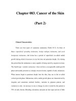
Fig. 4.5a, b. Venous ulcers. a. Brown pigmentation of sta-
sis dermatitis around the ulcer. b. Lipodermatosclerotic
leg; varicosities are seen in the medial area of the foot
04_031_052 01.09.2004 13:55 Uhr Seite 41
Endothelial damage, therefore, is the result
of edema with subsequent impaired oxygena-
tion and interference of metabolic activity
(and, perhaps, peaks of venous pressure). Inter-
cellular adhesion molecules seem to play a sig-
nificant role in the pathologic process, as re-
flected by their expression on endothelial cells
[78–81]. This process is followed by endothelial-
leukocyte adhesion and the trapping of white
cells within the capillaries. Loss of endothelial
integrity, together with the increasing presence
of white blood cells, may lead to the destruction
of adjacent tissue, protracted inflammation,
and fibrosis [82–86].
In addition to the above, numerous hypothe-
ses have been put forward to explain the ex-
act mechanism of skin damage and the de-
velopment of ulceration in the presence of
venous insufficiency.
Two presented below are worth mentioning:
5 Pericapillary fibrin cuffs are a prom-
inent histological feature of venous
insufficiency. In 1982, Browse and
Burnard [87, 88] suggested that ve-
nous hypertension, transmitted to
the capillary network, results in the
distention of capillary walls and the
widening of capillary pores. Subse-
quently, fibrinogen molecules leak
into the extracellular fluid, forming
complexes of fibrin around the cap-
illaries. The pericapillary fibrin layer
is claimed to form a mechanical bar-
rier, which prevents the transfer of
oxygen and nutrients, leading to
progressive damage to the skin and
subcutaneous tissues.
However, other researchers have in-
dicated that the fibrin cuffs do not
function as a barrier for oxygen
transport [89]. If so, these cuffs only
seem to reflect abnormal microcir-
culation with transmural deposition
of plasmatic macromolecules.
5 Falanga and Eaglstein [90] have
suggested that growth factors may
be trapped by certain macromole-
cules leaking through the capillary
pores into the dermis. Therefore,
growth factors are unable to partici-
pate and function in the processes
of tissue repair.
Location. The above discussion may help to
explain the distribution of venous ulcers. Since
venous pressure and the subsequent detrimen-
tal effect on tissue is maximal distally, venous
ulcers tend to occur in the lower calf. The me-
dial malleolus is more commonly affected than
the lateral. This finding is attributed to the
anatomy of the venous system, in which a larg-
er mass of venous vessels is located medially.
Therefore, the medial aspects of the legs are
subjected to higher venous pressures. Never-
theless, not infrequently these ulcers may ap-
pear above the lateral malleolus as well [91].
Lateral venous ulcers usually reflect the pres-
ence of an incompetent lesser saphenous vein,
with or without deep venous insufficiency.Oth-
er characteristics of venous ulcers are detailed
in Chap. 5.
4.3.3.2 Ulcers in Peripheral Arterial
Disease
Most patients with peripheral arterial disease
are over the age of 70. In contrast to venous ul-
cers, arterial ulcers are increasing in number.
People live longer nowadays, and peripheral ar-
terial disease is becoming more prevalent.Arte-
rial ulcers are estimated to constitute about
10% of leg ulcers [64]. Mixed ulcers, of arterial
and venous disease, are said to affect approxi-
mately 10–15% of patients with leg ulcers [63,
64].
Mechanisms of Formation. In many cases,
so-called ‘arterial’ ulcers develop following
physical trauma [65]. The trauma may be mi-
nor, but it affects vulnerable, poorly vascular-
Chapter 4 Cutaneous Ulcer Formation
42
4
t
t
04_031_052 01.09.2004 13:55 Uhr Seite 42
ized tissue, which is not able to heal as normal-
ly vascularized healthy tissue does. Moreover,
the trauma site may become the portal of entry
for infectious agents, further aggravating ulcer-
ation.
In other cases, arterial ulcers may appear
without trauma, when critical limb ischemia
has developed. Beyond a certain degree of is-
chemia, there is a complex chain of events that
may end in necrosis.
The definition of critical limb ischemia,
according to the Trans-Atlantic Inter-Society
Consensus Document on the Management of
Peripheral Arterial Disease (The TASC Work-
ing Group 2000), is based on a patient having
chronic ischemic rest pain, ulcers, or gangrene
attributable to objectively proven arterial oc-
clusive disease [92, 93]. The suggested inclusion
criteria in TASC for critical leg ischemia were
absolute ankle pressure below 50–70 mmHg or
reduced toe pressure (<30–50 mmHg).
Atherosclerosis of large arteries is the funda-
mental process in the pathogenesis of chronic
critical limb ischemia. It results in occlusion or
severe narrowing of vessels, with subsequent
reduction of blood flow and decreased perfu-
sion to distal regions. Other parameters such as
low blood pressure or the presence of anemia
may influence the degree of ischemia, and
hence the likelihood of progression to necrosis.
Location. Since a high percentage of arterial
ulcers are caused by trauma, arterial ulceration
(above the threshold of critical limb ischemia)
may develop anywhere on the lower calves. Ul-
cers tend to appear in the lateral or pretibial ar-
ea of the leg or on the dorsum of the foot (Fig.
4.6). Note that they may appear in the malleo-
lar region as well.
If critical limb ischemia has developed, it
may be manifested by distal necrosis of the toes
or forefoot (Fig. 4.7). This condition has a poor
prognosis, and amputation may be required.
The dorsum of the feet and heels may be affect-
ed as well.
Other characteristics of arterial ulcers are
detailed in Chap. 5.
4.3.3.3 Peripheral Arterial Disease
and Hypertensive Ulcers
Hypertensive ulcers were described by Marto-
rell in 1945 as ulcers located in the pretibial or
lateral area of the leg. These ulcers were said to
occur mainly in hypertensive women above the
age of 60 [94]. Some suggest that the so-called
Martorell’s ulcer represents a special variant of
an arterial leg ulcer, which should not be re-
garded as a separate entity. Others doubt the
validity of this clinical term, based on nonspe-
cific histologic features in leg ulcers clinically
diagnosed as ‘Martorell’s ulcers’ [95, 96]. In any
case, the elderly population is prone to develop-
ing hypertension, as well as atherosclerotic
changes within blood vessels.
4.3Mechanisms of Formation of Specific Types
43
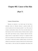
Fig. 4.6. An ulcer in peripheral arterial disease
Fig. 4.7. Arterial occlusion with significant ischemia,
pending ulceration
04_031_052 01.09.2004 13:55 Uhr Seite 43
4.3.3.4 Embolus
An acute, rapid development of limb ischemia
is caused by emboli. An atheromatous plaque
that becomes detached from a blood vessel wall
is a relatively large embolus that occludes a
large vessel and generally affects a specific ana-
tomic region. Cholesterol emboli, on the other
hand, are microemboli composed of cholesterol
crystals, 100–200 µm, which may occlude many
small arteries with the induction of multiple le-
sions [97, 98].
4.3.4 Leukocytoclastic Vasculitis
Note that leukocytoclastic vasculitis may be in-
duced by several types of infections, most com-
monly Streptococcus group A, Mycobacterium
leprae, and the hepatitis B and C virus [99].
Sometimes leukocytoclastic vasculitis may ap-
pear following the use of certain drugs (see
Chap. 16).
4.3.5 Connective Tissue
and Multisystem Diseases
Cutaneous ulcers appear in connective tissue
diseases and multisystem diseases. A classical
example is systemic lupus erythematosus
(SLE), which may present in several forms. In
most cases, ulcers in connective tissue diseases
are attributed to vasculitis. For example, the in-
cidence of cutaneous ulcers in idiopathic SLE
patients is about 5%. The ulcers are usually lo-
cated in malleolar or pretibial areas [100, 101]
due to the vasculitic process. Vasculitis may al-
so result in gangrene of the finger tips. Howev-
er, SLE may also lead to a secondary form of
anti-phospholipid syndrome with the subse-
quent development of cutaneous ulcers. Simi-
larly, the presence of cryoglobulins in SLE may
lead to the formation of cutaneous ulcers locat-
ed in the extremities.
In rheumatoid arthritis, various forms of cu-
taneous ulcers may be seen: leg ulcers or digital
necrosis, due to the vasculitic process, similar
to those of SLE; ulceration of subcutaneous
nodules; and pyoderma gangrenosum, which
may be found in rheumatoid arthritis. Pro-
longed glucocorticoid therapy may be detri-
mental to the quality of the skin in these cases,
thus further hindering the repair of cutaneous
ulcers.
Vasculitic involvement may induce ulcera-
tion in other connective tissue diseases, such as
dermatomyositis, Sjögren’s syndrome, or scler-
oderma. However, there may be other reasons
for ulceration in connective tissue disease. For
example, Raynaud’s phenomenon, which may
be associated with connective tissue diseases,
may result in digital ulceration. Similarly, the
gradual damage to the quality of the skin in
scleroderma predisposes to ulceration.
4.3.6 Hypercoagulable States
Some of the ‘hypercoagulable’ conditions listed
in Table 4.1, such as coumadin-induced necro-
sis, heparin necrosis, or disseminated intravas-
cular coagulation, are characterized by the de-
velopment of micro-thrombi [102]. The histo-
logic hallmark of these cases is the presence of
fibrin thrombi (see Chap. 6). The occlusion of
blood vessels by fibrin thrombi may manifest
clinically as cutaneous ulceration.
Other conditions listed in Table 4.1, i.e., pro-
tein C deficiency,activated protein C resistance,
protein S deficiency,and anti-thrombin III defi-
ciency, classified under the heading ‘thrombo-
philia’, may lead to vascular thrombosis. In
many cases, the mechanism leading to ulcera-
tion is not direct. Thrombophilia may result in
deep vein thrombosis which, in itself, predis-
poses to chronic venous ulceration [103, 104].
However,fibrin thrombi have been described in
such cases as well [105]. Most of these cases
have been associated with coumadin or hepar-
in therapy.
Conditions such as hyperhomocystinemia
have been implicated in the formation of deep
venous thrombosis with the subsequent devel-
opment of venous ulcers [106]. To the best of
our knowledge, it has never been described in
the literature as having directly caused a cuta-
neous ulcer through the formation of fibrin
thrombi.
Chapter 4 Cutaneous Ulcer Formation
44
4
04_031_052 01.09.2004 13:55 Uhr Seite 44
4.3.7 Metabolic Disorders:
Diabetes Mellitus
Diabetic ulcers are included in Table 4.1 under
the term ‘metabolic ulcers’. The metabolic ab-
normalities in diabetes may lead to the forma-
tion of ulcers by several mechanisms, as de-
tailed below.
4.3.7.1 Peripheral Arterial Disease
and Atherosclerosis
(Macroangiopathy)
Peripheral vascular disease is more common in
people with diabetes than in the rest of the pop-
ulation. In the presence of additional risk fac-
tors such as smoking, hyperlipidemia, or hy-
pertension, the incidence is even higher.
The prevalence of peripheral arterial disease
in diabetic patients is between 20% and 40%,
and it is regarded as a sign of premature aging
of blood vessels [107–109]. A distinguishing
feature of diabetes is that the ulcers tend to oc-
cur more distally than they do in non-diabetic
patients with peripheral arterial disease [110].
Diabetic ulcers due to peripheral arterial
disease may therefore appear anywhere on the
lower calves, usually on the lateral or pretibial
aspect of the leg, dorsum of the foot, or malleo-
lar region. As in peripheral arterial disease, ne-
crosis of a distal toe or foot may develop if there
is severe ischemia of a diabetic limb. In ad-
vanced cases, widespread calcification may de-
velop along the length of the media of the arte-
rial wall. Hence, Doppler measurement of ankle
blood pressure (and consequently ABI meas-
urement) may indicate high pressures, which
does not accurately reflect the true degree of is-
chemia of the limb [110, 111].
4.3.7.2 Neuropathy
Neuropathy in diabetes affects sensory, motor,
and autonomic fibers. It is estimated that al-
most 30% of type-2 diabetic patients have neu-
ropathy, while it affects 50% of patients over the
age of 60 years [112]. Ulceration of the soles of
diabetic patients is, in most cases, attributed to
neuropathy [113, 114].
The detrimental effects of sensory, motor,
and autonomic neuropathy are as follows:
5 Sensory neuropathy results in anes-
thesia and loss of protective sensa-
tion.
5 Motor neuropathy results in diffi-
culty in activating certain muscle
groups, resulting in inadequate dis-
tribution of pressure on the sole
while walking. Areas subjected to
repetitive focal pressure may ulcer-
ate or, alternatively, may develop a
callus, which predisposes to ulcera-
tion. The consequences of motor
neuropathy are reflected in the pres-
ence of typical foot deformities seen
in diabetic neuropathy, such as pro-
trusion of the metatarsal heads.
Mal perforant is a common neuro-
pathic ulcer of the sole, which ap-
pears over the metatarsal heads
[110].
5 Autonomic neuropathy is associated
with dry skin and further contrib-
utes to fissuring and callus forma-
tion. In addition, it leads to arteriov-
enous shunting which, although ac-
companied by increased blood flow,
reduces nutritive cutaneous capil-
lary flow [115, 116].
The above-mentioned processes may mis-
lead the physician, due to the following phe-
nomena:
5 Sensory neuropathy may conceal
symptoms of intermittent claudica-
tion and rest pain.
5 An ischemic foot may nevertheless
be warm and pink on clinical exam-
ination, due to autonomic neuropa-
thy [115, 117, 118].
4.3Mechanisms of Formation of Specific Types
45
t
t
04_031_052 01.09.2004 13:55 Uhr Seite 45
The neuropathic process leads to the formation
of ulcers on the sole or on the lateral and medi-
al regions of the foot in diabetic patients
(Fig. 4.8). Typically, a neuropathic ulcer of the
sole is surrounded by circumscribed callus for-
mation. Neuropathy and decreased sensation
render the patient even more prone to trauma
and subsequent ulceration, which may occur
anywhere in the distal regions of the limbs. In
some cases, the presence of neuropathy pre-
vents early identification of an ulcer by the af-
fected person, and appropriate intervention,
therefore, is not carried out.
4.3.7.3 Microangiopathy in Diabetes
Diabetic microangiopathy is characterized by
the thickening of basal membranes and in-
creased capillary permeability. In its advanced
stages, it results in compromised gas exchange,
a decrease in cutaneous pO
2
, and ischemia [110,
119].
The main clinical implications of microan-
giopathy with respect to skin ulcers are as
follows:
5 The ischemic changes described
above (together with macroangio-
pathy) cause additional damage to
the skin, thereby increasing the
probability of ulceration. The com-
bination of macroangiopathy and
microangiopathy seems to be the
reason why diabetic ulcerations
tend to be located more distally,
compared with ulceration in non-
diabetic peripheral arterial disease.
5 Microangiopathic involvement of
the vasa nervosum results in diabet-
ic neuropathy.
Note: The effect of microangiopathy is most ob-
vious in the kidneys and the retina. The possible
influence of these vascular changes on ulcer for-
mation in the diabetic leg is questionable and
has not yet been fully evaluated. It is reasonable
to assume that they affect capillary function [111,
120].
4.3.7.4 Other Factors:
Osteoarthropathy,
Cheiroarthropathy
Charcot’s osteoarthropathy describes a de-
structive process of the joints, occurring in dia-
betic neuropathy. It creates excessive focal pres-
sure on the sole of the foot, predisposing it to
ulcer formation. Another process is known as
cheiroarthropathy, in which there is a thicken-
ing of the skin with limitation of joint mobility
and an abnormal gait, with subsequent inap-
propriate weight distribution on the sole of the
foot [121, 122].
4.3.7.5 Reduced Resistance
to Infections
Infection is a frequent complication of dia-
betes, which aggravates tissue damage. Dia-
betes is associated with decreased phagocytic
activity and decreased function of leukocytes
Chapter 4 Cutaneous Ulcer Formation
46
4
Fig. 4.8. A neuropathic ulcer in diabetes
t
04_031_052 01.09.2004 13:55 Uhr Seite 46
[123]. Chemotaxis of leukocytes and phagocy-
tosis are impaired in poorly controlled diabetes
[110]. Hyperglycemia has been found to inhibit
the cellular transport of vitamin C into fibro-
blasts and leukocytes, with reduced chemotaxis
of leukocytes [124].
4.3.7.6 Location of Ulcers in Diabetes
In view of the above-mentioned pathologic
characteristics of diabetes, even minor trauma
or otherwise negligible superficial infection
may be sufficient to induce ulceration.
In a diabetic patient, ulcers may be located
as follows:
5 Lateral or pretibial regions of the
leg, dorsum of the foot, or malleolar
regions, due to peripheral arterial
disease and subsequent damage to
the skin and subcutaneous tissue
5 Distal toes (Fig. 4.9) or distal forefoot,
due to the severe ischemia of periph-
eral arterial disease
5 Neuropathy predisposes to ulcera-
tion mainly on the sole. Neverthe-
less, the decreased sensation com-
bined with increased susceptibility
to trauma may occur anywhere on
the distal limb. Osteoarthropathy
further contributes to the formation
of plantar ulcers.
In summary, the classical diabetic ulcer appears
on the sole. However, in view of the combination
of several detrimental factors including macro-
angiopathy, microangiopathy, neuropathy, and
reduced resistance to infections, ulcers in dia-
betes can, in fact, occur anywhere on the lower
leg.
4.3.8 Hematologic Abnormalities
4.3.8.1 Hemolytic Anemia
and Cutaneous Ulcers
Most of the literature in the field of hemolytic
anemia and cutaneous ulcers relates to sickle
cell disease. Blood vessels occluded by the
sludging of sickled erythrocytes are the histo-
logic hallmark of an ulcer in sickle cell anemia.
Sickle cells are relatively rigid, with a re-
duced ability to alter their shape. It seems that
the reduced deformability of sickled erythrocy-
tes is a major factor leading to vascular occlu-
sion and ulceration [125]. These features of
sickled erythrocytes may significantly decrease
blood flow, especially in capillary beds subject-
ed to venous stasis [126]. Below a certain level
of blood flow, there is a clumping of sickled
erythrocytes with subsequent obstruction of
blood vessels [125, 127]. The vascular occlusion
leads to ulceration. When the level of oxygen is
reduced, these processes are more pronounced.
The causes of ulceration in other types of
anemia such as thalassemia, hereditary sphero-
cytosis, or pyruvate kinase deficiency are not
fully understood. For example, leg ulcers are
rare in α-thalassemia, but relatively common in
severe β-thalassemia [128]. It is reasonable to
assume that, in these cases, there is also a di-
minished deformability of abnormal erythroc-
ytes. The tendency for ulcers to appear in the
gaiter area of the lower limbs suggests that
there is an element of venous stasis that con-
tributes to a reduction in blood flow.
In certain types of hemolytic anemia such as
hereditary spherocytosis, cutaneous ulcers
have been reported to improve and heal follow-
ing a splenectomy [129, 130]. In other cases of
anemia, such as in β-thalassemia, no beneficial
4.3Mechanisms of Formation of Specific Types
47
Fig. 4.9. An ulcer on the toe of a diabetic patient
t
04_031_052 01.09.2004 13:55 Uhr Seite 47
effect of a splenectomy has been observed [131].
A possible explanation for the above observation
regarding hereditary spherocytosis has been
suggested: During their passage through the
spleen, red blood cells may lose a membrane lip-
id. This change may lead to the entrapment of
cells in the microvasculature, resulting in stasis
with impaired oxygenation and the formation of
cutaneous ulcers. A splenectomy prevents this
sort of damage to red blood cells; their improved
function and increased capacity of deformabil-
ity leads to healing of the ulcers [125, 132].
4.3.9 Nutritional Disorders
In most cases, malnutrition is not a direct cause
of ulceration. However, malnutrition does
interfere with wound healing and has a detri-
mental effect on the general condition of the
patient. This issue is discussed in detail in
Chap. 18.
Conditions in which malnutrition may in-
duce ulceration directly are:
5 Vitamin C deficiency
5 Noma (cancrum oris, necrotizing
ulcerative gingivitis)
5 Tropical ulcer (tropical sloughing
phagedena)
Vitamin C deficiency results in impaired colla-
gen synthesis with subsequent poor wound
healing. The classical clinical descriptions of
scurvy by Lind [133] documented the appear-
ance of ulcers on affected skin, induced mostly
by minor trauma. Boulinguez et al. [134] docu-
mented three patients with scurvy presenting
with ecchymotic purpura and hemorrhagic ul-
cers of the lower limbs.
However, vitamin C is an important factor
not only in those relatively rare patients whose
ulcers are caused directly by vitamin C defi-
ciency. It is also very important to identify pa-
tients with cutaneous ulcers (caused by other
etiologies) who happen to be deficient in vita-
min C. In these cases, vitamin C supplementa-
tion may improve wound healing.
In the latter two conditions, i.e., noma and
tropical ulcer, the specific mechanisms leading
to ulceration have not yet been identified, but it
appears that opportunistic infection, related to
the state of malnutrition, plays a significant role
in their pathogenesis.
4.3.10 Other Causes
Epithelial tumors and leg ulcers are discussed
in Chap. 6, and a detailed review of drugs and
cutaneous ulcers is presented in Chap. 16.
References
1. Shai A, Halevy S: Direct triggers for ulceration in
patients with venous insufficiency. Int J Dermatol
(in press)
2. Reed BR, Clark RAF: Cutaneous tissue repair. Prac-
tical implications of current knowledge. II. J Am
Acad Dermatol 1985; 13:919–941
3. Robson MC, Stenberg BD, Heggers JP: Wound heal-
ing alterations caused by infection. Clin Plast Surg
1990; 17: 485–492
4. Rietchel RL, Fowler JF: The role of age, sex and col-
or of skin in contact dermatitis. In: Rietchel RL,
Fowler JF (eds) In: Fisher’s Contact Dermatitis, 4th
edn. Baltimore: Williams & Wilkins.1995; pp 41–65
5. Dooms-Goossens A, Degreef H, Parijs M, et al: A
retrospective study of patch test results from 163
patients with stasis dermatitis or leg ulcers. Der-
matologica 1979; 159 : 93–100
6. Wilson CL, Cameron J, Powell SM, Cherry G, Ryan
TJ: High incidence of contact dermatitis in leg-ul-
cer patients-implications for management. Clin
Exp Dermatol 1991; 16: 250–253
7. Rietchel RL, Fowler JF: Contact dermatitis and oth-
er reactions to metals. In: Rietchel RL, Fowler JF
(eds) In: Fisher’s Contact Dermatitis, 4th edn. Bal-
timore. Williams & Wilkins.1995; pp 808–885
8. Lee HS, Goh CL: Occupational dermatosis among
chrome platers. Contact Dermatitis 1988; 18:89–93
9. Tanaka T, Miyachi Y, Horio T: Ulcerative contact
dermatitis caused by sodium silicate. Coexistence
of primary irritant contact dermatitis and contact
urticaria. Arch-Dermatol 1982; 118 : 518–520
10. Mochida K, Hisa T,Yasunaga C, et al: Skin ulcera-
tion due to povidone-iodine. Contact Dermatitis
1995; 33: 61–62
11. Fisher AA: Unique reactions of scrotal skin to topi-
cal agents. Cutis 1989; 44 : 445–447
12. Krafchik BR: Eczematous dermatitis. In: Schachner
LA, Hansen RC (eds) Pediatric Dermatology, 2nd
edn. New York: Churchill Livingstone. 1995;
pp 704–705
Chapter 4 Cutaneous Ulcer Formation
48
4
t
04_031_052 01.09.2004 13:55 Uhr Seite 48
13. Rijswijk LV: Epidemiology. In: Morison MJ: The
prevention and treatment of pressure ulcers. 1st
edn. Edinburgh: Mosby.2001; pp 7–15
14. Kanj LF, Wilking SV, Phillips TJ: Pressure ulcers. J
Am Acad Dermatol 1998; 38 : 517–536
15. Allman RM: Epidemiology of pressure sores in dif-
ferent populations. Decubitus 1989; 2 : 30–33
16. Young JS, Burns PE, Bowen AM, et al: Spinal cord
injury statistics: experience of the regional spinal
cord injury systems. National Spinal Cord Injury
Data Research Center. Phoenix. 1982
17. Nixon J: The pathophysiology and aetiology of
pressure ulcers. In: Morison MJ (ed) The preven-
tion and treatment of pressure ulcers, 1st edn.
Edinburgh: Mosby. 2001; pp 17–36
18. Garfin SR, Pye SA, Hargens AR, Akeson WH: Sur-
face pressure distribution of the human body in
the recumbent position. Arch Phys Med Rehabil
1980; 61: 409–413
19. Kosiak M: Etiology and pathology of ischemic ul-
cers. Arch Phys Med Rehabil 1959; 40: 62–69
20. Daniel RK, Priest DL, Wheatley DC: Etiologic fac-
tors in pressure sores: an experimental model.
Arch Phys Med Rehabil 1981; 62: 492–498
21. Falanga V: Chronic wounds: pathophysiologic and
experimental considerations. J Invest Dermatol
1993; 100: 721–725
22. Kosiak M: Etiology of decubitus ulcers. Arch Phys
Med Rehabil 1961; 42: 19–29
23. Dinsdale SM: Decubitus ulcers: role of pressure
and friction in causation. Arch Phys Med Rehabil
1974; 55 : 147–152
24. Exton-Smith AN, Sherwin RW: The prevention of
pressure sores: significance of spontaneous bodily
movements. Lancet 1961; 2 : 1124–1126
25. Barbenel JC, Jordan MM, Nicol SM, et al: Incidence
of pressure sores in the Greater Glasgow Health
Board Area. Lancet 1977; 2 :548–550
26. Reuler JB, Cooney TG: The pressure sore: pathoph-
ysiology and principles of management. Ann
Intern Med 1981; 94 : 661–666
27. Bennet L, Bok YL: Pressure versus shear in pressure
sore causation. In: Lee BY: Chronic Ulcers of the
Skin, 1st edn. New York: McGraw-Hill.1986; pp 39–56
28. Bennett L, Kavner D, Lee BK, et al: Shear vs pres-
sure as causative factors in skin blood flow occlu-
sion.Arch Phys Med Rehabil 1979; 60: 309–314
29. Reichel SM: Shearing force as a factor in decubitus
ulcers in paraplegics. JAMA 1958; 166 : 762–763
30. Agris J, Spira M: Pressure ulcers: prevention and
treatment. Clin Symp 1979; 31: 1–32
31. The National Pressure Ulcer Advisory Panel. Pres-
sure ulcers prevalence, cost and risk assessment:
consensus development conference statement. De-
cubitus 1989; 2 : 24–28
32. Crickx B: Erysipelas: evolution under treatment,
complications. Ann Dermatol Venereol 2001; 128:
358–362
33. Ahrenholz DH: Necrotizing soft-tissue infections.
Surg Clin North Am 1988; 68: 199–214
34. Fustes-Morales A, Gutierrez-Castrellon P, Duran-
Mckinster C et al: Necrotizing fasciitis: Report of 39
pediatric cases. Arch Dermatol 2002; 138 : 893–899
35. Stevens DL: The flesh-eating bacterium: What’s
next? J Infect Dis 1999; 179 [Suppl 2] : S366–S374
36. Bosshardt TL, Henderson VJ, Organ CH Jr: Necro-
tizing soft tissue infections. Arch Surg 1996; 131 :
846–852
37. Umbert IJ,Winkelmann RK, Oliver GF, et al: Necro-
tizing fasciitis: a clinical, microbiologic, and histo-
pathologic study of 14 patients. J Am Acad Derma-
tol 1989; 20:774–781
38. Lee PK, Zipoli MT,Weinberg AN,Swartz MN, John-
son RA: Pyodermas: staphylococcus aureus, strep-
tococcus and other gram-positive bacteria. In:
Freedberg IM, Eisen AZ, Wolff K, Austen KF, Gold-
smith LA, Katz SI (eds) Fitzpatrick’s Dermatology
in General Medicine, 6th edn. New York: McGraw-
Hill. 2003; pp 1856–1878
39. Sanchez MR: Syphilis. In: Freedberg IM, Eisen AZ,
Wolff K, Austen KF, Goldsmith LA, Katz SI (eds)
Fitzpatrick’s Dermatology in General Medicine, 6th
edn. New York: McGraw-Hill. 2003; pp 2163–2188
40. Varela P,Alves R,Velho G, et al: Two recent cases of
tertiary syphilis. Eur J Dermatol 1999; 9 :371–373
41. Merkert R: Generalized ulcers. Noduloulcerative
syphilis (malignant syphilis, lues maligna). Arch
Dermatol 1997; 133 : 1027–1028, 1030–1031
42. Don PC, Rubinstein R, Christie S: Malignant syphi-
lis (lues maligna) and concurrent infection with
HIV. Int J Dermatol 1995; 34: 403–407
43. Gawkrodger DJ: Mycobacterial infections. In:
Champion RH, Burton JL, Burns DA, Breathnach
SM (eds) In: Rook/Wilkinson/Ebling Textbook of
Dermatology, 6th edn. Oxford: Blackwell Scientific
Publications. 1998; pp 1181–1214
44. Lantos G, Fisher BK, Contreras M: Tuberculous ul-
cer of the skin. J Am Acad Dermatol 1988; 19 :
1067–1072
45. Marechal V, Maradeix S, Pouaha J, et al: Cas pour
diagnostic: Ulceration atone de la cheville. Ann
Dermatol Venereol 2002; 129 : 439–440
46. Rao PT, Jena SK: Surgical treatment of plantar ul-
cers in leprosy. Int Orthop 1986; 10:75–78
47. Cross H, Kulkarni VN, Dey A, et al: Plantar ulcera-
tion in patients with leprosy. J Wound Care 1996; 5:
406–411
48. Mitchell PD: The threshold for protective sensation
that prevents neuropathic ulceration on the plantar
aspect of the foot: a study of leprosy patients in a
rural community in India. Lepr Rev 2001; 72 :
143–150
49. Boucher P, Sebille A: Frequency of the association
of perforating plantar leprotic lesions and truncal
involvement of the posterior tibial nerve.Acta Lep-
rol 1983; 1: 177–182
50. Feenstra W, Van de Vijver S, Benbow C, et al: Can
people affected by leprosy at risk of developing
plantar ulcers be identified? A field study from cen-
tral Ethiopia. Lepr Rev 2001; 72: 151–157
References
49
04_031_052 01.09.2004 13:55 Uhr Seite 49
51. Morton RS: The treponematoses. In: Champion
RH, Burton JL, Burns DA, Breathnach SM (eds) In:
Rook/Wilkinson/Ebling Textbook of Dermatology.
6th edn. Oxford: Blackwell Scientific Publications.
1998; pp 1237–1275
52. Ramesh V, Saxena U, Mukherjee A, et al: Multiple
ulcers in an elderly man.Necrotizing erythema no-
dosum leprosum (ENL) (necrotizing ENL). Arch
Dermatol 1992; 128: 1643, 1646
53. Bernadat JP, Faucher JF, Huerre M: Diffuse leprom-
atous leprosy disclosed by cutaneous vasculitis.
The Lucio phenomenon. Ann Dermatol Venereol
1996; 123: 21–23
54. Souza CS, Roselino AM, Figueiredo F, et al: Lucio’s
phenomenon: clinical and therapeutic aspects. Int
J Lepr Other Mycobact Dis 2000; 68:417–425
55. Barbarulo AM, Metha N, Bucalo B, et al: Recurrent
disseminated herpes zoster and cytomegalic peri-
anal ulcer: a case report and review of the litera-
ture. Cutis 2001; 67: 43–46
56. Pariser RJ: Histologically specific skin lesions in
disseminated cytomegalovirus infection. J Am Ac-
ad Dermatol 1983; 9 : 937–946
57. Rodot S, Lacour JP, van Elslande L, et al: Ecthyma
gangrenosum caused by Klebsiella pneumoniae.
Int J Dermatol 1995: 34 : 216–217
58. Cohen PR, Grossman ME: Recognizing skin lesions
of systemic fungal infections in patients with
AIDS. Am Fam Physician 1994; 49 : 1627–1634
59. Ginter G, Rieger E, Soyer HP, et al: Granulomatous
panniculitis caused by Candida albicans: a case
presenting with multiple leg ulcers.J Am Acad Der-
matol 1993; 28 : 315–317
60. Orem J, Mpanga L, Habyara E, et al: Disseminated
aspergillus fumigatus infection: case report. East
Afr Med J 1998; 75 :436–438
61. Phillips TJ, Dover JS: Leg ulcers. J Am Acad Derma-
tol 1991; 25:965–987
62. Nelzen O, Bergqvist D, Lindhagen A: Venous and
non-venous leg ulcers: clinical history and appear-
ance in a population study. Br J Surg 1994; 81 :
182–187
63. Baker SR, Stacey MC, Singh G, et al: Aetiology of
chronic leg ulcers. Eur J Vasc Surg 1992; 6: 245–251
64. Hafner J: Management of arterial leg ulcers and
combined (mixed) venous-arterial leg ulcers. Curr
Probl Dermatol 1999; 27: 211–219
65. Husni EA: Skin ulcers secondary to arterial and ve-
nous disease.Lee BY: Chronic Ulcers of the Skin,1st
edn. New York: McGraw-Hill. 1986; pp 93–101
66. Junger M,Hahn M,Klyscz T, et al: Microangiopathy
in the pathogenesis of chronic venous leg ulcers in-
sufficiency. Curr Probl Dermatol 1999; 27:124–129
67. Incandela L, Belcaro G, Cesarone MR, et al: Micro-
angiopathy and venous ulceration: Topical treat-
ment with Essaven gel. A placebo-controlled, ran-
domized study. Angiology 2001; 52 [Suppl 3] :
S17–S21
68. Bollinger A, Leu AJ, Hoffmann U, et al: Microvascu-
lar changes in venous disease: An update. Angiolo-
gy 1997; 48 : 27–32
69. Nicolaides AN: Investigation of chronic venous
insufficiency. A consensus statement. Circulation
2000; 102 : e126–e163
70. Bollinger A, Jager K, Geser A, et al: Transcapillary
and interstitial diffusion of Na-fluorescein in
chronic venous insufficiency with white atrophy.
Int J Microcirc Clin Exp 1982; 1:5–17
71. Partsch H: Pathogenesis of the venous leg ulcer.
Hautarzt 1985; 36 :196–202
72. Junger M, Steins A, Hahn M, et al: Microcirculatory
dysfunction in chronic venous insufficiency (CVI).
Microcirculation 2000; 7 : S3–S12
73. Fagrell B: Microcirculatory disturbances – The fi-
nal cause for venous leg ulcers? Vasa 1982; 11 :
101–103
74. Leu AJ,Yanar A,Pfister G, et al: Microangiopathy in
chronic venous insufficiency. Dtsch Med Wo-
chenschr 1991; 116:447–453
75. Pierard-Franchimont C, Letawe C, Fumal I, et al:
Gravitational syndrome and tensile properties of
skin in the elderly. Dermatology 1998; 197: 317–320
76. Olszewski W: Pathophysiology and clinical obser-
vations of obstructive lymphedema of the limbs.
In: Clodius L (ed) Lymphedema. Stuttgart: Georg
Thieme Verlag. 1977; pp 79–102
77. Casley-Smith JR,Casley-Smith JR: Pathology of oe-
dema – effect of oedema. In: Casley-Smith JR, Cas-
ley-Smith JR (eds) Modern Treatment for Lym-
phoedema, 5th revised edn. Adelaide: The Lym-
phoedema Association of Australia, Inc. 1997;
pp 60–73
78. Peschen M: Cytokines in progressing stages of
chronic venous insufficiency. Curr Probl Dermatol
1999; 27: 13–19
79. Hahn TL, Whitfield R, Salter J, et al: Evaluation of
the role of intercellular adhesion molecule 1 in a ro-
dent model of chronic venous hypertension. J Surg
Res 2000; 88 : 150–154
80. Peschen M, Lahaye T, Hennig B, et al: Expression of
the adhesion molecules ICAM-1, VCAM-1, LFA-1
and VLA-4 in the skin is modulated in progressing
stages of chronic venous insufficiency. Acta Derm
Venereol 1999; 79: 27–32
81. Junger M, Steins A, Hahn M, et al: Microcirculatory
dysfunction in chronic venous insufficiency. Mi-
crocirculation 2000; 7 : S3–S12
82. Valencia IC, Falabella A, Kirsner RS, et al: Chronic
venous insufficiency and venous leg ulceration. J
Am Acad Dermatol 2001; 44 : 401–421
83. Coleridge-Smith PD, Thomas P, Scurr JH, et al:
Causes of venous ulceration: a new hypothesis? Br
Med J 1988; 296 : 1726–1727
84. Thomas PR, Nash GB, Dormandy JA: White cell ac-
cumulation in dependent legs of patients with ve-
nous hypertension: a possible mechanism for
trophic changes in the skin. Br Med J (Clin Res Ed)
1988; 296: 1693–1695
85. Scott HJ, Coleridge Smith PD, Scurr JH: Histologi-
cal study of white blood cells and their association
with lipodermatosclerosis and venous ulceration.
Br J Surg 1991; 78: 210–211
Chapter 4 Cutaneous Ulcer Formation
50
4
04_031_052 01.09.2004 13:55 Uhr Seite 50
86. Bradbury AW, Murie JA, Ruckley CV: Role of the
leukocyte in the pathogenesis of vascular disease.
Br J Surg 1993; 80: 1503–1512
87. Browse NL, Burnand KG: The cause of venous ul-
ceration. Lancet 1982; 2 : 243–245
88. Burnand KG,Whimster I, Naidoo A, et al: Pericapil-
lary fibrin in the ulcer-bearing skin of the leg: the
cause of lipodermatosclerosis and venous ulcera-
tion. Br Med J 1982; 285 : 1071–1072
89. Neumann HA, van den Broek MJ, Boersma IH, et
al: Transcutaneous oxygen tension in patients with
and without pericapillary fibrin cuffs in chronic
venous insufficiency, porphyria cutanea tarda and
non-venous leg ulcers.Vasa 1996; 25 : 127–133
90. Falanga V, Eaglstein WH: The “trap” hypothesis of
venous ulceration. Lancet 1993; 341 : 1006–1008
91. Wienert V: Epidemiology of leg ulcers. Curr Probl
Dermatol 1999; 27: 65–69
92. Dormandy JA, Rutherford RB: Management of pe-
ripheral arterial disease (PAD). TASC Working
Group. J Vasc Surg 2000; 31 [Suppl 1]: S1–S296
93. de Graaff JC, Ubbink DT, Legemate DA, et al: Eval-
uation of toe pressure and transcutaneous oxygen
measurements in management of chronic critical
leg ischemia: A diagnostic randomized clinical
trial. J Vasc Surg 2003; 38 :528–534
94. Martorell F: Las ulceras supramaleolares par arte-
riolitis de los grandes hypertensos. Actas Inst Poli-
clinico (Barcelona) 1945; 1: 6–9
95. Hafner J, Schaad I, Schneider E, et al: Leg ulcers in
peripheral arterial disease (arterial leg ulcers):
Impaired wound healing above the threshold of
chronic critical limb ischemia. J Am Acad Derma-
tol 2000; 43:1001–1008
96. Leu HJ: Hypertensive ischemic leg ulcer (Marto-
rell’s ulcer): a specific disease entity? Int Angiol
1992; 11: 132–136
97. Falanga V, Fine MJ, Kapoor WN: The cutaneous
manifestations of cholesterol crystal embolization.
Arch Dermatol 1986; 122 : 1194–1198
98. Kalter DC, Rudolph A, McGavran M: Livedo reticu-
laris due to multiple cholesterol emboli. J Am Acad
Dermatol 1985; 13 : 235–242
99. Soter NA: Cutaneous necrotizing venulitis.In:
Freedberg IM, Eisen AZ, Wolff K, Austen KF, Gold-
smith LA, Katz SI (eds) Fitzpatrick’s Dermatology
in General Medicine, 6th edn. New York: McGraw-
Hill. 2003; pp 1727–1735
100. Goslen JB: Autoimmune ulceration of the leg. Clin
Dermatol 1990; 8: 92–117
101. Rowell NR, Goodfield MJD: The’connective tissue
diseases’. In: Champion RH, Burton JL, Burns DA,
Breathnach SM (eds) Rook/Wilkinson/Ebling Text-
book of Dermatology, 6th edn. Oxford: Blackwell
Scientific Publications. 1998; pp 2437–2575
102. Ackerman AB, Chongchitnant N, sanchez Y, Guo Y,
Benin B, Reichel M, Randall MB (eds) Proceeding
to specific diagnoses. Basic patterns and analysis of
them. In: Histologic Diagnosis of Inflammatory
Skin Diseases: An Algorithmic Method Based on
Pattern Analysis, 2nd edn. Baltimore: Williams &
Wilkins. 1997; pp 107–144
103. Bradbury AW, MacKenzie RK, Burns P, et al:
Thrombophilia and chronic venous ulceration. Eur
J Vasc Endovasc Surg 2002; 24 : 97–104
104. MacKenzie RK, Ludlam CA, Ruckley CV, et al: The
prevalence of thrombophilia in patients with
chronic venous leg ulceration. J Vasc Surg 2002; 35 :
718–722
105. Barnhill RL, Busam KJ: Vascular diseases. In: Elder
D, Elenitsas R, Jaworsky C, Johnson B (eds) Lever’s
Histopathology of the Skin; 8th edn. Philadelphia:
Lippincott-Raven. 1997; pp 185–208
106. den Heijer M, Keijzer MB: Hyperhomocysteinemia
as a risk factor for venous thrombosis. Clin Chem
Lab Med 2001; 39:710–713
107. Schaper NC, Nabuurs-Franssen MH, Huijberts MS:
Peripheral vascular disease and type 2 diabetes
mellitus. Diabetes Metab Res Rev 2000; 16 [Suppl
1] : S11–S15
108. Osmundson PJ, O’Fallon WM, Zimmerman BR, et
al: Course of peripheral occlusive arterial disease
in diabetes. Vascular laboratory assessment. Dia-
betes Care 1990; 13: 143–152
109. Beach KW, Bedford GR, Bergelin RO, et al: Progres-
sion of lower-extremity arterial occlusive disease
in type II diabetes mellitus. Diabetes Care 1988; 11:
464–472
110. Zinnagl N: Conservative therapy of diabetic foot.
Curr Probl Dermatol 1999; 27 : 235–241
111. Krone W, Muller-Wieland D: Special problems of
the diabetic patient. In: Dormandy JA, Stock G
(eds) Critical leg ischemia: its pathophysiology and
management. Berlin Heidelberg New York: Spring-
er-Verlag. 1990; pp 145–157
112. Young MJ, Boulton AJM, MacLeod AF, et al: A mul-
ticentre study of the prevalence of diabetic periph-
eral neuropathy in the United Kingdom hospital
clinic population. Diabetologia 1993; 36 : 150–154
113. Murray HJ, Boulton AJ: The pathophysiology of di-
abetic foot ulceration. Clin Podiatr Med and Surg
1995; 12: 1–17
114. Boulton AJ: Peripheral neuropathy and the diabet-
ic foot. Foot 1992; 2 : 67–72
115. Boulton AJ: The diabetic foot. Med Clin North Am
1988; 72: 1513–1530
116. Boulton AJ, Scarpello JH, Ward JD: Venous oxygen-
ation in the diabetic neuropathic foot: evidence of
arteriovenous shunting? Diabetologia 1982; 22:6–8
117. Slater R, Ramot Y, Rapoport M: Diabetic foot ul-
cers: Principles of assessment and treatment. Isr
Med Assoc J 2001; 3: 59–62
118. Sykes MT, Godsey JB: Vascular evaluation of the
problem diabetic foot. Clin Podiatr Med Surg 1998;
15 : 49–83
119. Incandela L, Belcaro G, Cesarone MR, et al: Micro-
vascular alterations in diabetic microangiopathy:
Topical treatment with Essaven gel. A placebo-
controlled, randomized study. Angiology 2001; 52
[Suppl 3] : S35–S41
References
51
04_031_052 01.09.2004 13:55 Uhr Seite 51
120. LoGerfo FW, Coffman JD: Vascular and microvas-
cular disease of the foot in diabetes. N Engl J Med
1984; 311: 1615–1619
121. Frykberg RG, Mendeszoon E: Management of the
diabetic Charcot foot. Diabetes Metab Res Rev
2000; 16 [Suppl 1]: S59–S65
122. Delbridge L, Perry P, Marr S, et al: Limited joint
mobility in the diabetic foot. Relationship to neu-
ropathic ulceration. Diabet Med 1988; 5:333–337
123. Ehrlichman RJ, Seckel BR, Bryan DJ, et al: Com-
mon complications of wound healing: prevention
and management. Surg Clin North Am 1991; 71 :
1323–1351
124. Barbul A, Purtill W: Nutrition in wound healing.
Clin Dermatol 1994; 12 : 133–140
125. Peachey RD: Leg ulceration and haemolytic anae-
mia: an hypotheses. Br J Dermatol 1978; 98 : 245–
249
126. Gabuzda TG: Sickle cell leg ulcers: current patho-
physiologic concepts. Int J Dermatol 1975; 14:
322–325
127. Richards RS, Bowen CV, Glynn MF: Microsurgical
free flap transfer in sickle cell disease. Ann Plast
Surg 1992; 29 : 278–281
128. Daneshmend TK, Peachey RD: Leg ulcers in α-tha-
lassaemia (haemoglobin H disease). Br J Dermatol
1978; 98 : 233–235
129. Lawrence P, Aronson I, Saxe N, et al: Leg ulcers in
hereditary spherocytosis. Clin Exp Dermatol 1991;
16 : 28–30
130. Peretz E, Hallel-Halevy D, Grunwald M, Halevy S:
Hereditary spherocytosis with leg ulcers which
healed after splenectomy. Eur J Dermatol 1997; 7 :
527–528
131. Gimmon Z, Wexler MR, Rachmilewitz EA: Juvenile
leg ulceration in β-thalassemia major and interme-
dia. Plast Reconst Surg 1982; 69 :320–325
132. Palek J: Hereditary spherocytosis. In: Williams JW,
Beutler E, Erslev AJ,et al (eds): Hematolgy,4th edn.
New York: McGraw-Hill. 1990; pp 558–569
133. Hirschmann JV, Raugi GJ: Adult scurvy. J Am Acad
Dermatol 1999; 41 : 895–906
134. Boulinguez S, Bouyssou-Gauthier M, De-Vencay P,
et al: Scurvy presenting with ecchymotic purpura
and hemorrhagic ulcers of the lower limbs. Ann
Dermatol Venereol 2000; 127 :510–512
Chapter 4 Cutaneous Ulcer Formation
52
4
04_031_052 01.09.2004 13:55 Uhr Seite 52
5.1 Diagnostic Approach:
Overview
This chapter focuses on the clinical determina-
tion of an ulcer’s etiology, based on history and
physical examination.As mentioned in the pre-
vious chapter, determining the cause of a cuta-
neous ulcer can be a somewhat complex, multi-
staged process, demanding a high level of ex-
pertise in medicine and dermatology.
The correlation between an ulcer and its
underlying cause is sometimes apparent. For
example, when a cutaneous ulcer appears in a
patient with ulcerative colitis or rheumatoid ar-
thritis, a physician is expected to consider the
possibility of pyoderma gangrenosum. Similar-
ly, the rapid spreading of cutaneous ulcers in a
patient with chronic renal insufficiency should
alert the clinician to the possibility of calciphy-
laxis.
Determining Etiology:
History and Physical Examination
5
Contents
5.1 Diagnostic Approach: Overview 53
5.2 Incidence by Age: Common Causes
of Ulcers in Adults and Children 54
5.2.1 Adults 54
5.2.2 Children 54
5.3 Typical Location
of Various Cutaneous Ulcers 56
5.3.1 Lower Legs 56
5.3.2 Fingers and Toes 59
5.3.3 Soles 59
5.3.4 Facial Ulcers 59
5.3.5 Genital Ulcers 60
5.4 The Ulcer’s Appearance
and Its Surroundings 61
5.4.1 The Ulcer’s Margin 61
5.4.2 The Skin that Surrounds the Ulcer 62
5.5 The Primary Lesion from Which
the Ulcer Originates 63
5.5.1 Ulcers Originating from a Plaque
or a Nodule 63
L
ike all other arts, the Science
of Deduction and Analysis
is one which can only be acquired
by long and patient study, nor
is life long enough to allow any
mortal to attain the highest
possible perfection in it.
(A Study in Scarlet,
Arthur Conan Doyle)
’’
5.5.2 Ulcers that May Originate
from a Vesicle or a Pustule 63
5.5.3 Erythematous Area
that Gradually Darkens 63
5.6 Infectious Ulcers
in Various Geographical Areas 64
5.7 Additional Points 65
5.8 Addendum: Details Regarding Venous
and Arterial Ulcers 66
5.8.1 Venous Ulcers 66
5.8.2 Arterial Ulcers 67
References 67
05_053_070 01.09.2004 13:56 Uhr Seite 53
Yet, in some cases, a cutaneous ulcer may be
the presenting sign of diseases such as systemic
lupus erythematosus (SLE) and SLE-like syn-
dromes [1–3], systemic scleroderma [4], or We-
gener’s granulomatosis [5–7]. A cutaneous ul-
cer may also appear as a presenting sign in he-
molytic anemia [8].
The underlying disease is not always evident
or ‘handed to the physician on a silver platter’.
However,in many cases the information may be
readily obtained from the patient’s history or
physical examination.
It would be difficult to build an algorithmic
flow-chart to establish an ulcer’s etiology, since
too many parameters are involved. Neverthe-
less, we will present here a systematic approach,
based on data obtainable from the patient’s his-
tory and physical examination.
The clues to follow are:
5 Clue 1 Incidence by age
5 Clue 2 Typical location of var-
ious cutaneous ulcers
5 Clue 3 The ulcer’s appearance
and its surroundings
5 Clue 4 The primary lesion
from which the ulcer
originates
5 Clue 5 Incidence of infectious
ulcers in various geo-
graphic regions
5 More clues Additional points to
consider
5.2 Incidence by Age:
Common Causes of Ulcers
in Adults and Children
5.2.1 Adults
There are certain diseases (see below) that
cause more than 95% of cutaneous ulcers in the
general adult population.
It is therefore reasonable, as a first step, to
check whether the ulcer belongs to one of
the following diagnoses:
5 Venous ulcers
5 Ulcers due to peripheral arterial
disease
5 Diabetic ulcers
5 Livedoid vasculitis (atrophie
blanche)
5 Ulcers developing in the course of
cellulitis
5 Pressure ulcers
In most cases, it is highly recommended that
clinical diagnoses be supported by an ancillary
laboratory investigation (e.g., Doppler flowme-
ter examination of leg arteries; Doppler ultra-
sonography of the lower-limb venous system).
If the diagnosis is doubtful, other possibilities
should be explored.
Note that even in cases where the etiology
seems to be obvious, but the ulcer does not heal
within a reasonable period (up to 3–4 months),
the diagnosis should be reconsidered and a
thorough investigation should be undertaken.
This should follow the schemes recommended
in Chap. 6.
5.2.2 Children
The differential diagnosis of cutaneous ulcers
in children is presented below in Table 5.1. In
contrast to adults, the etiology of cutaneous ul-
cers in children is quite different. For example,
diabetic ulcers in long-standing diabetes mel-
litus type I are extremely rare in childhood [9].
Similarly, venous ulcers are rare in childhood;
when present, they are associated with venous-
lymphatic malformations.
In most cases, cutaneous ulcers in children
reflect an infectious process, the nature of
which is often related to the geographic region
(see Sect. 5.6). Ecthyma appearing in warm, hu-
mid areas or leishmaniasis are classical exam-
ples of the above concept. Infected insect bites
Chapter 5 Determining Etiology
54
5
t
t
05_053_070 01.09.2004 13:56 Uhr Seite 54
not infrequently, result in ulceration in chil-
dren. Other infectious processes, such as tuber-
culosis and atypical mycobacterium infections
(Fig. 5.1), or Buruli ulcers should be considered,
depending on the circumstances and clinical
findings.
Table 5.1 presents infectious diseases that
commonly affect children; other infectious pro-
cesses, less common, are not mentioned in Ta-
ble 5.1. Certain diseases may affect both adults
and children without having any particular
predilection for a specific age group; etiologies
of cutaneous ulcers are presented in Table 4.1.
Another group of ulcers in pediatrics are
those caused by physical injuries, including
burns and cold injuries. In a healthy child, trau-
matic ulcerations tend to recover rapidly.
An ulcer in a child that does not heal within
a few weeks may be a manifestation of a sys-
temic disorder such as hemolytic anemia or a
connective tissue disease. Periarteritis nodosa
results in peripheral gangrene more frequently
in children than in adults [10].
In addition to the above, other conditions
should be considered when dealing with ulcer-
ations in the pediatric age group. These condi-
tions, while relatively uncommon, include Ka-
wasaki disease, pyoderma gangrenosum (PG),
and hypercoagulability states such as protein C
deficiency or protein S deficiency. In addition,
cutaneous metastases of malignancies occur-
ring during childhood (such as neuroblastoma,
leukemia, or lymphoma) may ulcerate [25].
5.2Incidence by Age: Common Causes of Ulcers
55
Fig. 5.1. A cutaneous ulcer appearing on the cheek of a
3-year-old child; Mycobacterium avium intracellulare
was isolated from the ulcer
Table 5.1. Causes of cutaneous ulcers in children
Infection
Ecthyma
Infected insect bite
Leishmaniasis
Tuberculosis and atypical mycobacterium
infections
Buruli ulcer
Sexually transmitted diseases
a
[11]
Physical injuries
Traumatic wounds
Burns
Cold injuries (frost bites and pernio)
b
[12]
Jaquet’s erosive diaper dermatitis [13]
Extravasation injury
c
[14]
Artifactual injury
d
[12]
Hemolytic anemia [8, 15–20]
Sickle cell anemia
Thalassemia
Hereditery spherocytosis
Connective tissue diseases/
multi-system diseases
e
[5, 6, 21–23]
Systemic lupus erythematosus
Scleroderma
Dermatomyositis
Periarteritis nodosa
Others
Pressure ulcers
f
Spider bites, scorpion bites
a. Sexually transmitted diseases may be acquired by
children by one of the following [11]:
¼ Sexual activity (in adolescents)
¼ Sexual abuse
¼ Transplacental infection or infection following
passage through birth canal
b. While lesions of pernio appear to be more common
in children, they rarely ulcerate in this age group
[12].
c. Extravasation injury is relatively common in neona-
tal intensive care units [14].
d. In contrast to adults,artifactual ulcers in young chil-
dren may be caused by a parent or a care giver, and
not always by the patients themselves [12].
e. Raynaud’s phenomenon and cryoglobulinemia may
also result in cutaneous ulcers in children [24], usu-
ally as a finding associated with a connective tissue
disease.
f. Pressure ulcers may occur in bed-ridden children, or
in cases where the child is subjected to another sort
of continous pressure; e.g. improperly-fitted pros-
thesis [12].
05_053_070 01.09.2004 13:56 Uhr Seite 55
Kawasaki Disease. Kawasaki disease may
manifest as severe peripheral ischemia with
subsequent gangrene. It is more common in in-
fants under seven months [25].
Pyoderma Gangrenosum. Powell and Perry
[26] documented eight (4%) of 180 cases of PG
in which the lesions appeared before the age of
15 years.There are isolated reports of PG occur-
ring in infants [26, 27].
Prolidase Deficiency. Prolidase deficiency is
a rare metabolic disease causing chronic cuta-
neous ulcers. This possibility should be consid-
ered when dealing with children who develop
chronic ulcers [28, 29].
5.3 Typical Location
of Various Cutaneous Ulcers
The location of an ulcer may provide valuable
information. Table 5.2 presents some of the
pathologic processes that tend to affect specific
locations. Note that ulcers may occur in ‘unex-
pected’ sites, so not all possibilities are intro-
duced in the table.
5.3.1 Lower Legs
Venous, diabetic,and arterial ulcers,as discussed
in the previous chapter, appear on the lower
legs. Not infrequently, there is a tendency to la-
bel cutaneous ulcers of lower legs as ‘venous’ or
‘arterial’. In some cases, the correct diagnosis
can be revealed only following a thorough in-
vestigation.
It is reasonable to assume that the compo-
nent of hydrostatic pressure, which plays a ma-
jor role in ulcerations of venous insufficiency,
may also be a significant factor in various other
ulcerative conditions, such as connective tissue
diseases, vasculitis, and hypercoguability states.
Ulcers in hemoglobinopathies (sickle cell ane-
mia, thalassemia) also tend to appear on the low-
er third of the tibia [15–18], most probably for the
same reason.
As a region generally exposed to trauma and
infection, the lower legs tend to be more prone
to ulcers attributed to infection as well.
Cutaneous Ulcers of Lower Legs in Connec-
tive Tissue/Multi-System Diseases. Cutaneous
ulcers in systemic lupus erythematosus (SLE)
tend to appear in the malleolar region [4]. Sim-
ilar leg ulcers may be seen in rheumatoid ar-
thritis.
In periarteritis nodosa, cutaneous or subcu-
taneous nodules which may undergo ulceration
appear along the course of superficial arteries,
i.e., around the knee, the anterior and distal
shin, and the dorsum of the foot [10].
In Wegener’s granulomatosis, nodules that
may ulcerate tend to appear most commonly in
crops along the extensor surface of extremities.
The lower extremities have been found to be
the most common location affected in
Wegener’s granulomatosis, involved in more
than 70% of cases [6].
Hypercoagulable States. Hypercoagulable
states, such as protein C deficiency, protein S
deficiency, or activated protein C resistance,
may present as leg ulcers [37–40]. In contrast,
coumarin necrosis tends to occur in regions
with increased subcutaneous fat, such as the
thighs, breasts, and buttocks [68, 69].
Pyoderma Gangrenosum. The lower ex-
tremity is a common site of involvement of
pyoderma gangrenosum; particularly the ante-
rior tibial surface. Nevertheless, pyoderma gan-
grenosum may appear anywhere [42].
Epitheliomatous Tumors. Basal cell carcino-
ma or squamous cell carcinoma may develop
on the legs [43–47]. Such an ulcer may pose a
diagnostic challenge. As will be discussed in
Chap. 6, a biopsy should be considered in the
case of any cutaneous ulcer that does not heal
within a reasonable time.
Pressure Ulcers. Pressure ulcers usually de-
velop over bony prominences. In about 30% of
cases they are located on the lower legs [70, 71].
Chapter 5 Determining Etiology
56
5
05_053_070 01.09.2004 13:56 Uhr Seite 56
5.3Typical Location of Various Cutaneous Ulcers
57
Lower legs
Most common causes
Venous ulcers
Peripheral arterial disease
Diabetic ulcers
Infectious
Ulcers developing in the course of cellulitis
Ecthyma [30]
Yaws [31]
Tropical ulcer [32]
Connective tissue diseases
Systemic lupus erythematosus [4]
Rheumatoid arthritis
Periarteritis nodosa [33]
Wegener’s granulomatosis [6]
Temporal arteritis [34]
Anti-phospholipid syndrome [35]
Hemolytic anemia [15–18, 36]
Hemoglobinopathies
Sickle cell anemia
Thalassemia
Leukocytoclastic vasculitis
Drugs
Infections
Malignancy
Hypercoagulabale state
Protein S deficiency [37]
Protein C deficiency [38]
Activated protein C resistance [39, 40]
Others
Atrophie blanche: [41]
Kaposi sarcoma (classic type)
Pressure sores
Pyoderma gangrenosum [42]
Basal cell carcinoma [43–45]
Squamous cell carcinoma [45, 46]
Malignant melanoma
Fingers and toes
Cold exposure
Frost bite, perniosis [48]
Raynaud’s phenomenon
Cryoglobulinemia
Connective tissue/multi-system diseases [4, 10]
Systemic lupus erythematosus
Dermatomyositis
Periarteritis nodosa
Systemic scleroderma
Arterial occlusion
Peripheral arterial disease
Embolus [49, 50]
Diabetes
Infectios diseases
Venereal diseases (such as syphilis) [51]
Tularemia,
Swimming pool granuloma
Malignancy
Essential thrombocytosis
Polycytemia vera
Leukemia, Lymphoma [52]
Table 5.2. Locations of cutaneous ulcers
05_053_070 01.09.2004 13:56 Uhr Seite 57
Chapter 5 Determining Etiology
58
5
Table 5.2. Locations of cutaneous ulcers (Continued)
Genital ulcers; Perianal ulcers
Classical venereal diseases
Syphilitic chancre
Chancroid
Lymphgranuloma venereum
Granuloma inguinale
Herpes genitalis
Herpetic or CMV infection
in immunocompromised patients [56, 57]
Other infections
Infected insect bite
Leishmania
Ecthyma gangrenosum [58]
Fournier’s gangrene [59, 60]
Following injury/dermatitis
Trauma
Papaverine injections [61]
Contact dermatitis [64]
Jaquet’s erosive diaper dermatitis [13]
Malignancy
Tumor located on the genital area
Ucerations of the scrotum, or vagina
in leukemik patients [65, 66]
Others
Erosive/ulcerative lichen planus
Behçet’s disease [67]
Suppositories containing ergotamine [62, 63]
Soles
Neuropathic ulcers
Diabetes
Leprosy
Deformities of the foot
e.g. prominent metatarsal head
Others
Ulcerative lichen planus [53–55]
Ulcers on the face
Epithelial tumors
Basal cell carcinoma
Squamous cell carcinoma
Cold exposure
Frost bite
Infections
Infected insect bites
Leishmania
Tularemia
Anthrax
Tuberculosis (Lupus vulgaris)
Others
Pyoderma gangrenosum
(In the form of malignant pyodermia)
Noma
Malignant melanoma
05_053_070 01.09.2004 13:56 Uhr Seite 58
5.3.2 Fingers and Toes
When ulcers appear on the fingers or toes, two
main points should be clarified: (a) Is there a
history of exposure to cold? (b) Are there other
characteristics of connective tissue diseases?
Exposure to Cold. Frostbite and pernio
(chilblains) are associated with exposure to
cold. Frostbite usually affects fingers, toes, ears,
cheeks, and nose. In pernio, the proximal pha-
langes of the fingers and toes and the plantar
surface of the toes are usually involved [48].
In addition, Raynaud’s disease and cryoglu-
bulinemia, which can manifest as ulceration
following exposure to cold, may be secondary
to collagen diseases or malignancy.
Connective Tissue/Multi-system Diseases.
SLE, dermatomyositis, systemic scleroderma,
and periarteritis nodosa may be associated
with necrosis of the fingers or toes, occasional-
ly as the presenting sign. Other characteristics
of connective tissue disease, such as a butterfly
rash or arthritis, may point to a diagnosis.
Arterial Pathology. Angiopathy due to pe-
ripheral arterial disease, embolus, or diabetes,
may result in necrotic toes. Abrupt appearance
of a blue toe (even prior to necrosis and ulcer-
ation), may be a manifestation of embolization
that may require limb-salvaging surgery. Cho-
lesterol emboli may result from surgical inter-
ventions such as angiography, bypass surgery,
and vascular injuries [49, 50].
Venereal Diseases. In some venereal diseas-
es the primary lesion may appear on a finger
[51]. In addition, in some infectious diseases
(e.g., tularemia, swimming pool granuloma)
the primary inoculation site may be an exposed
cutaneous area such as fingers or hands.
Malignancy. Digital ulceration may occur in
lymphoma or leukemia. In some cases the ul-
ceration is attributed to the presence of cryo-
globulins [52]. Digital ulceration may also oc-
cur in thrombocythemia and polycythemia ve-
ra due to stasis of the blood and secondary
thrombosis.
5.3.3 Soles
When ulcers develop on the soles, the most
common etiologies are neuropathy or defor-
mities of the foot (see Chap. 4, sect. 4.3.7.2, on
diabetic neuropathy). For example, a promi-
nent metatarsal head produces excessive pres-
sure on tissues at a specific site, with subse-
quent formation of a cutaneous ulcer. Typical
clinical features of neuropathic ulcer, such as
callus formation around the ulcer, should be
sought. Deformities of the foot should be
looked for on physical examination and by X-
ray.
5.3.4 Facial Ulcers
The face, as a highly exposed region of the hu-
man body, is subjected to ulcerations originat-
ing from infectious processes, such as leishma-
niasis (Fig. 5.2). Tularemia and anthrax are also
documented as occurring on the face. Insect
bites, if they become infected, may cause cuta-
neous ulcers.
Solar exposure and actinic damage may in-
duce formation of basal cell carcinoma or squa-
mous cell carcinoma. Severe exposure to cold
may affect the ears and nose and may cause
frostbite ulcerations.
Other unique clinical conditions that affect
the face are noma and pyoderma gangrenosum
(in the form of malignant pyodermia).
5.3Typical Location of Various Cutaneous Ulcers
59
Fig. 5.2. An ulcerated lesion of leishmaniasis
05_053_070 01.09.2004 13:56 Uhr Seite 59
5.3.5 Genital Ulcers
Classical venereal diseases, manifested by geni-
tal ulcers are syphilitic chancre, chancroid,
lymphogranuloma venereum, granuloma in-
guinale, and herpes genitalis. Note, however,
that venereal diseases may involve extragenital
areas. Common sites are lips, fingers, and the
perianal region. Syphilitic chancres, for exam-
ple, have been reported to appear on the
tongue,a tonsil, or a nipple; they may in fact oc-
cur anywhere on the abdomen, trunk, or ex-
tremities [51].
Not infrequently, venereal diseases may also
affect the perianal region. Other conditions
mentioned in Table 5.2 may induce both genital
ulcers, perianal ulcers, or both.
Cutaneous ulcers that usually affect other re-
gions in the body may also occur in the genital
area. Thus, other etiologies should be consid-
ered, such as trauma, malignancy, and even less
likely conditions such as infected insect bite or
leishmaniasis [72]. As mentioned in Sect. 5.2,
sexually transmitted diseases may also affect
children.
5.3.5.1 Further Comments
Fournier’s gangrene is an extensive fulminant
infection of the genital area, anorectal area, and
perineum which may extend to the abdominal
wall. Rapid progression of tissue necrosis of
these areas necessitates prompt antibiotic treat-
ment together with debridement of necrotic
tissue.
Ulcerations of the scrotum and vaginal ul-
cers have been reported in leukemic patients
[65, 66].
Behçet’s disease is commonly characterized
by the presence of genital ulcers. In men they
tend to appear on the scrotum (rarely on the
penis); in women they are located on the labia,
vulva, and vaginal wall [67].
Ulcerations may develop in diaper derma-
titis due to secondary infection. A unique form
of diaper dermatitis appears in children, as
Jacquet’s erosive diaper dermatitis. It consists
of erosions and ulcers appearing on the labia or
penis. Jacquet’s erosive diaper dermatitis has
been attributed to home diapering, accompa-
nied by the use of various chemicals for wash-
ing. It is less frequent nowadays, with the in-
creasing use of disposable diapers [13].
5.3.5.2 Differential Diagnosis
of Venereal Ulcers
The typical syphilitic chancre lesion begins as a
red macule that gradually evolves into an in-
flammatory papule. It may undergo further
changes and become (not necessarily) an indu-
rated ulceration, described in the literature as
‘button-like’ and approximately 1–2 cm in di-
ameter. A typical chancre is slightly elevated,
well defined, and surrounded by a red margin.
In more than 20% of cases there is more than
one chancre [51].
The diagnosis may be supported by the
patient’s reporting having had sexual inter-
course within a period of time that fits the clin-
ical diagnosis.
Note that lesions of late/tertiary syphilis may
undergo ulceration. Pseudo chancre redux is a
gummatous lesion on the penis.
Other clues for the diagnosis of venereal
ulcers are:
5 Level of pain
5 Consistency of the ulcer
5 Clinical characteristics of draining
lymph nodes
Level of Pain. A primary chancre is typically
painless, whereas chancroid and genital herpes
are characterized by particularly painful ul-
cers. Granuloma inguinale and lymphogranu-
loma venereum do not tend to cause pain [73,
74].
Consistency of the Ulcer. A syphilitic chan-
cre is firm at its margin (ulcus durum), where-
as ulcers in chancroid are soft (ulcus molle).
Draining Lymph Nodes. Lymph nodes in
chancroid and lymphogranuloma venereum
Chapter 5 Determining Etiology
60
5
t
05_053_070 01.09.2004 13:56 Uhr Seite 60
are tender and painful. They may undergo sup-
puration. Lymph nodes tend not to be painful
in syphilis. Granuloma inguinale is not charac-
terized by lymphadenopathy.
The groove sign has been described in lym-
phogranuloma venereum. The enlarged ingui-
nal and femoral nodes are separated by a de-
pression caused by the presence of Poupart’s
ligament.
5.4 The Ulcer’s Appearance
and Its Surroundings
One should distinguish between morphological
features that can be used as diagnostic clues
and those that are irrelevant to proper identifi-
cation of the ulcer’s cause. The general appear-
ance of the ulcer surface and its depth are less
relevant with respect to etiology.
Surface Area. The presence of a purulent
discharge or a crust on the ulcer’s surface, in
most cases, does not contribute to the diagnos-
tic process. Any cutaneous ulcer may become
secondarily infected irrespective of the under-
lying disease causing it and produce a purulent
or seropurulent discharge. Similarly, cutaneous
ulcers of any origin may become crusted.
A pale pink surface is characteristic of ulcers
caused by arterial peripheral disease. The pale-
ness is attributed to decreased vascularization.
These ulcers do not present healthy,red-to-pur-
ple granulation tissue.
Depth of Ulcer. Ulcers of the same cause
may appear at different degrees of severity (due
to secondary infection, patient’s immune status
etc.). Thus, information regarding the ulcer’s
depth does not usually help the physician try-
ing to determine the etiology.
In most cases, the clinical appearance of the ul-
cer itself does not contribute valuable informa-
tion to the diagnostic process. The main mor-
phological characteristics that may direct the
physician to the underlying cause are (a) the
appearance of the ulcer’s margin and (b) the
appearance of the skin around the ulcer.
5.4.1 The Ulcer’s Margin
Peripheral Discoloration. The search for pe-
ripheral discoloration and the presence of
undermining is of utmost importance. Periph-
eral purple discoloration, i.e., a purple halo
around the ulcer’s margin, classically appears
in pyoderma gangrenosum. It is also docu-
mented in infectious ulcers, such as ecthyma
gangrenosum [74], or Meleny’s ulcer [75]. Pyo-
derma gangrenosum-like ulcers may also appe-
ar in vasculitis [76, 77], connective tissue/mul-
ti-system diseases [78–80], anti-phospholipid
syndrome [81], lymphocytic leukemia [82], and
cutaneous cryptococcosis [83].
It is reasonable to assume that the blue, pur-
ple, and red discoloration around the ulcer
margin is the end result of a combination of
certain processes:
5 Damage to blood vessels; decreased
perfusion with subsequent impaired
oxygenation of blood within the
ulcer’s margin
5 Level of inflammation, accompanied
by dilatation of blood vessels
Peripheral bluish/violet discoloration may also
be seen in ulcers appearing in peripheral arte-
rial disease (Fig. 5.3).
5.4The Ulcer’s Appearance and Its Surroundings
61
t
Fig. 5.3. Bluish discoloration around an ulcer caused by
peripheral arterial disease
05_053_070 01.09.2004 13:56 Uhr Seite 61
Undermining. Undermining is defined as
the spread of the ulceration with involvement
and destruction of tissue, located deep but pe-
ripheral to the ulcer margin, beyond the appar-
ently normal skin. Pressure ulcers are com-
monly undermined in stages III or IV. Pyoder-
ma gangrenosum ulcers are typically under-
mined as well. Yet undermined margins are
known to develop in other types, such as ster-
oid ulcer [84] and Buruli ulcer [85]. Tubercu-
lous chancre is described as undermined [86].
Meleney’s ulcer is also known as a ‘chronic
undermining burrowing ulcer’.
There are other features that may help to
identify the ulcer’s etiology. Self-inflicted ulcers
have angular margins and an ‘artificial’ shape
(Fig. 5.4); they often present a geometric out-
line, clearly demarcated from its surroundings.
Circumscribed callus formation around the ul-
cer is the hallmark of a neuropathic ulcer
(Fig. 5.5).
5.4.2 The Skin that Surrounds
the Ulcer
The appearance of the skin around the ulcer
may provide valuable information regarding
etiology. For example, vasculitic ulcers can be
identified by their being surrounded by areas of
palpable purpura.
Other clinical clues are:
5 Atrophy: Atrophic skin, with hair
loss, accompanied by pallor or cya-
nosis, may imply the presence of pe-
ripheral arterial disease.
5 Stasis dermatitis: Venous ulcers are
usually surrounded by stasis derma-
titis with brown to purple pigmen-
tation and varicose veins.
5 Livedo reticularis: This may appear
in vasculitis, connective tissue dis-
eases, atrophie blanche, anti-phos-
pholipid syndrome, thrombocythe-
mia and polycythemia vera. Livedo
reticularis has also been document-
ed in bacterial infections such as
syphilis and tuberculosis [87] and in
three patients with multiple choles-
terol emboli [88].
5 Dermatitis and excoriations: Der-
matitis and excoriations around an
ulcer may suggest the possibility
that the ulcer developed because of
a bacterial infection, following re-
peated scratching of the skin.
Chapter 5 Determining Etiology
62
5
Fig. 5.4. A self-inflicted ulcer. Note the artificial, linear
margin
Fig. 5.5. A neuropathic ulcer surrounded by callus for-
mation
t
t
05_053_070 01.09.2004 13:56 Uhr Seite 62
5 Erythema: In the case of an ulcer
surrounded by an inflammatory er-
ythematous area, one should deter-
mine which component developed
first: If the redness appeared follow-
ing the development of the ulcer, it
suggests that the ulcer area under-
went secondary infection. On the
other hand, ulcers may develop in
the course of cellulitis, when the in-
fectious process itself results in ul-
ceration of the skin.
5 Ivory plaque: In atrophie blanche,
the ulcer is located within ivory
white plaque, ranging from less than
one cm to a few centimeters in size.
The scar-like plaques are commonly
described in the literature as ‘star-
like’ (Fig. 5.6). Telangiectases and
areas of hyperpigmentation are
found peripherally.
5.5 The Primary Lesion from Which
the Ulcer Originates
A cutaneous ulcer is not a primary lesion. An
ulcer does not develop de novo, from an intact
normal skin; it is preceded by an initial lesion
from which it evolves.
Knowing the identity of the primary lesion
from which the ulcer developed may be an im-
portant clue in the diagnostic process. The pri-
mary lesion can be identified by (a) examining
the affected area, on which one of the primary
lesions or its remnant can be recognized, and
(b) asking the patient how the ulcer began.
Three categories can thus be discerned:
5 A plaque or nodule
5 A vesicle or pustule
5 A cutaneous infarct, which begins as
a pink to dusky red macule, pro-
gressively becoming a black necrot-
ic area
5.5.1 Ulcers Originating
from a Plaque or a Nodule
Plaques and nodules may develop in the course
of certain inflammatory diseases (e.g., pannic-
ulitis), infectious diseases (e.g., deep fungal in-
fections), or other pathologic processes (see Ta-
ble 5.3). The lesions may ulcerate due to a grad-
ual obliteration of small blood vessels in the
course of the basic pathologic process.
5.5.2 Ulcers that May Originate
from a Vesicle or a Pustule
In most cases, an ulcer that develops from a
vesicle or pustule is due to an infectious pro-
cess (see Table 5.3). However, pyoderma gan-
grenosum ulcer may also appear initially as a
small pustule (Fig. 5.7)
5.5.3 Erythematous Area
that Gradually Darkens
The finding of an erythematous, ecchymotic
area that gradually becomes darker, developing
5.5Primary Lesion from Which the Ulcer Originated
63
t
Fig. 5.6. Scar-like plaque around an atrophie blanche
ulcer
t
05_053_070 01.09.2004 13:56 Uhr Seite 63
a black eschar-like covering, is the hallmark of
blood vessel occlusion and a cutaneous infarct.
The black eschar represents devitalized tissue,
similar to that of deep burn wounds.
Note that certain medical conditions such as
spider bites or relatively severe cutaneous in-
fections may manifest initially as a vesicle or a
pustule (Fig. 5.8), but later the involved area
may become black and necrotic as a result of
the destructive vascular process.
5.6 Infectious Ulcers
in Various Geographical Areas
The occurrence of ulcerative lesions of infec-
tious etiology varies according to the geo-
Chapter 5 Determining Etiology
64
5
Fig. 5.7. Ulcerated pyoderma gangrenosum. Note initial
pustular lesions
Table 5.3. The primary lesion from which the ulcer originates
Ulcers originating from a plaque or a nodule
¼ Ulcerating panniculitis
¼ Some connective tissue diseases (e.g. ulcerating rheumatoid nodule of rheumatoid arthritis)
¼ Ulcers developing in malignant lesions such as lymphoma (and mycoses fungoides), leukemia,
skin metastases, or Kaposi sarcoma.
¼ Deep fungal infections
¼ In some bacterial infections such as ayphilis, yaws, tuberculosis, atypical mycobacterial infections
and leprosy.
¼ Leishmaniasis
Ulcers that may originate from a vesicle or a pustule
Infections
¼ Ecthyma
¼ Ecthyma gangrenosum
¼ Tularemia
¼ Anthrax
¼ Tropical ulcer
¼ Ecthymateous varicella zoster
Pyoderma gangrenosum
Spider bites
Erythematotic area that gradually darken
¼ Vasculitis and connective tissue diseases
¼ Livedoid vasculitis (atrophie blanche)
¼ Hypercoagulabile states
¼ Arterial occlusion; e.g., in peripheral arterial disease or embolus; it may occur in diabetes as well,
due to vascular damage
¼ Spider bites
¼ Pressure ulcers
05_053_070 01.09.2004 13:56 Uhr Seite 64
graphical region, both in children and adults.A
full history should include information regard-
ing overseas travel.
Bacterial diseases such as Buruli ulcer and
yaws appear in warm tropical regions with high
humidity. Nowadays yaws appears mainly in
Africa, although there are sporadic reports
from Asia and Central America. It usually ap-
pears in children; 70% of yaws infections ap-
pear before the age of 15 [31]. (Subcutaneous
nodules may ulcerate in late yaws.)
Ecthyma is more frequent in humid areas,
especially among children (or elderly popula-
tions).
The geographic distribution of leprosy var-
ies. It is endemic only in certain tropical and
subtropical regions of Asia, Africa, and Central
America [89].
The possibility of certain infectious diseases,
such as certain types of deep fungi or leishma-
niasis (Fig. 5.9),should be considered according
to the geographic location.
Tropical ulcer and noma develop mainly in
malnourished children, in geographical areas
subject to hunger; they have been documented
in Africa and the Far East.
5.7 Additional Points
Severity of Pain. Severely painful ulcers oc-
cur in peripheral arterial disease, thromboan-
giitis obliterans (Buerger’s disease). Ulcers
caused by embolus, atrophie blanche, vascu-
litis, and connective tissue diseases are also
painful. Pyoderma gangrenosum tends to be
painful.
Neuropathic ulcers are painless. Venous ul-
cers are usually not associated with significant
pain. If an ulcer labeled as venous causes severe
pain, the diagnosis should be reconsidered.
In infectious ulcers the level of pain varies.
Buruli ulcer is typically painless, while Mele-
ney’s ulcers are reported to be painful.
Splenomegaly and Cutaneous Ulcers
Splenomegaly and cutaneous ulcers tend to
appear in the following conditions:
5 Hemolytic anemia
5 Felty’s syndrome
5 Malignant diseases (lymphoma, leu-
kemia)
5 Disseminated infectious disease
5 Prolidase deficiency: splenomegaly
appears in 30% of patients
Ulcers in Linear Distribution. Certain con-
ditions are characterized by a linear distribu-
tion of cutaneous ulcers. The classical descrip-
tions of a linear distribution of lesions are as-
sociated with sporotrichosis (where the distri-
bution is determined by lymphatic drainage);
hence, a linear pattern of distribution may also
be referred to as a ‘sporotrichoid pattern’.
5.7Additional Points
65
Fig. 5.8. Remnants of a blister surrounding an ulcerated
spider bite
Fig. 5.9. Ulcerated lesions of leishmaniasis developing
from a nodule (seen peripherally)
t
05_053_070 01.09.2004 13:56 Uhr Seite 65