Wound Healing and Ulcers of the Skin - part 7 ppsx
Bạn đang xem bản rút gọn của tài liệu. Xem và tải ngay bản đầy đủ của tài liệu tại đây (645.39 KB, 28 trang )
11.5 Silver
11.5.1 General Comments
Silver and silver compounds, known for their
antibacterial effect, have been used in medicine
since the nineteenth century [37, 38]. Silver ni-
trate and later silver sulfadiazine have been
used in recent decades as the treatments of
choice for burns.
The bacteriostatic properties of silver ions
were evaluated in vitro by Deltch et al. [39] us-
ing a woven nylon cloth coated with metallic
silver. The antibacterial effects were shown to
be proportional to the concentration of silver
ions around the organisms tested. In vivo tests
[40–42] have demonstrated the antibacterial ef-
fect of silver in a variety of organisms, includ-
ing Staphylococcus aureus, Esherichia coli,
Pseudomonas aeruginosa,and Proteus mirabi-
lis. The bacteriocidal action of silver is propor-
tional to the amount of silver and its rate of re-
lease [38]. Silver denatures nucleic acids, there-
by inhibiting bacterial replication [43, 44].
Compared to the situation with antibiotic
substances, bacteria show a relatively low ten-
dency to develop resistance to silver or silver
compounds [45, 46]. Furthermore, silver is ef-
fective against Candida species [38, 47] by
interfering with the normal synthesis of the
yeast cell wall. Wright et al. reported the effec-
tiveness of topical silver against fungal infec-
tions in burns [48].
There is also evidence that silver ions can
damage host tissue by interfering with fibro-
blast proliferation, thus possibly impairing
wound-healing processes [49–51]. Data on the
possible toxicity of silver sulfadiazine are dis-
cussed below.
Currently, silver compounds may be used in
the treatment of cutaneous ulcers in the form of
silver sulfadiazine. Novel modes of dressings
incorporating silver have been introduced, such
as Actisorb (discussed in Chap. 8).
11.5.2 Silver Sulfadiazine
Silver sulfadiazine (SSD) is prepared as a water-
soluble cream in a concentration of 1%. It is
composed of silver nitrate and sodium sulfadi-
azine, both having antibacterial qualities [52].
SSD is commonly used in the management
of burns and cutaneous ulcers. It seems to be
effective against a wide range of pathogenic
bacteria, including Staphylococcus aureus, Esh-
erichia coli, Proteus, Enterococci and, to some
extent, Pseudomonas strains [52–54]. However,
the presence of Pseudomonas strains resistant
to silver sulfadiazine has been documented
[54]. SSD has some effect against methicillin-
resistant Staphylococcus aureus [55, 56]. As is
the case with other silver compounds, SSD also
shows a certain degree of activity against some
yeast and fungi [52, 53].
Contraindications. In cases of documented
sensitivity to sulfa compounds or G6PD defi-
ciency, SSD is contraindicated. In addition,
since sulfonamides are known to be possible
inducers of kernicterus, silver sulfadiazine is
contraindicated in pregnancy or during the
first 2 months of life.
Adverse Effects. When SSD is used for cuta-
neous ulcers, the most common side effect is al-
lergic contact dermatitis, manifested by red-
ness and itching [57, 58]. In most cases, the sen-
sitivity is to the vehicle component and not to
the active ingredient. Usually, these reactions
are well tolerated and can be easily managed by
avoiding topical application or by using steroid
topical preparations, if needed. Other adverse
effects of SSD have been reported following its
use for widespread burns, including transient
leukopenia [59, 60] and methemoglobinemia
[61].
Silver Sulfadiazine and Cutaneous Ulcers.
SSD is applied twice a day to cutaneous ulcers,
and care must be taken to remove all traces of
the substance from the ulcer bed when chang-
ing the dressing. Following the topical use of
SSD a proteinaceous gel forms over the wound
surface area, which must be distinguished from
a purulent discharge.
Silver Toxicity. Not surprisingly, as with oth-
er antiseptic compounds, the antimicrobial ac-
tivity of silver is associated with some degree of
toxicity to host tissues. In vitro studies of kera-
Chapter 11 Topical Antibacterial Agents
154
11
11_151_158 01.09.2004 14:02 Uhr Seite 154
tinocyte cultures have demonstrated significant
toxicity against human keratinocytes [20, 51]. In
vivo studies on the effect of SSD on epithelial-
ization have shown contradictory results. In
most studies, in fact, SSD has not been found to
delay epithelialization [62–64].A significant de-
lay in wound contraction following the use of
SSD has been documented [64, 65].
Clinical Studies. Several clinical studies on
cutaneous ulcers comparing the effect of SSD
with that of saline cleansing plus non-adherent
dressing showed no statistically significant dif-
ferences in wound healing [66]. However, other
studies did show a beneficial effect of SSD.
Bishop et al. [67] conducted a prospective,
randomized study on the healing of venous ul-
cers, comparing the effect of SSD with that of
tripeptide-copper complex or placebo. SSD was
found to be significantly more effective than
the other two preparations in reducing the ul-
cer area.
Van den Hoogenenband documented better
healing results of chronic leg ulcers treated by
split-thickness skin grafting when silver sulfa-
diazine had been applied over a period of five
days before the grafting procedure [68].
The unique study quoted above in reference
to povidone-iodine [28] also included an arm
involving the use of SSD: In 17 of the patients
who had two chronic leg ulcers of similar na-
ture, one of the two ulcers was treated with hy-
drocolloid dressings and saline rinse, while the
other ulcer was treated similarly, but with the
addition of SSD applied underneath the hydro-
colloid dressing. They measured the surface ar-
eas of the ulcers after three and six weeks of
treatment. Those ulcers treated with SSD
showed a modest improvement over those
treated with hydrocolloid alone.
Final Comment. The information presented
above should be considered when SSD is ap-
plied; it should be used for only a limited peri-
od of time. Most of the antibacterial substanc-
es in this chapter should be used for limited pe-
riods of time, basically with the aim of cleans-
ing the wound and protecting against infection.
Once the ulcer is clean, more definitive treat-
ment should be used.
Examples of dressings containing silver:
5 Acticoat with Silcryst® nanocrystals
– Smith & Nephew
5 Actisorb plus® – Johnson & Johnson
(a charcoal dressing)
5 Actisorb silver 220® – Johnson &
Johnson (a charcoal dressing)
5 Aquacel AG® – Convatec
5 Contreet foam® – Coloplast
5 Contreet hydrocolloid® – Coloplast
11.6 Other Antiseptics
11.6.1 Antiseptic Dyes
Antiseptic dyes have been used for many years
to disinfect wounds and chronic skin ulcers
[69]. Substances such as gentian violet (crystal
violet) or brilliant green are known to have
antibacterial properties against gram-positive
and gram-negative bacteria. Gentian violet was
reported to be effective in the eradication of
methicillin-resistant Staphylococcus aureus
strains from pressure ulcers [70]. Brilliant
green was also shown to be especially effective
against dermatophytes and yeasts [69].
However, both substances have been found
to be potent inhibitors of wound healing. Neid-
ner at al. [71] found that both dyes reduced
granulation tissue formation to 5% of the nor-
mal amount. There are also reports of signifi-
cant tissue damage caused by gentian violet,
and of its inhibitory effect on wound healing
[72–74]. In addition, necrotic skin reactions
have been documented following the use of
gentian violet [75], and there have been reports
of a possible carcinogenic effect of antiseptic
dyes [75,76].Therefore,these dyes are contrain-
dicated in the treatment of cutaneous ulcers.
Among other antiseptic dyes are eosin, a flu-
orescent dye, used in a concentration of 0.5%,
which has an antibacterial effect and does not
interfere with wound healing [69]. Fuchsin is a
mixture of rosaniline and pararosaniline. It has
an antimycotic effect [69] and is used only in
the form of ‘solutio castellani cum colore’.
11.6Other Antiseptics
155
t
11_151_158 01.09.2004 14:02 Uhr Seite 155
There are no evidence-based clinical data re-
garding the use of eosin or fuchsin on cutane-
ous ulcers.
11.6.2 Burow’s Solution
Burow’s solution, named after Karl August von
Burow (1809–1874), has been used since the
nineteenth century [77]. At present, it is em-
ployed mainly as a local otological preparation
for the treatment of discharging ear. In its dilut-
ed form, it may be applied to the skin as a wet
dressing to oozing areas, including secreting
cutaneous ulcers [78, 79]. It is composed of alu-
minum acetate, prepared from aluminum sul-
fate and acetic acid, and purified water. It con-
tains about 0.65% aluminum salts [78]. The so-
lution must be freshly prepared and used with-
in a few days.
The solution is said to have an antiseptic ef-
fect, which may be attributed to its acidity.Being
hygroscopic, it can absorb secretions. This qual-
ity further supports its use on secreting cutane-
ous ulcers.
In vitro studies have demonstrated that
Burow’s solution may have a certain inhibitory
effect on bacteria such as Pseudomonas aerugi-
nosa, Staphylococcus aureus,and Proteus mi-
rabilis [80], as well as on species of fungi and
yeasts [81].
In a double-blind, randomized study com-
paring the effect of Burow’s solution with that
of gentamicin sulfate in the treatment of otor-
rhea, no significant difference was observed
between the preparations. In contrast to gen-
tamicin, however, development of resistant or-
ganisms was not found following treatment
with Burow’s solution [82].At present, there are
no adequate data regarding the efficacy of
Burow’s solution on cutaneous ulcers.
11.7 Conclusion
Under certain circumstances,one may consider
using the substances discussed in this chapter
to cleanse cutaneous ulcers. Be aware, however,
of possible damage to the wound tissues, or
possible impairment of wound healing that
may follow the use of these substances.
There may be a price to pay in order to con-
trol infection and achieve a cleaner ulcer.
Therefore, this treatment is meant to be used
for only short periods of time, and once the ul-
cer is clean, other forms of treatment should be
employed.
References
1. Damour O, Hua SZ, Lasne F, et al: Cytotoxicity eval-
uation of antiseptics and antibiotics on cultured hu-
man fibroblasts and keratinocytes. Burns 1992; 18:
479–485
2. Gasset AR, Ishii Y: Cytotoxicity of chlorhexidine.
Can J Ophthalmol 1975; 10 : 98–100
3. Faddis D, Daniel D, Boyer J: Tissue toxicity of anti-
septic solutions.A study of rabbit articular and per-
iarticular tissues. J Trauma 1977; 17 : 895–897
4. Kimbrough RD: Review of the toxicity of hexachlo-
rophene. Arch Environ Health 1971; 23:119–122
5. Eaglstein WH, Falanga V: Chronic wounds.Surg Clin
North Am 1997; 77 : 689–700
6. Murray PR, Rosenthal KS,Kobayashi GS, et al: Steril-
ization, disinfection, and antisepsis. In: Murray PR,
Rosenthal KS, Kobayashi GS, Pfaller MA (eds) Medi-
cal Microbiology, 3rd edn. St. Louis: Mosby. 1988;
pp 74–78
7. Disinfectants and preservatives. In: Kathleen Parfitt
(ed) Martindale – The Complete Drug Reference,
32nd edn. London: Pharmaceutical Press. 1999;
pp 1116–1117
8. Lineaweaver W, McMorris S, Soucy D, et al: Cellular
and bacterial Toxicities of topical antimicrobials.
Plast Reconstr Surg 1985; 75 : 394–396
9. O’Toole EA, Goel M, Woodley DT: Hydrogen perox-
ide inhibits human keratinocyte migration. Derma-
tol Surg 1996; 22 :525–529
10. Tur E, Bolton L, Constantine BE: Topical hydrogen
peroxide treatment of ischemic ulcers in the guinea
pig: blood recruitment in multiple skin sites. J Am
Acad Dermatol 1995; 33 : 217–221
11. Christensen OB, Anehus S: Hydrogen peroxide
cream: an alternative to topical antibiotics in the
treatment of impetigo contagiosa.Acta Derm Vener-
eol (Stockh) 1994; 74:460–462
12. Disinfectants and preservatives. In: Kathleen Parfitt
(ed) Martindale – The Complete Drug Reference,
32nd edn. London: Pharmaceutical Press. 1999;
pg 1123
13. Southwood T,Lamb CM, Freeman J: Ingestion of po-
tassium permanganate crystals by a three-year-old
boy. Med J Aust 1987; 146:639–640
14. Middleton SJ, Jacyna M, McClaren D, et al: Haemor-
rhagic pancreatitis – a cause of death in severe po-
Chapter 11 Topical Antibacterial Agents
156
11
11_151_158 01.09.2004 14:02 Uhr Seite 156
tassium permanganate poisoning. Postgrad Med J
1990; 66:657–658
15. Rutala WA: Antisepsis,disinfection,and sterilization
in hospitals and related institutions. In: Murray PR,
Baron EJ, Pfaller MA, Tenover FC, Yolken RH (eds)
Manual of Clinic Microbiology, 6th edn. Washing-
ton: ASM Press. 1995; pp 227–245
16. Disinfectants and preservatives. In: Kathleen Parfitt
(ed): Martindale – The Complete Drug Reference,
32nd edn. London: Pharmaceutical Press. 1999;
pp 1123–1124
17. Georgiade NG, Harris WA: Open and closed treat-
ment of burns with povidone iodine. Plast Reconstr
Surg 1973; 52 : 640–644
18. Burks RI: Povidone-iodine solution in wound treat-
ment. Phys Ther 1998; 78 : 212–218
19. Smoot EC 3rd, Kucan JO,Roth A, et al: In vitro toxic-
ity testing for antibacterials against human kerati-
nocytes. Plast Reconstr Surg 1991; 87:917–924
20. Cooper ML, Laxer JA, Hansbrough JF: The cytotoxic
effects of commonly used topical antimicrobial
agents on human fibroblasts and keratinocytes. J
Trauma 1991; 31:775–784
21. Kozuka T: Patch testing to exclude allergic contact
dermatitis caused by povidone-iodine. Dermatolo-
gy 2002; 204: 96–98
22. Nishioka K, Seguchi T,Yasuno H, et al : The results of
ingredient patch testing in contact dermatitis elicit-
ed by povidone-iodine preparations. Contact Der-
matitis 2000; 42 : 90–94
23. Erdmann S, Hertl M, Merk HF: Allergic contact der-
matitis from povidone-iodine. Contact Dermatitis
1999; 40:331–332
24. Niedner R: Cytotoxicity and sensitization of povi-
done-iodine and other frequently used unti-infec-
tive agents. Dermatology 1997; 195:89–92
25. Lopez Saez MP, de Barrio M, Zubeldia JM, et al:
Acute lgE-mediated generalized urticaria-angioede-
ma after application of povidone-iodine. Allergol
Immunopathol (Madr) 1998; 26 23–26
26. Waran KD, Munsick RA: Anaphylaxis from povi-
done-iodine. Lancet 1995; 345 :1506
27. Pierard-Franchimont C, Paquet P, Arrese JE, et al:
Healing rate and bacterial necrotizing vasculitis in
venous leg ulcers. Dermatology 1997; 194:383–387
28. Fumal I, Braham C, Paquet P, et al: The beneficial
toxicity paradox of antimicrobials in leg ulcer heal-
ing impaired by a polymicrobial flora: a proof of
concept study. Dermatology 2002; 204 : 70–74
29. O’Meara SM, Cullum NA, Majid M, Sheldon TA:
Systemic review of antimicrobial agents used for
chronic wounds. Br J Surg 2001; 88 : 4–21
30. Donohue K, Rausch H, Falanga V: Wound bed prep-
aration. In: Rovee DT, Maibach HI (eds) The Epider-
mis in Wound Healing. Boca Raton,CRC Press. 2004;
pp 255-264.
31. Kirsner RS,Martin LK, Drosou A: Wound microbiol-
ogy and the use of antibacterial agents. In: Rovee
DT, Maibach HI (eds) The Epidermis in Wound
Healing. Boca Raton: CRC Press. 2004; pp 155-182.
32. Dychdala GR: Chlorine and chlorine compounds. In:
Block SS (ed) Disinfection, Sterilization and Preserva-
tion. 4th edn. Philadelphia: Lea & Febiger. 1991;
pp 131–151
33. Leaper DJ: Eusol: Still awaiting proper clinical trials.
Br Med J 1992; 304: 930–931
34. Mertz PM,Alvarez OM, Smerbeck RV, et al: A new in
vivo model for the evaluation of topical antiseptics
on superficial wounds. Arch Dermatol 1984; 120 :
58–62
35. Brennan SS, Foster ME, Leaper DJ: Antiseptic toxic-
ity in wound healing by secondary intention. J Hosp
Infect 1986; 8: 263–267
36. Brennan SS, Leaper DJ: The effect of antiseptics on
the healing wound: a study using the rabbit ear
chamber. Br J Surg 1985; 72 :780–782
37. Spadaro JA, Chase SE, Webster DA: Bacterial inhibi-
tion by electrical activation of percutaneous silver
implants. J Biomed Mater Res 1986; 20: 565–577
38. Lansdown AB: Silver I. Its antibacterial properties
and mechanism of action. J Wound Care 2002; 11:
125–130
39. Deitch EA, Marino AA, Gillespie TE, et al: Silver-ny-
lon: a new antimicrobial agent. Antimicrob Agents
Chemother 1983; 23:356–359
40. Colmano G, Edwards SS, Barranco SD: Activation of
antibacterial silver coatings on surgical implants by
direct current: preliminary studies in rabbits. Am J
Vet Res 1980; 41 : 964–966
41. Tsai WC, Chu CC, Chiu SS, et al: In vitro quantitative
study of newly made antibacterial braided nylon su-
tures. Surg Gynecol Obstet 1987; 165 : 207–211
42. Chu CC, Tsai WC,Yao JY, et al: Newly made antibac-
terial braided nylon sutures. I. In vitro qualitative
and in vivo preliminary biocompatibility study. J
Biomed Mater Res 1987; 21 :1281–1300
43. Wysor MS, Zollinhofer RE: On the mode of action
of silver sulphadiazine. Pathol Microbiol 1972; 38 :
296–308
44. Modak SM, Fox CL Jr: Binding of silver sulfadiazine
to the cellular components of Pseudomonas aerugi-
nosa. Biochem Pharmacol 1973; 22 : 2391–2404
45. Lowbury EJ, Babb JR, Bridges K, et al: Topical chem-
oprophylaxis with silver sulphadiazine and silver ni-
trate chlorhexidine creams: emergence of sulphona-
mide-resistant. Gram-negative bacilli. Br Med J 1976;
1 : 493–496
46. Fuller FW, Parrish M, Nance FC: A review of the do-
simetry of 1% silver sulphadiazine cream in burn
wound treatment. J Burn Care Rehabil 1994; 15:
213–223
47. Wlodkowski TJ, Rosenkranz HS: Antifungal activity
of silver sulphadiazine. Lancet 1973; 2 : 739–740
48. Wright JB, Lam K, Hansen D,et al: Efficacy of topical
silver against fungal burn wound pathogens. Am J
Infect Control 1999; 27:344–350
49. Hidalgo E, Dominguez C: Study of cytotoxicity
mechanisms of silver nitrate in human dermal fi-
broplasts. Toxicol Lett 1998; 98:169–179
References
157
11_151_158 01.09.2004 14:02 Uhr Seite 157
50. Hidalgo E, Bartolome R, Barroso C, et al: Silver ni-
trate: antimicrobial activity related to cytotoxicity
in cultured human fibroblasts. Skin Pharmacol Appl
Skin Physiol 1998; 11:140–151
51. McCauley RL, Li YY, Poole B, et al: Differential inhi-
bition of human basal keratinocyte growth to silver
sulfadiazine and mafenide acetate. J Surg Res 1992;
52 :276–285
52. Antibacterials. In: Kathleen Parfitt (ed) Martindale
– The Complete Drug Reference. 32nd edn. London:
Pharmaceutical Press. 1999; pp 247, 248
53. Monafo WW, Freedman B: Topical therapy for
burns. Surg Clin North Am 1987; 67: 133–145
54. Pirnay JP, De Vos D, Cochez C, et al: Molecular epi-
demiology of Pseudomonas aeruginosa coloniza-
tion in a burn unit: persistence of a multidrug-re-
sistant clone and a silver sulfadiazine-resistant
clone. J Clin Microbiol 2003; 41 : 1192–1202
55. Yoshida T, Ohura T, Sugihara T, et al: Clinical effica-
cy of silver sulfadiazine (AgSD: Geben cream) for ul-
cerative skin lesions infected with MRSA. Jpn J Anti-
biot 1997; 50 :39–44
56. Marone P, Monzillo V, Perversi L, et al: Comparative
in vitro activity of silver sulfadiazine, alone and in
combination with cerium nitrate, against staphylo-
cocci and gram-negative bacteria. J Chemother
1998; 10:17–21
57. Degreef H, Dooms-Goossens A: Patch testing with
silver sulfadiazine cream. Contact dermatitis 1985;
12 : 33–37
58. McKenna SR, Latenser BA, Jones LM, et al: Serious
silver sulphadiazine and mafenide acetate derma-
titis. Burns 1995; 21 : 310–312
59. Fuller FW, Engler PE: Leukopenia in non-septic
burn patients receiving topical 1% silver sulfadia-
zine cream therapy: a survey. J Burn Care Rehabil
1988; 9:606–609
60. Thomson PD, Moore NP, Rice TL, et al: Leukopenia
in acute thermal injury: evidence against topical sil-
ver sulfadiazine as the causative agent. J Burn Care
Rehabil 1989; 10:418–420
61. Chou TD, Gibran NS, Urdahl K, et al: Methemoglob-
inemia secondary to topical silver nitrate therapy – a
case report. Burns 1999; 25:549–552
62. Geronemus RG, Mertz PM, Eaglstein WH: Wound
healing. The effects of topical antimicrobial agents.
Arch Dermatol 1979; 115:1311–1314
63. Glesinger R, Cohen AD, Bogdanov-Berezovsky A, et
al: A randomized controlled trial of silver sulfadia-
zine, biafine, and saline-soaked gauze in the treat-
ment of superficial partial-thickness burn wounds
in pigs. Acad Emerg Med 2004; 11:339–342
64. Watcher MA, Wheeland RG: The role of topical
agents in the healing of full thickness wounds. J Der-
matol Surg Oncol 1989; 15 : 1188–1195
65. Phillips TJ, Dover JS: Leg ulcers. J Am Acad Derma-
tol 1991; 25: 965–987
66. Blair SD,Backhouse CM,Wright DDI,et al: Do dress-
ings influence the healing of chronic venous ulcers?
Phlebology 1988; 3 :129–134
67. Bishop JB, Phillips LG, Mustoe TA, et al: A prospec-
tive randomized evaluator-blinded trial of two po-
tential wound healing agents for the treatment of ve-
nous stasis ulcers. J Vasc Surg 1992; 16 :251–257
68. Van Den Hoogenband HM: Treatment of leg ulcers
with split- thickness skin grafts. J Dermatol Surg
Oncol 1984; 10: 605–608
69. Niedner R, Schopf E: Wound infections and antibac-
terial therapy. In: Westerhof W (ed) Leg Ulcers: Di-
agnosis and Treatment. Amsterdam: Elsevier. 1993;
pp 293– 303
70. Saji M, Taguchi S, Uchiyama K, et al: Efficacy of gen-
tian violet in the eradication of methicillin-resistant
Staphylococcus aureus from skin lesions. J Hosp In-
fect 1995; 31 :225–228
71. Niedner R, Schopf E: Inhibition of wound healing by
antiseptics. Br J Dermatol 1986; 115 [Suppl] :41–44
72. Mobacken H: Gentian violet and wound repair.J Am
Acad Dermatol 1986; 15 : 1303
73. Bjornberg A, Mobacken H: Necrotic skin reactions
caused by 1% gentian violet and brilliant green. Acta
Derm Vevereol 1972; 52 : 55–60
74. Mobacken H, Zederfeldt B: Influence of a cationic
triphenylmethane dye on granulation tissue growth
in vivo. An experimental study in rats. Acta Derm
Venereol 1973; 53: 167–172
75. Balabanova M, Popova L, Tchipeva R: Dyes in der-
matology. Clin Dermatol 2003; 21 : 2–6
76. Disinfectants and preservatives. In: Kathleen Parfitt
(ed) Martindale – The Complete Drug Reference,
32nd edn. London: Pharmaceutical Press. 1999;
pp 1111–1112
77. Goldwyn RM: Carl August Burow. Plast Reconstr
Surg 1984; 73 : 687–690
78. Wilkinson JD: Formulary of topical applications. In:
Champion RH, Burton JL, Ebling FJC (eds)
Rook/Wilkinson/Ebling Textbook of Dermatology.
5th edn. Oxford: Blackwell Scientific Publications.
1992; pp 3122
79. Supplementary drugs and other substances. In:
Kathleen Parfitt (ed) Martindale – The Complete
Drug Reference, 32nd edn. London: Pharmaceutical
Press. 1999; pp 1547
80. Thorp MA, Kruger J, Oliver S, et al: The antibacteri-
al activity of acetic acid and Burow’s solution as top-
ical otologic preparations. J Laryngol Otol 1998; 112:
925– 928
81. Stern JC, Shah MK, Lucente FE: In vitro effectiveness
of 13 agants in otomycosis and review of the litera-
ture. Laryngoscope 1988; 98 :1173–1177
82. Clayton MI, Osborne JE, Rutherford D, et al: A dou-
ble-blind, randomized, prospective trial of a topical
antiseptic versus a topical antibiotic in the treat-
ment of otorrhoea. Clin Otolaryngol 1990; 15 : 7–10
Chapter 11 Topical Antibacterial Agents
158
11
11_151_158 01.09.2004 14:02 Uhr Seite 158
12.1 Introduction
Attempts to develop skin substitutes that may
function as normal, healthy integument have
been made for many years in the treatment of
burns, surgical wounds, cutaneous ulcers, and
other skin defects. The accepted term for skin
substitutes originally derived from living tissues
is ‘biological dressings’. This term is used
regardless of whether the substitutes contain liv-
ing cells or not.
The classic, simple technique of applying bi-
ologic dressings is to use autologous split-
thickness or full-thickness skin grafts, surgical-
ly excised from the patient’s own healthy skin.
Skin grafting is known to have been used some
3000 years ago in India [1–3] and there are iso-
lated reports of its use during the nineteenth
century [1–3]. The first documentation of skin
grafting in humans in the ‘early modern’ medi-
Skin Grafting
12
Contents
12.1 Introduction 159
12.2 Split-Thickness Skin Graft
and Full-Thickness Skin Graft 160
12.3 Preparing a Cutaneous Ulcer
for Grafting 160
12.4 Forms of Autologous Grafting 161
12.5 Conclusion 162
References 163
skin for skin and all that a man has
he will give for his life.
(Job II: 4)
’’
cal literature is attributed to Reverdin in 1869
[4]. The procedure of grafting became com-
monly accepted, especially for burns, following
the invention of the dermatome by Padgett and
Hood, reported in 1939 [5].
Grafting autologous skin is still a commonly
accepted method of covering a cutaneous sur-
face denuded by a variety of causes, such as cu-
taneous ulcers [6–14].
Possible forms of skin grafting are as
follows:
5 Autograft (or ‘autologous graft’): a
graft originating from one part of
the body and transplanted onto an-
other area (from patient’s own
healthy skin)
5 Isograft (or ‘isogeneic graft’): Iso-
grafting usually relates to laboratory
animals belonging to the same species
and sharing an identical genetic
makeup. In human beings, an isograft
is any sort of graft transferred from
one genetically identical twin to the
other.
5 Allograft (or ‘allogeneic graft’; previ-
ously termed ‘homograft’): a graft
from one person to another, who do
not have identical genetic character-
istics; in general, it is transferred
from one individual to another of
the same species
5 Xenograft (syn.‘heterograft’): a graft
taken from an individual of one spe-
cies and transplanted onto an indi-
vidual of another species. (The term
zoograft has a similar meaning and
refers to a graft from an animal to a
human.)
t
12_159_164* 01.09.2004 14:03 Uhr Seite 159
It follows that the most common type of skin
grafts today are autografts. These will be dealt
with in this chapter. The use of allografting is
becoming more and more common, both in the
form of allogeneic keratinocyte grafting and as
composite grafting. That topic will be covered
in Chap. 13. There is also use of xenografts, i.e.,
skin grafts from an animal – commonly a pig –
which may have some use as a temporary bio-
logic dressing, to be applied to extensively de-
nuded areas, such as in large burn wounds.
12.2 Split-Thickness Skin Graft
and Full-Thickness Skin Graft
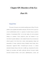
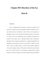
The graft may be in the form of a split-thick-
ness skin graft or a full-thickness skin graft. A
split-thickness skin graft contains epidermis
and a certain amount of dermis, while a full-
thickness skin graft contains epidermis and the
whole dermis (Figs. 12.1, 12.2)
A full-thickness skin graft offers better pro-
tection from trauma. It does not contract as
much as a split-thickness skin graft and gener-
ally looks more natural after healing; thus, it is
often used for aesthetic reasons. However, a
full-thickness graft requires a well-vascular-
ized recipient bed. Because of this limitation, it
is not commonly used in cutaneous ulcers. On
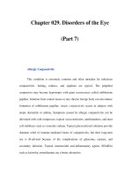
the other hand, a split-thickness skin graft re-
sults in a better ‘take’, even when applied to tis-
sue in which vascularization is not optimal and
relatively reduced (Fig. 12.3). This feature makes
it more appropriate for use in the management
of cutaneous ulcers.
The thicker the graft, the smaller the extent
of contraction of the grafted wound. Similarly,
wounds covered with thin split-thickness skin
grafts contract less than open wounds [15].
12.3 Preparing a Cutaneous Ulcer
for Grafting
Grafting should be done only onto a viable
wound surface. Prior to the application of the
skin graft, the ulcer bed should be debrided to
remove any necrotic tissue. Vital granulating
tissue should be exposed, thereby enabling cells
Chapter 12 Skin Grafting
160
12
Fig. 12.1. Histological representation of a full-thickness
skin graft
Fig. 12.2. Histological representation of a split-thickness
skin graft
Fig. 12.3. A split-thickness graft is placed on a cutaneous
ulcer. Longitudinal incisions in the graft were made in
order to facilitate drainage of secretions and prevent
their accumulation under the graft, which would pre-
vent its ‘taking’
12_159_164* 01.09.2004 14:03 Uhr Seite 160
in the graft to attach to the ulcer’s surface and
its blood supply. Note that the presence of more
than 10
5
bacteria per gram of tissue should be
regarded as infection (see Chap. 10).
12.4 Forms of Autologous Grafting
A simple autograft, applied as a layer, whether
done with a dermatome, a scalpel, or a special
grafting knife, may provide appropriate biolog-
ical coverage. However, it must be remembered
that the harvesting of an autograft results in a
wound in the healthy donor skin, analogous to a
second-degree burn. The donor wound, apart
from being painful, may require a considerable
amount of effort and time to heal. Therefore,
several techniques have been developed to re-
duce the required surface area of the donor skin.
Techniques in use for applying autologous
grafts are:
5 Taking one sheet of grafted skin to
cover all the denuded area: A split-
thickness graft is harvested with a
dermatome; a full-thickness graft is
usually obtained using a small scal-
pel.
5 Small pieces: One way of decreasing
the required area of donor skin is to
apply smaller pieces of donor skin,
instead of one large sheet that cov-
ers the entire area of the ulcer.
These grafts are placed onto the ul-
cer bed at regular intervals, to allow
drainage of secretions.
5 Pinch grafting was documented as
early as 1869 by Reverdin [4, 16].
The skin is anesthetized, a small
portion is lifted up on the point of a
needle, and the top is cut off with a
scalpel. Pinch grafts should be of
full thickness, 3–5 mm in diameter.
The grafts are evenly placed on the
ulcer bed, with free spaces of 5 to 10
mm between each of the grafts.
Pinch grafting has been document-
ed several times in the past 20 years
as a possible treatment method for
chronic skin ulcers [17–23].
5 Punch grafts, obtained by using a
punch biopsy instrument, represent
another modification of full-thick-
ness autografting. This procedure
enables a smaller area of donor skin
to be used, assuming that epithelial-
ization will take place and advance
peripherally from each punch. The
punch method is still used [24]. The
punch grafts, which may be 3–5 mm
in diameter, are placed onto the
ulcer’s surface at regular intervals.
5 Mesh grafting (Fig. 12.4): A mechan-
ical device is used to cut multiple
slits in the graft, thereby allowing it
to be stretched, so that it can ex-
pand and cover a larger surface ar-
ea. This procedure is commonly
used for burns, where large areas of
donor grafts may be needed, but not
for cutaneous ulcers.
Ahnlide and Bjellerup [17] used pinch grafting
for 145 therapy-resistant leg ulcers. Three
months following the procedure, the average
healing rate was 36%. Poskitt et al. [23] present-
ed a randomized trial comparing autologous
pinch grafting (25 patients) with porcine der-
12.4Forms of Autologous Grafting
161
t
t
Fig. 12.4. Mesh grafting
12_159_164* 01.09.2004 14:03 Uhr Seite 161
mis dressings (28 patients). Sixty-four percent
(64%) of ulcers treated by autologous pinch
grafting were healed at six weeks and 74% by 12
weeks, compared with ulcers treated by porcine
dermis, where healing rates were 29% and 46%,
respectively, after 6 and 12 weeks.
While pinch grafting and punch grafting are
usually intended for relatively small ulcers,
some suggest that mesh grafting may be used
for larger ulcers. Kirsner et al. [9] documented
29 patients with 36 leg ulcers of various etiology,
treated by meshed split-thickness skin grafts.
The grafts were harvested with a Padget derma-
tome and expanded through meshing to one
and a half times their original size. The initial ‘-
take’ of the grafts was recorded as ‘excellent’. At
a mean follow-up of 11 months (three months to
three years) 52% of ulcers were healed.
The information above covers simple auto-
grafts. More advanced forms such as cultured
keratinocyte grafting and tissue engineering
are discussed in Chap. 13.
12.5 Conclusion
In a comprehensive Cochrane review, Jones and
Nelson [6] suggest that further research is need-
ed to compare the beneficial effects of ‘simple’
skin grafting with those of other modes of treat-
ment intended for venous leg ulcers. This con-
clusion may actually be implemented for other
types of cutaneous ulcers as well.
The ‘take’ of the graft and the final result de-
pend on the ulcer’s condition in terms of vascu-
larization, absence of infection, and appropri-
ate preparation of the ulcer bed, as well as on
the patient’s general condition.
In our experience, a skin graft may provide
suitable coverage for a cutaneous ulcer, result-
ing in healing. However, in some cases, the graft
does not ‘take’well for the same reasons that re-
sulted in the ulceration in the first place (e.g.,
poor vascularization) and the ulcer does not
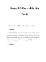
heal. Moreover, even in cases where closure of a
cutaneous ulcer is achieved by skin grafting,
the final clinical result is not satisfactory – in
most cases because there is no adequate prolife-
ration of granulation tissue (Fig. 12.5). The orig-
inal ulcer site usually remains as a depression
in the skin, with inadequate subcutaneous tis-
sue covered by a thin, very vulnerable cutane-
ous layer. Hence, autologous skin grafting of
cutaneous ulcers is commonly followed by re-
ulceration.
In view of the above, advanced modalities
such as keratinocyte grafting, composite grafts,
or preparations containing growth factors,
which may stimulate proliferation of granula-
tion tissue, may be used (see Chaps. 13, 14, and
15). The use of advanced modalities (e.g.,
growth factors) may indeed result in complete
healing of a treated ulcer, even without skin
grafting. However, in many cases this stimulus
will not suffice for healing and closure – espe-
cially with relatively large chronic ulcers. It may
well be that the solution to the problem of cer-
tain ulcers will lie in a combination of such ad-
vanced modalities together with skin grafting.
Chapter 12 Skin Grafting
162
12
Fig. 12.5. Cutaneous ulcers following grafting and par-
tial (b) and complete (a) healing. Note that the area is
slightly depressed due to decreased production of gran-
ulation tissue during active stages of healing
12_159_164* 01.09.2004 14:03 Uhr Seite 162
References
1. Ratner D: Skin grafting. From here to there. Derma-
tol Clin 1998; 16: 75–90
2. Hauben DJ, Baruchin A, Mahler D: On the history of
the free skin graft. Ann Plast Surg 1982; 9: 242–245
3. Kelton PL: Skin grafts and skin substitutes. Selected
Readings in Plastic Surgery 1982; 9:1–23
4. Reverdin JL: Greffe epidermique, experience faite
dans le service de monsieur le docteur Guyon, a
l’Hopital Necker. Bull Imp Soc Chir Paris 1869; 10:
511–515
5. Padgett EC: Skin grafting in severe burns.Am J Surg
1939; 43: 626
6. Jones JE, Nelson EA: Skin Grafting for venous leg ul-
cers (Cochrane Review). The Cochrane Library, is-
sue 4. 2000; Oxford: Update Software
7. Fisher JC: Skin grafting. In: Georgiade GS, Riefkohl
R, Levin LS (eds): Plastic, Maxillofacial and Recon-
structive Surgery. 3rd edn. Baltimore: Williams &
Wilkins. 1996; pp 13–18
8. Kirsner RS, Eaglstein WH,Kerdel FA: Split-thickness
skin grafting for lower extremity ulcerations. Der-
matol Surg 1997; 23 :85–91
9. Kirsner RS, Mata SM, Falanga V, et al: Split-thickness
skin grafting of leg ulcers. Dermatol Surg 1995; 21 :
701–703
10. Berretty PJ, Neumann HA, Janssen de Limpens AM,
et al: Treatment of ulcers on legs from venous hyper-
tension by split-thickness skin grafts. J Dermatol
Surg Oncol 1979; 5 :966–970
11. Michaelides P, Camisa C: The treatment of ulcers on
legs with split- thickness skin grafts : report of a
simple technique. J Dermatol Surg Oncol 1979; 5 :
961–965
12. Van den Hoogenband HM: Treatment of leg ulcers
with split-thickness skin grafts. J Dermatol Surg On-
col 1984; 10 : 605–608
13. Harrison PV: Split-skin grafting of varicose leg ul-
cers: a survey and the importance of assessment of
risk factors in predicting outcome from the proce-
dure. Clin Exp Dermatol 1988; 13 : 4–6
14. Ruffieux P, Hommel L, Saurat JH: Long-term assess-
ment of chronic leg ulcer treatment by autologous
skin grafts. Dermatology 1997; 195:77–80
15. Rudolph R: The effect of skin graft preparation on
wound contraction. Surg Gynecol Obstet 1976; 142:
49–56
16. Reverdin JL: Sur la greffe epidermique. Arch Gen
Med Paris 1872; 19 : 276–303
17. Ahnlide I, Bjellerup M: Efficacy of pinch grafting in
leg ulcers of different aetiologies. Acta Derm Vener-
eol 1997; 77 : 144–145
18. Steele K: Pinch grafting for chronic venous leg ulcers
in general practice. J R Coll Gen Pract 1985; 35 :
574–575
19. Christiansen J, Ek L, Tegner E: Pinch grafting of leg
ulcers. A retrospective study of 412 treated ulcers in
146 patients. Acta Derm Venereol (Stockh) 1997; 77 :
471–473
20. Millard LG, Roberts MM, Gatecliffe M: Chronic leg
ulcers treated by the pinch graft method. Br J Der-
matol 1977; 97 : 289–295
21. Oien RF, Hansen BU,Hakansson A: Pinch grafting of
leg ulcers in primary care. Acta Derm Venereol
(Stockh) 1998; 78 : 438–439
22. Ceilley RI, Rinek MA, Zuehlke RL: Pinch grafting for
chronic ulcers on lower extremities. J Dermatol Surg
Oncol 1977; 3 : 303–309
23. Poskitt KR, James AH, Lloyd-Davies ER, et al: Pinch
skin grafting or porcine dermis in venous ulcers: a
randomised clinical trial. Br Med J 1987; 294 :
674–676
24. Mol MA, Nanninga PB, Van Eendenburg JP, et al:
Grafting of venous leg ulcers. An intraindividual
comparison between cultured skin equivalents and
full-thickness skin punch grafts. J Am Acad Derma-
tol 1991; 24:77–82
References
163
12_159_164* 01.09.2004 14:03 Uhr Seite 163
13.1 Overview
The accepted term for skin substitutes that are
originally derived from living tissues, whether
they contain living cells when applied to the
wound surface or not,is ‘biological dressings’.In
view of the limitations of skin grafting, as dis-
cussed in the previous chapter, a variety of sub-
stitutes have been developed. We distinguish
herein between ‘non-living’ substitutes, which
do not contain living cells, and those containing
living cells, termed ‘living’ skin substitutes.
The products described below may be con-
sidered ‘tissue-engineered’ skin, according to
the accepted definition, namely, skin products
composed mainly of cells, skin products com-
posed of extracellular matrix materials, or a
combination of both [1, 2]. We shall focus our
discussion mainly on skin products intended to
be used for skin ulcers.
Skin Substitutes and Tissue-Engineered
Skin Equivalents
13
Contents
13.1 Overview 165
13.2 ‘Non-Living’ Skin Substitutes 165
13.2.1 General Functions 165
13.2.2 Allogeneic Cadaver Skin 165
13.2.3 Xenografts 166
13.2.4 Naturally Occurring Collagen Matrix
and Collagen-Containing Dressings 166
13.2.5 Conclusion 168
13.3 ‘Living’ Skin Substitutes 168
13.3.1 General 168
13.3.2 Epidermal: Keratinocyte Grafts 169
13.3.3 Dermal Grafting 172
13.3.4 Composite Grafts 172
13.4 Summary 173
References 174
13.2 ‘Non-Living’ Skin Substitutes
13.2.1 General Functions
Under the heading ‘non-living skin substitutes’
we include biological dressings originally de-
rived from living tissues, but which, when ap-
plied to denuded cutaneous areas, do not con-
tain living cells (see Table 13.1). A variety of
non-living skin substitutes have been used in
the treatment of burns and surgical wounds.
Non-living skin substitutes function as highly
effective biological dressings. They fulfill the
main purposes of an optimal dressing, i.e., pro-
vision of a moist environment, prevention of
water loss (indeed, dermal skin substitutes
were primarily developed for the treatment of
burns), and protection against external infec-
tions or trauma. Their use is usually accompa-
nied by significant pain reduction.
Moreover, several research studies suggest
that a layer of acellular dermis may serve as a
template for the regeneration of a viable dermis
[3–5]. Several types, described below, have been
discussed in the literature. They have been pro-
posed for use mainly in surgical wounds or
burns and their efficacy in cutaneous ulcers has
yet to be validated. Some are in current clinical
use and some are still under research.
13.2.2 Allogeneic Cadaver Skin
Allogeneic cadaver skin may be used as a bio-
logical dressing. Devitalization of the allograft
obviates its antigenic effect. The graft can be
preserved by various techniques, such as cryo-
preservation with glycerol [6] or by freeze-dry-
ing [7]. Another possibility is to produce an
13_165_176 01.09.2004 14:04 Uhr Seite 165
acellular dermal matrix by the removal of the
epidermis and the cells in the dermis [3]. Cha-
karbarty et al. [8] used ethylene oxide steriliza-
tion followed by immersion of the skin in
1 mol/l sodium chloride for eight h to achieve
acellularization. The current clinical use of
cryopreserved or acellular allografts is mainly
in the management of burn wounds [9–12].
There are few reports on the use of allogene-
ic cadaver skin substitutes as an option for the
treatment of chronic cutaneous ulcers. In 1999,
Snyder et al. [13] documented treatment with
cadaveric allografts in 27 patients with 34 leg
ulcers of various etiologies. In 65% of patients
the ulcers healed by secondary intention, and
the average healing time was 113.9 days.
13.2.3 Xenografts
The most common source of xenografts (syn.:
heterografts or zoografts) is porcine skin, since
it is similar to human skin. Sterility is achieved
by irradiation [14]; thus, the graft is actually
non-living. The use of xenografts is well docu-
mented for burns,surgical wounds,and cutane-
ous ulcers [15–22]. The products defined as nat-
urally occurring collagen matrix, described be-
low, are actually processed xenografts.
13.2.4 Naturally Occurring
Collagen Matrix and
Collagen-Containing Dressings
Collagen has unique biologic and physical
properties that, with appropriate processing
and manufacturing, make it an ideal dressing
product. Collagen is found in abundance in
supporting tissues such as skin, fascia, tendons,
and bones. Its structure is organized and
aligned, and it forms strong fibers [23].
Certain scientific observations lend further
support to the medical use of collagen in the
management of wounds and wound healing:
5 A collagen matrix may serve as a
skeleton or scaffolding on which the
new tissue gradually forms [24, 25].
5 It has been suggested that attach-
ment of fibroblasts to the implanted
collagen enhances new collagen
synthesis in the healing wound [26].
5 A collagen matrix can efficiently
provide the basic requirements of a
dressing. Being tough in texture, it
protects the ulcer and its surround-
ings from mechanical trauma. Its
Chapter 13 Skin Substitutes and Tissue-Engineered Skin
166
13
Table 13.1. Non-living skin substitutes
Non-living skin substitutes Commercial products Comments
Products processed from AlloDerm® Cadaver skin and xenografts are not widely
fresh cadaver skin used by commercial companies, but rather
by laboratories and skin banks in medical
centers
Naturally occurring CollatamFascie® These products may be regarded as modifica-
collagen matrix E-Z-derm® tions of xenografts (manufactured from
(processed xenografts) Integra® bovine or porcine tissue)
Oasis®
SkinTemp®
Synthetic collagen Biobrane®
dressings Fibracol Plus®
Promogran® Promogran also contains oxidized regenerated
cellulose to neutralize the matrix metallo-
proteinases in the ulcer bed
t
13_165_176 01.09.2004 14:04 Uhr Seite 166
permeability may vary depending
on its method of manufacture, but
in most cases it can provide a moist
environment, which is desirable for
the healing process.
The following discussion distinguishes between
two forms of collagen dressings. The first re-
lates to the use of collagen in its native form,
i.e., as a naturally occurring collagen matrix,
while the second relates to synthetic dressings
that contain collagen.
13.2.4.1 Naturally Occurring Collagen
Matrix
The use of natural collagen matrix represents a
relatively advanced modification of the biolog-
ical dressing. In practice, the products de-
scribed below are sheets of xenografts (porcine
or bovine) that have been processed to make
them suitable for use on denuded skin areas.
There are several advantages to such prod-
ucts:
5 The collagen is in its natural form,
which preserves the normal struc-
ture and alignment of the fibers (as
opposed to dressings containing
collagen, which have undergone
more complex processing). The
manufacturers claim that the for-
mer product acts as a more natural
scaffolding, which ‘takes’ better to
the wound surface.
5 Some of these products may contain
growth factors that signal host cells
within the wound bed to attach and
proliferate on the collagen template
[27].
In this group of biological dressings, more ad-
vanced modifications have been developed us-
ing non-living skin substitutes, whose structure
is that of a cross-linked matrix of collagen. A
graft composed of bovine collagen matrix with
chondroitin-6 sulfate covered by a silicone layer
(Integra®), has been shown to have a beneficial
effect on burn wounds at an initial stage; a
meshed autograft is applied a few weeks later
[9, 25]. Another product, composed of lyophi-
lized type-1 bovine collagen (SkinTemp®), has
been used for surgical wound healing by secon-
dary intention [28]. Bovine collagen products
are thought to form a network-like architectu-
ral structure, which enhances organization of
fibroblasts and newly forming collagen bun-
dles. Degradation products of bovine collagen
are considered to act as chemotactic factors,
which further enhance wound repair processes
[29, 30]. Another product (CollatamFascie®) is
a type-1 collagen derived from bovine Achilles
tendons, which has been shown to enhance
healing in acute wounds and chronic cutaneous
ulcers [14].
An acellular collagen matrix derived from
porcine small intestine (Oasis®) has been intro-
duced as a substitute skin covering [31, 32]. The
intestine is processed to remove the serosal,
smooth-muscle, and mucosal layers while re-
taining the submucosa. The final product is an
acellular, collagenous sheet. Brown-Etris et al.
[33] reported on 15 patients with leg ulcers
treated with acellular matrix derived from por-
cine small intestinal submucosa. Wound clo-
sure was reported in seven patients within
4–10 weeks.
E-Z-derm® is another biosynthetic acellular
dressing made of xenogeneic collagen matrix. It
is made of porcine collagen that has been
chemically cross-linked with an aldehyde. This
process is claimed to impart durability to the
product, enabling its storage at room tempera-
ture.
13.2.4.2 Collagen-Containing
Dressings
In dressings containing collagen, the collagen
has undergone more complex processing (as
compared with naturally occurring collagen),
so that in essence the product is synthetic. In
13.2‘Non-Living’ Skin Substitutes
167
t
t
13_165_176 01.09.2004 14:04 Uhr Seite 167
spite of the theoretical advantages of using col-
lagen as a dressing, results of initial experi-
ments using collagen dressings were disap-
pointing [34–36].
Other early developments of dressing mate-
rials containing collagen peptides were lami-
nates such as Biobrane®. This consisted of a
laminate of silicone rubber with nylon, linked
with porcine collagen peptides [37]. Biobrane®
was found to be an effective dressing material
that provided adequate covering for surgical or
burn wounds [38–40].
Another collagen-containing dressing, Fib-
racol®, was relatively effective in the treatment
of diabetic foot ulcers when compared with
regular gauze moistened with normal saline
[41], and in the treatment of pressure ulcers,
compared with calcium-sodium alginate dress-
ings [42].
Recently, a new collagen-containing dressing
was introduced (Promogran®). In addition to
bovine collagen (55%), with its above-men-
tioned advantages, Promogran® also contains
oxidized regenerated cellulose (45%). The latter
is said to bind (and thereby neutralize) matrix
metalloproteinases in the ulcer bed, thus obvi-
ating their proteolytic effects on the growth
factors and matrix proteins (Fig. 13.1). Ghatnek-
ar et al. [43] found that Promogran®, combined
with good wound care, was more cost-effective
than good wound care alone in the treatment of
diabetic foot ulcers. Veves et al. [44] conducted
a randomized, controlled trial comparing Pro-
mogran® and standard treatment with a mois-
tened gauze in the management of diabetic foot
ulcers. The results, after 12 weeks of treatment,
were not statistically significant. However,
among 95 patients with ulcers of less than six
months’ duration, 43 (45%) of those treated
with Promogran® underwent healing, com-
pared with 29 (33%) of those treated by mois-
tened gauze. The beneficial effect of Promogran
on venous leg ulcers has also been demonstrat-
ed by Vin et al. [45].
13.2.5 Conclusion
At present, non-living skin substitutes are con-
sidered to function as efficient biological dress-
ings for cutaneous ulcers. However, the overall
impression is that they do not actively stimulate
or enhance wound healing, as do living substi-
tutes.
Whether non-living skin replacements con-
tribute further to wound healing, as compared
with synthetic dressings, is questionable. Some
investigators suggest, as mentioned above, that
the acellular dermis may serve as a template for
dermal regeneration. Some of these non-living
substitutes are said to contain cytokines [27],
which may render them more effective than
synthetic dressings. Nevertheless, controlled
research studies must be undertaken on the
various types of non-living skin substitutes be-
fore this assumption can be confirmed. By the
same token, further studies are needed in order
to obtain a more accurate evaluation of the effi-
cacy of newer combinations of collagen-con-
taining dressings.
13.3 ‘Living’ Skin Substitutes
13.3.1 General
Recently, more sophisticated techniques have
been developed in an attempt to create skin
equivalents and to re-establish the appropriate
physiological microenvironment needed for
optimal wound repair. In the following discus-
sion, distinctions should be made between sub-
Chapter 13 Skin Substitutes and Tissue-Engineered Skin
168
13
Fig. 13.1. Application of a collagen-containing dressing
(Promogran) to a cutaneous ulcer
13_165_176 01.09.2004 14:04 Uhr Seite 168
types of living equivalents, i.e., those containing
epidermal components, those containing der-
mal components,and composite grafts contain-
ing both dermal and epidermal components.
13.3.2 Epidermal: Keratinocyte Grafts
The use of viable epidermal cells is thought to
contribute to the wound repair process by ac-
tively stimulating the ulcer to heal. In 1975,
Rheinwald and Green [46] described a method
for culturing keratinocytes from single-cell
suspensions of human epidermal cells. Their
breakthrough opened new vistas in the field of
13.3‘Living’ Skin Substitutes
169
Fig. 13.2. Graft pad containing cultured keratinocytes in
a Petri dish
Fig. 13.3. Trimming a graft pad of cultured keratinocytes
to the appropriate size preparatory to the grafting
Fig. 13.4. Placing the graft on the ulcer
Fig. 13.5. Placing a gauze pad, moistened with saline, on
the graft pad
Fig. 13.6. Placing additional gauze pads to protect the
graft
13_165_176 01.09.2004 14:04 Uhr Seite 169
skin research and led to clinical applications in-
volving cultured keratinocyte grafting (Fig.
13.2–13.6). At present, it is possible to produce
multi-layered stratified skin equivalents that
very closely resemble natural skin.
Initially,keratinocyte grafts were autologous
grafts derived from the patient’s skin on burn
wounds [47–54]. They were later introduced in
the management of cutaneous leg ulcers [55,
56]. At a still later stage, the method was modi-
fied by using allogeneic cultured keratinocyte
grafts, derived from the foreskins of newborns
[57–62]. The first group of leg ulcer patients
treated by allogeneic grafts was described by
Leigh et al. [58] in 1987, comprising 51 patients
with 70 ulcers of various etiologies. The mean
duration of the ulcers prior to the grafting pro-
cedure was 16 years. Beneficial effects were
manifested either by the appearance of islands
of epithelium on the ulcer bed (in 29% of the
ulcers) or by enhanced migration of epithelium
from the periphery of the ulcer (seen in 44% of
the ulcers).
Since then, other studies have shown that
topically applied allogeneic keratinocyte grafts
accelerate the healing of chronic cutaneous ul-
cers, with similar findings: significant reduc-
tion in the ulcer area within three weeks follow-
ing grafting, and complete healing of 65–83% of
treated ulcers within a few weeks. In most of the
ulcers, even those that did not undergo com-
plete healing, granulation tissue formed (with a
subsequent decrease in ulcer depth), with en-
hanced epithelialization and reduction in the
ulcer surface area [59–62].
The main advantage of allogeneic keratino-
cyte grafts compared with autologous keratino-
cyte grafts lies in the fact that newborn kerati-
nocytes are thought to be more effective in pro-
moting healing. This effect is attributed mainly
to the increased secretion of growth factors by
younger keratinocytes compared with adult
cells (see below) [58, 59, 62–65]. Having said
this, it is our experience that even autologous
keratinocyte grafts will, in many cases,promote
wound healing, as shown by the proliferation of
granulation tissue in the ulcer bed and advanc-
ing epithelialization.
While neonatal keratinocytes can be pre-
pared from newborn foreskins, refrigerated and
used immediately for grafting when needed,
the preparation of an autologous keratinocyte
graft is a more complex procedure that requires
either a biopsy specimen from the patient’s skin
or a sample of his/her hair follicles, obtained by
hair plucking [66, 67]. It takes a period of 2–3
weeks before there is sufficient epithelium to
cover the ulcer surface area. This process is car-
ried out either in specialized medical centers or
by commercial companies that prepare the au-
tologous grafts for grafting from samples of the
patient’s skin or hair follicles (see Table 13.2).
Mechanism of Healing. In view of the fact
that patients with cutaneous ulcers report sig-
nificant pain relief within a few hours of kera-
tinocyte grafting, it seems that the graft func-
tions as a semi-occlusive dressing that prevents
dehydration and reduces pain. However, an al-
logeneic graft does not serve as a permanent
Chapter 13 Skin Substitutes and Tissue-Engineered Skin
170
13
Table 13.2. Living skin substitutes
Living skin substitutes Commercial product Comments
Keratinocyte grafts BioSeed® Autologous-obtained from skin biopsy
Epibase® Autologous-obtained from skin biopsy
Epicel® Autologous-obtained from skin biopsy
Epidex® Autologous-obtained by plucking hair
Dermal grafting Dermagraft®
Transcyte®
Composite grafting Apligraf®
OrCel®
13_165_176 01.09.2004 14:04 Uhr Seite 170
alternative cover over the ulcer, since it remains
on the ulcer bed for only a short period. After a
few hours, rejection of the graft occurs. The
graft is then replaced by autologous cells, as
demonstrated by DNA fingerprinting and the
disappearance of Y chromosomes in sex-
matched grafts [51].
Accumulating evidence suggests that the
main factor in wound healing using allogeneic
keratinocyte grafts lies in the presence of solu-
ble growth factors produced by transplanted
newborn keratinocytes [58, 59, 62–65]. The ef-
fect of growth factors is manifested clinically by
the formation of healthy granulation tissue on
the ulcer bed and the gradual epithelialization
of its surface.
Beneficial Effect. Ideally, in order to assess
the efficacy of keratinocyte grafting, one would
require patients with symmetrically located
(i.e., one on each leg) cutaneous ulcers; one ul-
cer would be grafted while the other would be
treated by placebo in a controlled, double-blind
procedure.
In most of the reported studies, this proce-
dure has not been compared with an alternative
therapeutic modality. Moreover, some of the
studies contain methodological errors. Jones
and Nelson [65] have indicated that, not infre-
quently, research studies have failed to present
baseline data such as a history of previous ul-
ceration, which may affect the prognosis. How-
ever, the overall impression in the medical liter-
ature nowadays is that allogeneic keratinocyte
grafting, containing an epithelial component of
living keratinocytes does, in fact, enhance
wound healing in chronic cutaneous ulcers.
Recently, Bolivar-Flores and Kuri-Harcuch
[68] documented complete healing in 10 pa-
tients with recalcitrant leg ulcers of various eti-
ologies, using frozen allogeneic keratinocyte
grafting.
Three research studies have also compared
cultured keratinocyte allografts to control dress-
ings [69–71]. Teepe et al. [69] compared the ben-
eficial effect of cryopreserved allogeneic keratin-
ocyte grafting vs. hydrocolloid dressings in 43
patients with 47 cutaneous ulcers. They demon-
strated enhanced healing rates and increased re-
duction of ulcer size in the grafted group. How-
ever, there was no difference between the two
groups in the number of healed ulcers at six
weeks.
Lindgren et al. [70] did not demonstrate
superiority of cryopreserved allogeneic kera-
tinocyte grafting in 15 patients with chronic ve-
nous leg ulcers compared with 12 patients in a
control group. The authors suggested that this
observation (contradicting their previous ex-
periments using fresh keratinocytes) might be
attributed to the effect of cryopreservation.
Jones and Nelson [65] summarized the data
of the relevant trials in a Cochrane review, and
found no demonstrable benefit of allogeneic
keratinocyte grafting over control dressings.
In our clinical experience, keratinocyte
grafting does provide some degree of improve-
ment in most cases, even in ulcers that do not
heal completely. Improvement is manifested by
granulation tissue formation, epithelialization
advancing from the ulcer margin, and a signifi-
cant reduction in the ulcer surface area.
In relatively small ulcers of up to 2–3 cm in
diameter, the use of allogeneic cultured kerati-
nocytes can provide the ‘push’ needed to pro-
mote complete healing and closure of the le-
sion.On the other hand, in larger ulcers it is un-
reasonable, in most cases, to expect complete
healing following allogeneic keratinocyte graft-
ing. In this particular situation, one may con-
sider using repeated combinations of allogene-
ic keratinocyte grafts with autologous grafts
taken from the patient himself. Alternatively,
one could consider using other advanced mo-
dalities, discussed elsewhere in this book (see
Chap. 20).
We have found that allogeneic keratinocyte
grafting is preferable to surgical autologous
split- or full-thickness skin grafts, since the for-
mer induces granulation tissue formation,
which fills the wound cavity and reduces its
depth.
Nowadays, cultured keratinocyte grafting is
commercially available and is performed in
many medical centers (mainly burn centers).
These products are also produced and market-
ed by commercial companies (See Table 13.2).
13.3‘Living’ Skin Substitutes
171
13_165_176 01.09.2004 14:04 Uhr Seite 171
13.3.3 Dermal Grafting
Viable dermal substitutes may be considered a
step beyond non-viable forms. However, to the
best of our knowledge, there are no document-
ed studies comparing the effectiveness of the
viable dermal substitute with non-viable der-
mis.
Dermagraft® is a viable dermal substitute
which consists of a bioabsorbable mesh, upon
which cryopreserved fibroblasts derived from
neonatal foreskins are cultured.The cultured fi-
broblasts are screened for the presence of infec-
tious organisms.
The neonatal fibroblasts proliferate within
the polymer mesh, and secrete dermal matrix
proteins (collagens, tenascin, vitronectin) and
growth factors [72, 73]. Once the graft has been
placed on the wound, it may provide an extra-
cellular matrix and growth factors, synthesized
by the transplanted fibroblasts. The mesh con-
sists of biodegradable material which dissolves
gradually within 3–4 weeks.As previously men-
tioned, following dermal grafting (provided the
ulcer is ready), keratinocyte grafts can be
placed on the fibroblast-containing dermal
bed.
Each unit of Dermagraft® is supplied frozen.
It is packaged in a clear bag, within a saline-
based cryoprotectant, containing 10% dime-
thylsulfoxide and bovine serum. A Dermagraft
piece, measuring 5 × 7.5 cm, is intended for sin-
gle-use application.
Studies on diabetic foot ulcers treated with
this dermal graft have shown improved healing
compared with controls [9, 74–77]. Recently,
Marston et al. [78] conducted a randomized,
controlled, single-blind, multi-centered study,
enrolling 314 patients suffering from chronic
diabetic foot ulcers. The efficacy of Derma-
graft® (130 patients) was compared with that of
a moist saline gauze covering (115 patients).
Both groups received standard care such as
pressure-reducing footwear.Following 12 weeks
of treatment, complete wound closure was
achieved in 30% of patients treated with Der-
magraft®, compared with 18.3% of patients in
the control group.
Another unique randomized, controlled,sin-
gle-blind, clinical trial was introduced in 1996
by Gentzkow et al. [74]. The rate of complete
healing of diabetic foot ulcers was higher in pa-
tients treated with Dermagraft® than in pa-
tients in the control group.The article also eval-
uated the desirable frequency of Dermagraft®
application, suggesting that a weekly applica-
tion is better than less frequent applications.
Transcyte® is a product made of neonatal fi-
broblasts. The cells are cultured on nylon mesh
for 4–6 weeks. The product eventually consists
of dense cellular tissue (with human matrix
proteins) and contains high levels of growth
factors. Transcyte® is supplied in a cassette con-
taining two transparent units, each measuring
13 × 19 cm. The product is intended for burn
wounds. To date, the use of Transcyte® on
chronic cutaneous ulcers has not been reported.
13.3.4 Composite Grafts
Composite grafts represent a combination of
dermal substitute and epidermal grafts. Re-
search has shown that epidermal grafts are
more beneficial when applied to a dermal bed.
We distinguish here between composite grafts
containing devitalized dermis and those with
viable dermis in which fibroblasts are embed-
ded.
Composite Grafts Combining Epidermal
Cells with Devitalized Dermis. As described
above, a dermal matrix (even acellular dermis)
may function as a template for cutaneous re-
generation [3, 4]. The addition of allogeneic or
autologous cultured keratinocytes may further
contribute to healing. Some suggest that the
dermal component protects the basal layer of
the epidermis and affects the biological pro-
cesses of epithelial proliferation, differentia-
tion, migration, and wound healing.
In practice, following the absorption and
‘take’ of acellular dermal grafts such as those
mentioned earlier in this chapter (Integra®,
SkinTemp®, CollatamFascie®), cultured kerati-
nocytes may be seeded onto these dermal
grafts, approximately two weeks after the der-
mal graft has been applied. However, the benefi-
Chapter 13 Skin Substitutes and Tissue-Engineered Skin
172
13
13_165_176 01.09.2004 14:04 Uhr Seite 172
cial effect of devitalized dermis with epidermal
cells is not well documented. More advanced
methods, combining dermal components with
living fibroblasts, together with cultured kerati-
nocytes, tend to yield better results (see below).
Cutaneous Ulcers Treated by Composite
Grafts with Viable Dermis. The incorporation of
viable fibroblasts improves the epidermal re-
sponse of the grafted skin [78–83]. Most studies
described below, using composite grafts, in-
volved improvised products. Some of the com-
posite grafts used were ephemeral and did not
manage to penetrate the current pharmaceutical
market, while others became the prototype for
the composite grafts currently being marketed.
While the general impression of the prod-
ucts discussed in this section is positive, there
are insufficient data to assess their efficacy.
There are more controlled studies and data re-
lated to those in current use (i.e.,Apligraf®), de-
scribed in Chap. 14.
Following are studies on cutaneous ulcers
implementing the use of composite grafts
that are not commercially available:
5 Mol et al. [84] compared the efficacy
of punch grafts with that of com-
posite grafts (allogeneic fibroblasts
covered by autologous cultured ke-
ratinocytes), with both types being
applied to the ulcer site in the same
patient. The median healing time of
ulcers covered by composite grafts
was 18 days whereas that of ulcers
treated by punch grafts was 15 days.
5 Bovine collagen matrix used by
Burke et al. [25], as described above,
was modified by the incorporation
of fibroblasts into the collagen-gly-
cosaminoglycan substrate and by
replacing the silastic layer with hu-
man keratinocytes. Clinical efficacy
was documented in burns [85] and
chronic cutaneous ulcers [86].
5 Harvima et al. [87] conducted an
open study comparing the effect of
cultured allogeneic keratinocytes
with that of fibroblast-gelatin
sponge placed on the ulcer bed once
weekly. This study involved 22 dia-
betic patients with chronic ulcers
and 16 patients with leg or ankle ul-
cers of different etiologies. From the
diabetic group, four randomly se-
lected patients (five ulcers) received
combined treatment. The patients
were first treated by fibroblast-gela-
tin sponge, up to the point where
the ulcer area was reduced by 50%.
These ulcers were then treated with
keratinocyte cultures. In general, the
treatment was effective. All but one
diabetic ulcer healed. Nevertheless,
the investigators did not find a sig-
nificant difference between the cul-
tured keratinocyte treatment alone
and the combined treatment.
13.4 Summary
There is a wide range of skin substitutes that
can be used in the treatment of chronic cutane-
ous ulcers. From the current information avail-
able, it is difficult to evaluate precisely the effi-
cacy of each of the methods reviewed in this
chapter. Some studies lack basic data about the
patients, such as the presence of ulceration in
the past, or information regarding the clinical
appearance of the ulcer. Other studies involved
an insufficient number of patients to draw sig-
nificant conclusions,while others were not con-
trolled.
Nevertheless, the general impression that
arises from these studies is that in the treat-
ment of cutaneous ulcers, skin substitutes con-
taining living cells are likely to be superior to
skin substitutes that do not contain living cells.
This being the case, advanced treatment mo-
dalities such as keratinocyte cultures or com-
posite grafts could present a reasonable treat-
ment option when dealing with clean ulcers
with relatively healthy granulation tissue.
13.4Summary
173
t
t
13_165_176 01.09.2004 14:04 Uhr Seite 173
References
1. Bello YM, Falabella AF, Eaglstein WH: Tissue-Engi-
neered Skin. Current status in wound healing. Am J
Clin Dermatol 2001; 2:305–313
2. Eaglstein WH, Falanga V: Tissue engineering for
skin: an update. J Am Acad Dermatol 1998; 39:
1007–1010
3. Wainwright DJ: Use of an acellular allograft dermal
matrix (AlloDerm) in the management of full thick-
ness burns. Potential as a template for the recon-
struction of viable dermis. Burns 1995; 21:243–248
4. Livesey SA, Herndon DN, Hollyoak MA, et al: Trans-
planted acellular allograft dermal matrix. Trans-
plantation 1995; 60:1–9
5. Gustafson CJ, Kratz G: Cultured autologous kerati-
nocytes on a cell-free dermis in the treatment of
full-thickness wounds. Burns 1999; 25:331–335
6. Hussmann J, Russell RC, Kucan JO, et al: Use of gly-
cerolized human allografts as temporary and per-
manent cover in adults and children. Burns 1994; 20
[Suppl 1] : S61–S66
7. Abbott WM, Hembree JS: Absence of antigenicity in
freeze-dried skin allografts. Cryobiology 1970; 6:
416– 418
8. Chakrabarty KH, Dawson RA, Harris P, et al: Devel-
opment of autologous human dermal- epidermal
composites based on sterilized human allodermis
for clinical use. Br J Dermatol 1999; 141 : 811–823
9. Phillips TJ: New skin for old: Developments in bio-
logical skin substitutes. Arch Dermatol 1998; 134:
344–349
10. Herndon DN: Perspectives in the use of allograft. J
Burn Care Rehabil 1997; 18:S6
11. Greenleaf G, Hansbrough JF: Current trends in the
use of allograft skin for patients with burns and re-
flections on the future of skin banking in the United
States. J Burn Care Rehabil 1994; 15: 428–431
12. Balasubramani M, Kumar TR, Babu M: Skin substi-
tutes: a review. Burns 2001; 27 :534–544
13. Snyder RJ, Simonson DA: Cadaveric allograft as ad-
junct therapy for nonhealing ulcers. J Foot Ankle
Surg 1999; 38 : 93–101
14. Ruszczak Z, Schwartz RA: Modern aspects of wound
healing: an update. Dermatol Surg 2000; 26 : 219–229
15. Davis DA,Arpey CJ: Porcine heterografts in dermat-
ologic surgery and reconstruction. Dermatol Surg
2000; 26:76–80
16. Bromberg BE, Song IC: Homograts and heterografts
as skin substitutes.Am J Surg 1966; 112 : 28–33
17. Culliton P, Kwasnik RE, Novicki D, et al: The efficacy
of porcine skin grafts for treating non-healing cuta-
neous ulcers. Part I: clinical studies. J Am Podiatry
Assoc 1978; 68:1–10
18. Elliott RA Jr, Hoehn JG: Use of commercial porcine
skin for wound dressings. Plast Reconstr Surg 1973;
52 : 401–405
19. Ersek RA, Lorio J: The most indolent ulcers of the
skin treated with porcine xenografts and silver ions.
Surg Gynecol Obstet 1984; 158:431–436
20. Kaplan EG, Kaplan GS, Kaplan DM, et al: Superficial
ulcer treatment utilizing hyperbaric oxygen and
porcine skin grafts. J Foot Surg 1978; 17:144–149
21. Kudo K,Yokota M,Fujioka Y: Immediate reconstruc-
tion using a scalp- forehead flap for the entire upper
lip defect with the application of lyophilized porcine
skin to surgical wounds. A case report of a malig-
nant melanoma in the upper lip and oral mucosa. J
Maxillofac Surg 1983; 11:275–278
22. Poskitt KR, James AH, Lloyd- Davies ER, et al: Pinch
skin grafting or porcine dermis in venous ulcers: a
randomised clinical trial. Br Med J 1987; 294 :674–676
23. Piez KA: Structure and assembly of the native colla-
gen fibril. Connect Tissue Res 1982; 10 : 25–36
24. Dedhar S,Ruoslahti E, Pierschbacher MD: A cell sur-
face receptor complex for collagen type I recognizes
the Arg-Gly-Asp sequence. J Cell Biol 1987; 104 :
585–593
25. Burke JF, Yannas IV, Quinby WC Jr, et al: Successful
use of a physiologically acceptable artificial skin in
the treatment of extensive burn injury. Ann Surg
1981; 194:413–428
26. Postlethwaite AE, Seyer JM, Kang AH: Chemotactic
attraction of human fibroblasts to type I, II, and III
collagens and collagen-derived peptides. Proc Natl
Acad Sci USA 1978; 75: 871–875
27. Voytik-Harbin SL, Brightman AO, Kraine MR, et al:
Identification of extractable growth factors from
small intestinal submucosa. J Cell Biochem 1997; 67 :
478–491
28. Kolenik SA 3rd, McGovern TW, Leffell DJ: Use of a
lyophilized bovine collagen matrix in postoperative
wound healing. Dermatol Surg 1999; 25 :303–307
29. Motta G, Ratto GB, De Barbieri A, et al: Can heterol-
ogous collagen enhance the granulation tissue
growth? An experimental study. Ital J Surg Sci 1983;
13 : 101–108
30. Leipziger LS, Glushko V, DiBernardo B, et al: Dermal
wound repair: role of collagen matrix implants and
synthetic polymer dressings. J Am Acad Dermatol
1985; 12: 409–419
31. Hodde JP, Badylak SF, Brightman AO, et al: Glyco-
saminoglycan content of small intestinal submuco-
sa: A bioscaffold for tissue replacement. Tissue Engi-
neering 1996; 2 : 209–217
32. McPherson TB, Badylak SF: Characterization of Fi-
bronectin derived from porcine small intestinal
submucosa. Tissue Engineering 1998; 4 :75–83
33. Brown-Etris M, Punchello M, Shields D: Final report
on a pilot study to evaluate porcine small intestinal
submucosa as a covering for partial thickness
wounds. WOCN 32nd Annual Conference. June
2000; Toronto
34. Mian E, Martini P,Beconcini D,et al: Healing of open
skin surfaces with collagen foils. Int J Tissue React
1992; 14 [Suppl] :27–34
35. Lorenzetti OJ, Fortenberry B, Busby E, et al: Influ-
ence of microcrystalline collagen on wound healing.
I. Wound closure of normal excised and burn ex-
cised wounds of rats, rabbits and pigs. Proc Soc Exp
Biol Med 1972; 140 : 896–900
Chapter 13 Skin Substitutes and Tissue-Engineered Skin
174
13
13_165_176 01.09.2004 14:04 Uhr Seite 174
36. Chvapil M: Considerations on manufacturing prin-
ciples of a synthetic burn dressing: a review. J
Biomed Mater Res 1982; 16:245–263
37. Phillips TJ, Dover JS: Leg Ulcers. J Am Acad Derma-
tol 1991; 25: 965–987
38. Zapata- Sirvent R, Hansbrough JF, Carroll W, et al:
Comparison of Biobrane and Scarlet red dressings
for treatment of donor site wounds. Arch Surg 1985;
120 : 743–745
39. McHugh TP, Robson MC, Heggers JP, et al: Thera-
peutic efficacy of Biobrane in partial- and full-
thickness thermal injury.Surgery 1986; 100 : 661–664
40. Zachary L, Heggers JP, Robson MC, et al: The use of
topical antimicrobials combined with Biobrane in
burn wound infections. J Trauma 1982; 22:833–836
41. Donaghue VM, Chrzan JS, Rosenblum BI, et al: Eval-
uation of a collagen-alginate wound dressing in the
management of diabetic foot ulcers. Adv Wound
Care 1998; 11: 114–119
42. Alvarez O, Fahey C, Papantonio C, et al: Experience
with a collagen-alginate wound dressing in the
management of chronic venous ulcers. Symposium
on Advanced Wound Care and Medical research for-
um on Wound Repair. 1999
43. Ghatnekar O,Willis M, Persson U: Cost-effectiveness
of treating deep diabetic foot ulcers with Promog-
ran in four European countries. J Wound Care 2002;
11 : 70–74
44. Veves A, Sheehan P, Pham HT: A randomized, con-
trolled trial of Promogran (a collagen/oxidized re-
generated cellulose dressing) vs standard treatment
in the management of diabetic foot ulcers. Arch
Surg 2002; 137: 822–827
45. Vin F, Teot L, Meaume S: The healing properties of
Promogran in venous leg ulcers.J Wound Care 2002;
11 : 335–341
46. Rheinwald JG, Green H: Serial cultivation of strains
of human epidermal keratinocytes: The formation
of keratinizing colonies from single cells. Cell 1975;
6 : 331–343
47. O’Connor NE, Mulliken JB, Banks-Schlegel S, et al:
Grafting of burns with cultured epithelium pre-
pared from autologous epidermal cells. Lancet 1981;
1:75–78
48. Gallico GG 3rd, O’Connor NE, Compton CC, et al:
Permanent coverage of large burn wounds with au-
tologous cultured human epithelium. N Engl J Med
1984; 311:448–451
49. Teepe RG, Ponec M, Kreis RW, et al: Improved graft-
ing method for the treatment of burns with autolo-
gous cultured human epithelium (letter). Lancet
1986; 1:385
50. Teepe RG, Ponec JA, Kempenaar B: Clinical, histo-
logical and ultrastructural aspects of cultured epi-
thelium. In: Proceedings of the International Sym-
posium on the clinical Use of Cultured Epithelium
in Surgery and Dermatology. Leiden, March, 1987
51. De Luca M, Albanese E, Bondanza S, et al: Multicen-
tre experience in the treatment of burns with autol-
ogous and allogenic cultured epithelium, fresh or
preserved in a frozen state. Burns 1989; 15 : 303–309
52. Eldad A, Burt A, Clarke JA, et al: Cultured epithelium
as a skin substitute. Burns Incl Therm Inj 1987; 13:
173–180
53. Bettex-Galland M, Slongo T, Hunziker T, et al: Use of
cultured keratinocytes in the treatment of severe
burns. Z Kinderchir 1988; 43:224–228
54. Gao ZR, Hao ZQ, Nie LJ, et al: Coverage of full skin
thickness burns with allograft inoculated with au-
togenous epithelial cells. Burns Inc Therm Inj 1986;
12 : 220–224
55. Hefton JM, Caldwell D, Biozes DG, et al: Grafting of
skin ulcers with cultured autologous epidermal
cells. J Am Acad Dermatol 1986; 14 :399–405
56. Leigh IM, Purkis PE: Culture grafted leg ulcers. Clin
Exp Dermatol 1986; 11:650–652
57. Teepe RG, Keobrugge EJ, Ponec M, et al: Fresh ver-
sus cryopreserved cultured allografts for the treat-
ment of chronic skin ulcers. Br J Dermatol 1990; 122 :
81–89
58. Leigh IM,Purkis PE,Navsaria HA,et al: Treatment of
chronic venous ulcers with sheets of cultured allog-
eneic keratinocytes. Br J Dermatol 1987; 117:591–597
59. Phillips TJ, Gilchrest BA: Cultured allogeneic kera-
tinocyte grafts in the management of wound heal-
ing: prognostic factors. J Dermatol Surg Oncol 1989;
15 : 1169–1176
60. Beele H, Naeyaert JM, Goeteyn M, et al: Repeated
cultured epidermal allografts in the treatment of
chronic leg ulcers of various origins. Dermatologica
1991; 183 : 31–35
61. Marcusson JA, Lindgren C, Berghard A: Allogeneic
cultured keratinocytes in the treatment of leg ulcers.
Acta Derm Venereol (Stockh) 1992; 72 : 61–64
62. De Luca M,Albanese E, Cancedda R, et al: Treatment
of leg ulcers with cryopreserved allogeneic cultured
epithelium. A multicenter study. Arch Dermatol
1992; 128 : 633– 638
63. Stanulis- Praeger BM, Gilchrest BA: Growth factor
responsiveness declines during adulthood for hu-
man skin-derived cells. Mech Ageing Dev 1986; 35:
185–198
64. Sauder DN, Stanulis-Praeger BM, Gilchrest BA: Au-
tocrine growth stimulation of human keratinocytes
by epidermal cell-derived thymocyte activating fac-
tor: Implications for skin aging. Arch Dermatol Res
1988; 280 :71–76
65. Jones JE, Nelson EA: Skin grafting for venous leg ul-
cers (Cochrane Review). In: The Cochrane Library,
Issue 4, 2000. Oxford: Update Software
66. Limat A, Mauri D, Hunziker T: Successful treatment
of chronic leg ulcers with epidermal equivalents
generated from cultured autologous outer root
sheath cells. J Invest Dermatol 1996; 107 : 128–135
67. Limat A, French LE, Blal L, et al: Organotypic cul-
tures of autologous hair follicle keratinocytes for the
treatment of recurrent leg ulcers. J Am Acad Derma-
tol 2003; 48:207–214
68. Bolivar-Flores YJ, Kuri-Harcuch W: Frozen allogene-
ic human epidermal cultured sheets for the cure of
complicated leg ulcers. Dermatol Surg 1999; 25:
610–617
References
175
13_165_176 01.09.2004 14:04 Uhr Seite 175
69. Teepe RG, Roseeuw DI, Hermans J, et al: Random-
ized trial comparing cryopreserved cultured epider-
mal allografts with hydrocolloid dressings in heal-
ing chronic venous ulcers. J Am Acad Dermatol 1993;
29 :982–988
70. Lindgren C, Marcusson JA, Toftgard R: Treatment of
venous leg ulcers with cryopreserved cultured allog-
eneic keratinocytes: a prospective open controlled
study. Br J Dermatol 1998; 139 :271–275
71. Duhra P, Blight A, Mountford E, et al: A randomized
controlled trial of cultured keratinocyte allografts
for chronic venous ulcers. J Dermatol Treat 1992; 3 :
189–191
72. Jones I, Currie L, Martin R: A guide to biological
skin substitutes. Br J Plast Surg 2002; 55: 185–193
73. Hansbrough JF, Morgan J, Greenleaf G, et al: Devel-
opment of a temporary living skin replacement
composed of human neonatal fibroblasts cultured in
Biobrane, a synthetic dressing material. Surgery
1994; 115: 633–644
74. Gentzkow G, Iwasaki SD, Hershon KS, et al: Use of
Dermagraft: a cultured human dermis to treat dia-
betic foot ulcers. Diabetes care 1996; 19:350–354
75. Pollak RA, Edington H, Jensen JL, et al: A human
dermal replacement for the treatment of diabetic
foot ulcers.Wounds 1997; 9: 175–183
76. Hanft JR, Surprenant MS: Healing of chronic foot ul-
cers in diabetic patients treated with a human fibro-
blast-derived dermis. J Foot Ankle Surg 2002; 41 :
291–299
77. Hansbrough J: Status of cultured skin replacement.
Wounds 1995; 7: 130–136
78. Marston WA, Hanft J, Norwood P, et al: The efficacy
and safety of Dermagraft in improving the healing
of chronic diabetic foot ulcers. Diabetes Care 2003;
26: 1701–1705
79. Bell E, Ehrlich HP,Sher S,et al: Development and use
of a living skin equivalent. Plast Reconstr Surg 1981;
67:386–392
80. MacKenzie IC, Fusenig NE: Regeneration of orga-
nized epithelial structure.J Invest Dermatol 1983; 81 :
189S– 194S
81. Cuono CB, Langdon R, Birchall N, et al: Composite
autologous-allogeneic skin replacement: Develop-
ment and clinical application. Plast Reconstr Surg
1987; 80:626–637
82. Bell E, Ehrlich HP, Buttle DJ, et al: Living tissue
formed in vitro and accepted as skin-equivalent tis-
sue of full thickness. Science 1981; 211 : 1052–1054
83. Krejci NC, Cuono CB, Langdon RC, et al: In vitro re-
constitution of skin: Fibroblasts facilitate keratino-
cyte growth and differentiation on acellular reticu-
lar dermis. J Invest Dermatol 1991; 97: 843–848
84. Mol MA, Nanninga PB, van Eendenburg JP, et al:
Grafting of venous leg ulcers. An intraindividual
comparison between cultured skin equivalents and
full-thickness skin punch grafts. J Am Acad Derma-
tol 1991; 24:77–82
85. Hansbrough JF, Dore C, Hansbrough WB: Clinical
trials of a living dermal tissue replacement placed
beneath meshed, split thickness skin grafts on ex-
cised burn wounds. J Burn Care Rehabil 1992; 13 :
519–529
86. Boyce ST, Glatter R, Kitzmiller WJ: Case studies:
Treatment of chronic wounds with cultured skin
substitutes: a pilot study. Ostomy Wound Manage
1995; 41 : 26–28, 30, 32
87. Harvima IT, Virnes S, Kauppinen L, et al: Cultured
allogeneic skin cells are effective in the treatment of
chronic diabetic leg and foot ulcers. Acta Derm Ve-
nereol (Stockh) 1999; 79 :217–220
Chapter 13 Skin Substitutes and Tissue-Engineered Skin
176
13
13_165_176 01.09.2004 14:04 Uhr Seite 176
14.1 General Structure
and Mechanism of Action
The term ‘human skin equivalents’ (HSE) refers
to living composite grafts composed of a der-
mal component containing human living fibro-
blasts and an epidermal component containing
living keratinocytes. HSE is also referred to as
‘graftskin’, ‘bio-engineered tissue’, ‘bilayered
cellular matrix’ or ‘bilayered skin substitute’.
The manufacturing of HSE is based on an
original idea mooted by Bell [1] in 1981. Two
available commercial HSE products are FDA
approved: Apligraf® and OrCel®. Apligraf® is
the product with which the most experience
Human Skin Equivalents:
When and How to Use
14
Contents
14.1 General Structure and Mechanism
of Action 177
14.2 Product Description 178
14.2.1 Apligraf® 178
14.2.2 OrCel® 178
14.3 Indications 178
14.4 Instructions for Use 179
14.4.1 Preparing the Ulcer Bed 179
14.4.2 Steps to Take Prior to Applying
the Product to the Ulcer Bed 179
14.4.3 Grafting Procedure 180
14.4.4 Dressing the HSE Layer 180
14.4.5 Following Grafting 180
14.5 Contraindications 181
14.6 Efficacy 181
14.7 Concluding Remark 181
References 183
has been gathered in the treatment of cutane-
ous ulcers. Therefore,most of the following dis-
cussion is based on research studies investigat-
ing Apligraf®. Further types of commercial
composite grafts are expected to be available in
forthcoming years. As described in Chap. 13,
some studies have been carried out with HSE
products not in commercial use and intended
for research purposes only.
HSE products manufactured in vitro from
neonatal foreskins represent a modification of
allogeneic grafting. They have an internal der-
mal layer containing living human fibroblasts
and an external epidermal layer containing liv-
ing keratinocytes. Histologically, the currently
available HSE products do not contain Langer-
hans’ cells, macrophages, melanocytes, lym-
phocytes, blood vessels, or hair follicles [2, 3].
Having the above-mentioned qualities, an
HSE product functions as an effective biolog-
ical dressing, providing an optimal environ-
ment for wound repair. Its dermal layer may
serve as a structural template for dermal gener-
ation. Fibroblasts incorporated in the dermal
layer, as well as the epidermal cells, induce se-
cretion of growth factors, by which HSE are
considered to exert their healing effect [4–7]. In
fact, positive immunostaining has been shown
within the dermal component of Apligraf® for
platelet-derived growth factor (PDGF), trans-
forming growth factor-beta (TGF-β), and basic
fibroblast growth-factor (FGF-2) [4]. OrCel®,
according to the manufacturer, contains several
growth factors including fibroblast growth fac-
tor (FGF), granulocyte-macrophage colony
stimulating factor (GM-CSF), and keratinocyte
growth factor (KGF). It has been suggested that
the new allogeneic cells incorporated in HSE
may initiate a cascade of growth factors pro-
duced within the treated ulcer [5, 8].
14_177_184* 01.09.2004 14:04 Uhr Seite 177
Apligraf® has also been shown to produce
certain antibacterial peptides, such as human
β-defensin 2, a substance active against gram-
negative bacteria, yeasts, and fungi [4]. In any
case, the allogeneic cells do not survive on the
transplanted ulcer bed, although their rejection
may be delayed in conditions of abnormal im-
mune status [9].
14.2 Product Description
14.2.1 Apligraf®
The supporting internal dermal layer of Apli-
graf® consists of living human fibroblasts em-
bedded in a bovine type-1 collagen. The exter-
nal epidermal layer contains living keratinocy-
tes. The behavior of Apligraf® is similar to that
of human skin, and its epidermal layer is able to
produce differentiated stratum corneum. In vi-
tro, it demonstrates self-healing capacities fol-
lowing injury.
Each unit of Apligraf® is kept in a sealed
polyethylene bag in a plastic dish, over a layer
of an agar-like material. The product should be
kept in the sealed bag until used, within a tem-
perature range of 20°–31°C, in order to ensure
its viability. Each sheet is manufactured as a cir-
cular disc measuring 7.5 cm in diameter and is
0.75 mm thick.
According to the manufacturer’s informa-
tion, the agar-based Apligraf® medium con-
tains agarose,
L-glutamine, hydrocortisone, bo-
vine serum albumin, bovine insulin, human
transferrin, triiodothyronine, ethanolamine, O-
phosphoryletanolamine, adenine, selenious ac-
id, DMEM powder,HAM’s F-12 powder,sodium
bicarbonate, calcium chloride, and water for in-
jection.All components of the product undergo
thorough screening for various potential infec-
tious agents, including viruses (HIV and hepa-
titis A,B,and C), bacteria (such as syphilis), and
fungi [3]. The cells are also screened for iden-
tification of chromosomal abnormalities and
biochemical defects. The product is packed so
that its dermal, glossy layer is placed face down
on the agar substrate, while its epidermal (dull)
layer faces up.
14.2.2 OrCel®
The product OrCel® is made of two separate
compartments of a porous, type-I bovine colla-
gen sponge. It contains cultured allogeneic ke-
ratinocytes and fibroblasts, both originating
from human neonatal foreskin [10]. It measures
approximately 6 × 6 cm (36 cm
2
).
The donor’s cells are screened for viruses
(HTLV I and II, Hepatitis B, HIV I and II, EBV
and HHV-6), bacteria, fungi, yeast, mycoplas-
ma, and tumorigenicity. The final product
should fulfill the required criteria as to mor-
phology, cell density, cell viability, sterility, and
physical container integrity.
14.3 Indications
HSE products induce repair in ulcers that have
been considered recalcitrant to conventional
therapy.
Apligraf® is FDA approved for the condi-
tions listed below, i.e., for venous ulcers and
diabetic ulcers:
5 Venous ulcers that fulfill the follow-
ing criteria:
– Of partial or full thickness
– Non-infected
– Not showing significant
improvement following
one month of conventional
therapy
5 Diabetic ulcers that fulfill the fol-
lowing criteria:
– Of full thickness
– Non-infected
– With neuropathic etiology
– Not showing significant improve-
ment following three weeks of
conventional therapy
– Not extending into tendons, mus-
cles or bones
– Located on the plantar, medial, or
lateral aspects of the foot (ex-
cluding the heel)
Chapter 14 Human Skin Equivalents: When and How to Use
178
14
t
14_177_184* 01.09.2004 14:04 Uhr Seite 178
However, Apligraf® has also been reported as
beneficial for other types of cutaneous ulcers,
such as those caused by polyarteritis nodosa,
those due to sarcoidosis, and epidermolysis
bullosa lesions [11–13]. Apligraf® has also been
used for excised burn wounds [14].
Currently, OrCel® is FDA approved for:
5 Split-thickness donor sites of burn
patients
5 Surgical wounds and donor sites as-
sociated with mitten-hand defor-
mities secondary to recessive dys-
trophic epidermolysis bullosa
Note that OrCel® has recently been reported to
be beneficial in the treatment of diabetic neu-
ropathic foot ulcers [15] (see Table 14.1).
14.4 Instructions for Use
The main principles described below are based
on our experience with Apligraf®. However, sim-
ilar techniques should be implemented when
dealing with other HSE products. The instruc-
tions also apply, at least in their main principles,
to dermal grafting products (such as Derma-
graft®). There are unique guidelines for the
grafting of each product, and manufacturers’ in-
structions should be followed precisely and
thoroughly.
14.4.1 Preparing the Ulcer Bed
The product should be applied onto a clean, vi-
able granulation tissue. Debridement is re-
quired in order to obtain an optimal substrate
for grafting.
The ulcer bed should be prepared as fol-
lows:
5 Prior to debridement, the ulcer
should be cleansed thoroughly with
a saline solution.
5 Any necrotic tissue present should
be removed by scalpel and forceps.
Shaving the ulcer’s surface until a
minor degree of bleeding can be
seen is required to create an opti-
mal, vascular substrate for the HSE
treatment [8, 16]. The principles that
apply here are the same as those for
preparing the ulcer bed for the ap-
plication of preparations containing
growth factors [17].
5 Shaving the upper layer of the ulcer
bed, superficially and horizontally,
may be performed with a scalpel or
with a fine curette. A fine curette
can be used for the removal of
layers near the ulcer’s margin – its
shape enabling an accurate outlin-
ing. Debridement of the ulcer’s mar-
gin is extremely important, since
peripheral epithelialization can be
stimulated by this procedure. This
issue is detailed in Chap. 9.
5 Bleeding following debridement
may be dealt with by applying gen-
tle pressure with a sterile gauze. Fol-
lowing debridement, the ulcer
should be cleansed with saline or
Ringer’s lactate solution.
14.4.2 Steps to Take Prior to Applying
the Product to the Ulcer Bed
Check the expiry date of the product to be
used. Assure that the product has been kept
under appropriate conditions prior to grafting
(temperature range according to manufactu-
rer’s instructions). Note: Products should be
used within a limited period following removal
from their packaging (i.e., 15 min for Apligraf®).
14.4Instructions for Use
179
t
t
14_177_184* 01.09.2004 14:04 Uhr Seite 179