Wound Healing and Ulcers of the Skin - part 8 pdf
Bạn đang xem bản rút gọn của tài liệu. Xem và tải ngay bản đầy đủ của tài liệu tại đây (425 KB, 28 trang )
References
1. Bell E, Ehrlich HP, Buttle DJ, et al: Living tissue
formed in vitro and accepted as skin-equivalent tis-
sue of full thickness. Science 1981; 211:1052–1054
2. Falanga V, Margolis D,Alvarez O, et al: Rapid healing
of venous ulcers and lack of clinical rejection with
an allogeneic cultured human skin equivalent. Arch
Dermatol 1998; 134:293–300
3. Wilkins LM, Watson SR, Prosky SJ, et al: Develop-
ment of a bilayered living skin construct for clinical
applications. Biotechnol Bioeng 1994; 43 :747–756
4. Schmid P:Apligraf – phenotypic characteristics and
their potential implications for the treatment of dia-
betic foot ulcers. A satellite symposium at the 36th
annual meeting of the European association for the
study of diabetes (EASD). Jerusalem, September
2000
5. Phillips TJ, Manzoor J, Rojas A, et al: The longevity
of a bilayered skin substitute after application to ve-
nous ulcers. Arch Dermatol 2002; 138 :1079–1081
6. Navsaria HA, Myers SR, Leigh IM, et al: Culturing
skin in vitro for wound therapy. Trends Biotechnol
1995; 13:91–100
7. Martin P, Hopkinson-Woolley J, McCluskey J:
Growth factors and cutaneous wound repair. Prog
Growth Factor Res 1992; 4 : 25–44
8. Falanga V: How to use Apligraf to treat venous ul-
cers. Skin & Aging 1999; 7 :30–36
9. Fine JD: Skin bioequivalents and their role in the
treatment of inherited epidermolysis bullosa. Arch
Dermatol 2000; 136: 1259–1260
10. Still J, Glat P, Silverstein P, et al: The use of a collagen
sponge/living cell composite material to treat donor
sites in burn patients. Burns 2003; 29: 837–841
11. Pennoyer JW, Susser WS, Chapman MS, et al: Ulcers
associated with polyarteritis nodosa treated with bi-
oengineered human skin equivalent (Apligraf). J
Am Acad Dermatol 2002; 46 :145
12. Streit M, Bohlen LM,Braathen LR: Ulcerative sarcoi-
dosis successfully treated with apligraf. Dermatolo-
gy 2001; 202: 367–370
13. Falabella AF,Valencia IC, Eaglstein WH,et al: Tissue-
engineered skin (Apligraf) in the healing of patients
with epidermolysis bullosa wounds. Arch Dermatol
2000; 136:1225–1230
14. Waymack P, Duff RG, Sabolinski M: The effect of a
tissue engineered bilayered living skin analog, over
meshed split-thickness autografts on the healing of
excised burn wounds. Burns 2000; 26 :609–619
15. Lipkin S, Chaikof E, Isseroff Z, et al: Effectiveness of
bilayered cellular matrix in healing of neuropathic
diabetic foot ulcers: results of a multicenter pilot
trial. Wounds 2003; 15: 230–236
16. Brem H, Balledux J, Sukkarieh T, et al: Healing of ve-
nous ulcers of long duration with a bilayered living
skin substitute: results from a general surgery and
dermatology department. Dermatol Surg 2001; 27:
915–919
17. Steed DL, Donohoe D, Webster MW, et al: Effect of
extensive debridement and treatment on the healing
of diabetic foot ulcers. J Am Coll Surg 1996; 183:
61–64
18. Falanga V,Sabolinski M: A bilayered living skin con-
struct (Apligraf) accelerates complete closure of
hard to heal venous ulcers. Wound Repair Regen
1999; 7:201–207
19. Pham HT, Rosenblum BI, Lyons TE, et al: Evaluation
of a human skin equivalent for the treatment of dia-
betic foot ulcers in a prospective, randomized, clini-
cal trial. Wounds 1999; 11 : 79–86
References
183
Fig. 14.6. After eight days
Fig. 14.7. After 12 days
14_177_184* 01.09.2004 14:05 Uhr Seite 183
15.1 Overview
The identification of topical growth factors and
the development of their use in treating chronic
ulcers of the skin represents a major break-
through of recent years in the field of wound
healing. Advanced dressing modalities have
been reviewed in previous chapters. However,
while various dressing materials are intended to
provide an optimal environment for the healing
of an ulcer, growth factors can do much more.
Growth factors actually provide a significant
stimulus for the healing of cutaneous ulcers:
They not only function as an external cover that
may provide optimal conditions for repair, but
also actually initiate and enhance the wound
healing process. The effect of certain therapeu-
tic modalities in wound healing involving living
cell grafting, such as cultured keratinocyte grafts
or composite grafts, is attributed, in part, to the
stimulation of various cells within the treated
ulcer to secrete endogenous growth factors,
thereby enhancing the healing process [1–5].
15.2 What Are Growth Factors?
Growth factors are a specific subgroup of cyto-
kines, whose main activity is the induction of
mitosis. They are secreted by a wide range of
cells including macrophages, fibroblasts, endo-
thelial cells, and platelets [6].
Many cytokines have been identified as hav-
ing a role in wound healing. These include
platelet-derived growth factor (PDGF), fibro-
blast-derived growth factor (FGF), epidermal
growth factor (EGF), tumor necrosis factor
(TNF), granulocyte-macrophage colony-stimu-
lating factor (GM-CSF), insulin-like growth fac-
tor (IGF), transforming growth factors (TGF) α
and β, and many others.
Growth Factors
15
Contents
15.1 Overview 185
15.2 What Are Growth Factors? 185
15.3 Beneficial Effects of Growth Factors
on Acute Wounds
and Chronic Cutaneous Ulcers 186
15.4 Recombinant Human Platelet-Derived
Growth Factor: rhPDGF (Becaplermin) 186
15.5 Research Studies Using Recombinant
Human PDGF 187
15.6 PDGF: Indications
and Contraindications 187
15.7 Mode of Using PDGF Gel Preparation 188
15.8 Topical Use of Other Growth Factors 188
15.8.1 Granulocyte-Macrophage
Colony-Stimulating Factor 189
15.8.2 Epidermal Growth Factor 189
15.9 Anti-Infective Effects of Growth Factors 190
15.10 Summary and Future Research 190
References 190
I
ndiana Jones pours the water
over the wound and everyone
watches in astonishment as
the wound and the blood stain
disappear before their eyes.
(From the screenplay
‘Indiana Jones and the Last Crusade’
by J. Boam, story by Lucas & Meyjes)
’’
15_185_192 01.09.2004 14:05 Uhr Seite 185
Growth factors exert their effect on cells
through cell-surface receptors. They may bind
to one or several receptors. In the process of
wound healing, endogenous growth factors co-
ordinate cellular migration including chemo-
taxis of inflammatory cells. They have a mito-
genic effect on epithelial cells, by inducing their
proliferation and differentiation with enhance-
ment of epithelial regeneration. They also exert
a mitogenic effect on mesodermal cells, mani-
fested as stimulated angiogenesis and granula-
tion tissue formation.Growth factors also influ-
ence and regulate the degradation and forma-
tion of collagen [6–15].
Note that the current terminology used for
growth factors does not adequately present an
accurate description of their biological activity.
In most cases, the original naming of each
growth factor is associated with the circum-
stances of its biochemical identification, de-
rived from what has been considered previous-
ly as its ‘source cell’.
The equivocal terminology associated with
growth factors in the scientific literature exists,
in fact, to a much wider extent. Thus, apart
from being secreted by platelets, platelet-de-
rived growth factor (PDGF) is also secreted by a
wide range of cells including macrophages, fi-
broblasts, and endothelial cells.
Various peptides that activate cellular reac-
tions similar to those of growth factors may be
called interleukins or colony-stimulating fac-
tors; yet, by the same token, it would also be sci-
entifically justified to refer to these peptides as
growth factors.
15.3 Beneficial Effects
of Growth Factors
on Acute Wounds
and Chronic Cutaneous Ulcers
In animal models, research studies have shown
that growth factors may enhance the process of
healing in acute wounds [12, 16–21]. Several
studies have demonstrated the beneficial ef-
fects of growth factors on the healing of acute
wounds in human beings, i.e., split-thickness
donor sites [22, 23] or punch wounds to normal
skin [24].
Preparations containing growth factors are
used mainly for chronic cutaneous ulcers. Cur-
rent evidence indicates that in chronic cutane-
ous ulcers, for reasons that are not fully under-
stood, the process of wound healing is arrested.
Hence, a chronic ulcer remains in an ongoing
inflammatory phase, rather than proceeding
through the phases of healing, as occurs in a
‘healthy’ acute wound [25, 26].
Studies of wound fluid in chronic cutaneous
ulcers have revealed an increased protease ac-
tivity with the breakdown of growth factors
[27–31]. The reduced activity of growth factors
in chronic ulcers may partly explain why these
ulcers sometimes fail to heal.The use of prepar-
ations containing growth factors in the treat-
ment of chronic cutaneous ulcers may over-
come this stagnatory state, thereby stimulating
the repair process and facilitating wound heal-
ing.
Today, advances in molecular biology have
enabled the production of large amounts of
growth factors by recombinant DNA technolo-
gies. Hence, specific growth factors may be
used to enhance wound healing. In this process,
there is interaction between various growth
factors and the induction of stimulatory or in-
hibitory effects. Therefore, a specific growth
factor may act at several stages in the course of
healing of an ulcer or a wound. However, when
used individually, certain growth factors have
not been found not to promote healing in prac-
tice, since they affect only specific sites in the
chain of processes. A few cytokines, such as
TGF β,GM-CSF, and PDGF have been known to
influence several key steps in the wound heal-
ing process [6, 7, 12, 13, 32–36]. Currently, only
PDGF is commercially available. Hence, PDGF
will be discussed in detail below.
15.4 Recombinant Human
Platelet-Derived Growth Factor:
rhPDGF (Becaplermin)
Initial observations have demonstrated that a
platelet-derived growth factor (PDGF), stored
in α granules of circulating platelets, is released
following injury as part of the process of blood
clotting. However, in the human body, PDGF is
Chapter 15 Growth Factors
186
15
15_185_192 01.09.2004 14:05 Uhr Seite 186
secreted by a wide range of cells, including
macrophages, fibroblasts, and endothelial cells.
It regulates numerous aspects of wound healing
including chemotaxis of inflammatory cells
and mitogenesis in mesodermal and epithelial
cells and enhances epithelial regeneration
[36–39].
Of the growth factors currently under inves-
tigation, PDGF has shown beneficial effects in
phase III clinical trials, and it is the only one
commercially available (Regranex gel®). The
commercial topical preparation is produced us-
ing recombinant DNA technology. The gene for
the β chain of PDGF is inserted into the yeast
Saccharomyces cerevisiae – a process that en-
ables production of high amounts of this com-
pound [40]. It is manufactured in tubes of 7.5 g
or 15 g as an aqueous-based sodium carboxy-
methyl cellulose topical gel containing 0.01%
recombinant human PDGF.
In clinical use, PDGF has been shown to in-
crease the formation of granulation tissue
(manifested by the continuous flattening of rel-
atively deep cutaneous ulcers) and the concur-
rent coverage of the wound surface by layers of
regenerative epithelium. Several research stud-
ies [41–46] in which it was compared with a
placebo gel have shown it to be of benefit in di-
abetic foot ulcers. In 1997, it was approved for
clinical use in North America and in the Euro-
pean Union for treating diabetic neuropathic
ulcers of the lower extremities.
Currently, there are many ongoing studies
investigating the effect of PDGF on other types
of wounds and chronic ulcers. Promising re-
sults of its use in the management of pressure
ulcers [47–49] and radiation ulcers [50] have al-
so been reported. Recently,Wieman document-
ed the beneficial effect of PDGF, together with
good wound care based on compression thera-
py, in the treatment of patients with chronic ve-
nous leg ulcers [51].
15.5 Research Studies Using
Recombinant Human PDGF
Wieman et al. [41] conducted a multicenter,
double-blind, placebo-controlled study in 1998
that included 382 patients with chronic neuro-
pathic diabetic ulcers that had been present for
more than eight weeks. Following 20 weeks of
treatment with recombinant human PDGF, 50%
of the treated ulcers healed, compared with a
35% healing rate in the control group treated
with a placebo preparation. In addition, the
time needed to achieve complete wound clo-
sure was reduced by 32%.
In another study, Steed et al. [42] also dem-
onstrated the efficacy of rhPDGF. Twenty-nine
of 61 patients (48%) with diabetic neuropathic
ulcers treated with rhPDGF healed within 20
weeks, compared with a healing rate of 25% (14
of 57 patients) in placebo-treated ulcers.
In 1999, Smiell et al. [44] summarized the
combined results of four multicenter, random-
ized studies that evaluated the efficacy of
rhPDGF.A total of 922 patients with diabetic ul-
cers in the lower legs were treated once a day
with a topical preparation containing either
100 µg/g rhPDGF, 30 µg/g rhPDGF, or placebo.
Patients were treated until complete healing
was achieved, or for a period of 20 weeks. A
program of good ulcer care was given to all
treatment groups, including initial sharp
debridement (and additional debridement if
necessary throughout the research project), a
non-weight-bearing regimen, systemic antibio-
tics when needed (for infected wounds), and
moist saline dressings. The 100-µg/g rh PDGF
preparation was shown to significantly de-
crease the time to achieve complete healing
compared with the placebo gel.
Other research studies indicating the benefi-
cial effect of PDGF on diabetic ulcers have also
been published [45, 46]. Note that PDGF has
been shown to have a beneficial effect not only
on chronic cutaneous ulcers, but also on acute
wounds. Cohen and Eaglestein [24] conducted
a double-blind controlled study, in which PDGF
was applied to punch biopsy wounds on nor-
mal skin of healthy volunteers and was found to
speed up the healing rate of the treated wounds.
15.6 PDGF: Indications
and Contraindications
PDGF is intended for use only on a clean (or a
relatively clean) ulcer. In any case, superficial
15.6PDGF: Indications and Contraindications
187
15_185_192 01.09.2004 14:05 Uhr Seite 187
debridement is needed (see below). For the
time being, it has been approved for use only on
non-infected diabetic foot ulcers.
Contraindications to the use of PDGF, as
presented by the manufacturer are:
5 Infected ulcers
5 Known hypersensitivity to a compo-
nent of the preparation (e.g., the
parabens)
5 Neoplasm in the application site
15.7 Mode of Using
PDGF Gel Preparation
The ulcer should be thoroughly rinsed prior to
the application of a PDGF preparation. The
superficial outer layer of a cutaneous ulcer
should be debrided and removed prior to the
application of PDGF gel. Debridement should
extend (very superficially) to viable healthy tis-
sue until a minor degree of bleeding (pinpoint
bleeding) is achieved, and vital granulating tis-
sue is exposed (see Chap.9, Section 9.4.1.1). This
creates a more vascular bed, providing a better
substrate for the wound-healing process. In ad-
dition, the superficial debridement removes a
fibrin superficial layer (which, although almost
invisble, may still prevent the preparation
from coming into direct contact with the ulcer
bed).
In a retrospective study conducted by Steed
et al. [52], better healing rates were achieved in
centers where ulcers were debrided more fre-
quently. Some suggest that superficial, very deli-
cate debridement may be repeated every 7–10
days; however, extreme care should be taken not
to remove the newly forming epithelial layer.
After any minor bleeding has ceased, a very
thin layer (approximately 0.2 cm thick) of
PDGF gel is applied to the ulcer. The prepara-
tion should be spread over the ulcer surface
with an application device, such as a tongue de-
pressor, in order to obtain an even and continu-
ous layer (Fig. 15.1). Subsequently, the wound
should be covered with gauze.
The amount of preparation required depends
on the ulcer’s surface area. The manufacturer’s
directions suggest that each square inch of sur-
face area requires a length of approximately 2/3 -
inch of gel preparation, squeezed from a stan-
dard tube (7.5 g or 15 g). In metric terms, each
square centimeter of the ulcer’s surface area re-
quires a length of 0.25 cm of gel preparation
from a standard tube. The physician should re-
evaluate the required amount of preparation
needed, depending on the current surface area,
every 1–2 weeks.
The preparation should be changed once
daily; provided that the previously applied
preparation is rinsed off with normal saline so-
lution each time. Note that PDGF gel should be
kept in the refrigerator. Room temperature may
damage the preparation. It should not be kept
in the freezer.
15.8 Topical Use
of Other Growth Factors
Although PDGF is the only growth factor that
has been licensed for use, other growth factors
have been shown to be effective when used on
experimental wounds or cutaneous ulcers. In
view of the large extent of this issue, we discuss
below only growth factors whose effects have
been documented on humans. For example, in
randomized, double-blind placebo-controlled
research studies,Robson et al. [53] have demon-
Chapter 15 Growth Factors
188
15
t
Fig. 15.1. Application of the preparation onto the ulcer’s
surface with a tongue depressor
15_185_192 01.09.2004 14:05 Uhr Seite 188
strated a beneficial effect of recombinant basic
fibroblast growth factor (FGF) on patients with
stage III/IV pressure sores. FGF has also been
shown to accelerate wound healing in burns,
split-thickness skin graft donor-site wounds,
and chronic cutaneous ulcers [23]. Similarly,
topically applied recombinant human keratino-
cyte growth factor-2 was shown to accelerate
the healing of venous ulcers [54].
Other studies have examined the effect of
various growth factors including TGF-β,insu-
lin-like growth factors, and interleukin-1-β on
acute wounds and chronic cutaneous ulcers [55,
56]. Nevertheless, at present, the two growth
factors that have been studied the most inten-
sively (in addition to PDGF) are GM-CSF and
EGF.
15.8.1 Granulocyte-Macrophage
Colony-Stimulating Factor
In vivo research studies have shown that rhGM-
CSF may enhance wound healing by affecting
several healing mechanisms, including the in-
duction of myofibroblast differentiation, the
mobilization of white blood cells, and the stim-
ulation of proliferation and migration of epi-
thelial cells [32, 57].
Perilesional GM-CSF. A few case reports
have suggested that using perilesional rhGM-
CSF on chronic cutaneous ulcers may be effec-
tive [58– 60]. More solid evidence may be de-
rived from two randomized, double-blind, pla-
cebo-controlled studies. Da Costa et al. [61]
documented the effect of rhGM-CSF injected
subcutaneously adjacent to the ulcer margin in
25 patients with chronic venous ulcers. In eight
of 16 (50%) patients treated with GM-CSF, there
was complete healing within eight weeks, com-
pared with a healing rate of 11% (one of nine)
in control patients, treated with injections of
saline solution.
In another double-blind, placebo-controlled
study conducted by Da Costa et al. [62], 60 pa-
tients with chronic venous leg ulcers were treat-
ed by perilesional injections of rhGM-CSF.
Complete healing was achieved within 12–14
weeks in 57% of patients treated with a 200-mg
preparation of GM-CSF and in 61% of patients
treated with a 400-mg preparation of GM-CSF,
but in only 19% of the placebo group.
Topical GM-CSF. The most convincing study
documenting the beneficial effect of topical
GM-CSF has been provided by Jaschke et al.
[63], in which 52 venous ulcers were treated
with a topical preparation containing 0.5–1.0 g/
cm 2–3 times weekly. Ninety percent of the ul-
cers healed completely, with an average healing
time of 19 weeks. Several other studies have al-
so given credence to the hypothesis that topical
GM-CSF is of benefit [64–66].
15.8.2 Epidermal Growth Factor
Epidermal growth factor (EGF) is a single poly-
peptide chain consisting of 53 amino acids orig-
inating mainly from macrophages and monocy-
tes. It was the first growth factor isolated from
urine, saliva, breast milk, and amniotic fluid;
this was followed by its biochemical identifica-
tion [67–69].Initial animal studies conducted in
the 1980s showed that EGF induces epidermal
proliferation and angiogenesis [70, 71].
In 1989, Brown et al. [22] conducted a ran-
domized, double-blind study on 12 patients,
each with two skin graft donor sites.In each pa-
tient, one donor site was treated with silver-sul-
fadiazine, while the other was treated with sil-
ver-sulfadiazine containing EGF. There was im-
proved healing at the donor sites treated with
the silver-sulfadiazine/EGF combination, com-
pared with those treated with silver-sulfadia-
zine only.
In 1992, Falanga et al. [72] used an aqueous
solution of 10 g/ml human recombinant EGF,
applied twice daily to venous ulcers. The prep-
aration was applied for up to 10 weeks or until
complete healing was achieved. Of the 18 pa-
tients treated with the EGF solution, complete
healing was achieved in six (35%), while only
two of the 17 patients (11%) in the control group
were healed completely. The median reduction
in ulcer size was 73% in the EGF group, com-
pared with 33% in the control group. Though
the above data look promising, there have been
no well-documented studies of EGF since 1992.
15.8Topical Use of Other Growth Factors
189
15_185_192 01.09.2004 14:05 Uhr Seite 189
Other members of the EGF family are li-
gands of the receptor EGF-R that share similar
proliferative activity in the epidermis. These
are transforming growth factor α (TGF-α), am-
phiregulin, and heregulin. Yet, for the time be-
ing, EGF is the only member of this group
whose effects on wounds and ulcers have been
documented.
15.9 Anti-Infective Effects
of Growth Factors
In addition to their effect on healing, growth
factors may also possess certain features that
assist human tissues in coping with infection.
Some aspects of this issue are obvious:
Through induction of angiogenesis, for exam-
ple, and improved vascularization of affected
tissues, the ability to overcome infection is in-
creased.
There may be other ways in which growth
factors enhance the immune function of pa-
tients. For example, granulocyte colony-stimu-
lating factor (G-CSF) has been shown to have a
beneficial effect on foot infection in diabetic
patients, which is attributed to improvement in
neutrophil function [73]. De Lalla et al. [74]
demonstrated that the administration of G-CSF
for three weeks as an adjunctive therapy in
limb-threatening diabetic foot infections was
associated with better clinical outcomes, i.e.,
fewer cases of infection leading to the need to
amputate the affected limb. The question as to
whether growth factors actually secrete any ac-
tive anti-bacterial substances requires further
research.
15.10 Summary and Future Research
Advances in the field of molecular biology have
enabled the production of highly purified re-
combinant human proteins. For the time being,
PDGF is the only growth factor commercially
available. Its beneficial effects on cutaneous
ulcers have been demonstrated in numerous
clinical trials. In addition to PDGF, several oth-
er growth factors are currently being investi-
gated.
Future research may focus on:
5 Combining growth factors with skin
grafts, various skin substitutes, and
tissue engineering products
5 Matching and adapting growth fac-
tors to specific types of cutaneous
ulcers or wounds, depending on
their etiology or clinical appearance
5 Identifying and using the anti-infec-
tive potential that certain growth
factors may possess, thereby extend-
ing their use to infected cutaneous
ulcers
References
1. Leigh IM,Purkis PE,Navsaria HA,et al: Treatment of
chronic venous ulcers with sheets of cultured allo-
genic keratinocytes. Br J Dermatol 1987; 117 :591–597
2. Phillips TJ,Gilchrest BA: Cultured allogenic keratin-
ocyte grafts in the management of wound healing:
prognostic factors. J Dermatol Surg Oncol 1989;
15 :1169–1176
3. Stanulis-Praeger BM, Gilchrest BA: Growth factor re-
sponsiveness declines during adulthood for human
skin-derived cells.Mech Ageing Dev 1986; 35: 185–198
4. Sauder DN, Stanulis-Praeger BM, Gilchrest BA: Au-
tocrine growth stimulation of human keratinocytes
by epidermal cell-derived thymocyte activating fac-
tor: Implications for skin ageing.Arch Dermatol Res
1988; 280 :71–76
5. Phillips TJ, Manzoor J, Rojas A, et al: The longevity
of a bilayered skin substitute after applications to
venous ulcers. Arch Dermatol 2002; 138 :1079–1081
6. Harding K: Introduction to growth factors. In: Meet-
ing the challenge of managing the diabetic foot: use
of growth factor therapy. Proceedings from a sym-
posium preceding the 35th Annual Meeting of the
European Association for the Study of Diabetes.
Antwerp. 1999; pp 31–40
7. Falanga V, Shen J: Growth factors, signal transduc-
tion and cellular responses. In: Falanga V (ed) Cuta-
neous Wound Healing, 1st edn. London: Martin Du-
nitz. 2001; pp 81–93
8. Robinson CJ: Growth factors: therapeutic advances
in wound healing. Ann Med 1993; 25 :535–538
9. Lawrence WT, Diegelmann RF: Growth factors in
wound healing. Clin Dermatol 1994; 12:157–169
10. Sporn MB, Roberts AB: A major advance in the use
of growth factors to enhance wound healing. J Clin
Invest 1993; 92:2565–2566
11. Rohovsky S,D’Amore PA: Growth factors and angio-
genesis in wound healing. In: Ziegler TR, Pierce GF,
Chapter 15 Growth Factors
190
15
t
15_185_192 01.09.2004 14:05 Uhr Seite 190
Herndon DN (eds) Growth Factors and Wound
Healing: Basic Science and Potential Clinical Appli-
cations. Berlin Heidelberg New York: Springer-Ver-
lag. 1997; pp 8–26
12. Greenhalgh DG: The role of growth factors in
wound healing. J Trauma 1996; 41 :159–167
13. Jaschke E, Zabernigg A, Gattringer C: Recombinant
human granulocyte-macrophage colony-stimulat-
ing factor applied locally in low doses enhances
healing and prevents recurrence of chronic venous
ulcers. Int J Dermatol 1999; 38 :380–386
14. Declair V: The importance of growth factors in
wound healing. Ostomy Wound Manage 1999; 45:
64–68, 70–72, 74
15. Gibbs S, Silva-Pinto AN, Murli S, et al: Epidermal
growth factor and keratinocyte growth factor diffe-
rentially regulate epidermal migration, growth, and
differentiation.Wound Rep Reg 2000; 8:192–203
16. Mustoe TA, Pierce GF, Thomason A, et al: Accelerated
healing of incisional wounds in rats induced by trans-
forming growth factor β. Science 1987; 237: 1333–1336
17. Leitzel K, Cano C, Marks JG Jr, et al: Growth factors
and wound healing in the hamster. J Dermatol Surg
Oncol 1985; 11 : 617–622
18. McGee GS, Davidson JM, Buckley A, et al: Recombi-
nant basic fibroblast growth factor accelerates
wound healing. J Surg Res 1988; 45: 145–153
19. Hebda PA, Klingbeil CK,Abraham JA,et al: Basic fibro-
blast growth factor stimulation of epidermal wound
healing in pigs. J Invest Dermatol 1990; 95:626–631
20. Wu L, Mustoe TA: Effect of ischemia on growth fac-
tor enhancement of incisional wound healing. Sur-
gery 1995; 117 :570–576
21. Greenhalgh DG, Sprugel KH, Murray MJ,et al: PDGF
and FGF stimulate healing in the genetically diabet-
ic mouse.Am J Pathol 1990; 136 : 1235–1246
22. Brown GL, Nanney LB, Griffen J, et al: Enhancement
of wound healing by topical treatment with epider-
mal growth factor. N Engl J Med 1989; 321:76–79
23. Fu X, Shen Z, Chen Y, et al: Recombinant bovine ba-
sic fibroblast growth factor accelerates wound heal-
ing in patients with burns, donor sites and chronic
dermal ulcers. Chin Med J (Engl) 2000; 113:367–371
24. Cohen MA, Eaglstein WH: Recombinant human
platelet-derived growth factor gel speeds healing of
acute full-thickness punch biopsy wounds. J Am Ac-
ad Dermatol 2001; 45: 857–862
25. Bello YM, Phillips TJ: Recent advances in wound
healing. JAMA 2000; 83: 716–718
26. Konig M, Peschen M, Vanscheidt W: Molecular biol-
ogy of chronic wounds. In: Hafner J, Ramelet AA,
Schmeller W, Brunner UV (eds) Management of Leg
Ulcers. Current Problems in Dermatology, vol 27.
Basel: Karger. 1999; pp 8–12
27. Castronuovo JJ Jr, Ghobrial I, Giusti AM,et al: Effects
of chronic wound fluid on the structure and biolog-
ical activity of becaplermin (rhPDGF-BB) and beca-
plermin gel. Am J Surg 1998; 176 : 61S–67S
28. Pierce GF, Tarpley JE, Tseng J, et al: Detection of
platelet- derived growth factor (PDGF)-AA in ac-
tively healing human wounds treated with recombi-
nant PDGF-BB and absence of PDGF in chronic
nonhealing wounds. J Clin Invest 1995; 96 :1336–1350
29. Staiano-Coico L, Higgins PJ, Schwartz SB, et al:
Wound fluids: a reflection of the state of healing. Os-
tomy Wound Manage 2000; 46 [1 A Suppl]: 85S–93S
30. Trengove NJ, Stacey MC, MacAuley S, et al: Analysis
of the acute and chronic wound environments: the
role of proteases and their inhibitors. Wound Rep
Reg 1999; 7 : 442–452
31. Yager DR, Zhang LY, Liang HX, et al: Wound fluids
from human pressure ulcers contain elevated matrix
metalloproteinase levels and activity compared to
surgical wound fluids. J Invest Dermatol 1996; 107:
743–748
32. Kaplan G,Walsh G,Guido LS,et al: Novel responses of
human skin to intradermal recombinanat granulo-
cyte/macrophage-colony-stimulating factor: Langer-
hans cell recruitement, keratinocyte growth, and en-
hanced wound healing. J Exp Med 1992; 175: 1717–1728
33. Kiritsy CP, Lynch AB, Lynch SE: Role of growth fac-
tors in cutaneous wound healing: a review. Crit Rev
Oral Biol Med 1993; 4:729–760
34. Robson MC, Mustoe TA, Hunt TK: The future of re-
combinant growth factors in wound healing. Am J
Surg 1998; 176(2A Suppl): 80S–82S
35. Limat A,French LE: Therapy with growth factors.In:
Hafner J, Ramelet AA, Schmeller W, Brunner UV
(eds) Management of Leg Ulcers. Current Problems
in Dermatology, vol 27. Basel: Karger. 1999; pp 49–56
36. Wieman TJ: Clinical efficacy of becaplermin (rhPDGF-
BB) gel. Am J Surg 1998; 176 [Suppl 2A] :74S–79S
37. Ross R, Raines EW, Bowen-Pope DF: The biology of
platelet-derived growth factor.Cell 1986; 46:155–169
38. Lynch SE, Colvin RB, Antoniades HN: Growth fac-
tors in wound healing: Single and synergistic effects
on partial thickness porcine skin wounds. J Clin In-
vest 1989; 84 : 640–646
39. Ross R: Platelet-derived growth factor. Annu Rev
Med 1987; 38 :71–79
40. Yarborough P, Bennet MS, Cannon B, et al: New
Product Bulletin – Regranex® (becaplermin) gel.
American Pharmaceutical Association. 1998
41. Wieman TJ, Smiell JM, Su Y: Efficacy and safety of a
topical gel formulation of recombinant human
platelet- derived growth factor-BB (becaplermin) in
patients with chronic neuropathic diabetic ulcers.
Diabetes Care 1998; 21:822–827
42. Steed DL: Clinical evaluation of recombinant human
platelet-derived growth factor for the treatment of
lower extremity diabetic ulcers. Diabetic ulcer study
group. J Vasc Surg 1995; 21 :71–78
43. D’Hemecourt PA, Smiell JM, Karim MR: Sodium
carboxymethylcellulose aqueous-based gel vs. beca-
plermin gel in patients with nonhealing lower ex-
tremity diabetic ulcers. Wounds 1998; 10 : 69–75
44. Smiell JM, Wieman TJ, Steed DL, et al: Efficacy and
safety of becaplermin (recombinant human plate-
let-derived growth factor-BB) in patients with non-
healing, lower extremity diabetic ulcers: a combined
analysis of four randomized studies. Wound Rep
Reg 1999; 7 : 335–346
References
191
15_185_192 01.09.2004 14:05 Uhr Seite 191
45. Embil JM, Papp K, Sibbald G, et al: Recombinant hu-
man platelet-derived growth factor-BB (becapler-
min) for healing chronic lower extremity diabetic
ulcers: an open-label clinical evaluation of efficacy.
Wound Rep Reg 2000; 8: 162–168
46. Mannari RJ, Payne WG, Ochs DE, et al: Successful
treatment of recalcitrant diabetic heel ulcers with
topical becaplermin (rh PDGF-BB) gel. Wounds
2002; 14 : 116–121
47. Rees RS, Robson MC, Smiell JM, et al: Becaplermin
gel in the treatment of pressure ulcers: a phase II
randomized, double-blind, placebo-controlled
study. Wound Rep Reg 1999; 7 : 141–147
48. Kallianinen LK, Hirshberg J, Merchant B, et al: Role
of platelet-derived growth factor as an adjunct to
surgery in the management of pressure ulcers. Plast
Reconstr Surg 2000; 106 : 1243–1248
49. Robson MC,Phillips LG,Thomason A,et al: Platelet-
derived growth factor BB for the treatment of
chronic pressure ulcers. Lancet 1992; 339:23–25
50. Wollina U, Liebold K, Konrad H: Treatment of chron-
ic radiation ulcers with recombinant platelet-derived
growth factor and a hydrophilic copolymer mem-
brane.J Eur Acad Dermatol Venereol 2001; 15: 455–457
51. Wieman TJ: Efficacy and safety of recombinant hu-
man platelet-derived growth factor-BB (becapler-
min) in patients with chronic venous ulcers: a pilot
study. Wounds 2003; 15: 257–264
52. Steed DL, Donohoe D, Webster MW, et al: Effect of
extensive debridement and treatment on the healing
of diabetic foot ulcers. Diabetic ulcer study group. J
Am Coll Surg 1996; 183 :61–64
53. Robson MC, Phillips LG, Lawrence WT, et al: The
safety and effect of topically applied recombinant
basic fibroblast growth factor on the healing of
chronic pressure sores.Ann Surg 1992; 216 :401–408
54. Robson MC, Phillips TJ, Falanga V, et al: Random-
ized trial of topically applied repifermin (recombi-
nant human keratinocyte growth factor-2) to accel-
erate wound healing in venous ulcers. Wound Rep
Reg 2001; 9 :347–352
55. Robson MC, Smith PD: Topical use of growth factors
to enhance healing. In: Falanga V (ed) Cutaneous
Wound Healing, 1st edn. London: Martin Dunitz.
2001; pp 379–398
56. Nayeri F, Stromberg T, Larsson M, et al: Hepatocyte
growth factor may accelarate healing in chronic leg
ulcers: a pilot study. J Dermatolog Treat 2002; 13: 81–86
57. Groves RW, Schmidt-Lucke JA: Recombinant human
GM-CSF in the treatment of poorly healing wounds.
Adv Skin Wound Care 2000; 13:107–112
58. Da Costa RM, Aniceto C, Jesus FM, et al: Quick heal-
ing of leg ulcers after molgramostim. Lancet 1994;
344 :481–482
59. Halabe A, Ingber A, Hodak E, et al: Granulocyte-
macrophage colony stimulating factor – a novel
therapy in the healing of chronic ulcerative lesions.
Med Sci Res 1995; 23: 65–66
60. Voskaridou E, Kyrtsonis MC, Loutradi-Anagnostou
A: Healing of chronic leg ulcers in the hemoglobi-
nopathies with perilesional injections of granulo-
cyte macrophage colony-stimulating factor. Blood
1999; 93: 3568–3569
61. Da Costa RM, Jesus FM, Aniceto C, et al: Double-
blind randomized placebo-controlled trial of the
use of granulocyte-macrophage colony- stimulating
factor in chronic leg ulcers. Am J Surg 1997; 173 :
165–168
62. Da Costa RM, Jesus FM, Aniceto C, et al: Random-
ized, double-blind, placebo-controlled, dose-ranging
study of granulocyte-macrophage colony stimulat-
ing factor in patients with chronic venous leg ulcers.
Wound Repair Regen 1999; 7:17–25
63. Jaschke E, Zabernigg A, Gattringer C: Low dose re-
combinant human granulocyte macrophage colony
stimulating factor in the local treatment of chronic
wounds. Paper presented at: GM-CSF: New Applica-
tions for Wound Healing. Sixth European Confer-
ence on Advances in Wound Management. Amster-
dam: October 2, 1996
64. Raderer M, Kornek G, Hejna M, et al: Topical granu-
locyte-macrophage colony- stimulating factor in
patients with cancer and impaired wound healing
[letter]. J Natl Cancer Inst 1997; 89 :263
65. Pieters RC, Rojer RA, Saleh AW, et al: Molgramostim
to treat SS – sickle cell leg ulcers. Lancet 1995; 345 :528
66. Robson MC, Hill DP, Smith PD, et al: Sequential cy-
tokine therapy for pressure ulcers: clinical and me-
chanistic response.Ann Surg 2000; 231 :600–611
67. Starkey RH, Cohen S, Orth DN: Epidermal growth
factor: Identification of a new hormone in Human
Urine. Science 1975; 189: 800–802
68. Tranuzzer RW, Macaulay SP, Mast BA, et al: Epider-
mal growth factor in wound healing: A model for the
molecular pathogenesis of chronic wounds. In: Zie-
gler TR, Pierce GF, Herndon DN (eds) Growth Fac-
tors and Wound Healing: Basic Science and Poten-
tial Clinical Applications. Berlin Heidelberg New
York: Springer-Verlag. 1997; pp 206–228
69. Carpenter G, Cohen S: Epidermal growth factor. J
Biol Chem 1990; 265 :7709–7712
70. Laato M, Niinikoski J, Lebel L, et al: Stimulation of
wound healing by epidermal growth factor. A dose-
dependent effect. Ann Surg 1986; 203 :379–381
71. Franklin JD, Lynch JB: Effects of topical applications
of epidermal growth factor on wound healing. Ex-
perimental study on rabbit ears. Plast Reconstr Surg
1979; 64 :766–770
72. Falanga V,Eaglstein WH,Bucalo B,et al: Topical use of
human recombinant epidermal growth (h-EGF) in
venous ulcers. J Derm Surg Oncol 1992; 18 :604–606
73. Gough A, Clapperton M, Rolando N, et al: Random-
ized placebo-controlled trial of granulocyte-colony
stimulating factor in diabetic foot infection. Lancet
1997; 350 :855–859
74. De Lalla F, Pellizzer G, Strazzabosco M, et al: Ran-
domized prospective controlled trial of recombi-
nant granulocyte colony-stimulating factor as ad-
junctive therapy for limb- threatening diabetic foot
infection. Antimicrob Agents Chemother 2001; 45 :
1094–1098
Chapter 15 Growth Factors
192
15
15_185_192 01.09.2004 14:05 Uhr Seite 192
16.1 Overview
This chapter deals with the association
between medications and the wound healing
process.
Schematically, three major categories of
medications may be considered:
5 Drugs that directly ulcerate the skin
(whether by injection, topical use, or
systemic administration)
5 Drugs that interfere with the natural
wound healing process
5 Drugs that affect skin quality in
general
This classification into three categories is
somewhat artificial, and in many cases there is
an overlap between the various types of effects
listed above for a given drug. For example, it is
reasonable to assume that each medication that
directly causes ulcers (e.g., a drug that induces
systemic lupus erythematosus), interferes with
the healing of existing ulcers as well.
Drugs such as calcium blockers, which may
cause leg edema,may serve as another example.
Edema, in itself, has a generally adverse effect
on the skin.As a result, the skin is more vulner-
able,and even trivial trauma can result in ulcer-
ation. At the same time, since the skin’s overall
quality is adversely affected, it is reasonable to
assume that its ability to heal is diminished,
even for pre-existing ulcers. Nevertheless, for
the sake of simplicity,in this chapter we restrict
ourselves to the three-category classification.
A major issue presented is the category of
medications that directly cause ulceration. In
Drugs,Wound Healing and Cutaneous Ulcers
16
Contents
16.1 Overview 193
16.2 Ulceration at the Injection Site 194
16.2.1 Injections for Therapeutic Purposes –
Subcutaneous or Intramuscular 194
16.2.2 Injection for Therapeutic Purposes –
Extravasation 196
16.2.3 Accidental Injections 196
16.2.4 Drug Abuse 196
16.2.5 Self-Inflicted Ulcers 197
16.3 Direct Cutaneous Exposure 198
16.4 Systemic Drugs that Directly
Induce Ulceration 198
16.4.1 Causing or Aggravating Certain Diseases 198
16.4.2 Induction of Vasculitis 199
16.4.3 Vasospasm 199
16.4.4 Drugs Affecting Coagulability 199
16.4.5 Drugs Causing Bullae 200
16.4.6 Unidentified Mechanisms 200
16.5 Interference with Normal Mechanisms
of Wound Healing 200
16.5.1 Glucocorticoids 201
16.5.2 Non-Steroidal Anti-inflammatory Drugs 202
16.5.3 Anti-Neoplastic
and Immunosuppressive Drugs 202
16.5.4 Other Drugs that Interfere with Healing 202
16.6 Drugs that Adversely Affect
Skin Quality 202
16.6.1 Leg Edema 202
16.6.2 Skin Atrophy
or Scleroderma-Like Reactions 203
References 203
t
16_193_208 01.09.2004 14:06 Uhr Seite 193
some cases, the drug causes ulceration follow-
ing its injection into the skin at the injection
site. In addition, ulceration may develop follow-
ing cutaneous application of certain topical
preparations.
Certain drugs administered systemically
may cause ulceration directly, through various
mechanisms such as the induction of vasculitis,
the causing or aggravating of existing diseases,
or by affecting coagulation. In some instances
the mechanism leading to ulceration is not
known with certainty.
16.2 Ulceration at the Injection Site
Some drugs are known to cause ulceration
when injected into the skin.
Drugs may be injected for various reasons:
5 For therapeutic purposes
(see Table 16.1)
5 Accidental injections
5 Drug abuse
5 Self-inflicted (factitious) ulcers
(Drugs causing ulceration through accidental
injections, drug abuse, and self-inflicted ulcers
are detailed in Table 16.2).
16.2.1 Injections for Therapeutic
Purposes – Subcutaneous
or Intramuscular
Ulceration of the skin may occur at the injec-
tion site following subcutaneous or intramus-
Chapter 16 Drugs,Wound Healing and Cutaneous Ulcers
194
16
Table 16.1. Ulceration at the injection site – injections for therapeutic purposes
Subcutaneous or intramuscular injections Extravasation
Aseptic necrosis (embolia cutis medicamentosa) Doxorubicin hydrochloride (Adriamycin)
Phenylbutazone cis-Platinum
Local anesthetics 5-Fluorouracil
Corticosteroids Vinblastine
Vincristine
Formation of sterile abscess Actinomycin D
Paraldehyde Daunorubicin
Clindamycin
Intralesional BCG
Prophylactic plague vaccination
Induction of destructive vasculitis
Pentazocine
Interferon α, β, and γ
Vasospastic effect
Cocaine
Ergotamine
Interferon injections
Granulomatous reaction
Silicone injections
Other medications
Sclerosing agents
Papaverin injections
Heparin
t
16_193_208 01.09.2004 14:06 Uhr Seite 194
cular injections, as described below. Ulceration
may be due to a variety of mechanisms:
Aseptic Necrosis (embolia cutis medicamen-
tosa). Aseptic necrosis is a relatively rare, ad-
verse effect of injected drugs. It has been docu-
mented following i.m. injections of phenylbu-
tazone type analgesics and following injections
containing local anesthetics or corticosteroids
[1–4].
Soon after the injection, pain (which may be
very intense) occurs, followed by skin necrosis
of varying degrees in the affected area. The ne-
crosis, in this case, is thought to be the result of
an arterial occlusion, which could result from
either embolus of the injected drug or direct
compression of the artery by the injected mate-
rial adjacent to the affected vessel [2, 5].
Sterile Abscesses. Intramuscular injections
of paraldehyde or clindamycin have been re-
ported to result in the formation of sterile ab-
scesses with subsequent ulceration [1, 6]. A pa-
tient treated with multiple intramuscular injec-
tions is at a greater risk of developing this com-
plication [6].
Sterile abscesses have also been reported fol-
lowing injections containing other materials,
such as prophylactic plague vaccination [7].
Note that improper injection technique may
result in local infection, sometimes accompa-
nied by abscess formation and ulceration. How-
ever, this being the case, the abscess is not ster-
ile and this phenomenon is not related to the
injected drug.
Induction of Destructive Vasculitis. Injec-
tions of pentazocine [8] have induced oblitera-
tive vasculitis with subsequent ulceration. Note
that pentazocine, apart from being used for
genuine therapeutic indications, is commonly
used by drug abusers.
Certain immunomodulators injected i.m.,
such as interferon α,β,and γ, have also been de-
scribed as causing necrotizing vasculitis [9].
Note that recently, the most common drugs
documented as causing ulceration following
i.m. or s.c. injections are cytokines such as
interferon α, β,or γ, used as immunomodula-
tors [9–14]. With the increasing use of these
substances, there are increasing numbers of re-
ports of cutaneous ulceration following their
use.
A biopsy of the ulcer may reveal necrotizing
vasculitis [9]. However, this particular patholo-
gy is not necessarily seen in many other cases of
ulceration following s.c. or i.m.injections of im-
munomodulators. Other suggested possible
mechanisms of ulceration in these cases are vas-
ospastic effects of the drug or direct toxic effects
of the drug on the endothelium, with its subse-
quent formation of fibrin thrombi in deep der-
mal vessels [11, 14]. Note that certain immuno-
modulators such as interferon β-1b may result in
non-injection site ulceration as well [13].
Vasospastic Effect. As mentioned above, a
vasospastic effect has been suggested as a pos-
sible mechanism for the induction of ulcera-
tion following interferon injections [14]. Note
that digital blocks with adrenaline (epineph-
rine) are known to result in vasospasm, which
16.2Ulceration at the Infection Site
195
Table 16.2. Injected drugs – ulceration at the injection site
Accidental injections Drug abuse Self-inflicted ulcers
a
Phenytoin Cocaine Phenol
Diazepam Heroin Sodium hydroxide
Dopamine Pentazocine Kerosene
Methylphenidate Talc
Penicillin Fillers of analgesic tablets
Terbutaline sulfate Oily substances such as paraffin
a
Numerous materials have been documented, including those listed here.
16_193_208 01.09.2004 14:06 Uhr Seite 195
may result in severe ischemia [15, 16]. Local
anesthetic containing adrenaline should there-
fore not be used in acral areas such as the fin-
gers, toes, penis, and nose, which may be par-
ticularly affected by these preparations [17].
However, some researchers have pointed out
that there have been no documented reports of
ischemic necrosis of a digit from the appropri-
ate use of local anesthetics containing adrena-
line [18].
On the other hand, ulceration in drug abus-
ers may be induced by the vasospastic effects of
certain drugs such as cocaine (discussed be-
low). Ergotamine preparations (which may, on
occasion, be given as intramuscular injections)
are discussed in Sect. 16.4.3.
Granulomatous Reaction. Silicone injec-
tions may induce granulomatous reactions.
Typical reactions to silicone injections may
manifest as classic signs of inflammation such
as erythema, edema, and local sensitivity.How-
ever, more severe reactions with subcutaneous
atrophy, fibrosis, and ulceration have been doc-
umented [19, 20]. Currently, the FDA has not
approved liquid silicone injections for any pur-
pose.
Other Medications. Ulceration has been
documented following injections of sclerosants
for telangiectasia [21]. Penile ulcerations have
been documented following injections of pa-
paverine [22]. In the latter case,there is no clear
explanation for the development of ulceration.
The authors suggest that it may be attributed to
a combination of vascular trauma (due to the
needle) and the low pH of the papaverine solu-
tion.
Heparin, given subcutaneously, may also
cause ulceration. This issue is discussed below
in Sect. 16.4.4.
16.2.2 Injection for Therapeutic
Purposes – Extravasation
Extravasation injury is defined as leakage of
pharmacologic solutions into the skin and sub-
cutaneous tissue during intravenous adminis-
tration. It is not an uncommon event; the re-
ported incidence is 11% in children and 22% in
adults treated with intravenous drugs [23, 24].
Doxorubicin hydrochloride (Adriamycin) is the
most ‘notorious’ extravasated drug [25–29] in
terms of skin ulceration. Other cytotoxic drugs,
such as cis-platinum, 5-fluorouracil, vinblas-
tine, vincristine, actinomycin D, and daunoru-
bicin, are reported as causing ulceration when
extravasated [1, 30–33]. Extravasation of con-
trast medium may also result in cutaneous ul-
ceration [34].
The clinical course of a typical extravasation
injury has been documented mainly with re-
gard to extravasated doxorubicin [35, 36]. Ex-
travasation is initially followed by the appear-
ance of swelling and redness in the area of inju-
ry, accompanied by pain. Induration at the ex-
travasation site may develop into an ulcer with-
in a period of weeks or months.
The extent of ulceration depends on the:
5 Type of offending drug
5 Amount of extravasated material
5 Site of extravasation: more severe
reactions tend to develop on the
dorsum of the hand and in the area
of the antecubital fossa [1].
16.2.3 Accidental Injections
Accidental intra-arterial injections of various
drugs, including phenytoin, diazepam, dopa-
mine, methylphenidate, and penicillin, have
been documented as resulting in digital gan-
grene or cutaneous ulceration at the site of in-
jection [37–43].An accidental finger stab with a
needle used for administration of terbutaline
sulphate (a β-adrenergic drug) has been docu-
mented as causing local necrosis [44].
16.2.4 Drug Abuse
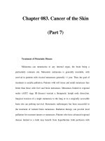
Cutaneous ulcers within injection sites of co-
caine and heroin have been documented
[45–47] (Fig. 16.1). Pentazocine ulcers are com-
Chapter 16 Drugs,Wound Healing and Cutaneous Ulcers
196
16
t
16_193_208 01.09.2004 14:06 Uhr Seite 196
mon among drug abusers [48]. Drugs such as
heroin may be adulterated with fillers contain-
ing other drugs (e.g., quinine, barbiturates,
mannitol) or certain chemicals, including dex-
trose, cocaine, caffeine, procaine, talc, baking
soda, starch, battery acid, butacaine, and nico-
tine [48].
There are several pathophysiological mecha-
nisms by which ulcers occur [49]:
5 The drug itself, e.g., vasospastic
properties of cocaine [46]
5 Irritant or caustic effects of adulter-
ants or excipients
5 The use of contaminated needles
with consequent infection
Kirchenbaum and Midenberg [49] reported the
following incidence of ulceration at injection
sites in the lower extremities of 56 drug abus-
ers: dorsal venous arch of the foot (68%), great-
er saphenous vein (14%), veins in the digits
(10%), and the spaces between the toes (7%).
‘Puffy foot syndrome’ was described in pa-
tients who repeatedly used their limbs for drug
injections, resulting in the blockage of lym-
phatics and the distortion of the venous return,
with an increased risk of ulceration [49].
16.2.5 Self-Inflicted Ulcers
In most cases, self-inflicted ulcers are caused by
continuous scratching, rubbing, or cutting of
the skin. Once an ulcer appears, the continual
‘fiddling’ with it by the patient interferes with
its healing.
Sometimes, self-inflicted ulcers are deliber-
ately induced by injecting certain materials, in-
cluding drugs, into the skin. Some injected
drugs may be identified,since they are incorpo-
rated in specific vehicles. Jackson et al. [50] re-
ported factitious ulcers caused by injections of
certain pulverized tablet materials, originating
from analgesic tablets or pentazocine hydro-
chloride tablets. Numerous materials such as
phenol [48], sodium hydroxide [51], and kero-
sene [52] have been documented as being used
for self-mutilation.
A lesion caused by the injection of an of-
fending material may initially appear in the
form of a nodule, or an abscess, which subse-
quently undergoes ulceration. In these cases,
histology may contribute to a diagnostic iden-
tification of the ulcer’s cause. The lesion may
show (but not always) giant cells or epitheloid
cells.
The unique characteristics of specific mate-
rials may also be reflected in the histology. For
example, oil-containing substances are identi-
fied by the ‘Swiss-cheese pattern’ (as seen in
paraffinoma), with numerous ovoid spaces of
varying size, filled with the oily substance [53].
Using polarized light, one may identify the
presence of inorganic birefringent materials.
Some of these, such as talc, which is used as a
filler in certain analgesic tablets, may be inject-
ed into the skin.
Substances that are doubly refractile on po-
larizing examination include:
5 Nylon sutures
5 Wo o d
5 Tal c
5 Starch powder
5 Silica
5 Beryllium
16.2Ulceration at the Infection Site
197
Fig. 16.1. A cutaneous ulcer in a drug addict following
an injection of heroin
t
t
16_193_208 01.09.2004 14:06 Uhr Seite 197
Other substances may be identified by chemical
or spectrophotometric methods [54].
Jackson et al. [50] introduced the electron-
probe microanalysis method to determine ac-
curately the presence and chemical nature of
foreign material. This technique can identify
certain pulverized tablet materials such as mi-
crocrystalline cellulose (found in pentazocine
hydrochloride tablets) or talc (used as a filler in
certain analgesic tablets), which may be inject-
ed into the skin and subcutaneous tissue.
16.3 Direct Cutaneous Exposure
The long-term use of suppositories containing
ergotamine is reported to have caused perianal
ulcers [55, 56]. Certain drugs, such as povidone-
iodine [57], may induce ulceration by causing
severe contact dermatitis (see Chap. 4).
In addition to the comments presented earli-
er regarding drug abuse, Tirney and Stadel-
mann [58] documented extensive facial necro-
sis, resulting from ischemia and subsequent in-
fection, following the intranasal impaction of
‘crack’ cocaine.
16.4 Systemic Drugs that Directly
Induce Ulceration
16.4.1 Causing or Aggravating Certain
Diseases
Some drugs tend to cause or aggravate certain
diseases in which cutaneous ulcers may be a
feature (Table. 16.3). The classical example of
such a disease is systemic lupus erythematosus
(SLE). The most common drugs related etio-
logically to SLE are hydralazine, procainamide,
β-blockers, phenytoin, and isoniazid [59].
These drugs, among many others, may result in
SLE-like syndromes that are indistinguishable
from SLE. In some cases, ulceration may devel-
op, similar to that seen in SLE. Certain other
medications may exacerbate pre-existing SLE:
griseofulvin, sulfonamides, testosterone, and
estrogens [60].
Despite the abundance of information in the
literature regarding medications that cause SLE
or SLE-like syndromes, there are no accurate
statistical data regarding the development of
cutaneous ulcers following treatment with such
Chapter 16 Drugs,Wound Healing and Cutaneous Ulcers
198
16
Cause or aggravate certain diseases
SLE
¼ Hydralazyne
Lichen planus
¼ Hydroxyurea
¼ Methyldopa
¼ Propanolol
¼ Lithium carbonate
Pyoderma gangrenosum
¼ Granulocyte colony-stimulating factor
¼ Isotretinoine
¼ Montelukast sodium
¼ Ibuprofen
Induce vasculitis
¼ Clyndamycin
¼ Non-steroidal anti-inflammatory drugs
¼ Propylthiouracil
¼ Diltiazem
¼ Oral contraceptives
¼ Levamisole
¼ Minocycline
¼ Hydralazine
¼ Warfarin
Affect coagulability
¼ Warfarin
¼ Heparin
¼ Low-molecular-weight-heparin
By unidentified mechanisms
¼ Hydroxyurea
¼ Methotrexate
¼ Bleomycin
Table 16.3. Systemic drugs that directly induce ulceration
16_193_208 01.09.2004 14:06 Uhr Seite 198
medications, and the information available is
limited to isolated case reports. Drug-induced
SLE associated with the development of ulcers
has been documented following the ingestion
of hydralazine [59, 61, 62]. In some cases, histol-
ogy shows that the hydralazine itself apparent-
ly induces vasculitis [62]. However, it should be
noted that other conditions associated with
SLE, such as Raynaud’s phenomenon or the
anti-phospholipid syndrome, may cause ulcer-
ation as well (see Chap. 4).
There are several other systemic diseases in
which ulceration may occur following the use of
certain drugs. The anti-phospholipid syndrome
has been reported following the use of chloro-
promazine, procaine-amide, quinidine, hydrala-
zine, phenytoin, and interferon [63]. Dermato-
myositis may also be precipitated by several
drugs, including penicillamine, NSAIDs, or car-
bamazepine [60]. Nevertheless, for the above
cases there are no data relating to the induction
of ulceration by drugs in these diseases.
The association between drugs, scleroderma
and cutaneous ulcers is discussed in Sect.
16.6.2.
Finally, certain ulcerative cutaneous pro-
cesses may be induced by certain drugs: Lichen
planus of the ulcerative type has been associat-
ed with the use of hydroxyurea [68], methyldo-
pa [65], propanolol [66], and lithium carbonate
[67]; pyoderma gangrenosum has been docu-
mented in association with the use of granulo-
cyte colony-stimulating factor, isotretinoin,
montelukast sodium (Singulair®), and ibuprof-
en [68–71]. In addition, sulpiride has been doc-
umented as a possible inducer of pyoderma
gangrenosum-like eruption [72].
6.4.2 Induction of Vasculitis
Drugs that most commonly cause leukocyto-
clasic vasculitis are penicillin, sulfonamides,
thiazides, allopurinol, and non-steroidal anti-
inflammatory agents [73]. However, specific
documentation as to drug-induced leukocyto-
clasic vasculitis manifested by ulceration is
rather infrequent. Certain medications have
been reported to induce vasculitis with subse-
quent formation of cutaneous ulcers. These in-
clude clindamycin [74], NSAIDs [75], propylthi-
ouracil [76], diltiazem [77], oral contraceptives
[78], levamisole [79], and minocycline [80].
However, in some of these reports the associa-
tion between the drug and vasculitis may be
questionable, and other factors may be in-
volved.
Hydralazine, as discussed above, has been
reported to induce SLE-reactions with vascu-
litis [62]. Vasculitis has also been documented
in several cases of warfarin administration
[81–83]. There are no accurate and well-estab-
lished epidemiological reports regarding medi-
cations that tend to induce vasculitis presenting
as cutaneous ulcers. As mentioned earlier in
this chapter, certain drugs may induce a local
vasculitic response when injected s.c. or i.m.
16.4.3 Vasospasm
Certain medications mentioned-above have
been reported as having a vasospastic effect
when administered by injection. Ergotamine
preparations (usually given orally) have also
been documented as resulting in severe vasos-
pasm with subsequent ulceration [84–87]. Most
of the existing reports have documented is-
chemic events of the lower extremities.
16.4.4 Drugs Affecting Coagulability
Skin necrosis may be induced by drugs that af-
fect coagulability, such as warfarin or heparin.
As noted below, the exact mechanism by which
these drugs induce ulceration has not yet been
fully established.
Warfarin-induced Skin Necrosis. The phe-
nomenon appears in 0.01%–0.1% of patients
treated with warfarin or its derivatives [88]. In
most cases, warfarin necrosis usually appears
within 3–6 days following ingestion of the
drug, but it can also appear within the first two
weeks. This process tends to develop in areas
containing relatively high amounts of subcuta-
neous fat, such as buttocks, thighs, and breasts.
This preference is attributed to the reduced
vascularity of adipose tissue. However, other
16.4Systemic Drugs that Directly Induce Ulceration
199
16_193_208 01.09.2004 14:06 Uhr Seite 199
areas may also be affected. The initial cutane-
ous finding is an erythematous area, usually
poorly demarcated, which gradually darkens to
blue-black and progresses towards a well-de-
marcated area of necrosis. Hemorrhagic blis-
ters may develop prior to the onset of ulcera-
tion [17, 89, 90]. Most of the affected patients
are adult women with a tendency to obesity,the
female-to-male ratio being 9 :1 [88]. In most
cases there is only a single lesion, but in one
third of patients multiple lesions have been
documented [88].
The initial mechanism of warfarin-induced
ulceration involves a hypercoagulable state [88,
90]. The histological findings support this as-
sumption: The early pathologic changes are mi-
crovascular damage with fibrin deposits in the
postcapillary venules and small veins. Later, ar-
eas of hemorrhage and diffuse necrosis are
seen in the dermis and subcutaneous tissue [91,
92]. However, in some cases, evidence of vascu-
litis has been documented following warfarin
administration [86, 87]. One may conclude that
there is probably more than one mechanism of
ulceration.
Predisposing factors to warfarin necrosis are
protein C deficiency or protein S deficiency [89,
93–99]. These associations further support the
assumption that the mechanism causing war-
farin necrosis is basically mediated via process-
es affecting coagulability.
Heparin-Induced Necrosis. Skin necrosis
usually develops within 6–8 days following
subcutaneous injection of heparin, but it can
also appear after two weeks [100, 101]. In most
cases the ulceration appears right at the injec-
tion site. Rarely, other sites may also be in-
volved. Lesions of heparin necrosis have been
reported to occur on the nose, hand, forearm,
ankle, and thigh [101]. The clinical and histo-
logic findings are indistinguishable from those
of warfarin necrosis [101–103], which implies
that similar mechanisms are responsible for
necrosis. There are several reports of necrosis
following the use of low-molecular-weight-
heparin [104–107].
6.4.5 Drugs Causing Bullae
As mentioned in Chap. 4, even a superficial ero-
sion (due to trauma, or bullous disease) may
become an ulcer following bacterial infection.
The probability of this occurring is much high-
er when there is an underlying problem such as
diabetes mellitus or prolonged glucocorticoid
therapy (as is the case with many patients suf-
fering from bullous diseases). Numerous drugs
may induce bullae; a detailed discussion, how-
ever, is beyond the scope of this chapter.
16.4.6 Unidentified Mechanisms
Apart from the groups of drugs presented
above, certain drugs may result in cutaneous
ulcers by mechanisms that are not currently
fully understood. Hydroxyurea is a relatively
frequent inducer of cutaneous ulcers [108–114].
It is reasonable to assume that hydroxyurea ul-
ceration is not attributed to its anti-neoplastic
effect, since other anti-neoplastic medications
from the same group are not known to cause
skin ulcers.
Similarly, methotrexate is known to induce
the formation of mucosal and, less commonly,
skin ulcers[115, 116]. Kaplan et al. [117] docu-
mented erosion of psoriatic plaques following
chronic methotrexate administration.
Bleomycin-induced digital gangrene has
been reported following i.m. injection [118]. In
this report, the exact mechanism was not clari-
fied, although the authors suggested that the
drug may have induced Raynaud’s phenome-
non.
16.5 Interference with Normal
Mechanisms of Wound Healing
In most cases, drugs belonging to this group do
not cause ulceration directly. These drugs may
interfere with the healthy physiological mecha-
nisms of wound healing, and their use may sig-
nificantly impede the healing of existing ulcers.
Glucocorticoids are the most notorious drugs of
this group, in respect to their influence on heal-
Chapter 16 Drugs,Wound Healing and Cutaneous Ulcers
200
16
16_193_208 01.09.2004 14:06 Uhr Seite 200
ing. Other drugs that may interfere with healing
are presented in Table 16.4.
Note that the use of some drugs in the pres-
ence of certain diseases such as diabetes mellit-
us or venous insufficiency may significantly in-
crease the risk of developing cutaneous ulcers.
Generally speaking, it is difficult to predict
the extent to which the medications discussed
below impede the healing of chronic cutaneous
ulcers. In the few cases described, when re-
search studies were planned in order to exam-
ine the detrimental effect of drugs on healing,
they focused on acute surgical wounds only.
16.5.1 Glucocorticoids
The detrimental effect of glucocorticoids on
wound healing has been recognized and docu-
mented for many years [119]. Not surprisingly,
the same qualities, namely anti-inflammatory,
immunosuppressive, and anti-proliferative,
that impart the therapeutic value of glucocorti-
coids in certain diseases are those that interfere
with the normal course of wound repair.
Glucocorticoids influence and interfere with
various aspects of wound healing. Laboratory
research studies have documented decreased
production of cytokines and growth factors,
suppression of inflammatory response and mo-
bilization of white cells, and the stabilization of
lysosomal membranes in white blood cells,
thereby decreasing phagocytic activity [120–
122]. Host defense mechanisms become less ef-
ficient, resulting in an increased susceptibility
to various infections.
Reduced proliferation of epidermal cells and
fibroblasts occurs, associated with impaired an-
giogenesis [123–126]; wound contraction is im-
paired and reduced synthesis of proteins and
collagen further delays the normal course of
wound healing and closure of cutaneous ulcers
[127, 128].
Research studies have shown that if gluco-
corticoids are administered within the first
three days following injury, the effect on wound
healing is much more significant than if they
are administered more than three days after the
trauma [129, 130]. Therefore, one may conclude
that the detrimental effect of glucocorticoids is
related mainly to their anti-inflammatory ef-
fects in the initial stages of injury.
In traumatic wounds in adults, it appears
that low-dosage glucocorticoids (less than
10 mg prednisone per day) do not have a dis-
cernible effect on wound healing. Moderate
dosages of 10–30 mg prednisone per day result
in a mild mechanical impairment of wound
strength. A dosage of 40 mg prednisone or
more per day has a direct inhibitory effect on
wound healing [121]. To the best of our knowl-
edge, the data presented above have not been
confirmed or re-evaluated in recent years by
up-dated research studies.
The above conclusions may be reached only
as regards acute traumatic wounds.When deal-
ing with chronic cutaneous ulcers, it is difficult
to assess the effects of glucocorticoids, and
there is no definite information on this subject
in the literature. Note that prolonged glucocor-
ticoid therapy results in skin atrophy, with spe-
cific consequent ramifications on the process of
wound healing (see below).
Through clinical experience we do know
that even low doses of steroids, when taken for
long periods, can result in such cutaneous dam-
age as atrophy of the skin. It is reasonable to as-
sume that those same mechanisms would inter-
fere with wound healing and skin repair.
The obvious conclusion that can be drawn
from the above information is that every effort
should be made to discontinue glucocorticoid
therapy (or at least reduce the dosage) in pa-
tients with chronic cutaneous ulcers.
16.5Interference with Normal Mechanisms
201
Table 16.4. Drugs that Interfere healing
Glucocorticoids
Non-steroidal anti-inflammatory drugs
Anti-neoplastic
and immunosuppressive drugs
Others
¼ colchicine
¼ penicillamine
16_193_208 01.09.2004 14:06 Uhr Seite 201
Two further comments should be made here:
5 To some extent, vitamin A may
counteract the effects of glucocorti-
coids on wound healing. This issue
is discussed in more detail in
Chap. 19.
5 The question as to whether topical
growth factors may prevent some of
the glucocorticoid effects on wound
healing is still unanswered. Con-
trolled research studies are needed.
16.5.2 Non-Steroidal
Anti-Inflammatory Drugs
Medications such as aspirin and ibuprofen have
been shown to impair collagen production and
to lower the tensile strength of healing wounds
[131, 132]. Whether the effect of non-steroidal
anti-inflammatory drugs (NSAID) on the heal-
ing of cutaneous wounds is similar to their ef-
fect on gastric mucosa requires further investi-
gation.
The extent to which the healing of cutaneous
ulcers is hampered by NSAID is questionable.
However, for patients with cutaneous ulcers,
one should consider avoiding the use of NSAID
and using alternative analgesic drugs. At
present, there are no data regarding whether
the use of advanced forms of NSAID may have
a certain advantage with respect to their effect
on healing.
16.5.3 Anti-Neoplastic
and Immunosuppressive Drugs
Observations that anti-neoplastic and immu-
nosuppressive drugs interfere with the normal
course of wound healing, with respect to surgi-
cal wounds, are well documented in the litera-
ture [133]. Animal experiments have demon-
strated that the extent of these detrimental ef-
fects depends on the specific medication used.
Severe impairment of healing was observed fol-
lowing the use of actinomycin D, bleomycin, or
BCNU,whereas the effect caused by vincristine,
cyclophosphamide, or 5FU was relatively mild
[134].
At present, the mechanism by which anti-
neoplastic drugs exert their effects on the heal-
ing of cutaneous ulcers is not fully understood.
Some of these effects may be attributed to nu-
tritional deficiencies and the catabolic effects
associated with these drugs [135–137]. In any
case, the identification and correction of nutri-
tional deficiencies may prevent the negative
consequences of anti-neoplastic treatment on
healing of wounds and chronic ulcers of the
skin.
The issue of extravasation injury following
intravenous injections of anti-neoplastic drugs
is discussed above.
16.5.4 Other Drugs that Interfere
with Healing
Colchicine has been shown to delay the normal
course of wound healing. This effect may be
due to its inhibitory effect on tubulin-depen-
dent cell functions, including interference with
fibroblasts, secretion of collagen precursors,
and the inhibition of wound contraction [121,
138, 139]. Another medication that has been de-
tected as a possible inhibitor of wound contrac-
tion and cutaneous healing is penicillamine
[121, 139].
16.6 Drugs that Adversely Affect
Skin Quality
16.6.1 Leg Edema
Calcium channel blockers, especially nifedi-
pine, may cause edema of the legs [140–142].
Edema adversely affects the quality of the skin
[143, 144], which can result in the formation of
leg ulcers and the aggravation of pre-existing
ulcers.The effect of calcium channel blockers is
highly significant in patients who suffer from
venous stasis or lymph vessel disease. In the
presence of cutaneous ulcers, whenever pos-
sible, one should consider discontinuing these
Chapter 16 Drugs,Wound Healing and Cutaneous Ulcers
202
16
t
16_193_208 01.09.2004 14:06 Uhr Seite 202
drugs. Luca and Romero [145] reported im-
provement of leg ulcers following the discon-
tinuation of nifedipine. The phenomenon of
drug-induced leg edema is commonly associat-
ed with the dihydropyridine calcium antago-
nists (nifedipine, felodipine, isradipine, nicar-
dipine, nisoldipine) (see Table 16.5) [146]. How-
ever, it may occur following the use of non-di-
hydropyridine calcium antagonists (i.e., verap-
amil and diltiazem) as well [147]. The edema
formation is caused, most probably, by arterio-
lar dilatation (which is greater than venous dil-
atation) and increased transcapillary pressure
gradients [146].
16.6.2 Skin Atrophy or Scleroderma-
Like Reactions
The effects of glucocorticoids on wound heal-
ing have been discussed above.As noted, gluco-
corticoid usage, whether systemic or topical,
has been known for many years to cause skin
atrophy [148]. Atrophic skin is poorly vascular-
ized and very vulnerable, and its ability to heal
wounds is reduced.
Prolonged penicillamine therapy may also
result in cutaneous atrophy [149].
Scleroderma-like reactions (Fig. 16.2) which
affect the quality of the skin may develop fol-
lowing the use of certain medications including
pentazocine, bleomycin, appetite suppressants,
and bromocriptine [150–152]. Exposure to vari-
ous agents (e.g., polyvinyl chloride, epoxy res-
ins, organic solvents, and pesticides) is also as-
sociated with scleroderma or scleroderma-like
changes [150, 151]. There are no clear data in the
literature regarding the formation of ulcers
under these circumstances.
References
1. Bork K: Ulcers and necroses; atrophies. In: Cutane-
ous Side Effects of Drugs, 1st edn.Philadelphia: WB
Saunders. 1988; pp 360–369
2. Kohler LD, Schwedler S, Worret WI: Embolia cutis
medicamentosa. Int J Dermatol 1997; 36 : 197
3. Kunzi T, Ramstein C, Pirovino M: Circumscribed
skin necrosis following intramuscular injection
(embolia cutis medicamentosa). Schweiz Rundsch
Med Prax 1995; 84 : 640–643
4. Muller-Vahl H: Aseptic tissue necrosis: a severe
complication after intramuscular injections. Dtsch
Med Wochenschr 1984; 109 :786–792
5. Gammel JA: Arterial embolism, an unusual com-
plication following the intramuscular administra-
tion of bismuth. JAMA 1927; 88 :998
References
203
Table 16.5. Drugs that adversely affect skin quality
Formation of leg edema
a
Dihydropyridine calcium antagonists
¼ Nifedipine
¼ Amlodipine
¼ Felodipine
¼ Isradipine
¼ Nicardipine
Drugs causing skin atrophy
or scleroderma-like reactions
b
¼ glucocorticoids
¼ penicillamine
¼ bleomycin
¼ appetite suppressants
¼ pentazocine
¼ bromocriptine
a. The use of non-dihydropyridine calcium antagonists
(i.e., verapamil and diltiazem) has also been reported
to induce leg edema [147].
b. Exposure to various agents (e.g.,polyvinyl chloride,ep-
oxy resins, organic solvents, and pesticides) is also as-
sociated with sclerodrema or scleroderma-like chang-
es [150, 151].
Fig. 16.2. Cutaneous ulcers on sclerodermoid skin
16_193_208 01.09.2004 14:06 Uhr Seite 203
6. Fiscina S: Injections followed by a sterile abscess.
Mil Med 1986; 151 :452
7. Smith AL, Khan F, De Mello W: Sterile abscess for-
mation following plague vaccination. J R Army
Med Corps 1997; 143:116–117
8. Cosman A, Feliciano WC, Wolff M: Pentazocine ul-
cers. Plast Reconst Surg 1977; 59 :255–259
9. Krainick U, Kantarjian H, Broussard S, et al: Local
cutaneous necrotizing lesions associated with
interferon injections. J Interferon Cytokine Res
1998; 18:823–827
10. Webster GF, Knobler RL, Lublin FD, et at: Cutane-
ous ulcerations and pustular psoriasis flare caused
by recombinant interferon β injections in patients
with multiple sclerosis. J Am Acad Dermatol 1996;
34 :365–367
11. Weinberg JM: Cutaneous necrosis associated with
recombinant interferon injection. J Am Acad Der-
matol 1998; 39:807–808
12. Oeda E, Shinohara K: Cutaneous necrosis caused
by injection of α-interferon in a patient with
chronic myelogenous leukemia. Am J Hematol
1993; 44 :213–214
13. Gaines AR, Varricchio F: Interferon β-lb injection
site reactions and necroses. Mult Scler 1998; 4 :70–73
14. Weinberg JM, Wolfe JT, Sood S, et al: Cutaneous
necrosis associated with recombinant interferon
injection. Report of three cases with interferon be-
ta-1b and review of the literature. Acta Derm Ve-
nereol (Stockh) 1997; 77 :146–148
15. Hinterberger JW, Kintzi HE: Phentolamine reversal
of epinephrine-induced digital vasospasm. How to
save an ischemic finger. Arch Fam Med 1994; 3:
193–195
16. Maguire WM, Reisdorff EJ, Smith D, Weigenstein
JG: Epinephrine-induced vasospasm reversed by
phentolamine digital block.Am J Emerg Med 1990;
8 :46–47
17. Bork K: Necrosis and ulcers. In: Cutaneous Side Ef-
fects of Drugs. 1st edn. Philadelphia: W.B Saunders.
1988; pp 220–230
18. Sylaidis P, Logan A: Digital blocks with adrenaline.
An old dogma refuted. J Hand Surg [Br] 1998; 23:
17–19
19. Rae V, Pardo RJ, Blackwelder PL, et al: Leg ulcers
following subcutaneous injection of a liquid sili-
cone preparation. Arch Dermatol 1989; 125:
670–673
20. Mastruserio DN, Pesqueira MJ, Cobb MW: Severe
granulomatous reaction and facial ulceration oc-
curring after subcutaneous silicone injection. J Am
Acad Dermatol 1996; 34: 849–852
21. Bihari I, Magyar E: Reasons for ulceration after in-
jection treatment of telangiectasia. Dermatol Surg
2001; 27:133–136
22. Borgstrom E: Penile ulcer as complication in self-
induced papaverine erections. Urology 1988; 32 :
416–417
23. Brown AS, Hoelzer DJ, Piercy SA: Skin necrosis
from extravasation of intravenous fluids in chil-
dren. Plast Reconstr Surg 1979; 64 :145–150
24. Riyami A: Complications of intravenous infusions.
J Ir Med Assoc 1968; 61: 23–25
25. Disa JJ, Chang RR, Mucci SJ,et at: Prevention of Ad-
riamycin induced full-thickness skin loss using
hyaluronidase infiltration. Plast Reconstr Surg
1998; 101: 370–374
26. Barden GA: Venous extravasation of doxorubicin
HCI with secondary skin ulceration. South Med J
1980; 73 :1543–1544
27. Mehta P, Najar N: Skin ulceration due to faulty ad-
riamycin administration. Clin Pediatr (Phila) 1978;
17 :663–664
28. Luedke DW, Kennedy PS,Rietschel RL: Histopatho-
genesis of skin and subcutaneous injury induced
by Adriamycin. Plast Reconstr Surg 1979; 63 :
463–465
29. Bowers DG Jr, Lynch JB: Adriamycin extravasation.
Plast Reconstr Surg 1978; 61 :86–92
30. Bertelli G: Prevention and management of ex-
travasation of cytotoxic drugs. Drug Saf 1995; 12:
245–255
31. Boman NL, Tron VA, Bally MB, et al: Vincristine in-
duced dermal toxicity is significantly reduced
when the drug is given in liposomes. Cancer
Chemother Pharmacol 1996; 37:351–355
32. Blair WF, Kilpatrick WC Jr, Saiki JH, Alter EJ: Ex-
travasation of chemotherapeutic agents. Clin Or-
thop 1980; 151 :228–230
33. Bairey O, Bishara J, Stahl B, et al: Severe tissue ne-
crosis after cisplatin extravasation at low concen-
tration: possible “immediate recall phenomenon”. J
Natl Cancer Inst 1997; 89: 1233–1234
34. Elam EA, Dorr RT, Lagel KE, Pond GD: Cutaneous
ulceration due to contrast extravasation. Experi-
mental assessment of injury and potential anti-
dotes. Invest Radiol 1991; 26: 13–16
35. Heckler FR: Current thoughts on extravasation in-
juries. Clin Plast Surg 1989; 16:557–563
36. Rudolph R, Stein RS, Pattillo RA: Skin ulcers due to
Adriamycin. Cancer 1976; 3: 1087–1094
37. Sintenie JB, Tuinebreijer WE, Kreis RW, et al: Digital
gangrene after accidental intra-arterial injection of
phenytoin (epanutin). Eur J Surg 1992; 158: 315–316
38. Joist A, Tibesku CO, Neuber M, et al: Gangrene of
the fingers caused by accidental intra-arterial in-
jection of diazepazn. Dtsch Med Wochenschr 1999;
124 :755–758
39. Steib A, Jeanpierre C, Mole JP, et al: Tissue necrosis
after accidental perivenous injection of dopamine.
Ann Fr Anesth Reanim 1984; 3: 328–329
40. De Myttenaere S, Heifetz M, Shilansky H, et al: Dif-
ferent treatments used in a case of gangrene due to
accidental intra-arterial injection of methylpheni-
date (rilatine). Anesth Analg (Paris) 1977; 34 :
405–410
41. Schanzer H, Gribetz I, Jacobson JH: Accidental
intra-arterial injection of penicillin G. A prevent-
able catastrophe. JAMA 1979; 242 :1289–1290
42. Schanzer H, Jacobson JH: Tissue damage caused by
the intramuscular injection of long-acting penicil-
lin. Pediatrics 1985; 75 :741–744
Chapter 16 Drugs,Wound Healing and Cutaneous Ulcers
204
16
16_193_208 01.09.2004 14:06 Uhr Seite 204
43. Wynne JM, Williams GL, Ellman BA: Accidental
intra-arterial injection. Arch Dis Child 1978; 53 :
396–400
44. Yasin M, Vishne TH, Hendel D: Local necrosis of
finger following stab with needle used to pump ter-
butaline sulfate (bricalin). Harefuah 2001; 140:
398–399
45. Bertoncini RL, Silveira PG, Ceola L, et at: Vascular
complications in intravenous drug addicts. Rev As-
soc Med Bras 1992; 38: 204–208
46. Abidin MR, Gillinov MA, Topol BM, et al: Injection
of illicit drugs into the granulation tissue of chron-
ic ulcers.Ann Plast Surg 1990; 24 :268–270
47. Dunne JH, Johnson WC: Necrotizing skin lesions in
heroin addicts.Arch Dermatol 1972; 105: 544–547
48. Fellner MJ, Ledesma GN: Leg ulcers secondary to
drug reactions. Clin Dermatol 1990; 8 : 144–149
49. Kirchenbaum SE, Midenberg ML: Pedal and lower
extremity complications of substance abuse. J Am
Podiatry Assoc 1982; 72 :380–387
50. Jackson RM, Tucker SB, Abraham JL, et al: Factitial
cutaneous ulcers and nodules: the use of electron-
probe microanalysis in diagnosis. J Am Acad Der-
matol 1984; 11: 1065–1069
51. Gschnait F, Brenner W, Berger E, et al: An unusual
case of repeated self-mutilation. Wien Kim Wo-
chenschr 1981; 93: 127–131
52. Qaryoute SM: Skin ulceration induced by kerosene
injection. Ann Plast Surg 1984; 12: 361–363
53. Shapiro PE: Non infectious granulomas. In: Elder
D, Elenitas R, Jaworsky C, Johnson B (eds) Lever’s
Histopathology of the Skin, 8th edn. Philadelphia:
Lippincott-Raven. 1997; pp 317–340
54. Ackerman AB: Nodular and diffuse dermatitis. In:
Histologic Diagnosis of Inflammatory Skin Diseas-
es: An Algoritmic Method Based on Pattern Analy-
sis. 1st edn. Baltimore Philadelphia: Lea & Febiger.
1978; pp 385– 495
55. Brandt O,Abeck D, Breitbart E, et al: Perianal ergo-
tismus gangraenosus. Hautarzt 1997; 48 : 199–202
56. Baptista AP, Mariano A, Machado A: Peranal ulcers
caused by ergotamine-containing suppositories.
Acta Med Port 1992; 5 :39–41
57. Mochida K, Hisa T, Yasunaga C, et al: Skin ulcera-
tions due to povidone-iodine. Contact Dermatitis
1995; 33:61–62
58. Tierney BP, Stadelmann WK: Necrotizing infection
of the face secondary to intranasal impaction of “-
crack” cocaine. Ann Plast Surg 1999; 43: 640–643
59. Bork K: Drug-induced Lupus Erythemtosus. In:
Bork K: Cutaneous Side Effects of Drugs. 1st edn.
Philadelphia: WB Saunders. 1988; pp 181–184
60. Breathnach SM, Hintner H: Patterns of clinical dis-
ease. Types of clinical reaction. In: Breathnach SM,
Hintner H: Adverse Drug Reactions and the Skin.
1st edn. Oxford: Blackwell Scientific Publications.
1992; pp 41–136
61. Kissin MW, Williamson RC: Hydrallazine-induced
SLE- like syndrome presenting as a leg ulcer. Br
Med J 1979; 2 :1330
62. Bernstein RM, Egerton-Vernon J, Webster J: Hy-
dralazine-induced cutaneous vasculitis. Br Med J
1980; 280 :156–157
63. Gibson GE, Su WP, Pittelkow MR: Antiphospholip-
id syndrome and the skin. J Am Acad Dermatol
1997; 36: 970–982
64. Renfro L, Kamino H, Raphael B, et al: Ulcerative li-
chen planus-like dermatitis associated with hy-
droxyurea. J Am Acad Dermatol 1991; 24 :143–145
65. Burry JN: Ulcerative lichenoid eruption from
methyldopa (letter).Arch Dermatol 1976; 112: 880
66. Massa MC, Jason SM, Gradini R, et at: Lichenoid
drug eruption secondary to propranolol. Cutis
1991; 48:41–43
67. Srebrnik A, Bar-Nathan EA, Ilie B, et al: Vaginal ul-
cerations due to lithium carbonate therapy. Cutis
1991; 48:65–66
68. Ross HJ, Moy LA, Kaplan R, et al: Bullous pyoder-
ma gangrenosum after granulocyte colony-stimu-
lating factor treatment. Cancer 1991; 68 : 441–443
69. Gangaram HB, Tan LP, Gan AT, et al: Pyoderma
gangrenosum following treatment with isotreti-
noine. Br J Dermatol 1997; 136: 636–637
70. Cohen AD, Grossman N, Halevy S: Pyoderma gan-
grenosum induced by montelukast sodium – con-
firmation by interferon-γ (IFN-γ) release test. In:
32nd Annual European society for dermatology re-
search (ESDR) meeting. Geneva: September, 2002
71. Cohen AD, Cagnano E, Halevy S: Ibuprofen-in-
duced pyoderma gangrenosum. J Am Acad Derma-
tol (in press)
72. Srebrnik A, Shachar E, Brenner S: Suspected induc-
tion of a pyoderma gangrenosum-like eruption
due to sulpiride treatment. Cutis 2001; 67 :253–256
73. Soter NA: Cutaneous necrotizing venulitis. Freed-
berg IM, Eisen AZ, Wolff K, Austen KF, Goldsmith
LA, Katz SI, Fitzpatrick TB (eds) In: Fitzpatrick’s
Dermatology in General Medicine. 5th edn. New-
York: McGraw-Hill. 1999; pp 2044–2053
74. Lambert WC, Kolber LR, Proper SA: Leukocyto-
clastic angiitis induced by clindamycin. Cutis 1982;
30 :615–619
75. Veraguth AJ, Hauselmann HJ, Hunziker T, Gerber
NJ: Vasculitic skin lesions caused by nonsteroidal
anti-inflammatory agents. Schweiz Med Wochen-
schr 1992; 122: 923–929
76. Houston BD, Crouch ME, Brick JE, et al: Apparent
vasculitis associated with propylthiouracil use. Ar-
thritis Rheum 1979; 22 :925–928
77. Carmichael AJ, Paul CJ: Vasculitic leg ulcers asso-
ciated with diltiazem. Br Med J 1988; 297 :562
78. Mosovich B, Biton A, Avinoach I: Vasculitis with
cutaneous necrosis induced by oral contraceptive.
Harefuah 1991; 120 :451–453
79. Menni S, Pistritto G, Gianotti R, et al: Ear lobe bilat-
eral necrosis by levamisole-induced occlusive vas-
culitis in a pediatric patient. Pediatr Dermatol
1997; 14:477–479
References
205
16_193_208 01.09.2004 14:06 Uhr Seite 205
80. Schrodt BJ, Kulp-Shorten CL, Callen JP: Necrotiz-
ing vasculitis of the skin and uterine cervix asso-
ciated with minocycline therapy for acne vulgaris.
South Med J 1999; 92:502–504
81. Altmeyer P, Welke S, Renger A, et al: Zur pathoge-
nese der sogenannten Cumarin-Nekrose. Aktuel
Dermatol 1976; 2 : 65–68
82. Slutzki S, Bogokowsky H, Gilboa Y, et al: Coumadin
induced skin necrosis. Int J Dermatol 1984; 23 :
117–119
83. Tanay A, Yust I, Brenner S, et al: Dermal vasculitis
due to coumadin hypersensitivity. Dermatologica
1982; 165: 178–185
84. Stoebner PE, Gaspard C, Meynadier J, et al: Tongue
necrosis provoked by ergotamine tartrate and dis-
closing a giant cell arteritis. Eur J Dermatol 1999; 9:
652–653
85. Zavaleta EG, Fernandez BB, Grove MK, et al: St.
Anthony’s fire (ergotamine induced leg ischemia)
– a case report and review of the literature.Angiol-
ogy 2001; 52: 349–356
86. Bongard O, Bounameaux H: Severe iatrogenic er-
gotism: incidence and clinical importance. Vasa
1991; 20:153–156
87. Payne B, Sasse B, Franzen D, et al: Manifold mani-
festations of ergotism. Schweiz Med Wochenschr
2000; 130:1152–1156
88. DeFranzo AJ, Marasco P, Argenta LC: Warfarin-in-
duced necrosis of the skin. Ann Plast Surg 1995; 34 :
203–208
89. Chan YC, Valenti D, Mansfield AO, et al: Warfarin
induced skin necrosis. Br J Surg 2000; 87: 266–272
90. Ad-El DD, Meirovitz A, Weinberg A, et al: Warfarin
skin necrosis: local and systemic factors. Br J Plast
Surg 2000; 53: 624–626
91. Cole MS, Minifee PK,Wolma FJ: Coumarin necrosis
– a review of the literature. Surgery 1988; 103 :
271–277
92. Faraci PA, Deterling RA Jr, Stein AM, et al: Warfar-
in induced necrosis of the skin. Surg Gynecol Ob-
stet 1978; 146 :695–700
93. Makris M, Bardhan G, Preston FE: Warfarin in-
duced skin necrosis associated with activated pro-
tein C resistance. Thromb Haemost 1996; 75 :
523–524
94. McGehee WG, Klotz TA, Epstein DJ, et al: Coumar-
in necrosis associated with hereditary protein C
deficiency.Ann Int Med 1984; 101 : 59–60
95. Broekmans AW: Hereditary protein C deficiency.
Haemostasis 1985; 15:233–240
96. Comp P, Elrod J, Karzenski S: Warfarin-induced
skin necrosis. Semin Thromb Hemost 1990; 16:
293–298
97. Rose VL, Kwaan HC, Williamson K, et al: Protein C
antigen deficiency and warfarin necrosis. Am J
Clin Pathol 1986; 86: 653–655
98. Ng T, Tillyer ML: Warfarin-induced skin necrosis
associated with Factor V Leiden and protein S defi-
ciency. Clin Lab Haematol 2001; 23 : 261–264
99. Broekmans AW, Bertina RM, Loeliger EA, et al: Pro-
tein C and the development of skin necrosis during
anticoagulant therapy (letter). Thromb Haemost
1983; 49 :251
100. Breathnach SM, Hintner H: Anticoagulants, fibrin-
olytic agents and antiplatlet drugs. In: Adverse
Drug Reactions and the Skin, 1st edn. Oxford:
Bleckwell Scientific Publications. 1992; pp 247–253
101. Gold JA, Watters AK, O’Brien E: Coumadin versus
heparin necrosis. J Am Acad Dermatol 1987; 16:
148–150
102. White PW, Sadd JR, Nensel RE: Thrombotic com-
plications of heparin therapy: including six cases
of heparin- induced necrosis. Ann Surg 1979; 190:
595–608
103. Levine LE, Bernstein JE, Soltani K, et al: Heparin-
induced cutaneous necrosis unrelated to injection
sites: a sign of potentially lethal complications.
Arch Dermatol 1983; 119: 400–403
104. Santamaria A,Romani J,Souto JC, et al: Skin necro-
sis at the injection site induced by low-molecular-
weight heparin: case report and review. Dermatol-
ogy 1998; 196 :264–265
105. Balestra B, Quadri P, Dermarmels-Biasiutti F, et al:
Low-molecular-weight heparin-induced thrombo-
cytopenia and skin necrosis distant from injection
sites. Eur J Haematol 1994; 53: 61–63
106. Montserrat I, Lopez D, Zuazu-Jausoro I, et al: Low-
molecular-weight subcutaneous heparin-induced
skin necrosis. Blood Coag Fibrinol 1990; 1:751–752
107. Drew PJ, Smith MJ, Milling MA: Heparin-induced
skin necrosis and low molecular weight heparins.
Ann R Coll Surg Engl 1999; 81 :266–269
108. Sirieix ME, Debure C, Baudot N, et al: Leg ulcers
and hydroxyurea: Forty-one cases. Arch Dermatol
1999; 135: 818–820
109. Velez A, Garcia-Aranda JM, Moreno JC: Hydroxyu-
rea-induced leg ulcers: is macroerythrocytosis a
pathogenic factor? J Eur Acad Dermatol Venereol
1999; 12:243–244
110. Best PJ, Daoud MS, Pittelkow MR, et al: Hydroxyu-
rea-induced leg ulceration in 14 patients. Ann
Intern Med 1998; 128 :29–32
111. Weinlich G, Fritsch P: Leg ulcers in patients treated
with hydroxyurea for myeloproliferative disorders:
what is the trigger? Br J Dermatol 1999; 141 :171–172
112. Ravandi-Kashani F, Cortes J,Cohen P, et al: Cutane-
ous ulcers associated with hydroxyurea therapy in
myeloproliferative disorders. Leuk Lymphoma
1999; 35: 109–118
113. Demircay Z, Comert A, Adiguzel C: Leg ulcers and
hydroxyurea: report of three cases with essential
thrombocythemia. Int J Dermatol 2002; 41:872–874
114. Cohen AD,Hallel-Halevy D, Hatskelzon L, et al: Lon-
gitudinal melanonychia associated with hydroxyu-
rea therapy in a patient with essential thrombocyto-
sis. J Eur Acad Dermatol Venereol 1999; 13: 137–139
115. Lawrence CM, Dahl MG: Two patterns of skin ul-
ceration induced by methotrexate in patients with
psoriasis .J Am Acad Dermatol 1984; 11:1059–1065
Chapter 16 Drugs,Wound Healing and Cutaneous Ulcers
206
16
16_193_208 01.09.2004 14:06 Uhr Seite 206
116. Ben-Amitai D, Hodak E, David M: Cutaneous ul-
ceration: an unusual sign of methotrexate toxicity
– first report in a patient without psoriasis. Ann
Pharmacother 1998; 32: 651–653
117. Kaplan DL, Olsen EA: Erosion of psoriatic plaques
after chronic methotraxate administration. Int J
Dermatol 1988; 27: 59–62
118. Surville-Barland J, Caumes E,Ankri A,et al: Bleom-
ycin-induced digital gangrene. Eur J Dermatol
1998; 8:221
119. Green JP: Steroid therapy and wound healing in
surgical patients. Br J Surg 1965; 52: 523–525
120. Anstead GM: Steroids, retinoids, and wound heal-
ing. Adv Wound Care 1998; 11:277–285
121. Pollack SV: Wound healing: A review. 4. Systemic
medications affecting wound healing. J Dermatol
Surg Oncol 1982; 8 : 667–672
122. Mogford JE, Mustoe TA: Experimental models of
wound healing. In: Falanga V (ed) Cutaneous Wound
Healing. London: Martin Dunitz. 2001; pp 109–122
123. Lenco W, McKnight M, MacDonald AS: Effects of
cortisone acetate, methylprednisolone, and me-
droxyprogesterone on wound contracture and epi-
thelization in rabbits. Ann Surg 1975; 181 : 67–73
124. Durant S, Duval D, Homo-Delarche F: Factors in-
volved in the control of fibroblast proliferation by
glucocorticoids: a review. Endocr Rev 1986; 7:
254–269
125. Hashimoto I, Nakanishi H, Shono Y, et al: Angios-
tatic effects of corticosteroid on wound healing of
the rabbit ear. J Med Invest 2002; 49: 61–66
126. Chedid M, Hoyle JR, Csaky KG, et al: Glucocorti-
coids inhibit keratinocyte growth factor produc-
tion in primary dermal fibroblasts. Endocrinology
1996; 137:2232–2237
127. Stadelmann WK, Digenis AG, Tobin GR: Impedi-
ments to wound healing. Am J Surg 1998; 176 [-
Suppl 2A]: 39S–47S
128. Autio P, Oikarinen A, Melkko J, et al: Systemic glu-
cocorticoids decrease the synthesis of type I and
type III collagen in human skin in vivo, whereas
isotretinoin treatment has little effect. Br J Derma-
tol 1994; 131: 660–663
129. Edwards LC, Dunphy JE: Wound healing. 2. Injury
and abnormal repair. N Engl J Med 1958; 259:
275–285
130. Sandberg N: Time relationship between adminis-
tration of cortisone and wound healing in rats.Ac-
ta Chir Scand 1964; 127: 446–455
131. Kulick MI, Smith S, Hadler K: Oral ibuprofen: eval-
uation of its effect on peritendinous and the break-
ing strength of a tenorrhaphy. J Hand Surg [Am]
1986; 11:110–120
132. Lee KH: Studies on the mechanism of action of sa-
licylates. 3. Effect of vitamin A on the ‘wound heal-
ing retardation action of aspirin. J Pharm Sci 1968;
57 :1238–1240
133. Falcone RE, Nappi JF: Chemotherapy and wound
healing. Surg Clin North Am 1984; 64: 779–794
134. Cohen SC, Gabelnick HL, Johnson RK, Goldin A:
Effects of antineoplastic agents on wound healing
in mice. Surgery 1975; 78 :238–244
135. McGrath P: Reflections on nutritional issues asso-
ciated with cancer therapy. Cancer Pract 2002; 10:
94–101
136. Capra S, Ferguson M, Ried K: Cancer: impact of
nutrition intervention outcome – nutrition issues
for patients. Nutrition 2001; 17: 769–772
137. Donaldson SS, Lenon RA: Alterations of nutrition-
al status: impact of chemotherapy and radiation
therapy. Cancer 1979; 43 [Suppl] :2036–2052
138. Alster Y, Varssano D, Loewenstein A, Lazar M: De-
lay of corneal wound healing in patients treated
with colchicine. Ophthalmology 1997; 104: 118–119
139. Joseph HL, Anderson GL, Barker JH, et al: Inhibi-
tion of wound contraction with colchicine and D-
penicillamine. J Surg Res 1996; 61: 197–200
140. Williams GH: Hypertensive vascular diseases. In:
Fauci As, Braunwald E, Isselbacher KJ, Wilson JD,
Martin JB, Kasper DL, Hauser SL, Longo DL (eds)
Harrison’s Principles of Internal Medicine,14th
edn. New York: McGraw-Hill. 1998; pp 1380–1394
141. Parfitt K (ed) Nifedipine. In: Martindale – The
complete drug reference, 32nd edn. London: Phar-
maceutical Press. 1999; pp 916–922
142. Lewis JG: Adverse reactions to calcium antagonists.
Drugs 1983; 25:196–222
143. Pierard-Franchimont C, Letawe C, Fumal I, et al:
Gravitational syndrome and tensile properties of
skin in the elderly. Dermatology 1998; 197: 317–320
144. Casley-Smith JR, Casley-Smith JR: Pathology of oe-
dema- Effect of oedema. In: Casley-Smith JR, Cas-
ley-Smith JR (eds) Modern Treatment for Lym-
phoedema, 5th revised edn. Adelaide: The Lym-
phoedema Association of Australia. 1997; pp 60–73
145. Luca S,Romeo S: Edema and skin ulcers of the low-
er limbs as a collateral effect of nifedipine. A clini-
cal case report. Minerva Cardioangiol 1999; 47 :
219–222
146. Weir MR: Dihydropyridine calcium antagonists.In:
Izzo JL, Black HR (eds) Hypertension Primer. The
Essentials of High Blood Pressure, 2nd edn. Dallas:
American Heart Association (Lippincott Williams
& Wilkins). 1999; pp 379–381
147. Levine TB, Sica DA: Non-dihydropyridine calcium
antagonists. In: Izzo JL, Black HR (eds) Hyperten-
sion Primer. The Essentials of High Blood Pres-
sure. 2nd edn. Dallas: American Heart Association
(Lippincott Williams & Wilkins). 1999; pp 382–384
148. Valencia IC, Kerdel FA: Topical glucocorticoides.
In: Freedberg IM, Eisen AZ, Wolff K, Austen KF,
Goldsmith LA, Katz SI (eds) Fitzpatrick’s Derma-
tology in General Medicine, 6th edn. New York:
McGraw-Hill. 2003; pp 2324–2328
149. Lever WF, Schaumburg-Lever G: Eruptions due to
drugs. In: Histopathology of the Skin, 7th edn.Phil-
adelphia: J.B. Lippincott. 1990; pp 284–297
References
207
16_193_208 01.09.2004 14:06 Uhr Seite 207
150. Rowell NR, Goodfield MJD: The ‘connective tissue
diseases’. In: Champion RH, Burton JL, Burns DA,
Breathnach SM (eds) Rook/Wilkinson/Ebling Text-
book of Dermatology, 6th edn. Oxford: Blackwell
Scientific Publications. 1998; pp 2437–2575
151. Tu JH, Eisen AZ: Scleroderma. In: Freedberg IM, Ei-
sen AZ, Wolff K, Austen KF, Goldsmith LA, Katz SI,
Fitzpatrick TB (eds) Fitzpatrick’s Dermatology in
General Medicine, 5th edn. New York: McGraw-
Hill. 1999; pp 2023–2033
152. Leshin B, Piette WW, Caplan RM: Morphea after
bromocriptine therapy. Int J Dermatol 1989; 28 :
177–179
Chapter 16 Drugs,Wound Healing and Cutaneous Ulcers
208
16
16_193_208 01.09.2004 14:06 Uhr Seite 208