Atlas of the Diabetic Foot - part 3 pdf
Bạn đang xem bản rút gọn của tài liệu. Xem và tải ngay bản đầy đủ của tài liệu tại đây (766.57 KB, 22 trang )
42 Atlas of the Diabetic Foot
POSTOPERATIVE HALLUX VALGUS AFTER SECOND
TOE REMOVAL
FIRST RAY AMPUTATION
CALLUS UNDER BONE PROMINENCE
CALLUS OVER PROMINENT METATARSAL HEADS
HEMORRHAGIC CALLUS
ULCER UNDER A CALLUS AREA
ULCER UNDER HALLUX
HEEL CRACKS
BILATERAL CHOPART DISARTICULATION
NEUROPATHIC ULCER
INGROWN NAILS (ONYCHOCRYPTOSIS)
Anatomical Risk Factors for Diabetic Foot Ulceration 43
PES PLANUS (FLAT FOOT)
A 73-year-old female patient with type 2
diabetes diagnosed at the age of 55 years
and treated with insulin since the age of
65 years, attended the diabetic foot clinic
because of a small superficial painful ulcer
over her medial malleolus. The patient
complained of dysesthesias (she had a cold
or warm sensation in her feet), and she had
hypertension for which she had been treated
with enalapril since the age of 55 years.
The ulcer was noticed 4 weeks previously
and had been caused by an external minor
trauma.
On examination, bilateral pes planus
with minor hyperkeratosis over the first
metatarsal head was found (Figure 3.1).
The ankle brachial index, peripheral pulses,
vibration perception threshold, and monofil-
ament (5.07) sensation were all normal. The
ulcer was debrided on a weekly basis, and
it healed in 4 weeks.
Pes planus (or flat foot) is characterized
by diminished longitudinal and transverse
concavities of the foot. Diminished plan-
tar transverse concavity is associated with
an increase in frontal transverse convex-
ity of the tarsometatarsal joint line (Lis-
franc joint line) and divergence of the five
metatarsal bones. The load transfer is dis-
placed to the medial border of the mid-
tarsal region. However, there is evidence
that flat feet protect against loading of the
metatarsal heads, although they are poor
shock absorbers. Pes planus may cause
bunionette formation and plantar heel spur
pain, but other foot problems are uncom-
mon. Foot orthotics and arch supports do
not alter the osseous relationships and are
ineffective in many patients. Surgical treat-
ment is rarely indicated in adults.
Keywords: Pes planus; malleoli ulcer;
infection
PES PLANUS DEFOR-
MITY — BUNIONETTE
A 74-year-old male patient with type 2
diabetes diagnosed at the age of 61 years
attended the outpatient diabetic foot clinic
for chiropody treatment. On examination,
he was found to have mild callus formation
Figure 3.1 Pes planus
44 Atlas of the Diabetic Foot
at the plantar and the lateral area of the
fifth metatarsal head (Figures 3.2 and 3.3).
Bilateral pes planus (flat foot) deformity
of his feet and a bony prominence at the
lateral aspect of the fifth metatarsal head
(a bunionette or tailor’s bunion) were also
found (see Figure 3.2). Blackening of the
nail of the hallux was due to a subungual
hematoma. Pedal pulses were palpable and
the patient had severe peripheral neuropa-
thy. The patient had the callus removed and
was instructed in appropriate foot care. In
addition, he was advised to wear suitable
shoes with a wide toe box.
Pes planus or flat foot is the commonest
foot deformity (prevalence is about 20%
in the adult population) and its prevalence
increases with the age. The majority of
flat feet are considered to be variations
of normal. People with this deformity are
able to walk as comfortably as people with
normal arches (see also Figure 3.1).
Keywords: Pes planus; flat foot; bunionette
PES CAVUS
A 64-year-old female patient with type 2
diabetes diagnosed at the age of 62 years
was referred to the outpatient diabetic foot
Figure 3.2 Pes planus with bunionette. Plantar aspect
Figure 3.3 Pes planus with bunionette. Dorsal aspect
Anatomical Risk Factors for Diabetic Foot Ulceration 45
Figure 3.4 Pes cavus
clinic for foot care. She had been treated
with insulin for the last 4 years. The patient
had a history of hypertension. No diabetic
complications were mentioned.
On examination, peripheral pulses were
bounding. She had severe peripheral neu-
ropathy (no sensation of pain, light touch,
temperature, vibration or 5.07 monofila-
ments) and dry skin. A high plantar arch
due to pes cavus was noted, which was
more apparent in the standing position.
Mild hallux valgus, clawing of the toes, and
callus formation over the inner aspect of the
first metatarsal heads as well as at the tip
of the second toe and the second metatarsal
head bilaterally were observed (Figure 3.4).
The patient had the callus removed, and the
nails cut and she was educated in foot care.
Suitable shoes and insoles were prescribed
and she was advised to attend the foot clinic
on a monthly basis for chiropody treatment.
Pes cavus is a deformity not necessarily
related to diabetes. Indeed, the patient
mentioned that her foot shape had been
the same before the diagnosis of diabetes
and her mother probably had the same
deformity.
Normally the inner edge of the mid-
foot is raised off the floor forming an arch,
which extends between the first metatarsal
and the calcaneus. When the arch of the
foot is higher than normal (pes cavus)
claw toes often develop. In cavus foot the
forefoot, and especially the first ray, is
drawn downwards and an abnormal dis-
tribution of plantar pressure upon stand-
ing and walking leads to callus formation
under the metatarsal heads. Cavus feet tend
to be stiffer than normal; some patients
may be prone to ankle strains. Patients
should be advised to wear appropriate shoes
(extra depth and broad at the toe box) and
46 Atlas of the Diabetic Foot
Figure 3.5 Bunionette with claw toes
orthotic, shock-absorbing insoles. Surgery
for the correction of the abnormality is
rarely recommended.
Keywords: Pes cavus
BUNIONETTE (TAILOR’S
BUNION)
A 54-year-old female diabetic patient atten-
ded the outpatient diabetic foot clinic for
regular chiropody treatment. She had severe
diabetic neuropathy with reduced sensation
of light touch, vibration, pain, temperature
and 5.07 monofilaments. Peripheral pulses
were normal. Muscle atrophy of the feet,
claw toes, mild hallux valgus, varus defor-
mity of the lesser toes, and an exostosis
of the lateral part of the fifth metatarsal
head (bunionette, Figure 3.5)werepresent.
Another exostosis was noted at the tuberos-
ity of the fifth metatarsal bone. Appropriate
shoes with a high and broad toe box were
prescribed, and the patient was educated in
correct foot care.
Bunionette, or tailor’s bunion, is often
associated with varus deformity of the
lesser toes. Ulceration over a bunionette
may occur in a patient who has no feel-
ing of pain, and an infection of the ulcer
may spread to the bursa and the underly-
ing bone.
Keywords: Bunionette
CLAW TOES
A 56-year-old male patient with type 2
diabetes diagnosed at the age of 44 years
attended the outpatient diabetes clinic. He
had been treated with insulin since the
age of 53 years, with excellent results
(HBA
1c
: 6.7%). He had background dia-
betic retinopathy.
Anatomical Risk Factors for Diabetic Foot Ulceration 47
Figure 3.6 Muscle atrophy with claw toes and hallux valgus
On examination, the patient had severe
diabetic neuropathy with complete loss of
sensation of pain, light touch and tempera-
ture; his vibration perception threshold was
40 V on both feet; Achilles tendon reflexes
were absent. Peripheral pulses were nor-
mal and the ankle brachial index was 1.2
bilaterally. Temperature of the feet was nor-
mal; the skin was dry, with normal hair and
nails, while mild vein distension was noted.
Severe atrophy of the intrinsic foot muscles
(lumbrical and interossei) — due to motor
neuropathy — resulted in an imbalance of
the foot muscles, and cocked-up toes (claw
toes) (Figure 3.6). Such an appearance is
so typical, that the diagnosis of peripheral
neuropathy can be made by inspection of
the feet alone.
A claw toe, the most common defor-
mity in diabetic patients, consists of dor-
siflexion of the metatarsophalangeal joint,
while the proximal interphalangeal and dis-
tal interphalangeal joints are in plantar flex-
ion (Figure 3.7). Shifting of the fat pads
underneath the metatarsal heads to the front
leaves the metatarsal heads exposed; high
plantar pressures develop under metatarsal
heads. This patient did not have problems
with his feet. He was educated in appropri-
ate foot care and instructed to wear suitable
footwear with a toe box large enough to
accommodate the deformity.
Figure 3.7 Claw toe
Keywords: Muscle atrophy; peripheral
neuropathy; claw toes
CLAW AND CURLY TOE
DEFORMITIES
A 68-year-old female patient with type 2
diabetes attended the outpatient diabetes
clinic for her usual follow-up. On exami-
nation, she had severe diabetic neuropathy
and palpable peripheral pulses. Claw toe
deformity of her left second and third toes
was noticed, as well as a curly fourth toe
(Figure 3.8). Subungual hemorrhage and
ingrown hallux nail, and hemorrhagic cal-
luses of the second and third toes were also
present. A hammer deformity was seen on
the second toe of her right foot. Protective
48 Atlas of the Diabetic Foot
Figure 3.8 Curly fourth toe with inward
malrotation. Claw toes
footwear was prescribed and the patient was
educated in foot care.
A curly toe consists of neutral position or
plantar flexion of the metatarsophalangeal
joint, and plantar flexion of the proximal
interphalangeal and distal interphalangeal
joints, by more than 5
◦
each (Figures 3.9
and 3.10). Inward or outward rotation may
be present. Curly toes may be either fixed
or flexible.
Keywords: Claw toe; curly toe; ham-
mer toe
VARUS DEFORMITY
OF TOES
In varus deformity of toes the third, fourth
and fifth toes drift medially. The nails of
Figure 3.9 Curly fourth toe
Figure 3.10 Curly fourth toe. Note inward
malrotation
the toes may cause superficial ulcers on the
adjacent toes. This patient was a 60-year-
old female with type 2 diabetes diagnosed
at the age of 51 years. She had severe
diabetic neuropathy; peripheral pulses were
normal, and she had never had a foot ulcer.
In addition to varus deformity, clawing of
her toes was present (Figure 3.11). Varus
deformity often co-exists with bunionette.
Keywords: Varus deformity of toes
HELOMA DURUM, BUNION,
BURSITIS, CLAW TOE
A 67-year-old male patient with type 2 dia-
betes attended the outpatient diabetic foot
Anatomical Risk Factors for Diabetic Foot Ulceration 49
Figure 3.11 Varus and claw toes deformity
clinic because he had developed painless
hyperkeratosis on the dorsum of his toes.
He had severe peripheral sensorimotor
neuropathy; peripheral pulses were normal.
Significant muscle atrophy was seen on
the dorsum of his feet (Figure 3.12). Mild
hallux valgus and claw toes deformity were
also present. As a result of a bunion (see
below) due to hallux valgus deformity, a
red and swollen bursa developed at the
medial aspect of both first metatarsal heads,
caused by pressure and friction exerted on
these areas by his shoes. Painless corns
were also present on the dorsum of the toes.
Such corns — called heloma durum or hard
corns — are a result of pressure and friction
on the deformed toes caused by wearing
low toe box shoes. Suitable shoes (with a
broad and high toe box) were prescribed in
order to accommodate the deformity. The
patient did well; heloma durum and bursitis
did not relapse.
A bunion is a bony prominence that
develops on the inner side of the foot, near
the base of the first toe. An infected ulcer
Figure 3.12 Heloma durum, bunion, bursitis
and claw toe
over a bunion or a heloma durum may
lead to infection spreading into a joint or
the bone.
Keywords: Heloma durum; bunion; bursi-
tis; claw toe
HELOMA MOLLE
A 54-year-old male patient with type 2
diabetes diagnosed at the age of 48 years
attended the outpatient diabetic foot clinic
for callus removal. He had severe dia-
betic neuropathy (loss of sensation of pain,
light touch, temperature, vibration and 5.07
monofilaments), and he complained of mild
pain on his left little toe.
On examination, a painful corn was seen
at the medial aspect of his left little toe
(Figure 3.13).
Corns are circular hyperkeratotic areas
which may be soft or hard. They have a pol-
ished or translucent center and may become
painful due to persistent pressure and fric-
tion. Soft corns develop in the interdigital
50 Atlas of the Diabetic Foot
Figure 3.13 Heloma molle
spaces; these are known as heloma molle,
and they are caused by pressure and fric-
tion from the adjacent toe bones. This type
of corn often has a soft consistency (in
contrast to a heloma durum) due to mois-
ture retention in the interdigital space. The
commonest location of a heloma molle is
the lateral side of the fourth toe, caused by
pressure and friction on the adjacent head
of the proximal phalanx of the fifth toe, but
it may also occur in the other interdigital
spaces. Osteoarthritic changes of the distal
interphalangeal joints often cause heloma
molle. Kissing heloma molles result when
the ends of the phalanges are too wide.
Tight shoes aggravate the problem. This
condition is especially common in women
who wear high-heel shoes, which shift the
body’s weight to the front of the foot,
squeezing the toes into a narrow, tapering
toe box.
Heloma molle, like heloma durum may
cause discomfort, and it may be compli-
cated by infection. The patient is advised to
wear wide shoes or shoes with a high toe
box. Surgical removal of small portions of
the bones or the exostoses that are involved
in the pathogenesis of the heloma molle is
the permanent treatment.
Keywords: Corns; heloma molle; heloma
durum
HALLUX VALGUS WITH
OVERRIDING TOE
A 69-year-old female patient with type 2
diabetes diagnosed at the age of 55 years
and treated with antidiabetic tablets was
referred to the outpatient diabetic foot clinic
because of a recurrent ulcer over her first
left metatarsal head. The patient had no
macroangiopathic complications; peripheral
neuropathy was found on examination.
Hallux valgus with fixed varus deformity
and clawing of second toe in supraductus
was noticed, together with callus formation
Figure 3.14 Hallux valgus with overriding toe
Anatomical Risk Factors for Diabetic Foot Ulceration 51
under her first metatarsal head and ulcera-
tion of its medial aspect (Figure 3.14).
Hallux valgus and the associated varus
posture of the first metatarsal bone cause
various deformities of the other toes, such
as varus, clawing and valgus formation. The
long and short extensor tendons of all the
toes shrink like bowstrings, causing sublux-
ation of the phalangeal bases. Contractures
of tendons and joint capsules result in fixa-
tion of the deformity. Due to the deformity
of the third and fourth toes the heads of the
three central metatarsal bones become low-
ered, resulting in their exposure and callus
formation. In more severe cases of hallux
valgus, the line of load is displaced progres-
sively towards the medial side of the foot,
and the longitudinal arch becomes lower,
leading to pes planovalgus.
Keywords: Overriding toe; hallux valgus
CONVEX TRIANGULAR
FOOT (HALLUX VALGUS
AND QUINTUS VARUS)
A 48-year-old female diabetic patient with
type 2 diabetes diagnosed 6 months before
her first visit, and treated with sulfonylurea,
was referred to the outpatient diabetic foot
clinic because of an ulcer on her right foot.
The diabetes had been adequately con-
trolled but the patient was already exhibit-
ing signs of diabetic complications, such
as background retinopathy and neuropathy.
On examination, she had a right convex
triangular foot, with an ulcer under the
head of the fifth metatarsal head following
callus formation at this site (Figure 3.15).
She had symptomatic diabetic neuropathy,
exemplified by a burning sensation in the
feet, which was especially exacerbated at
night; peripheral pulses were palpable and
the ankle brachial index was 1.0 bilaterally.
Small muscle atrophy of the feet was noted,
as well as dry skin and loss of feeling of
a 5.07 monofilament; vibration perception
threshold was 30 V.
A plain X-ray showed a convex triangu-
lar foot deformity (Figure 3.16). This defor-
mity is characterized by convergence of
first and fifth toes, and claw deformities
of the central three toes. The first and fifth
metatarsals are short and diverge. Both lon-
gitudinal and transverse plantar concavities
are accentuated, and the second and third
metatarsals are fixed in excessive equinus
Figure 3.15 Neuropathic ulcer under fifth metatarsal head
52 Atlas of the Diabetic Foot
Figure 3.16 Plain radiograph of a con-
vex triangular foot
from this level. Cavus feet balance on the
heel and the central part of the metatarsal
paddle. This deformity may cause high
pressures over the metatarsal paddle dur-
ing walking.
Debridement was performed and appro-
priate footwear and insoles were prescribed
(Figure 3.17). A suitable insole relieved
pressure strain from the sole of the patient’s
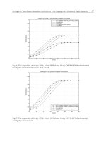
foot by redistributing pressures. High plan-
tar pressures can be seen on the graph pro-
duced by insole pressure sensors (Parotec
system, Germany) (Figure 3.18), when the
patient used her own shoes (Panel A), and
after the prescribed insole and shoe were
used (Panel B); pressures applied to the sole
of the patient’s foot during heel strike, mid-
support and push-off phase of walking with
the patient’s original shoe (left graph), and
with the custom-made insole (right graph)
areshowninPanelC.
After 6 weeks the ulcer heeled com-
pletely (Figure 3.19).
Keywords: Convex triangular foot; hallux
valgus; quintus varus
HALLUX VALGUS,
OVERRIDING TOE, CLAW
TOES, EDEMA
A 68-year-old female patient with type 2
diabetes diagnosed at the age of 45 years
attended the outpatient diabetic foot clinic
Anatomical Risk Factors for Diabetic Foot Ulceration 53
Figure 3.17 Extra-depth shoes and cus-
tom insoles
for routine chiropody treatment. She was
being treated with insulin. The patient
had hypertension, advanced background
retinopathy which had been treated with
laser in both eyes, and diabetic nephropa-
thy (urine protein: 2.6 g/24 h). On exami-
nation, she had severe diabetic neuropathy
and gross ankle edema due to nephropathy.
Peripheral pulses were normal and the ankle
brachial index was 1.1 on both feet. Mild
hallux valgus, claw toes, overriding of the
second to the third toe and lateral drip of the
toes were observed (Figures 3.20 and 3.21).
Callus formation at the inner aspect of the
first and on the second metatarsal heads
was noted. Fat pads on the first, second and
third metatarsal heads were displaced dis-
tally to the base of the proximal phalanges
due to clawing of the toes. A superficial
painful infected ulcer at the dorsum of the
second toe was also present, due to over-
riding and clawing of the toes. Debridement
of the callus was carried out. The patient
was put on clindamycin for 2 weeks. Treat-
ment with frusemide 40 mg daily was also
commenced to reduce edema. Extra depth
shoes and orthotic insoles were prescribed
in order to reduce the pressure on the plan-
tar area and the friction from the shoes on
the deformed toes.
The correct shoes and orthotic insoles
are often enough to reduce the risk for foot
ulceration in the majority of the patients
with foot deformities and loss of pro-
tective sensation. In addition, edema has
a detrimental effect on the foot at risk,
as it reduces local blood supply and has
been associated with increased risk for
ulceration. Therefore, reduction of ankle
edema is recommended for patients at risk
for ulceration.
Beyond diabetic nephropathy, other cau-
ses of ankle edema in diabetes include heart
failure and diabetic neuropathy. Edema due
to neuropathy is not rare. This form of
edema results from sympathetic denerva-
tion, which leads to loss of the vasomotor
reflex upon standing, arteriovenous shunt-
ing and increased capillary pressure. Neu-
ropathic edema responds to the admin-
istration of the sympathomimetic agent
ephedrine.
54 Atlas of the Diabetic Foot
Figure 3.18 In-shoe plantar pressure measure-
ments (A) when the patient used her own shoes;
and (B) after wearing the prescribed insole and
shoe. (C) Pressures on the sole of the patient’s
foot during walking in her own shoes (graph on
left), and when wearing the custom-made insole
(graph on right)
Keywords: Hallux valgus; toe overriding;
claw toes; edema
ONYCHOMYCOSIS;
HALLUX VALGUS AND
HAMMER TOE DEFORMITY
A 68-year-old female patient with dia-
betes diagnosed at the age of 50 years and
Figure 3.19 The neuropathic ulcer shown in
Figure 3.15 after it had healed following
6 weeks of treatment
treated with insulin, was referred to the
outpatient diabetic foot clinic because of
foot deformities and recurrent superficial
toe ulcers.
The patient had findings of periph-
eral neuropathy. Peripheral pulses were
palpable. No other diabetic complications
were present.
Onychomycosis was noticed and con-
firmed by direct microscopic examination
of nail specimens. The skin on her feet was
dry; hallux valgus and hammer toe defor-
mity of her second left toe were observed.
Tiny superficial ulcers on the dorsum of her
second and third toes due to shear pres-
sure were present, as well as a small ulcer
on the inner aspect of her great toe, and
Anatomical Risk Factors for Diabetic Foot Ulceration 55
Figure 3.20 Hallux valgus, over-
riding toe, claw toes and edema
a hemorrhagic callus on the tip of the left
great toe (Figures 3.22 and 3.23).
Mild hallux valgus and hammer toe
deformity on the right second and third toes
was apparent, with a superficial ulcer on
the dorsum of the second toe (Figure 3.24).
Hammer toe is a complex deformity con-
sisting of contraction (hyperflexion) of the
proximal interphalangeal joint, while the
metatarsophalangeal joint is either dorsi-
flexed or in the neutral position. The distal
interphalangeal joint may be in the neu-
tral position, hyperextended or in plantar
flexion (Figure 3.25). Hammer toe may be
flexible or rigid.
Overriding toe deformity often occurs
in the second and the fifth toes. The
cause of the overriding fifth toe is mainly
congenital, while a second overriding toe is
acquired and multifactorial. Elongation and
laxity of the plantar synovium bursa of the
metatarsal joint result in dorsal subluxation
of the affected joint. The second toe lacks
plantar interossei muscles, therefore lum-
brical muscles predominate, causing dor-
siflexion of the toe. Subluxation of the
metatarsophalangeal joint results in shrink-
age of the dorsal synovium bursa and the
dorsal interossei muscles. Further atrophy
of the intrinsic muscles contributes to the
development of the deformity which may
be fixed or flexible.
Debridement of the calluses and instruc-
tion in foot care was provided to this
patient, and shoes with a high toe box and
shock absorbing insoles were prescribed.
56 Atlas of the Diabetic Foot
Figure 3.21 Hallux valgus, overriding toe, claw toes and edema. Plantar aspect of the foot
illustrated in Figure 3.20
Keywords: Onychomycosis; hallux valgus;
hammer toe deformity
MALLET TOE
A mallet toe consists of plantar flexion of
the distal interphalangeal, and neutral posi-
tion of metatarsophalangeal and proximal
interphalangeal joints (Figure 3.26).
Toe deformities (hammer, claw, curly,
mallet toe and overriding of toes) are
unknown in non-shoe wearing populations.
Their incidence varies from 2 to 20%, and
increases with age. Women are affected
four to five times more often than men.
Most people have no underlying disease,
although neuromuscular diseases and in-
flammatory arthropathies may be accompa-
nied by such toe deformities.
Toe deformities are more common in
people with diabetes, due to muscle atrophy
and limited joint mobility. Deformities such
as those described above, when present
in a patient with loss of sensation due
to diabetic neuropathy, pose a risk for
the development of neuropathic ulcers, as
prominences are susceptible to skin-on-
shoe friction. Patients are instructed to
check their feet every day. Shoes with a
high toe box protect the deformed toes from
ulceration.
Anatomical Risk Factors for Diabetic Foot Ulceration 57
Figure 3.22 Hallux valgus, toe overriding and
onychomycosis
Figure 3.24 Mild hallux valgus and hammer
toe deformity on the right second and third toes,
with a superficial ulcer on the dorsum of the
second toe. Right foot of the patient whose feet
are shown in Figures 3.22 and 3.23
Figure 3.23 Hammer toe deformity of the second, third and fourth toes, hemorrhagic callus and
onychomycosis. Anterolateral view of the foot shown in Figure 3.22
58 Atlas of the Diabetic Foot
Figure 3.25 Hammer toe
Figure 3.26 Mallet toe
Keywords: Mallet toe; toe deformities
PROMINENT METATARSAL
HEADS AND CLAW TOES
A 65-year-old male patient with longstand-
ing type 2 diabetes attended the outpatient
diabetic foot clinic for callus removal and
treatment of ulcers on the tip of his second
and fifth right toes (Figure 3.27).
On examination, he had bounding pedal
pulses, and severe peripheral neuropathy.
Metatarsal heads were prominent, and claw
toes were present.
Claw toe deformities may cause promi-
nence of metatarsal heads with subsequent
Anatomical Risk Factors for Diabetic Foot Ulceration 59
Figure 3.27 Prominent metatarsal heads and claw toes
callus formation and ulceration. Ulcers may
develop at the tips of the claw toes, since
they are abnormally exposed to pressure
during walking.
Protective footwear (high toe box and
orthotic insoles) was provided to this
patient.
Keywords: Claw toes; prominent metatar-
sal heads
POSTOPERATIVE HALLUX
VALGUS AFTER SECOND
TOE REMOVAL
A 55-year-old female patient with type 2
diabetes diagnosed at the age of 40 years,
treated with insulin, and erratic glycemic
control, visited the diabetic foot clinic
because of recurrent callus formation. She
had background retinopathy, hypertension,
and severe peripheral neuropathy, and a
history of amputation of her left second
toe 3 years previously due to osteomyelitis
after a perforated ulcer.
After removal of her second toe, her
left great toe gradually dislocated to a val-
gus posture, underriding the adjacent (third)
toe (Figure 3.28). Gross callus formation
developed at the medioplantar aspect of the
first metatarsal head, which caused con-
stant discomfort during walking and danc-
ing (Figure 3.29). Callus was also noticed
over the third metatarsal head.
At the outpatient clinic the callosity
was removed and a full thickness ulcer
revealed. More callus built up quickly as
a result of the very active lifestyle of the
patient and her refusal to wear appropriate
footwear, she therefore had to attend the
clinic every week.
A plain X-ray showed disarticulation
of the left second toe, dislocation of the
60 Atlas of the Diabetic Foot
Figure 3.28 Hallux valgus and toe overriding after second toe disarticulation
Figure 3.29 Gross callus formation on the first and third metatarsal heads after second toe
disarticulation
Anatomical Risk Factors for Diabetic Foot Ulceration 61
metatarsophalangeal joint of the great toe,
medial pronation of the first metatarsal
head, and hallux valgus deformity with
rotation, together with dislocation of the
sesamoids, and arthritis; necrosis of the
head of the third metatarsal bone was also
evident (Figure 3.30).
She was referred to the orthopedic
department where her second metatarsal
was removed. The hallux valgus deformity
was corrected by arthrodesis of the metatar-
sophalangeal joint.
After the operation there was no signif-
icant callus development within the next
3 months (Figure 3.31).
Keywords: Hallux valgus; prophylactic
surgery; second ray amputation
FIRST RAY AMPUTATION
A 72-year-old male patient with type 2 dia-
betes diagnosed at the age of 56 years and
Figure 3.30 X-ray image of the foot illustrated in Figures 3.28 and 3.29. Disarticulation of the
left second toe, dislocation of the metatarsophalangeal joint of the great toe, medial pronation of
the first metatarsal head, and hallux valgus deformity with rotation, together with dislocation at the
sesamoids and arthritis; necrosis of the head of the third metatarsal bone is also evident
62 Atlas of the Diabetic Foot
Figure 3.31 Photograph of the foot
shown in Figures 3.28–3.30 3 months
after arthrodesis of the first metatar-
sophalangeal joint and second ray am-
putation. Note the absence of signifi-
cant callus formation
treated with sulfonylurea and metformin,
attended the outpatient diabetic foot clinic
because of a deep, infected neuropathic
ulcer under the first metatarsal head. His
diabetes control was acceptable (HBA
1c
:
7.6%). He had a history of hypertension and
dyslipidemia and was being treated with
a combination of angiotensin converting
enzyme inhibitor with diuretic and simva-
statin. He had neuropathic pain in his feet.
He described the pain as a burning sensa-
tion which worsened at night. On examina-
tion, an ulcer 3 × 3 cm in size and 1.5 cm
in depth surrounded by callus formation
was seen on the left first metatarsal head.
Its base was sloughy. Second left claw
toe deformity was also observed. Pedal
pulses were palpable, the ankle brachial
index was 1.0; the patient had findings
of severe peripheral neuropathy (loss of
sensation of light touch, pain, tempera-
ture, vibration, and 5.07 monofilaments; the
vibration perception threshold was 35 V on
both feet).
A plain radiograph revealed osteomyeli-
tis of the first metatarsal head extend-
ing to the base of the proximal pha-
lanx of the great toe. Cultures of the
base of the ulcer revealed the presence
of Staphylococcus aureus and Escherichia
coli. Based on the results of the swab cul-
ture he was given amoxicillin and clavu-
lanic acid for 2 weeks. After this time
a first ray amputation under local anes-
thesia was carried out. A culture of the
bone was negative for pathogens but patho-
logic examination of the resected bone
showed findings of chronic osteomyelitis
(granulated fibrous tissue with a predom-
inance of plasma cells and lymphocytes
and involucrum formation at the perios-
teum). The postoperative period was free
from complications and the wound healed
well in 2 weeks (Figure 3.32). Antibiotics
Anatomical Risk Factors for Diabetic Foot Ulceration 63
Figure 3.32 First ray amputation due to osteomyelitis
were discontinued 7 days after the opera-
tion and he was put on imipramine for the
neuropathic pain.
Removal of the great toe results in dys-
function of the foot during both stance
and propulsion. This disability is related
to the length of the removed metatarsal
shaft. Most surgeons preserve the longest
metatarsal shaft possible. The base of the
proximal phalanx should be preserved,
in order to keep the attachment of the
short flexor of hallux intact, thus keep-
ing sesamoids in place and maintaining
the windlass mechanism. This mechanism
protects the first metatarsal head from
overloading during the propulsion phase
of gait. In the case of an obligatory
removal of hallux — due to osteomyelitis of
the proximal phalanx — the surgeon should
preserve all uninvolved portions of the
metatarsal, except the avascular sesamoids
and their fibrocartilaginous plate. A hal-
lux disarticulation at the metatarsopha-
langeal joint exposes the head of the third
metatarsal to abnormally high pressure dur-
ing stance, and may displace the second toe
medially.
Keywords: First ray amputation; histology;
chronic osteomyelitis
CALLUS UNDER BONE
PROMINENCE
A 72-year-old male patient with type 2 dia-
betes attended the outpatient diabetes clinic
for his usual follow-up. His diabetes control
was fair with glibenclamide. He was free