DIABETIC NEUROPATHY: CLINICAL MANAGEMENT - PART 3 potx
Bạn đang xem bản rút gọn của tài liệu. Xem và tải ngay bản đầy đủ của tài liệu tại đây (2.43 MB, 52 trang )
6
Effectors—Sonic Hedgehog
and p38 Mitogen-Activated Protein Kinase
Sally A. Price, Rebecca C. Burnand, and David R. Tomlinson
SUMMARY
This chapter covers the identification of mitogen-activated protein kinases as early stage
transducers of the damaging effects of glucose on peripheral nerves. They are activated by
several metabolic consequences of hyperglycemia, in particular oxidative stress, osmotic stress,
and advanced glycation end products. Inhibition of one group of mitogen-activated protein
kinases––the p38 group—prevents the development of reduced nerve conduction velocity in
experimental diabetes; such inhibition can also be achieved by an aldose reductase inhibitor,
giving an explanation for the mechanism underlying the damaging effect of the polyol pathway.
The effect of treatment is also described with sonic hedgehog in preventing reduced nerve con-
duction velocity and normalising expression of genes coding for endoskeletal proteins, which
may be instrumental in preserving the integrity of the distal axon.
Key Words: Sonic hedgehog; p38 MAP kinase; nerve conduction; gene expression; axonal
endoskeleton.
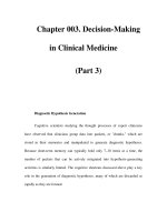
INTRODUCTION
The development of potential new therapies for diabetic neuropathy has been sporadic
over the last 20 years. In general, the process has been boosted by a prospective aetio-
logical mechanism reaching consensus among scientists together with the development
of drugs to counteract it. The polyol pathway and aldose reductase inhibitors provide a
classical example. As is shown in Fig. 1, interest in the polyol pathway rose dramatically
in the 1980s, peaking at around 1990; thereafter there has been a steady decline as clin-
ical findings indicated that the hypothesis was inapplicable to complications, at least as
a sole explanation of pathogenesis. Subsequently, no hypothesis has reached such a con-
sensus and the development of potential novel therapeutics has virtually stalled.
This chapter attempts to revitalize the process by proposing two new hypotheses to
explain the development of diabetic neuropathy. These are not mutually exclusive;
indeed it is instrumental that more than one set of pathogenetic mechanisms coexist and
act in concert. If these hypotheses are cogent, then new avenues for development of
therapeutics open up.
It has been obvious for many years that, if glucose itself is the damaging agent in the
initial aetiology of neuropathy, then there must be some processes that are sensitive to
From: Contemporary Diabetes: Diabetic Neuropathy: Clinical Management, Second Edition
Edited by: A. Veves and R. Malik © Humana Press Inc., Totowa, NJ
91
glucose and are interpolated between hyperglycemia and the onset of neurodegeneration.
We have made an extensive study of the way in which the mitogen-activated protein
kinases (MAPKs), and especially p38 MAPK, are activated directly by glucose and
indirectly by the osmotic and oxidative stresses that it induces in diabetes (1,2). In this
chapter is presented and discussed evidence for involvement of p38 MAPK in func-
tional changes and its inhibition as a therapeutic strategy considered.
The influence of long-term trophic support and its defects in diabetes on the devel-
opment of neuropathy have been examined (3). It is clear from this that more than one
neurotrophic factor is defective in diabetes and reversal of this possibly requires a pleio-
typic response characteristic of several factors. It is possible that agents that govern
multiple developmental changes may exert just such a broad–based influence. Such a
factor is sonic hedgehog (Shh) and this chapter begins with consideration of its poten-
tial influence and the novel therapeutic opportunities that it might present (4).
SONIC HEDGEHOG AND DIABETIC NEUROPATHY
The hedgehog proteins are a highly homologous family of proteins that are widely
expressed during development. There are three known mammalian homologues sonic
(Shh), desert (Dhh), and indian (Ihh). Treatment of the streptozotocin (STZ) rat model of
diabetes with a fusion protein containing human recombinant Shh and rat immunoglobin
G (Shh–IgG) ameliorates a range of diabetes-induced functional and structural disorders
of the peripheral nerve. For example, motor and sensory nerve conduction velocities in
the lower limbs are both increased to values comparable to that of nondiabetic animals
(4). In addition, deficits in nerve growth factor and the related peptide substance P, shown
in diabetic rats (5), are not present in rats treated with Shh–IgG (4).
92 Price et al.
Fig. 1. Publications per year on the sorbitol/polyol pathway as indexed by PubMed
( />There is a clear disruption in the gene expression of hedgehog genes in the periph-
eral nervous system of diabetic animals. The mRNA encoding Dhh is reduced in the
sciatic nerve of the diabetic rat (4). In addition, shh was downregulated in the dorsal
root ganglion (DRG) neurons of diabetic animals at 8 weeks duration of diabetes
(Burnand et al., unpublished observations). The mechanism by which treatment with
Shh–IgG restores functional deficits in the nerve is unknown.
The Hedgehog Family of Proteins
The name hedgehog comes from the spiky processes that cover the larval cuticle in
hh homozygotes. The hedgehog proteins (Hh) are a family of morphogens that act in a
dose dependent manner after being secreted from their tissue source; they exert their
effect by altering gene expression. The hedgehog gene (Hh) was first identified in
Drosophila embryos, as a gene encoding for a protein implicated in segment polarity
(6). Since then, most studies in Drosophila have focused on the role of hedgehog in
regulating the growth and patterning of the wing and other appendages (7).
Three mammalian hedgehog homologues have been found and are named Shh,
Dhh, and Ihh (8). Two homologues have been found in fish and are named echidna
and tiggywinkle hedgehog (9,10).
The multiple hh genes of vertebrates have presumably arisen by duplication and sub-
sequent divergence of a single ancestral hh gene. Although shh, ihh, and dhh are highly
homologous, shh is closer to ihh than dhh in sequence identity. Pathi et al. (11) have
shown that the three proteins have the ability to function similarly, but with different
potencies, hence the proteins can substitute for each other. They showed that the rank
order of potencies in each of the contexts they tested was Shh > Ihh > Dhh.
Shh is expressed in numerous tissues including the central nervous system, the
peripheral nervous system, limbs, somites, the skeleton, and skin. It has numerous
roles during mammalian development, directing pattern formation, and inducing cell
proliferation.
Humans or mice lacking Shh develop holoprosencephaly and cyclopia because of a
failure of separation of the lobes of the forebrain (12). Shh organises the developing
neural tube by establishing distinct regions of homeodomain transcription factor pro-
duction along the dorsoventral axis (13). These transcription factors, including Nkx,
Pax, and Dbx family members, specify neuronal identity. Shh acts directly on target
cells and not through other secreted mediating factors, to specify neuronal cell fate (14).
It also has important known patterning roles in the formation of other tissues including
the brain (15) and the eye (9). In addition to the many functions of Shh in determining
cell fate, it also has roles in controlling cell proliferation and differentiation in neuronal
and nonneuronal cell types.
The numerous responses to Shh are achieved by controlling the production, amount,
and biochemical nature of the signal itself, including covalent modification of Shh.
During development, the expression of Dhh mRNA is highly restricted. Its expres-
sion has been shown in the Sertoli cells of the developing testes (16,17) and in the
Schwann cells of the peripheral nerve (18). Male Dhh-null mice are sterile and fail to
produce mature spermatozoa (16). The peripheral nerves of Dhh-null mice are also
highly abnormal. The perineurial sheaths surrounding the nerve fascicles are abnormally
Effectors—Sonic Hedgehog and p38 MAPK 93
thin and extensive microfasicles consisting of perineurial like cells are formed within
the endoneurium. The nerve tissue barrier is permeable, and the tight junctional arrays
between, adjacent perineurial cells are abnormal and incomplete (18).
Ihh has two known roles in vertebrate development. The first is in the formation of
the endoderm where Ihh is critical for the differentiation of the visceral endoderm (19).
The second is in postnatal bone growth (20) where Ihh appears to coordinate growth and
morphogenesis, a suggestion has also been made proposing a role for Ihh in healing
long bone fractures (21).
Until recently, it was thought that hedgehog proteins directly bind to a single recep-
tor named Patched (Ptc). Ptc, located on the surface of responding cells, is a 1500 amino
acid glycoprotein that constitutes 12 membrane-spanning domains (22,23). Two human
homologues of Ptc have been identified named Ptc1 and Ptc2 (24). Ptc1 is the main
receptor for Shh, Ihh, and Dhh, the function of Ptc2 is unknown. It has been shown that
a number of isoforms of Ptc2 exist it is proposed that the expression of the different iso-
forms is associated with the “fine-tuning” of the Hh response (25).
Ptc is required for the repression of target genes in the absence of Hh. The Hh signal
induces target gene expression by binding to and inactivating Ptc. Inactivation of Ptc
allows smoothened to become active; Smo is a 115 kDa transmembrane protein that is
essential for transducing the Shh signal, only one human homolog is known. It is not yet
clear whether the inhibition of Smo by Ptc is the result of direct or indirect interaction.
Either way, the binding of Hh to Ptc results in a change that allows smoothened to trans-
duce the signal. In humans and mice, the loss of ptc function causes medulloblastomas,
tumors of the cerebullum, and other developmental abnormalities resulting from the
inappropriate expression of Shh target genes (26,27).
In addition to repressing target gene transcription, Ptc also regulates the movement
of Hh through tissues; the binding of Hh to Ptc limits the spread of Hh from its source.
In Drosophila producing mutant Ptc, Hh can be detected at distances greater than
those producing the wide-type protein (28). The binding of Shh to Ptc induces rapid
internalization of Shh into endosomes, the fate of Shh after internalization is not yet
known (29).
In 2002 it was shown that Shh also directly binds to another protein called megalin
(30). This single chain protein is approx 600 KDa and consists of a C-terminal cyto-
plasmic domain, a single transmembrane domain and an extremely large ectodomain
(31). Megalin functions as an endocytic receptor which mediates the endocytosis of lig-
ands including insulin (32), the presence of functional motifs at the C-terminal cyto-
plasmic domain suggest that this protein may also have a role in signal transduction
(33). The phenotypes of megalin deficient mice are consistent with phenotypes of mice
deficient in Shh and Smo (34,35).
The signal transduction pathway downstream to Ptc and Smo is not well under-
stood. Ultimately, it results in the nuclear translocation of the Gli proteins. The Gli
genes encode transcription factors that share five highly conserved tandem C
2
–H
2
zinc
fingers and a consensus histidine–cysteine linker sequence between the zinc fingers
(36). The Drosophila homolog is called cubitus interruptus (Ci). Ci is regulated
post-transcriptionally; the full length Ci protein consists of 155 amino acid residues
(Ci-155) (37,38).
94 Price et al.
In the absence of a Hh signal, Ci forms a tetrameric complex with proteins named:
Costal-2, Fused, and Suppressor of fused at the microtubules (39,40). In this complex
form Ci is cleaved to form a 75 amino acid residue (Ci
[rep]
) (41) that retains the zinc
finger domain and translocates to the nucleus to repress downstream target genes (42).
In some cells, proteolysis of Ci seems to be dependent on protein kinase-A mediated
phosphorylation (43). Transduction of the Hh signal inhibits proteolysis of Ci, result-
ing in an accumulation of the full-length protein. On translocation to the nucleus this
activator form stimulates transcription of target genes. In the absence of Hh signal not
all full length Ci is cleaved, a residual amount escapes but is prevented from activating
target genes by its retention in the cytoplasm and active nuclear export (41,44) thus, it
seems likely that there are many levels of control over Ci activity that remain to be
fully elucidated.
There are three known Gli homologues in mammals: Gli1 (also referred to as Gli), Gli
2, and Gli 3. All three Gli homologues have been tested for separate functional domains.
C-terminally truncated forms of both Gli2 and Gli3 that resemble the truncated form of
Ci have been shown to repress reporter gene expression in cell lines or Shh targets in vivo
(45,46). Gli1 does not seem to contain a represser domain, instead only functioning as a
transcription activator (46).
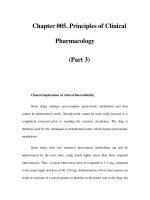
Effects on Indices of Diabetic Neuropathy
As previously mentioned, treatment of the diabetic rat with Shh–IgG reverses a
number of indices of diabetic neuropathy including, deficits in nerve conduction velocity.
Figure 2 shows sensory and motor nerve conduction velocity values at 8 and 12 weeks
duration of diabetes in the STZ model of diabetes. There are clear deficits in the dia-
betic animals that are reversed by treatment with Shh–IgG. Shh–IgG treatment had
no effect on body weight or glycemia in diabetic rats, implying that the severity of
diabetes was unaffected by Shh–IgG. Shh–IgG administration had no effect on the
concentration of polyol pathway components in peripheral nerve (Burnand et al.,
unpublished).
Shh protein signaling ultimately leads to the translocation of the Gli proteins to the
nucleus where they act as transcription factors. Therefore, the mechanism by which
Shh–IgG exerts its effects is likely to be transcription based. In both human and exper-
imental models of diabetes there are a wide range of structural changes in the periph-
eral nerve. These changes include a loss in the number of myelinated fibres and
paranodal demyelination (47,48). There is also a reduction in the capacity of peripheral
nerves to regenerate following injury (49,50). Actin, tubulin, and the neurofilament
proteins are the main cytoskeletal proteins essential for structural integrity of the axon.
Other accessory proteins including numerous actin binding proteins are present in the
peripheral nerve and produce a structure of extreme complexity and versatility.
Abnormalities in the production and processing of structural proteins have been widely
reported in diabetic neuropathy (5,51,52). Evidence gathered in our laboratory shows
that treatment of the diabetic rat with Shh–IgG reverses abnormalities in the gene
expression of a range of structural proteins as shown in Fig. 3. This restoration in gene
expression may form part of the mechanism by which treatment with Shh–IgG corrects
deficits in nerve conduction velocity in diabetic rats.
Effectors—Sonic Hedgehog and p38 MAPK 95
96 Price et al.
Fig. 2. Motor and sensory nerve conduction velocities in control (open columns), diabetic
(gray columns) and sonic hedgehog (diagonal hatching)-treated diabetic rats. Diabetes caused
significant (p < 0.01) slowing of both at 8 and 12 weeks, which was normalised by sonic hedge-
hog at both durations.
To date, the work conducted on the use of Shh–IgG as a potential therapeutic agent
in the treatment of diabetic neuropathy has resulted in positive outcomes. No adverse
side effects have been observed at 12 weeks duration of diabetes. A longer term study
is now necessary to determine the longer term potential of this promising new therapy.
MITOGEN-ACTIVATED PROTEIN KINASES
MAPKs are a family of enzymes involved in transducing signals derived from the
extracellular environment. There are three main subtypes of MAPKs: extracellular reg-
ulated kinases (ERKs), c-Jun N-terminal kinases (JNKs), and p38 MAPKs. All family
members are activated by dual phosphorylation of a consensus sequence, Thr-Xxx-Tyr
by MAPK kinases. Upstream of these are the MAPK kinase kinases, thereby forming
a three kinase cascade. There are fewer different kinases at each subsequent level of
the cascade, resulting in refinement of the signal. Specificity may be achieved by
stimulus-selective pathways, distinct cellular pools of kinases, or the presence of scaf-
fold proteins required for the interaction of certain kinases. Activated MAPKs can
phosphorylate targets within the cytoplasm, such as cytoskeletal proteins and other
kinases, or they may be translocated to the nucleus where they activate transcription
factors and mediate gene expression.
Extracellular Signal-Regulated Kinases
ERK1 was identified as a kinase activated by insulin, having a pivotal role in transduc-
ing mitogenic signals by converting tyrosine phosphorylation into the serine/threonine
phosphorylations that regulate downstream events (53). ERK2 and ERK3 were subse-
quently identified (54). ERK1 and ERK2 have 83% amino acid homology, are expressed
in most tissues to varying degrees, and are activated by growth factors, phorbol esters and
serum. ERK1/2 activation is typically triggered by receptor tyrosine kinases and G
protein-coupled receptors at the cell surface. These activate the small GTP-binding
protein Ras, allowing signaling through the Raf/MEK/ERK cascade. Downstream, ERK1/2
activates other kinases (e.g., RSKs, MSKs, and MNKs), membrane components (e.g.,
CD120a, Syk, and calnexin), cytoskeletal proteins (e.g., neurofilament) or nuclear
targets (e.g., SRC1, Pax6, NF-AT, Elk1, MEF2). ERK3 displays ubiquitous expression
and responds to various growth factors (54). It is only 42% identical to ERK1 and differs
from ERK1/2 in that it is a constitutively active nuclear kinase and does not phosphory-
late typical MAPK substrates (54,55). The fifth mammalian ERK kinase is designated
ERK5 or big MAPK1 (BMK1) because it is twice the size of the other ERK family mem-
bers and has a distinct C-terminal (56,57). Erk5 contributes to Ras/Raf signaling (56,58)
and is activated in response to growth factors and stress (56,59). ERK6 is a protein kinase
involved in myoblast differentiation (60) but is usually referred to as p38γ. ERK7 and
ERK8 have also been cloned recently (61,62).
Effectors—Sonic Hedgehog and p38 MAPK 97
Fig. 3. In dorsal root ganglia of rats with 12 weeks streptozotocin diabetes there was a gen-
eral reduction in gene expression (mRNA levels) for endoskeletal proteins; some of these reduc-
tions were normalised by treatment with sonic hedgehog. Coding: open circles—β-actin; filled
squares—γ-actin; filled circles—NFL, neurofilament light subunit; open squares—NFM, neuro-
filament medium subunit; half-filled circles—NFH, neurofilament heavy subunit; half-filled
squares—α-tubulin.
C-Jun N-Terminal Kinases
JNK was identified as the kinase that phosphorylated c-Jun after exposure of cells to
transforming oncogenes and ultraviolet light (63). It was thus recognized as an important
signaling cascade for modulating the activity of distinct nuclear targets. There are 10
mammalian isoforms of JNK arising from alternate splicing of the 3 JNK genes. The JNK
proteins are activated by MAP kinase kinases such as MKK4 and MKK7 and upstream of
these MAP kinase kinase kinases including MLKs and ASK. Scaffold proteins such as JIP
and β-arrestin 2 are also integral to the JNK signaling module, determining proximity and
specificity. JNK proteins differ in their associations with scaffold proteins and also in their
interaction with downstream targets. Defined substrates of JNK total at about 50–60 pro-
tein and include cytoskeletal proteins (e.g., neurofilament, tau, and microtubule associated
proteins), mitochondria (e.g., bim), and nuclear proteins (c-Jun, ATF-2, and Elk-1) (64).
Roles for the different isoforms of JNK are gradually becoming elucidated. It is known
that basal activity of JNK1 is far greater than that of JNK2 and JNK3. Coupled with the
fact that JNK1 knockout mice are defective/embryonically lethal, this suggests a greater
role for JNK1 under physiological conditions. JNK3 knockout mice are healthy and are
resistant to excitotoxic brain insults (65), suggesting a greater pathological role for this
isoform. In addition tissue specific effects of the role have been described. In most situa-
tions, inhibition of JNK is detrimental, however in cells such as cardiac myocytes and
sensory neurones inhibition of JNK may confer protection.
Mitogen-Activated Protein Kinase p38
The p38 MAPK signal transduction pathway is activated by proinflammatory
cytokines and environmental stresses such as osmotic shock, ultraviolet radiation, heat,
and chemicals (see refs. 66–68 for reviews). There are four members of the p38 MAPK
family: p38α (69,70), p38β (71), p38γ (72), and p38δ (73), each encoded by a different
gene. The p38 MAPKs are phosphorylated and activated by MKK3 and MKK6 at thre-
onine and tyrosine residues and can mediate signaling to the nucleus (74). A large num-
ber of substrates have been described for p38, these include the transcription factors
ATF-2, Elk-1, cAMP response element binding proteins (CREB), and cytoplasmic targets
such as tau, MAPKAPK-2. p38 MAPKs are widely expressed, with at least 3 of the
genes being expressed in the peripheral nervous system (S Price, personal observation).
The effect of p38 activation in response to cellular stress is diverse, although the major-
ity of reports favour a role in cell death rather than cell survival for neuronal cells. p38
signaling has been proposed to mediate apoptotic signaling in response to a variety of
stimuli in neurons including oxidative stress in primary forebrain cultures (75), mesen-
cephalic cells (76), and cortical neurons (77), and NGF withdrawal in PC12 cells (78).
Conversely, p38 activation was not observed following NGF withdrawal in primary cul-
tures of sympathetic neurons (79) and NGF has been shown to increase p38 activation
in DRG in vivo (80). This suggests that activation of p38 alone does not predict a detri-
mental outcome. High basal activity of p38 has been described in the adult rat brain
(81), although the physiological roles of p38 activation have been sparsely investigated.
Stress Kinases—Mechanism of Damage
In 1993, the Diabetes Control and Complications Trial Research Group concluded that
the incidence and severity of diabetic complications are increased by poor glycaemic
98 Price et al.
control, indicating that hyperglycemia is likely to be the major causative factor. Several
consequences are known to result from excess glucose these include hyperosmolarity,
increased polyol pathway flux, oxidative stress, formation of advanced glycation end
products (AGE), and activation of protein kinase C. These pathways are integrally linked
with each other and with a variety of other cellular pathways. MAPK activation is impli-
cated in all these pathways, suggesting a pivotal role in transducing the effects of high
glucose in diabetic neuropathy.
Uptake of extracellular glucose without the dependency for insulin is a common fea-
ture of tissues affected by diabetic macrovascular complications. One major conse-
quence is an increased flux through the polyol pathway (Fig. 4). In this pathway, aldose
reductase converts glucose to sorbitol, and this is subsequently converted to fructose by
sorbitol dehydrogenase. Excessive flux through the polyol pathway leads to accumula-
tion of the poorly membrane permeable metabolites sorbitol and fructose in diabetic rats
(82). One consequence is that cells may be subjected to osmotic stress. This mechanism
is thought to account for the formation of sugar-induced cataractogenesis in diabetic rat
lens (83). The contribution of osmotic stress resulting from increased polyol pathway
flux in peripheral nerve is less well defined (84).
Extracellular osmotic stress may also occur in diabetic nerves as these are subject to
serum hyperosmolarity. Demonstrated a reduction in axonal size in myelinated fibres
and suggested this was, at least in part, because of shrinkage as a result of increased tis-
sue osmolarity (85).
Hyperosmolarity activates MAPKs in a variety of cell types (69,86,87), and therefore
it is plausible that hyperosmotic stress can activate MAPKs in diabetic neuropathy. In
aortic smooth muscle cells from normal rats, glucose activates p38 by a PKC-δ isoform-
dependent mechanism (88). However, at higher levels of glucose, p38 is activated by
hyperosmolarity through a PKC independent pathway. This suggests that different path-
ways may be activated simultaneously by high glucose. Furthermore, p38 has been
shown to mediate the effects of hyperglycemia-induced osmotic stress in vivo in the rat
mesenteric circulation (89).
In recent years, oxidative stress has come to the forefront of hypotheses proposed to
be causative of diabetic neuropathy. Numerous studies have shown that antioxidants
such as vitamin E (90–92), DL-α-lipoic acid (93–95), and taurine (96,97) can prevent
abnormalities in diabetic nerve. Oxidative stress results from an imbalance in the pro-
duction of reactive oxygen species and cellular antioxidant defence mechanisms. The
increased free radical production may then result in oxidization of various cellular com-
ponents including lipids, proteins, and nucleic acids. Components that are modified by
ROS may have decreased activity leading to widespread dysfunction including distur-
bances in metabolism and defective signaling pathways.
Oxidative stress in diabetic nerve may result from a variety of mechanisms including
increased flux through the polyol pathway (Fig. 4), endoneurial hypoxia, hyperlipi-
daemia, increases in free fatty acids, activation of PKC, activation of receptors for AGE,
and glucose itself. The major source of oxidative stress in cells is the production of reac-
tive oxygen species (ROS) and reactive nitrogen species (RNS). Naturally occurring
ROS and RNS usually have oxygen or nitrogen based unpaired electrons resulting from
enzymatic or nonenzymatic reactions. Examples include superoxide anion, hydroxyl
radical, nitrogen oxide, and peroxynitrite. High glucose inhibits ATP synthase resulting
Effectors—Sonic Hedgehog and p38 MAPK 99
in slowing of electron transfer in the mitochondria and increased production of super-
oxide ions (98). Superoxide ions are normally converted to hydrogen peroxide and
water by the enzyme superoxide dismutase. Hydrogen peroxide is also produced by
enzymatic transfer of two electrons to molecular oxygen by enzymes such as monoamine
oxidase and urate oxidase. Hydrogen peroxide is reduced by glutathione peroxidase,
myeloperoxidase, and catalase or nonenzymatic decomposition occurs through the fen-
ton reaction, producing the highly reactive hydroxyl radical (OH). The activity of both
superoxide dismutase and catalase was found to be decreased (but not reaching statisti-
cal significance) in peripheral nerve after 6 weeks of diabetes (99,100). Longer dura-
tions of diabetes (3 or 12 months) failed to show a decrease in either gene expression or
activity of either enzyme, an increase in catalase expression was reported at 12 months.
These results suggest that changes in SOD and catalase may be dynamic in diabetic
nerve. Superoxides can also react with NO, forming peroxynitrite (ONOO-), which rap-
idly causes protein nitration or nitrosylation, lipid peroxidation, DNA damage, and cell
death. In sciatic nerve of rats given a peroxynitrite decomposition catalyst, immunore-
activity for nitrotyrosine and poly ADP-ribose (PARP) was present only in diabetic ani-
mals (101), indicating that nitrosative stress is indeed present in animal models of
diabetic neuropathy.
Glutathione is another important cellular antioxidant that acts as a non-enzymatic
reducing agent, helping to keep cysteine thiol side chains in a reduced state on the
surface of proteins. The reduction of oxidized glutathione to reduced glutathione
(GSH), catalysed by glutathione reductase is dependent on NADPH as a cofactor (Fig. 4).
Increased flux through the polyol pathway can cause depletion of GSH (102,103), pos-
sibly as a result of competition between aldose reductase and glutathione reductase for
NADPH resulting in NADPH deficiency (104,105) but more likely because of a
decrease in total glutathione (106,107). Increased polyol pathway flux can also create
oxidative stress because of the reaction of NADH with NADH oxidase and mitochon-
drial overloading with NADH. The significance of polyol-induced oxidative stress is
100 Price et al.
Fig. 4. Interconnecting pathways for oxidative stress. The polyol pathway consumes NADPH,
compromising the glutathione cycle, reducing levels of oxidized glutathione and impairing con-
version of hydrogen peroxide to water by glutathione peroxidase. This favours the Fenton reac-
tion generating super-hydroxyl radicals.
highlighted by the fact that an aldose reductase inhibitor can prevent diabetes induced
lipid peroxidation in peripheral nerve (107).
ROS and RNS such as hydrogen peroxide, superoxide, and peroxynitrite activate
ERK, JNK, and p38 in a variety of in vitro models (108–112), whereas MAPK activa-
tion is now well documented in these in vitro models, there is a lack of evidence for
MAPK activation by ROS and RNS in vivo. Recently, however (113), showed that
ConA induced liver failure in mice resulted in TNF-α induced ROS production leading
to sustained activation of JNK, which could be prevented by an antioxidant. Depleted
GSH was also a consequence whereas there was less depletion in JNK1–/– mice, thus
establishing a link between ROS activation and MAPK activation in vivo. β-adreno-
receptor stimulation in cardiac tissues was also found to increase superoxide production
and lipid peroxidation with concomitant activation of p38, JNK, and ERK. These
changes could be prevented with the antioxidant Tempol (114). The relationship
between high glucose, oxidative stress, and MAPK activation may only be apparent
with more chronic hyperglycemia because acute (3h) glucose infusion in rats resulted
in oxidative stress as measured by MDA and total glutathione but not in activation of
ERK1/2 or p38 in liver (115). It will be of great significance to establish the existence
of oxidative stress-induced MAPK activation in diabetic neuropathy.
AGE exert their cellular effects by interacting with cell surface receptors, the best
characterized of these is the receptor for advanced glycation end products (RAGE). In
rat pulmonary smooth muscle cells it was demonstrated that RAGE activation can
induce ERK1/2 activated p21 (ras) and nuclear factor kB (NFkB) signaling (116).
Subsequently a role for p38 in RAGE-induced NF-κB-dependent secretion of proin-
flammatory cytokines was established (117). RAGE-induced activation of JNK is not
well documented but has been shown in RAGE-amphoterin induced tumour growth
(118) and high-mobility group protein-1 (HMGB1—a novel inflammatory molecule)
induced RAGE activation in human microvascular endothelial cells (119).
MAPK Activation in Sensory Neurones
In vitro models cannot replicate the chronic conditions of diabetes because primary
cells slowly, but progressively die in culture. Furthermore, the interaction between
neuronal and non-neuronal cells and the supply of nutrients cannot be mimicked
directly. However cell culture models do provide a means of isolating components
known to be important in diabetes and reduce the use of in vivo models. In primary
cultures of dorsal root ganglia neurons, high glucose activated p38, and JNK but not
ERK in a concentration-dependent manner (10–200 mM) following 16 hours treatment
(1). Oxidative stress in the form of hydrogen peroxide or diethyl maleate resulted in
activation of p38 and ERK but not JNK. Treatment with high glucose and oxidative
stress had an additive effect on activation of p38, suggesting different mechanisms
of activation.
Exposure of DRG neurones to high glucose and oxidative stress also resulted in a
decrease in cell viability as indicated by lactate dehydrogenase and MTT assays, meas-
urements of intact plasma membranes and mitochondrial function, respectively.
Concomitant treatment with a specific ERK pathway inhibitor (U0126) or a specific p38
pathway inhibitor (SB20210) prevented activation of ERK or p38, respectively and
Effectors—Sonic Hedgehog and p38 MAPK 101
prevented the decrease in cell viability. This suggests that activation of p38 and ERK by
glucose or oxidative stress is detrimental in sensory neurones.
Commercially available specific inhibitors of the JNK pathway that are easily solu-
ble have been lacking and therefore less is known about the role of the JNK pathway in
sensory neurons. Treatment with the peptide inhibitor JNK inhibitor 1 (120) resulted in
death of primary cultures of DRG neurons (121). This inhibitor appeared to be selective
for JNK because c-Jun phosphorylation was prevented, whereas there was no effect on
other MAPKs. The recent development of new and more soluble JNK inhibitors may
help elucidate the effects of JNK signaling in sensory neurones in diabetic neuropathy.
MAPK Activation and Neuropathy in Diabetes
To investigate the effect of diabetes on MAPK activation, antibodies were used that
either recognize an epitope found on all forms of a particular MAPK (total, -T) or an
epitope specific to the phosphorylated (activate) form (phosphorylated, -P). Immuno-
histochemical studies on normal rats showed that ERK was expressed in both neurones
and satellite cells of the DRG, whereas ERK-P was found exclusively in satellite cells.
In the sciatic nerve ERK-T and ERK-P immunoreactivity was seen in both axons and
Schwann cells. Western blotting indicated that DRG from diabetic animals showed an
increase in ERK-P relative to ERK-T for both the p42 (ERK2) and p44 (ERK1) iso-
forms after 8, 10, or 12 weeks diabetes (1,122). The increase in ERK-P was because of
activation in the satellite cells in the DRG. Activated p44 ERK was also found to be sig-
nificantly increased in the sural nerve of 12 week STZ rats (123). However, no changes
were found in ERK-P in the sural nerve in a separate study with the same duration of
diabetes (122).
In control DRG, JNK-T staining was found predominantly in neurones. JNK-P
showed a similar distribution; staining was observed in the cytoplasm of neurones, but
was absent from the nuclei. In sciatic nerve, JNK immunohistochemistry was restricted
to axons. JNK staining has also been documented in the ventral horn and motoneuron
perikarya (122). In diabetic animals, Western blotting revealed increased JNK activation
(p46 and p54/56) in the DRG (1,121,122). Increased levels of JNK-P in sciatic and sural
nerve from 12 weeks STZ-rats were also observed (123). p54 JNK has also been shown
to be elevated in the DRG and sural nerve in an alternative model of type 1 diabetes, the
BB rat (122). Immunohistochemistry of diabetic DRG showed that JNK-P is translo-
cated from the cytoplasm to the nucleus of neurones. In axons of the sciatic nerve, stain-
ing is increased in large myelinated fibers. In other studies carried out in STZ-rats in the
same laboratory, only certain isoforms of JNK were shown to be activated (S. A. Price
and D. R. Tomlinson, personal observations) or were shown to be increased but not
reaching statistical significance. Activation of JNK was related to the duration of dia-
betes (increased activation with longer durations) and to the blood glucose levels
(increased with higher blood glucose levels). Activated c-Jun, a transcription factor
known to be downstream of JNK, displays a similar pattern of activation to that of JNK
in diabetic rats (122).
Immunohistochemistry demonstrated that p38-T was also located in neuronal cells in
the DRG of control rats (Fig. 5). Similar to JNK-T, immunoreactivity was largely
restricted to the cytoplasm and appeared to be absent from the nuclei and satellite cells.
102 Price et al.
Effectors—Sonic Hedgehog and p38 MAPK 103
Fig. 5. Bar charts and Western blots showing the effects of Insulin, fidarestat and the p38
mitogen-activated protein kinases inhibitor, SB239063 on activation of mitogen-activated pro-
tein kinases p38 in dorsal root ganglia. The Western blot shows the effect of diabetes (UD), com-
pared with controls (C), and fidarestat-treated diabetes (DF) on total (p38-T) and phosphorylated
p38 (p38-P).
p38-P was present in the cytoplasm of neurones but more intense staining was also
observed in the nuclei of some cells. In sciatic nerve p38-T was expressed in axons and
Schwann cells and p-38-P was expressed in axons. Western blotting showed an increase
in p38-P in the DRG of diabetic animals (1), accompanied by predominantly nuclear
staining observed with immunohistochemistry. p38-P staining was also increased in the
sciatic nerve of diabetic animals (124) and staining became visible in Schwann cells as
well as axons. In the L1 spinal cord, diabetes also promoted p38 activation in motoneu-
ron cell bodies, as identified by colocalization of choline acetyltransferase. There was also
intense p38 activation in microglia and diffuse labeling in neuronal and non-neuronal cells
of the gray matter. Interestingly, in sural nerve biopsies from diabetic patients there is
an increase in both p38-T and p38-P (1).
All changes that have been observed in diabetic animals could be reversed with insulin
and also the aldose reductase inhibitor, fidarestat, indicating that activation is a conse-
quence of hyperglycemia (Figs. 5 and 6). Treatment of STZ-diabetic rats with the second-
generation p38 inhibitor SB 239063 (20 mg/kg per day) prevents activation of p38 in DRG
and sciatic nerve and also deficits in nerve conduction velocity observed in untreated
diabetic rats (124). The inhibitor used specifically inhibits the α- and β-isoforms of p38
that are the isoforms predominantly expressed in neuronal tissue (125) and has no effect
on other MAP, tyrosine, or lipid kinases (126). Treatment of diabetic rats with the aldose
reductase inhibitor fidarestat or insulin also prevented activation of p38 (Figs. 5 and 6)
104 Price et al.
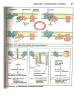
Fig. 6. Immunocytochemistry showing activated (phosphorylated) p38 in cytoplasm and,
especially, nuclei of both small and large neurone cell bodies in dorsal root ganglia. There was
marked activation in diabetes, which was specific, as is shown by the sections exposed to sec-
ondary, but no primary antibody. Both fidarestat and the p38 inhibitor, SB239063 prevented the
activation of p39 mitogen-activated protein kinase.
suggesting that p38 activation is a consequence of hyperglycemia and lies downstream
of the polyol pathway (124). This evidence supports the in vitro work by (1) and indi-
cates that MAPK activation is detrimental in diabetic neuropathy. Elucidation of the
down stream targets of p38 in sensory neurons may suggest therapeutic targets for dia-
betic neuropathy in the future.
CONCLUSION
This account makes it clear that one group of glucose transducers—the MAPKs—has
been identified. There can be little doubt of their involvement in early stages of the
registration of damaging effects of glucose to neurones and Schwann cells. This proba-
bly extends to other cell types and may contribute to vasculopathy, retinopathy, and
nephropathy. In so far as p38 MAPK might be pivotal, it is clear that aldose reductase
inhibitors that are as effective as fidarestat can remove that source of cellular damage.
This might be seen to negate the relevance of p38 because of the lack of clinical effi-
cacy of the aldose reductase inhibitors tested to date. However, it is clear that early inter-
vention is paramount and it is also likely that the level of inhibition may need to be
greater than has yet been achieved clinically.
It will be interesting to see the development of Shh analogues and to determine
whether these affect activation of MAPKs. If the two approaches to the consequences
of glucose intoxication are complimentary then we will have a clear gateway to what
some of us consider to be inevitable—multiple therapeutic approaches. This will add
interest as well as difficulty to clinical trials.
REFERENCES
1. Purves TD, Middlemas A, Agthong S, et al. A role for mitogen-activated protein kinases in
the aetiology of diabetic neuropathy. FASEB J 2001;15:2508–2514.
2. Tomlinson DR. Mitogen-activated protein kinases as glucose transducers for diabetic com-
plications. Diabetologia 1999;42:1271–1281.
3. Fernyhough P, Tomlinson DR. The therapeutic potential of neurotrophins for the treatment
of diabetic neuropathy. Diabetes Reviews 1999;7:300–311.
4. Calcutt NA, Allendoerfer KL, Mizisin AP, et al. Therapeutic efficacy of sonic hedgehog
protein in experimental diabetic neuropathy. J Clin Invest 2003;111:507–514.
5. Fernyhough P, Diemel LT, Hardy J, Brewster WJ, Mohiuddin L, Tomlinson DR. Human
recombinant nerve growth factor replaces deficient neurotrophic support in the diabetic rat.
Eur J Neurosci 1995;7:1107–1110.
6. Nusslein-Volhard C, Wieschaus E. Mutations affecting segment number and polarity in
Drosophila. Nature 1980;287:795–801.
7. Diaz-Benjumea FJ, Cohen SM. Wingless acts through the shaggy/zeste-white 3 kinase to
direct dorsal-ventral axis formation in the Drosophila leg. Development 1994;120:1661–1670.
8. Echelard Y, Epstein DJ, St-Jacques B, et al. Sonic hedgehog, a member of a family of putative
signaling molecules, is implicated in the regulation of CNS polarity. Cell 1993;75:1417–1430.
9. Ekker SC, Ungar AR, Greenstein P, et al. Patterning activities of vertebrate hedgehog
proteins in the developing eye and brain. Curr Biol 1995;5:944–955.
10. Currie PD, Ingham PW. Induction of a specific muscle cell type by a hedgehog-like pro-
tein in zebrafish. Nature 1996;382:452–455.
11. Pathi S, Pagan-Westphal S, Baker DP, et al. Comparative biological responses to human
Sonic, Indian, and Desert hedgehog. Mech Dev 2001;106:107–117.
Effectors—Sonic Hedgehog and p38 MAPK 105
106 Price et al.
12. Chiang C, Litingtung Y, Lee E, et al. Cyclopia and defective axial patterning in mice lack-
ing Sonic hedgehog gene function. Nature 1996;383:407–413.
13. Briscoe J, Pierani A, Jessell TM, Ericson J. A homeodomain protein code specifies pro-
genitor cell identify and neuronal fate in the ventral neural tube. Cell 2000;101:435–445.
14. Briscoe J, Chen Y, Jessell TM, Struhl G. A hedgehog-insensitive form of patched provides
evidence for direct long-range morphogen activity of sonic hedgehog in the neural tube.
Mol Cell 2001;7:1279–1291.
15. Kohtz JD, Baker DP, Corte G, Fishell G. Regionalizaton within the mammalian telen-
cephalon is mediated by changes in responsiveness to Sonic Hedgehog. Development
1998;125:5079–5089.
16. Bitgood MJ, Shen L, McMahon AP. Sertoli cell signaling by Desert hedgehog regulates the
male germline. Curr Biol 1996;6:298–304.
17. Bitgood MJ, McMahon AP. Hedgehog and Bmp genes are coexpressed at many diverse
sites of cell-cell interaction in the mouse embryo. Dev Biol 1995;172:126–138.
18. Parmantier E, Lynn B, Lawson D, et al. Schwann cell-derived Desert hedgehog controls
the development of peripheral nerve sheaths. Neuron 1999;23:713–724.
19. Becker S, Wang ZJ, Massey H, et al. A role for Indian hedgehog in extraembryonic endo-
derm differentiation in F9 cells and the early mouse embryo. Dev Biol 1997;187:298–310.
20. Vortkamp A, Pathi S, Peretti GM, Caruso EM, Zaleske DJ, Tabin CJ. Recapitulation of
signals regulating embryonic bone formation during postnatal growth and in fracture
repair. Mech Dev 1998;71:65–76.
21. Ito M, Yoshioka K, Akechi M, et al. JSAP1, a novel Jun N-terminal protein kinase (JNK)-
binding protein that functions as a scaffold factor in the JNK signaling pathway. Mol Cell
Biol 1999;19:7539–7548.
22. Hooper JE, Scott MP. The Drosophila patched gene encodes a putative membrane protein
required for segmental patterning. Cell 1989;59:751–765.
23. Nakano Y, Guerrero I, Hidalgo A, Taylor A, Whittle JR, Ingham PW. A protein with sev-
eral possible membrane-spanning domains encoded by the Drosophila segment polarity
gene patched. Nature 1989;341:508–513.
24. Motoyama J, Takabatake T, Takeshima K, Hui C. Ptch2, a second mouse Patched gene is
co-expressed with Sonic hedgehog. Nat Genet 1998;18:104–106.
25. Rahnama F, Toftgard R, Zaphiropoulos PG. Distinct roles of PTCH2 splice variants in
Hedgehog signalling. Biochem J 2004;378:325–334.
26. Goodrich LV, Milenkovic L, Higgins KM, Scott MP. Altered neural cell fates and medul-
loblastoma in mouse patched mutants. Science 1997;277:1109–1113.
27. Milenkovic L, Goodrich LV, Higgins KM, Scott MP. Mouse patched1 controls body size
determination and limb patterning. Development 1999;126:4431–4440.
28. Chen Y, Struhl G. Dual roles for patched sequestering and transducing Hedgehog. Cell
1996;87:553–563.
29. Incardona JP, Lee JH, Robertson CP, Enga K, Kapur RP, Roelink H. Receptor-mediated
endocytosis of soluble and membrane-tethered Sonic hedgehog by Patched-1. Proc Natl
Acad Sci USA 2000;97:12,044–12,049.
30. McCarthy RA, Barth JL, Chintalapudi MR, Knaak C, Argraves WS. Megalin functions as
an endocytic sonic hedgehog receptor. J Biol Chem 2002;277:25,660–25,667.
31. Oleinikov AV, Zhao J, Makker SP. Cytosolic adaptor protein Dab2 is an intracellular ligand
of endocytic receptor gp600/megalin. Biochem J 2000;347Pt 3:613–621.
32. Orlando RA, Rader K, Authier F, et al. Megalin is an endocytic receptor for insulin. J Am
Soc Nephrol 1998;9:1759–1766.
33. Hjalm G, Murray E, Crumley G, et al. Cloning and sequencing of human gp330, a Ca(2+)-
binding receptor with potential intracellular signaling properties. Eur J Biochem
1996;239:132–137.
34. Chen W, Burgess S, Hopkins N. Analysis of the zebrafish smoothened mutant reveals con-
served and divergent functions of hedgehog activity. Development 2001:128:2385–2396.
35. Litingtung Y, Chiang C. Specification of ventral neuron types is mediated by an antago-
nistic interaction between Shh and Gli3. Nat Neurosci 2000;3:979–985.
36. Ruppert JM, Kinzler KW, Wong AJ, et al. The GLI-Kruppel family of human genes. Mol
Cell Biol 1988;8:3104–3113.
37. Orenic TV, Slusarski DC, Kroll KL, Holmgren RA. Cloning and characterization of the
segment polarity gene cubitus interruptus Dominant of Drosophila. Genes Dev
1990;4:1053–1067.
38. Motzny CK, Holmgren R. The Drosophila cubitus interruptus protein and its role in the
wingless and hedgehog signal transduction pathways. Mech Dev 1995;52:137–150.
39. Sisson JC, Ho KS, Suyama K, Scott MP. Costal2, a novel kinesin-related protein in the
Hedgehog signaling pathway. Cell 1997;90:235–245.
40. Robbins E, Dobrzansky P, Haun K, et al. Efficacy of orally-administered CB-1093, an
NGF-inducing vitamin D receptor ligand, in the fimbria fornix lesion model (Abstract).
Society for Neuroscience Abstracts 1997;23:881.
41. Aza-Blanc P, Ramirez-Weber FA, Laget MP, Schwartz C, Kornberg TB. Proteolysis that is
inhibited by hedgehog targets Cubitus interruptus protein to the nucleus and converts it to
a repressor. Cell 1997;89:1043–1053.
42. Ohlmeyer JT, Kalderon D. Dual pathways for induction of wingless expression by protein
kinase A and Hedgehog in Drosophila embryos. Genes Dev 1997;11:2250–2258.
43. Jiang J, Struhl G. Protein kinase A and hedgehog signaling in Drosophila limb develop-
ment. Cell 1995;80:563–572.
44. Chen CH, von Kessler DP, Park W, Wang B, Ma Y, Beachy PA. Nuclear trafficking of
Cubitus interruptus in the transcriptional regualation of Hedgehog target gene expression.
Cell 1999;98:305–316.
45. Dai P, Akimaru H, Tanaka Y, Maekawa T, Nakafuku M, Ishii S. Sonic Hedgehog-induced
activation of the Gli1 promoter is mediated by GLI3. J Biol Chem 1999;274:8143–8152.
46. Shin SH, Kogerman P, Lindstrom E, Toftgard R, Biesecker LG. GLI3 mutations in human
disorders mimic Drosophila cubitus interruptus protein functions and localizaton. Proc
Natl Acad Sci USA 1999;96:2880–2884.
47. Thomas PK, Tomlinson DR. Diabetic and hypoglycaemic neuropathy. In Peripheral
Neuropathy 3 ed. Dyck PJ, Thomas PK, Griffin JW, Low PA, Poduslo JF, eds. W. B.
Saunders Co., Philadelphia, 1992, pp.1219–1250.
48. Yagihashi S. Nerve structural defects in diabetic neuropathy: Do animals exhibit similar
changes? Neurosci Res Commun 1997;21:25–32.
49. Longo FM, Powell HC, Lebeau J, Gerrero MR, Heckman H, Myers RR. Delayed nerve
regeneration in streptozotocin diabetic rats. Muscle Nerve 1986;9:385–393.
50. Ekstrom AR, Tomlinson DR. Impaired nerve regeneraton in streptozotocin-diabetic rats.
Effects of treatment with an aldose reductase inhibitor. J Neurol Sci 1989;93:231–237.
51. Mohiuddin L, Fernyhough P, Tomlinson DR. Reduced levels of mRNA encoding
endoskeletal and growth-associated proteins in sensory ganglia in experimental diabetes
mellitus. Diabetes 1995;44:25–30.
52. Scott JN, Clark AW, Zochodne DW. Neurofilament and tubulin gene expression in pro-
gressive experimental diabetes—failure of synthesis and export by sensory neurons. Brain
1999;122:2109–2117.
53. Boulton TG,Yancopoulos GD, Gregory JS, et al. An insulin-stimulated protein kinase sim-
ilar to yeast kinases involved in cell cycle control. Science 1990;249:64–67.
54. Boulton TG, Nye SH, Robbins DJ, et al. ERKs: a family of protein-serine/threonine
kinases that are activated and tyrosine phosphorylated in response to insulin and NGF. Cell
1991;65:663–675.
Effectors—Sonic Hedgehog and p38 MAPK 107
55. Boulton TG, Cobb MH. Identification of multiple extracellular signal-regulated kinases
(ERKs) with antipeptide antibodies. Cell Regul 1991;2:357–371.
56. Zhou G, Bao ZQ, Dixon JE. Components of a new human protein kinase signal transduc-
tion pathway. J Biol Chem 1995;270:12,665–12,669.
57. Lee JD, Ulevitch RJ, Han J. Primary structure of BMK1: a new mammalian map kinase.
Biochem Biophys Res Commun 1995;213:715–724.
58. English JM, Pearson G, Hockenberry T, Shivakumar L, White MA, Cobb MH.
Contribution of the ERK5/MEK5 pathway to Ras/ Raf signaling and growth control. J Biol
Chem 1999;274:31,588–31,592.
59. Hayashi M, Lee JD. Role of the BMK1/ERK5 signaling pathway: lessons from knockout
mice. J Mol Med 2004;82:800–808.
60. Lechner C, Zahalka MA, Giot JF, Moller NP, Ullrich A: ERK6, a mitogen-activated
protein kinase involved in C2C12 myoblast differentiation. Proc Natl Acad Sci U S A
1996;93:4355–4359.
61. Abe MK, Kuo WL, Hershenson MB, Rosner MR. Extracellular signal-regulated kinase 7
(ERK7), a novel ERK with a C-terminal domain that regulates its activity, its cellular local-
ization, and cell growth. Mol Cell Biol 1999;19:1301–1312.
62. Abe MK, Saelzler MP, Espinosa R III, et al. ERK8, a new member of the mitogen-activated
protein kinase family. J Biol Chem 2002;277:16,733–16,743.
63. Hibi M, Lin A, Smeal T, Minden A, Karin M. Identification of an oncoprotein-and UV-
responsive protein kinase that binds and potentiates the c-Jun activation domain. Genes
Dev 1993;7:2135–2148.
64. Waetzig V, Herdegen T. Neurodegenerative and physiological actions of c-Jun N-terminal
kinases in the mammalian brain. Neurosci Lett 2004;361:64–67.
65. Yang DD, Kuan CY, Whitmarsh AJ, et al. Absence of excitotoxicity-induced apoptosis in
the hippocampus of mice lacking the Jnk3 gene. Nature 1997;389:865–870.
66. Davis RJ. Transcriptional regulation by MAP kinases. Mol Reprod Dev 1995;42:459–467.
67. Cohen DM. Mitogen-activated prot ein kinase cascades and the signaling of hyperosmotic
stress to immediate early genes. Comp Biochem Physiol A Physiol 1997;117:291–299.
68. Whitmarsh AJ, Davis RJ. Signal transduction by MAP kinases: regulation by phosphory-
lation-dependent switches. Sci STKE 1999;E1.
69. Han J, Lee JD, Bibbs L, Ulevitch RJ. A MAP kinase targeted by endotoxin and hyperos-
molarity in mammalian cells. Science 1994;265:808–811.
70. Lee JC, Laydon JT, McDonnell PC, et al. A protein kinase involved in the regulation of
inflammatory cytokine biosynthesis. Nature 1994;372:739–746.
71. Jiang Y, Chen C, Li Z, et al. Characterization of the structure and function of a new
mitogen-activated protein kinase (p38β). J Biol Chem 1996;271:17,920–17,926.
72. Li Z, Jiang Y, Ulevitch RJ, Han J. The primary structure of p38 gamma: a new member of
p38 group of MAP kinases. Biochem Biophys Res Commun 1996;228:334–340.
73. Goedert M, Cuenda A, Craxton M, Jakes R, Cohen P. Activation of the novel stress-
activated protein kinase SAPK4 by cytokines and cellular stresses is mediated by SKK3
(MKK6); comparison of its substrate specificity with that of other SAP kinases. EMBO J
1997;16:3563–3571.
74. Raingeaud J, Whitmarsh AJ, Barrett T, Derijard B, Davis RJ. MKK3- and MKK6-regulated
gene expression is mediated by the p38 mitogen-activated protein kinase signal transduc-
tion pathway. Mol Cell Biol 1996;16:1247–1255.
75. McLaughlin B, Pal S, Tran MP, et al. p38 activation is required upstream of potassium cur-
rent enhancement and caspase cleavage in thiol oxidant-induced neuronal apoptosis.
J Neurosci 2001;21:3303–3311.
76. Choi WS, Eom DS, Han BS, et al. Phosphorylation of p38 MAPK induced by oxidative
stress is linked to activation of both caspase-8- and -9-mediated apoptotic pathways in
dopaminergic neurons. J Biol Chem 2004;279:20,451–20,460.
108 Price et al.
77. Wang JY, Shum AY, Ho YJ, Wang JY. Oxidative neurotoxicity in rat cerebral cortex neurons:
synergistic effects of H2O2 and NO on apoptosis involving activation of p38 mitogen-
activated protein kinase and caspase-3. J Neurosci Res 2003;72:508–519.
78. Xia Z, Dickens M, Raingeaud J. Davis RJ, Greenberg ME. Opposing effects of ERK and
JNK-p38 MAP kinases on apoptosis. Science 1995;270:1326–1331.
79. Eilers A, Whitfield J, Babij C, Rubin LL, Ham J. Role of the Jun kinase pathway in the reg-
ulation of c-Jun expression and apoptosis in sympathetic neurons. J Neurosci
1998;18:1713–1724.
80. Delcroix JD, Valletta JS, Wu C, Hunt SJ, Kowal AS, Mobley WC. NGF signaling in sen-
sory neurons: evidence that early endosomes carry NGF retrograde signals. Neuron
2003;39:69–84.
81. Mielke K, Brecht S, Dorst A, Herdegen T. Activity and expression of JNK1, p38 and ERK
kinases, c-Jun N-terminal phosphorylation, and c-jun promoter binding in the adult rat
brain following kainate-induced seizures. Neurosci 1999;91:471–483.
82. Gabbay KH, Merola LO, Field RA. Sorbitol pathway: presence in nerve and cord with sub-
strate accumulation in diabetes. Science 1966;151:209–210.
83. Kinoshita JH. A thirty year journey in the polyol pathway. Exp Eye Res 1990;50:567–573.
84. Oates PJ. Polyol pathway and diabetic peripheral neuropathy. Int Rev Neurobiol
2002;50:325–392.
85. Sugimura K, Windebank AJ, Natarajan V, Lambert EH, Schmid HHO, Dyck PJ.
Interstitial hyperosmolarity may cause axis cylinder shrinkage in streptozotocin diabetic
nerve. J Neuropathol Exp Neurol 1980;39:710–721.
86. Galcheva-Gargova Z, Derijard B, Wu IH, Davis RJ. An osmosensing signal transduction
pathway in mammalian cells. Science 1994;265:806–808.
87. Duzgun SA, Rasque H, Kito H, et al. Mitogen-activated protein phosphorylation in
endothelial cells exposed to hyperosmolar conditions. J Cell Biochem 2000;76:567–571.
88. Igarashi M, Wakasaki H, Takahara N, et al. Glucose or diabetes activates p38 mitogen-
activated protein kinase via different pathways. J Clin Invest 1999;103:185–195.
89. Schaffler A, Arndt H, Scholmerich J, Palitzsch KD. Amelioration of hyperglycemic and
hyperosmotic induced vascular dysfunction by in vivo inhibition of protein kinase C and
p38 MAP kinase pathway in the rat mesenteric microcirculation. Eur J Clin Invest
2000;30:586–593.
90. Nickander KK, Schmelzer JD, Rohwer DA, Low PA. Effect of α-tocopherol deficiency
on indices of oxidative stress in normal and diabetic peripheral nerve. J Neurol Sci
1994;126:6–14.
91. Karasu Ç, Dewhurst M, Stevens EJ, Tomlinson DR. Effects of anti-oxidant treatment on
sciatic nerve dysfunction in streptozotocin-diabetic rats; comparison with essential fatty
acids. Diabetologia 1995;38:129–134.
92. Tutuncu NB, Bayraktar M, Varli K. Reversal of defective nerve conduction with vitamin E
supplementation in type 2 diabetes: a preliminary study. Diabetes Care 1998;21:1915–1918.
93. Nagamatsu M, Nickander KK, Schmelzer JD, et al. Lipoic acid improves nerve blood flow,
reduces oxidative stress, and improves distal nerve conduction in experimental diabetic
neuropathy. Diabetes Care 1995;18:1160–1167.
94. Ziegler D, Hanefeld M, Ruhnau KJ, et al. Treatment of symptomatic diabetic peripheral
neuropathy with the anti-oxidant α-lipoic acid—a 3-week multicentre randomized con-
trolled trial (ALADIN study). Diabetologia 1995;38:1425–1433.
95. Garrett NE, Malcangio M, Dewhurst M, Tomlinson DR. α-Lipoic acid corrects neu-
ropeptide deficits in diabetic rats via induction of trophic support. Neurosci Lett
1997;222:191–194.
96. Pop-Busui R, Sullivan KA, Van Huysen C, et al. Depletion of taurine in experimental
diabetic neuropathy: implications for nerve metabolic, vascular, and functional deficits.
Exp Neurol 2001;168:259–272.
Effectors—Sonic Hedgehog and p38 MAPK 109
97. Obrosova IG, Fathallah L, Stevens MJ. Taurine counteracts oxidative stress and nerve growth
factor deficit in early experimental diabetic neuropathy. Exp Neurol 2001;172:211–219.
98. Brownlee M. Biochemistry and molecular cell biology of diabetic complications. Nature
2001;414:813–820.
99. Obrosova IG, Fathallah L, Greene DA. Early changes in lipid peroxidation and antioxida-
tive defense in diabetic rat retina: effect of DL-alpha-lipoic acid. Eur J Pharmacol
2000;398:139–146.
100. Stevens MJ, Obrosova I, Cao XH, Van Huysen C, Greene DA. Effects of DL-α-lipoic acid
on peripheral nerve conduction, blood flow, energy metabolism, and oxidative stress in
experimental diabetic neuropathy. Diabetes 2000;49:1006–1015.
101. Obrosova IG, Mabley JG, Zsengeller Z, et al. Role for nitrosative stress in diabetic neuropathy:
evidence from studies with a peroxynitrite decomposition catalyst. FASEB J 2005;19:401–403.
102. Gonzalez A-M, Sochor M, McLean P. The effect of an aldose reductase inhibitor (sorbinil)
on the level of metabolites in lenses of diabetic rats. Diabetes 1983;32:482–485.
103. Bravi MC, Pietrangeli P, Laurenti O, et al. Polyol pathway activation and glutathione redox
status in non-insulin-dependent diabetic patients. Metabolism 1997;46:1194–1198.
104. Cameron NE, Cotter MA, Basso M, Hohman TC. Comparison of the effects of inhibitors
of aldose reductase and sorbitol dehydrogenase on neurovascular function, nerve conduc-
tion and tissue polyol pathway metabolites in streptozotocin-diabetic rats. Diabetologia
1997;40:271–281.
105. Lee AY, Chung SS. Contributions of polyol pathway to oxidative stress in diabetic cataract.
FASEB J 1999;13:23–30.
106. Cameron NE, Cotter MA, Jack AM, Basso MD, Hohman TC. Protein kinase C effects on
nerve function, perfusion, Na
+
,K
+
-ATPase activity and glutathione content in diabetic rats.
Diabetologia 1999;42:1120–1130.
107. Obrosova IG, Van Huysen C, Fathallah L, Cao XC, Greene DA, Stevens MJ. An aldose
reductase inhibitor reverses early diabetes-induced changes in peripheral nerve function,
metabolism, and antioxidative defense. FASEB J 2002;16:123–125.
108. Guyton KZ, Liu Y, Gorospe M, Xu Q, Holbrook NJ. Activation of mitogen-activated pro-
tein kinase by H
2
O
2
. Role in cell survival following oxidant injury. J Biol Chem
1996;271:4138–4142.
109. Clerk A, Fuller SJ, Michael A, Sugden PH. Stimulation of “stress-regulated” mitogen-
activated protein kinases (stress-activated protein kinases/c-Jun N-terminal kinases and
p38-mitogen-activated protein kinases) in perfused rat hearts by oxidative and other
stresses. J Biol Chem 1998;273:7228–7234.
110. Kanterewicz BI, Knapp LT, Klann E. Stimulation of p42 and p44 mitogen-activated pro-
tein kinases by reactive oxygen species and nitric oxide in hippocampus. J Neurochem
1998;70:1009–1016.
111. Oh-hashi K, Maruyama W, Yi H, Takahashi T, Naoi M, Isobe K. Mitogen-activated protein
kinase pathway mediates peroxynitrite-induced apoptosis in human dopaminergic neurob-
lastoma SH-SY5Y cells. Biochem Biophys Res Commun 1999;263:504–509.
112. Go YM, Patel RP, Maland MC, et al. Evidence for peroxynitrite as a signaling molecule in flow-
dependent activation of c-Jun NH(2)-terminal kinase. Am J Physiol 1999;277:H1647–H1653.
113. Kamata H, Honda S, Maeda S, Chang L, Hirata H, Karin M. Reactive oxygen species pro-
mote TNFalpha-induced death and sustained JNK activation by inhibiting MAP kinase
phosphatases. Cell 2005;120:649–661.
114. Zhang GX, Kimura S, Nishiyama A, et al. Cardiac oxidative stress in acute and chronic iso-
proterenol-infused rats. Cardiovasc Res 2005;65:230–238.
115. Ling PR, Mueller C, Smith RJ, Bistrian BR. Hyperglycemia induced by glucose infusion
causes hepatic oxidative stress and systemic inflammation, but not STAT3 or MAP kinase
activation in liver in rats. Metabolism 2003;52:868–874.
110 Price et al.
116. Lander HM, Tauras JM, Ogiste JS, Hori O, Moss RA, Schmidt AM. Activation of the recep-
tor for advanced glycation end products triggers a p21(ras)-dependent mitogen-activated
protein kinase pathway regulated by oxidant stress. J Biol Chem 1997;272:17,810–17,814.
117. Yeh CH, Sturgis L, Haidacher J, et al. Requirement for p38 and p44/p42 mitogen-activated
protein kinases in RAGE-mediated nuclear factor-kappaB transcriptional activation and
cytokine secretion. Diabetes 2001;50:1495–1504.
118. Taguchi A, Blood DC, del Toro G, et al. Blockade of RAGE-amphoterin signalling sup-
presses tumour growth and metastases. Nature 2000;405:354–360.
119. Fiuza C, Bustin M, Talwar S, et al. Inflammation-promoting activity of HMGB1 on human
microvascular endothelial cells. Blood 2003;101:2652–2660.
120. Barr RK, Kendrick TS, Bogoyevitch MA. Identification of the critical features of a small
peptide inhibitor of JNK activity. J Biol Chem 2002;277:10,987–10,997.
121. Price SA, Hounsom L, Purves-Tyson TD, Fernyhough P, Tomlinson DR. Activation of JNK
in sensory neurons protects against sensory neuron cell death in diabetes and on exposure
to glucose/oxidative stress in vitro. Ann N Y Acad Sci 2003;1010:95–99.
122. Fernyhough P, Gallagher A, Averill SA, Priestley JV, Hounsom L, Patel J, Tomlinson DR.
Aberrant neurofilament phosphorylation in sensory neurons of rats with diabetic neuropathy.
Diabetes 1999;48:881–889.
123. Purves TD, Tomlinson DR. Are mitogen-activated protein kinases glucose transducers for
diabetic neuropathies? Int Rev Neurobiol 2002;50:83–114.
124. Price SA, Agthong S, Middlemas AB, Tomlinson DR. Mitogen-activated protein kinase
p38 mediates reduced nerve conduction velocity in experimental diabetic neuropathy:
interactions with aldose reductase. Diabetes 2004;53:1851–1856.
125. Jiang Y, Gram H, Zhao M, et al. Characterization of the structure and function of the fourth
member of p38 group mitogen-activated protein kinases, p38delta. J Biol Chem
1997;272:30,122–30,128.
126. Underwood DC, Osborn RR, Kotzer CJ, et al. SB 239063, a potent p38 MAP kinase
inhibitor, reduces inflammatory cytokine production, airways eosinophil infiltration, and
persistence. J Pharmacol Exp Ther 2000;293:281–288.
Effectors—Sonic Hedgehog and p38 MAPK 111
7
Neuronal and Schwann Cell Death
in Diabetic Neuropathy
James W. Russell, MD, MS, Rita M. Cowell, PhD,
and Eva L. Feldman,
MD, PhD
SUMMARY
The balance of evidence supports the concept that programmed cell death (PCD) occurs in
cells of the peripheral nervous system (PNS) in the presence of diabetes, elevated glucose lev-
els, or insulin deprivation. The morphological appearance of apoptosis, the severity of cell
death, and the mechanism of cell death might vary between different cell types in the PNS and
between different mammalian models of diabetes. However, most cells show evidence of mito-
chondrial (Mt) damage and some, if not all, the features of the original morphological descrip-
tions of apoptosis. PCD has mainly been described in cell culture and animal models of
diabetes, although there is also morphological evidence of apoptosis in Schwann cells from
human sural nerve. Evidence of PCD or organellar damage often exceeds the observed dorsal
root ganglion neuronal loss. Apoptosis represents only the final pathological observation in this
state of organellar failure or suboptimal organelle function. It is likely that even nonapoptotic
neurons exhibit impaired metabolic function and protein synthesis and this dysregulation will
in part induce neuropathy. One potential mechanism for induction of apoptosis in the PNS is
diabetes-induced generation of reactive oxygen species and dysregulation of Mt function.
During Mt dysfunction, several essential players of apoptosis, including procaspases and
cytochrome-c are released into the cytosol and result in the formation of multimeric complexes
that induce apoptotic cell death. Antioxidants and certain regulators of the inner Mt membrane
potential, for example B-cell lymphoma (BCL) proteins, uncoupling proteins, and growth fac-
tors might prevent apoptosis in the PNS. The primary precipitating events leading to apoptosis
in the PNS need to be clearly delineated if it is to be understood how to intervene or prevent the
most common complication of diabetes, namely neuropathy.
Key Words: Apoptosis; programmed cell death; diabetes; neuropathy; oxidative stress; mito-
chondria; growth factors; uncoupling proteins; BCL.
INTRODUCTION
Apoptosis or programmed cell death (PCD) is essential for the normal functioning and
survival of most cells including those in the peripheral nervous system (PNS). The mor-
phological appearance of apoptosis, the severity of cell death, and the mechanism of cell
death might vary between different cell types in the PNS and between different mam-
malian models of diabetes. However, most cells show evidence of mitochondrial (Mt)
From: Contemporary Diabetes: Diabetic Neuropathy: Clinical Management, Second Edition
Edited by: A. Veves and R. Malik © Humana Press Inc., Totowa, NJ
113
damage and some, if not all, the features of the original morphological descriptions of
apoptosis (1–3). PCD has mainly been described in cell culture and animal models of
diabetes, although there is also morphological evidence of apoptosis in Schwann cells
(SC) from human sural nerve. Reactive oxygen species and the resulting oxidative stress
play a pivotal role in apoptosis and are likely to primarily mediate their effect by caus-
ing dysregulation of Mt function. During Mt dysfunction, several essential players of
apoptosis, including procaspases and cytochrome-c are released into the cytosol and
result in the formation of multimeric complexes that induce apoptotic cell death.
Antioxidants and certain regulators of the inner Mt membrane potential, for example,
BCL proteins, uncoupling proteins (UCPs), and growth factors might prevent apoptosis
in the PNS. Despite disagreements over the nature of apoptosis in some cells in the PNS,
the actual importance of apoptosis in the PNS rests mainly in the pathways leading to
apoptosis, and how intervention in these pathways might result in a reduction in the
severity of peripheral neuropathy. This review will describe the pathological changes that
distinguish apoptosis from other forms of cell death, describe known mechanisms of
PCD, and finally discuss both evidence of PCD and mechanisms of PCD in the PNS.
DISTINGUISHING APOPTOSIS FROM NECROSIS
Cell death takes two distinct forms, necrosis and apoptosis. Necrosis is a degenera-
tive process that follows irreversible injury to the cell. Apoptosis, a Greek word that
refers to the dropping of leaves from the tree, is an active process requiring protein syn-
thesis for its execution and might perform either a homeostatic or a pathological role.
As a simple distinction, apoptosis requires activation of cell signaling whereas necrosis
does not. Apoptosis produces characteristic morphological changes including shrinkage
of the cell, cytoplasmic blebbing, rounding of the cell (loss of adhesion or anoikis), con-
densation of the nuclear chromatin and cytoplasm, fragmentation of the nucleus, and
budding of the whole cell to produce membrane-bounded bodies in which organelles are
initially intact (1–3). These bodies are phagocytosed and digested by adjacent cells
without evidence of inflammation. An important and overlooked characteristic is the
presence of cell shrinkage, hence the original term of shrinking necrosis (1,2). Other
distinguishing features between apoptosis and necrosis include rupture of lysosomes
and the internucleosome cleavage of DNA observed in apoptosis that does not resem-
ble the random DNA degradation observed in necrosis (4).
Despite the morphological and biochemical distinctions, it is important to realize
that under pathological conditions both apoptosis and necrosis might result from the
same process and that the difference in pathology might represent the degree of
response to the same stimulus. For example, intracellular adenosine triphosphate
(ATP) concentration might be critical in selection of the cell death pathway. A high
ATP concentration favors apoptosis, whereas a low concentration promotes necrosis
(5–8). The activity of poly(ADP-ribose) polymerase (PARP)-1 might be the pivotal
point in this cell death decision, and as in other pathological conditions is important in
the pathogenesis of diabetic complications (9–11). Although, poly ADP-ribosylation
contributes to DNA repair and helps to maintain the genome, under conditions of
oxidative stress there is overactivation of PARP that in turn consumes NAD(+),
depletes ATP, and culminates in cell necrosis. If the ATP remains relatively high then
PCD will occur by activation of caspases.
114 Russell et al.
CASPASES AND PCD
The name caspase is derived from the specificity of these cysteine proteases to cleave
their substrates after an aspartic acid. All caspases are synthesized as inactive zymogens
called procaspases. At the onset of apoptosis, caspases undergo intramolecular cleavage
and often this is followed by a second cleavage to remove the prodomain from the pro-
tease domain. The caspases form two primary groups, initiator caspases that include
caspase-2, -8, -9, and -10, and effector caspases. Initiator caspases are the proximal
death-inducing caspases that are activated in response to apoptotic stimuli; their primary
function is to activate downstream effector caspases by catalyzing a single proteolytic
cleavage. Activation of an effector caspase zymogen involves a specific intrachain
cleavage, which is mediated by a specific initiator caspase. As a consequence of the
intrachain cleavage, the catalytic activity of the effector caspase is enhanced by several
orders of magnitude, thus magnifying the cell death inducing effect. Classically, PCD is
induced by either extrinsic or intrinsic pathways.
EXTRINSIC PCD PATHWAY
The receptor-linked pathway is known as the extrinsic pathway and this pathway
requires binding of a ligand to a death receptor on the cell surface (4). In this system,
tumor necrosis factor (TNF) and Fas ligand (FasL) bind to their cell surface death recep-
tors, TNF receptor type 1 and Fas receptor, respectively. Once activated, these receptors
recruit the signal-producing moieties TNF receptor type 1-associated death domain,
Fas-associated death domain (4), and caspase-8 forming an oligomeric complex called
the death-inducing signaling complex. Formation of the death-inducing signaling com-
plex activates the initiator caspase, caspase-8, which then cleaves and activates the
effector caspase-3, resulting in PCD (12–14). Although the extrinsic pathway is less
well-characterized in diabetic complications, there is evidence of FasL activation in
association with diabetic neuropathy. Circulating soluble Fas and soluble FasL, two
transmembrane glycoproteins involved in apoptosis are significantly increased in dia-
betic patients with neuropathy compared with patients without complications or nondi-
abetic subjects. However, it is unclear if Fas has a neuronal origin (15).
INTRINSIC PCD PATHWAY
In contrast to the extrinsic pathway, the intrinsic pathway (Fig. 1) is mediated prima-
rily by Mt and Mt stress (3,16–21). One of the pivotal events in the process is Mt outer
membrane permeabilization. This leads to release of several Mt inducers, for example,
cytochrome-c, which are normally found in the space between the inner and outer Mt
membrane. Under high glucose conditions and in the diabetic state, Mt outer membrane
permeabilization is often preceded by hyperpolarization of the inner Mt membrane
potential (∆Ψ
Μ
), followed by a depolarization step, an event associated with induction of
PCD (16,20,22,23). In dorsal root ganglion (DRG) neurons, the hyperpolarization wave
is observed early after an added glucose load and this corresponds to early cleavage of
caspase-3 at the same point of time. One of the key events preceding apoptosis is a
change in the Mt permeability transition. Mt permeability transition is associated with
opening of the adenine nucleotide transporter (ANT)/voltage-dependent anion channel
(VDAC) spanning the inner and outer Mt membranes. This change results in osmotic
Neuronal and Schwann Cell Death in Diabetic Neuropathy 115
swelling that in turn disrupts the integrity of the outer Mt membrane (24), and is associ-
ated with release of proapoptotic factors into the cytoplasm that activate the caspase cas-
cade (17). In contrast, inhibition of the ANT/VDAC channel by bongkrekic acid or with
cyclosporine stabilizes the ∆Ψ
m
(20,25,26), and inhibits downstream cleavage of cas-
pase-3 indicating that stabilization of the ∆Ψ
M
is important in preventing PCD.
Moreover, kinetic data show that Mt undergo major changes in membrane permeability,
polarity, and volume before other well-recognized signs of apoptosis such as caspase
activation and chromatin condensation (18). All these changes have been described in
models of diabetic neuropathy (16,20,27–31).
116 Russell et al.
Fig. 1. Model for PCD pathways in neurons. Following inner Mt membrane depolarization,
cytochrome-c (Cyt C) is released and combines with cell death pathway components to form
the apoptosome complex, consisting of caspase-9 and apoptosis protease-activating factor 1
(Apaf-1). The formation of this complex leads to cleavage of caspase-9 and downstream acti-
vation of effector caspases-3, -6 and -7. The activation of the effector caspases is blocked by
inhibitor of apoptosis proteins (IAPs). The IAPs in turn might be inhibited by second Mt acti-
vator of caspase (SMAC/DIABLO) that is released by apoptotic stimulation of the Mt. The
effector caspases damage structural proteins, inhibit the DNA repair cycle, DNA transcription
and translation, and cleave poly-ADP-ribose-polymerase (PARP). Cleavage of PARP facili-
tates the degradation of DNA. Apoptosis inducing factor (AIF) is released from the Mt with
induction of apoptosis through a caspase independent pathway. AIF translocates to the nucleus
causing DNA fragmentation. Genes that regulate apoptosis (both activators and inhibitors) are
listed. (Reprinted from ref. 115.)