Endocrinology Basic and Clinical Principles - part 2 pdf
Bạn đang xem bản rút gọn của tài liệu. Xem và tải ngay bản đầy đủ của tài liệu tại đây (1.35 MB, 45 trang )
Chapter 2 / Receptors 33
Gilman AG. G proteins; transducers of receptor-generated signals.
Annu Rev Biochem 1987;56:615.
Horn F, Bettler E, Oliveira L, Campaign F, Cohen FE, Vriend G.
GCPRBD information system for G protein–coupled receptors.
Nucleic Acids Res 2003;31:294.
Ihle JN, Kerr IM. Jaks and stats in signaling by the cytokine receptor
superfamily. Trends Genet 1995;11:69.
Ingham PW, McMahon AP. Hedgehog signaling in animal develop-
ment: principles and paradigms. Genes Dev 2001;15:3059.
Jensen EV, Jacobson JU. Basic guide to the mechanisms of estrogen
action. Recent Prog Horm Res 1962;18:387.
Koesling D. Studying the structure and regulation of soluble
guanylyl cyclase. Methods 1999;19:485.
Kornfield S. Structure and function of the mannose-6-phosphate/
insulin-like growth factor II receptors. Annu Rev Biochem 1992;
61:307.
Langley JN. On nerve endings and on special excitable substances
in cells. Proc Roy Soc Lond 1906;78:170.
Mathews LS. Activin receptors and cellular signaling by the recep-
tor serine kinase family. Endocr Rev 1994;15:310.
McKenna NJ, O’Malley BW. Combinatorial control of gene expres-
sion by nuclear receptors and coregulators. Cell 2002;108:465.
Mumm JS, Kopan R. Notch signaling: from the outside in. Dev Biol
2000;228:151.
Pawson T, Scott JD. Signaling through scaffold, anchoring, and
adaptor proteins. Science 1997;278:2075.
Scatchard G. The attraction of proteins for small molecules and ions.
Ann NY Acad Sci 1949;51:660.
Schmidt JV, Bradfield CA. Ah receptor signaling pathways. Annu
Rev Cell Dev Biol 1996;12:55.
Schramm M, Selinger Z. Message transmission: receptor controlled
adenylate cyclase system. Science 1984;25:1350.
Semenza G. Signal transduction to hypoxia-inducible factor 1.
Biochem Pharmacol 2002;64:993.
Shenker A. G protein–coupled receptor structure and function: the
impact of disease-causing mutations. Baillières Clin Endocrinol
Metab 1995;9:427.
Shi Y, Massague J. Mechanism of TGFβ signaling from cell surface
to nucleus. Cell 2003;113:685.
Strange PG. Mechanism of inverse antagonism at G protein coupled
receptors. Trends Pharmacol Sci 2002;23:89.
Sutherland EW. Studies on the mechanism of hormone action. Sci-
ence 1972;177:401.
Toft D, Gorski J. A receptor molecule for estrogens: isolation from
the rat uterus and preliminary characterization. Proc Natl Acad
Sci USA 1966;55:1574.
van der Geer P, Hunter T, Lindberg RA. Receptor protein-tyrosine
kinases and their signal transduction pathways. Annu Rev Cell
Biol 1994;10:251.
Weigel N, Zhang Y. Ligand-independent activation of steroid hor-
mone receptors. J Mol Med 1998;76:469.
Willson TM, Moore JT. Genomics versus orphan nuclear receptors:
a half-time report. Mol Endocrinol 2002;16:1135.
Wodarz A, Nusse R. Mechanisms of Wnt signaling in development.
Annu Rev Cell Dev Biol 1998;14:59.
Chapter 3 / Second-Messenger Systems 35
35
From: Endocrinology: Basic and Clinical Principles, Second Edition
(S. Melmed and P. M. Conn, eds.) © Humana Press Inc., Totowa, NJ
3
Second-Messenger Systems
and Signal Transduction Mechanisms
Eliot R. Spindel, MD, PhD
CONTENTS
INTRODUCTION
SIGNALING THROUGH G PROTEIN–LINKED RECEPTORS
SIGNALING THROUGH RECEPTORS LINKED TO TYROSINE KINASES OR SERINE/THREONINE KINASES
SIGNALING THROUGH NITRIC OXIDE AND THROUGH RECEPTORS LINKED TO GUANYLATE CYCLASE
SIGNALING THROUGH LIGAND-GATED ION CHANNELS (ACETYLCHOLINE, SEROTONIN)
C
ROSS TALK BETWEEN SIGNALING SYSTEMS
DISEASES ASSOCIATED WITH ALTERED SIGNAL TRANSDUCTION
1. INTRODUCTION
1.1. Signal Transduction:
From Hormones to Action
Hormones are secreted, reach their target, and bind
to a receptor. The interaction of the hormone with the
receptor produces an initial signal that, through a series
of steps, results in the final hormone action. How does
the binding of a hormone to a receptor result in a cellular
action? For example, in times of stress, epinephrine is
secreted by the adrenal glands, is bound by receptors in
skeletal muscle, and results in the hydrolysis of glyco-
gen and the secretion of glucose. Signal transduction is
the series of steps and signals that links the receptor
binding of epinephrine to the hydrolysis of glycogen.
Signal transduction can be simple or complex. There
can be only one or two steps between receptor and
effect, or multiple steps. Common themes, however, are
specificity of action and control: the hormone produces
just the desired action and the action can be precisely
regulated. The multiple steps that are involved in signal
transduction pathway allows for precise regulation,
modulation, and a wide dynamic range.
There are two major mechanisms of signal transduc-
tion: transmission of signals by small molecules that
diffuse through the cells and transmission of signals by
phosphorylation of proteins. The diffusible small mol-
ecules that are used for signaling are known as second
messengers. Examples of second messengers are cylic
adenosine monophosphate (cAMP), calcium (Ca
2+
), and
inositol triphosphate (IP
3
). Equally important is the
transmission of hormonal signals by phosphorylation.
Hormone-induced phosphorylation of proteins is a key
way to activate or inactivate protein action. For example,
the interaction of epidermal growth factor (EGF) with
its receptor stimulates the phosphorylation of a tyrosine
residue in the EGF receptor (EGFR). This in turn trig-
gers the phosphorylation of other proteins in sequence,
finally resulting in the phosphorylation of a transcrip-
tion factor and increased gene expression. Enzymes that
phosphorylate are called kinases. Balancing kinases are
enzymes that remove phosphate groups from proteins;
these are called phosphatases. In a typical signal trans-
duction pathway, both second messengers and phos-
phorylation mechanisms are used. For example, cAMP
transmits its message by activating a kinase (camp-
dependent protein kinase A, or simply protein kinase A
[PKA]).
36 Part II / Hormone Secretion and Action
Some hormones produce effects without a membrane
receptor. The best examples of these are the steroid
hormones that bind to a cytoplasmic receptor and the
receptor then translocates to the nucleus to produce its
desired effects. Even these actions, however, are modi-
fied by the actions of kinases and phosphatases. Steroid
receptors are discussed in detail in Chapter 4.
Nature and evolution are parsimonious. Mechanisms
that originally evolved for the regulation of yeast are
also used for endocrine signaling in mammals. Simi-
larly, mechanisms used for regulation of embryonic
development are also used for endocrine signaling, and
mechanisms used for neuronal signaling are also used
for endocrine signaling. Thus, fundamental discoveries
about the growth of yeast, early embryonic develop-
ment, regulation of cancerous growth, and neurotrans-
mission in the brain have led to fundamental discoveries
of endocrine mechanisms of signal transduction. Simi-
lar receptors and signaling pathways underlie signaling
by neurotransmitters and by hormones. Growth and dif-
ferentiation factors trigger cell growth and development
by similar mechanisms as do hormones. Thus, signal
transduction is a major unifying area among endocrinol-
ogy, cell biology, developmental biology, oncology, and
neuroscience.
1.2. A Brief Overview
of Signal Transduction Mechanisms.
One approach to classifying signal transduction
mechanisms is as a function of the structure of the hor-
mone receptor. Thus, while both thyroid stimulating
hormone (TSH) and growth hormone (GH) are both
pituitary hormones, the TSH receptor is a seven-trans-
membrane G protein–coupled receptor linked to
cAMP, and the GH receptor is a single-transmembrane
kinase-linked receptor. The fact that both hormones
are pituitary hormones tells nothing about the signal
transduction mechanism. By contrast, knowledge of
the receptor structure involved provides some infor-
mation as to the potential mechanisms of signal trans-
duction and of the potential mediators involved.
Complicating matters, however, hormones can have
multiple receptors often with different signal transduc-
tion mechanisms. A good example of this is acetylcho-
line, which has more than a dozen receptors, some of
which are seven-transmembrane G protein–coupled
receptors and some of which are ligand-gated ion chan-
nels.
The major classes of membrane receptors are seven
transmembrane, single transmembrane, and four trans-
membrane. Within each of these classes of receptors,
there are multiple signal transduction mechanisms, but
certain unifying concepts emerge. The seven-transmem-
brane receptors are G protein linked, and initial signal-
ing is conducted by the activated G protein subunits.
The single-transmembrane receptors convey initial sig-
nals via phosphorylation events (sometimes direct,
sometimes induced by receptor dimerization), and the
four-transmembrane receptors are usually ion channels.
As discussed in Section 2, the seven transmembrane
receptors are linked to G proteins. G proteins are com-
posed of three subunits, and binding of the ligand to the
receptor G protein complex causes disassociation of
the G protein. The disassociated subunit then acts to
stimulate or inhibit second-messenger formation. Thus,
seven-transmembrane receptors signal through second
messengers such as cAMP, IP
3
, and/or calcium. Exam-
ples of G protein–linked hormones are parathyroid
hormone (PTH), thyrotropin-releasing hormone
(TRH), TSH, glucagons, and somatostatin. The four-
transmembrane receptors are typically ligand-gated ion
channels. Binding of the ligand to the receptor opens
an ion channel, allowing cellular entry of Na or Ca.
Examples of the four-transmembrane receptors are the
nicotinic receptors, the AMPA and kainate glutamate
receptors, and the serotonin type 3 receptor. The single-
transmembrane receptors form the most diverse class
of hormone receptors including both single and
multisubunit structures. These receptors signal through
endogenous enzymatic activity or by activating an as-
sociated protein that contains endogenous enzymatic
activity.
1.3. Hormone Action:
The End Result of Signal Transduction
After hormone binding, there are multiple signaling
steps until the hormone actions are achieved. Hormones
almost always have multiple actions, so there must be
branch points within the signal transduction cascade and
the ability to regulate independently these multiple
branches. This need for multiple, independently con-
trolled effects is one reason that signal transduction
pathways are so diverse and complicated. End effects of
the signal transduction cascade fall into three general
groups: enzyme activation, membrane effects, and acti-
vation of gene transcription. These individual actions
are covered in more detail in the specific chapters on
hormones, but it is important to understand the general
concepts of how signals link to the final action.
The classic example of hormone-induced enzyme
activation is epinephrine-induced glycogenolysis in
which binding of epinephrine to its receptor (β
2
-adren-
ergic receptor) stimulates formation of cAMP, which
activates a kinase (cAMP-dependent protein kinase,
PKA). PKA then phosphorylates the enzyme phospho-
rylase kinase, which, in turn, phosphorylates glycogen
Chapter 3 / Second-Messenger Systems 37
phosphorylase, which is the enzyme that liberates glu-
cose from glycogen. Phosphorylation is the most com-
mon mechanism by which hormonally induced signal
transduction activates enzymes.
One example of membrane action is cAMP regula-
tion of the cystic fibrosis transmembrane conductance
regulator (CFTR), which is a chloride channel that
opens in response to PKA-mediated phosphorylation.
Another important example of a membrane effect is
insulin-induced glucose transport, in which insulin
increases glucose transport by inducing a redistribution
of the Glut4 glucose transporter from intracellular stores
to the membrane.
Hormone-induced gene transcription is mediated by
hormone activation of transcription factors or DNA-
binding proteins. For steroid hormones and the thyroid
hormones, the hormone receptor itself is a DNA-bind-
ing protein. How these hormones interact with nuclear
receptors to stimulate gene transcription is discussed
in Chapter 4. As might be predicted from the preceding
paragraphs, membrane-bound receptors stimulate gene
transcription through phosphorylation of nuclear bind-
ing proteins. Typically, these factors are active only
when properly phosphorylated. Transcription factor
phosphorylation can be mediated by hormone-acti-
vated kinases such as PKA-induced phosphorylation
of the cAMP-responsive transcription factor CREB.
This is discussed in Section 2.2. GH or prolactin (PRL)
stimulates gene transcription by a series of steps lead-
ing to phosphorylation of the STAT transcription fac-
tors, which then bind and transactivate DNA.
2. SIGNALING THROUGH G
PROTEIN–LINKED RECEPTORS
2.1. Overview of G Proteins
As described in the previous chapter, the seven-
transmembrane receptors signal through G proteins.
The G proteins are composed of three subunits: α, β,
and γ. The α-subunit is capable of binding and hydro-
lyzing guanosine 5´ triphosphate (GTP) to guanosine
5´ diphosphate (GDP). As shown in Fig. 1, the trimeric
G protein with one molecule of GDP bound to the α-
subunit binds to the unliganded receptor. Binding of
ligand to the receptor causes a conformational shift such
that GDP disassociates from the α-subunit and GTP is
bound in its place. The binding of GTP produces a con-
formational shift in the α-subunit causing its disasso-
ciation into a βγ dimer and an activated α-subunit.
Signaling is achieved by the activated α-subunit bind-
ing to an effector molecule and by the free βγ dimer
binding to an effector molecule. Specificity of hor-
monal signaling is achieved by different α-subunits
coupling to different effector molecules. The α-subunit
remains activated until the bound GTP is hydrolyzed to
GDP. On hydrolysis of GTP to GDP, the α-subunit
reassociates with the βγ-subunit and returns to the
receptor to continue the cycle. The α-subunit contains
intrinsic guanosine 5´ triphosphatase (GTPase) activ-
ity (hence, the name G proteins), and how long the α-
subunit stays activated is a function of the activity of
the GTPase activity of the α-subunit. An important and
large family of proteins, the regulators of G protein
signaling (RGS) proteins bind to the free α-subunit and
greatly increase the rate of GTP hydrolysis to increase
the rate at which their ability to signal is terminated.
As shown in Fig. 2, the free βγ dimer can bind to and
activate G protein receptor kinases (GRKs) that play a
key role in desensitizing G protein–coupled receptors.
The activated GRK then phosphorylates the G protein–
coupled receptor, which then allows proteins known as
β-arrestins to bind to the receptor. The binding of the β-
arrestin to the receptor then blocks receptor function
both by uncoupling the receptor from the G protein and
by triggering internalization of the receptor. Besides the
βγ dimers, other signaling molecules can activate GRKs
to provide multiple routes to regulate G protein signal
transduction.
There are multiple subtypes of the α-, β- and γ-sub-
units. The subtypes form different families of the G
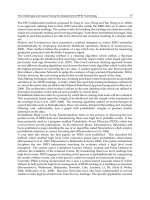
Fig. 1. The G protein cycle. The α-subunit with GDP bound
binds to the βγ dimer. The αβγ trimer then binds to the receptor.
Binding of ligand to the receptor causes a change in the G
protein’s conformation such that GDP leaves and GTP is bound.
Binding of GTP causes the α-subunit to disassociate from the βγ
dimer and assume its active conformation. The activated α-sub-
unit then activates effector molecules. The intrinsic GTPase
activity of the α-subunit hydrolyzes the bound GTP to GDP,
allowing the α-subunit to reassociate with the βγ dimer. The α-
subunit remains activated until the GTP is hydrolyzed. RGS
proteins bind to the activated α-subunit to increase the rate at
which GTP is hydrolyzed.
38 Part II / Hormone Secretion and Action
proteins. Most important are the subtypes of the α-sub-
units because they regulate the effector molecules that
the G protein activates. The major families of the G
proteins are G
S
, G
i
and G
q
. Specificity of hormone ac-
tion is achieved because only specific G proteins (com-
posed of the proper subunits) will couple to specific
hormone receptors and because the free βγ dimer and
the activated α-subunit subtypes will couple only to
specific effector molecules. The G
s
family couples to
and increases adenylyl cyclase activity and also opens
membrane K
+
channels; the G
i
family couples to and
inhibits adenylyl cyclase, opens membrane K
+
chan-
nels, and closes membrane Ca
2+
channels; and the G
q
family activates phospholipase Cβ (PLCβ) to increase
IP
3
, diacylglycerol (DAG), and intracellular Ca
2+
. The
signaling of these three families is discussed further in
Sections 2.2–2.4.
In addition to the trimeric G proteins discussed above,
there is also a class of small G proteins that consist of
single subunits, of which Ras, Rho and Rac are impor-
tant members. These proteins also hydrolyze GTP and
play a role in coupling tyrosine kinase receptors to ef-
fector molecules, as discussed in Section 3.
2.2. Hormonal Signaling Mediated by G
s
Hormones that signal through G
s
to activate adeny-
late cyclase and increase cAMP represent the first sig-
naling pathway as described by the pioneering work of
Sutherland and coworkers in the initial discovery of
cAMP. Elucidation of this pathway led to Nobel Prizes
for the discovery of cAMP and for the discovery of G
proteins. Examples of hormones that signal through this
pathway are TSH, luteinizing hormone, follicle-stimu-
lating hormone, adrenocorticotropic hormone, epi-
nephrine, and glucagons, among others. Signaling in
this pathway is outlined in Fig. 2. As described in
Section 2.1, the binding of hormone to the receptor-G
s
complex results in the active α-subunit binding to an
effector molecule, in this case adenylate cyclase. Ade-
nylate cyclase is a single-chain membrane glycopro-
tein with a molecular mass of 115–150 kDa. The
molecule itself has two hydrophobic domains, each
with six transmembrane segments. Binding of the acti-
vated α-subunit of G
s
results in catalyzing the forma-
tion of cAMP from ATP. Eight different isoforms of
adenylate cyclase have been described to date. These
isoforms differ in their distribution and regulation by
other factors such as calmodulin, βγ subunits, and speci-
ficity for α-subunit subtypes. Next cAMP binds to and
activates the cAMP-dependent PKA. PKA is a serine/
threonine kinase that phosphorylates proteins with the
recognition site Arg-Arg-X-(Ser or Thr)-X in which X
is usually hydrophobic. PKA is a heterotetramer com-
posed of two regulatory and two catalytic subunits. The
regulatory subunits suppress the activity of the cata-
lytic subunits. The binding of cAMP to the regulatory
subunits causes their disassociation from the catalytic
subunits, allowing PKA to phosphorylate its targets.
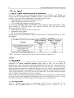
Fig. 2. Signaling by G
s
. Binding of ligand to the receptor causes formation of the activated α-subunit of G
s
. Activated Gα
s
then
activates adenylyl cyclase. Adenylyl cyclase forms cAMP from adenosine triphosphate. Two molecules of cAMP bind to each
regulatory subunit of inactive PKA and cause the regulatory subunits to disassociate from the catalytic subunits. The now-active
catalytic subunits can then phosphorylate their target proteins. The free βγ dimer also signals including triggering receptor desen-
sitization by activating GRK proteins to phosphorylate the receptor, which allows the binding of β-arrestin proteins.
Chapter 3 / Second-Messenger Systems 39
There are a number of PKA subtypes, but the key dif-
ference reflects the type I regulatory subunit (RI) vs the
type II (RII) subunit in which the RI subunit will disas-
sociate from PKA at a lower concentration of cAMP
than will the RII subunit. Recent reports have also dem-
onstrated that cAMP can also signal by activating other
proteins besides adenylate cyclase.
PKA phosphorylates multiple targets including
enzymes, channels, receptors, and transcription factors.
Enzymes can be activated or inhibited by the resulting
phosphorylation at Ser/Thr residues. An example of
regulation of glycogen phosphorylase was discussed in
Section 1.3. An example of a PKA-regulated channel is
the CFTR chloride channel that requires phosphoryla-
tion by PKA for chloride movement. PKA also phos-
phorylates seven-transmembrane receptors as part of
the mechanism of receptor desensitization similar to the
function of GRKs.
A key function of cAMP is its ability to stimulate
gene transcription. The basic concept is that cAMP
activates PKA, which phosphorylates a transcription
factor. The transcription factor then stimulates tran-
scription of the target gene. Several classes of cAMP-
activated transcription factors have been characterized.
These include CREB, CREM, and ATF-1. Probably
the most is known about CREB, so it is used here as an
example (Fig. 3). CREB is a 341-amino-acid protein
with two primary domains, a DNA-binding domain
(DBD) and a transactivation domain. The DBD binds
to specific DNA sequences in the target genes that are
activated by cAMP. When CREB is phosphorylated, it
recruits a coactivator protein, CREB-binding protein
(CBP). This positions CBP next to the basal transcrip-
tion complex, allowing interaction with the Pol-II tran-
scription complex to activate transcription. CBP also
stimulates gene transcription by a second mechanism
by functioning as a histone acetyltransferase. The trans-
fer of acetyl groups to lysine residues of histones is
another key mechanism to activate gene transcription.
As is almost always the case in signaling cascades,
there is important negative regulation of the CREB
pathway. A key element of the negative regulation is
mediated by phosphorylated-CREB-inducing expres-
sion of Icer, a negative regulator of CREB function.
Defects in CBP lead to mental retardation in a disease
called Rubinstein-Taybi syndrome (RTS), one of the
first diseases discovered that is caused by defects in
transcription factors.
2.3. Hormonal Signaling Mediated by G
i
Hormonal signaling through seven-transmembrane
receptors linked to G
i
is similar to that linked to G
s
except Gα
i
inhibits adenylyl cyclase rather than stimu-
lates it, as does Gα
s
. Thus, adenylyl cyclase activity
represents a balance between stimulation by Gα
s
and
inhibition by Gα
i
. Gα
i
also couples to calcium channels
(inhibitory) and potassium channels (stimulatory). Recep-
tors
that couple to G
i
include somatostatin, enkephalin,
and the α
2
-adrenergic receptor, among others. For G
i
signaling, the βγ dimer also plays key signaling roles by
activating potassium channels and inhibiting calcium
channels on the cell membrane.
2.4. Hormonal Signaling Mediated by G
q
Hormonal signaling through seven-transmembrane
receptors linked to G
q
proceeds by activation of PLCβ.
Examples of hormones that bind to G
q
include TRH,
gastrin-releasing peptide, gonadotropin-releasing hor-
mone, angiotensin II, substance P, cholecystokinin, and
PTH. Binding of hormone to its receptor leads to forma-
tion of active Gα
q
or Gα
12
, which then activates PLC to
hydrolyze phosphoinositides (Fig. 4) to form two sec-
ond messengers, IP
3
and DAG. IP
3
diffuses within the
cell to bind to specific receptors on the endoplasmic
reticulum (ER). The IP
3
receptor is a calcium channel,
and the interaction of IP
3
with its receptor opens the
channel and allows calcium to flow from the ER into the
cytoplasm, thus increasing free cytosolic calcium lev-
els. The IP
3
receptor is composed of four large sub-
units (≈310 kDa) that each bind a single molecule of IP
3
.
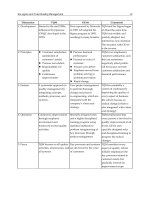
Fig. 3. Role of CREB in regulating gene transcription. PKA
phosphorylates CREB on Serine 133. CREB can be phosphory-
lated while in the cytoplasm (as shown) or while already bound
to DNA. The phosphorylation of CREB allows it to bind CBP,
which then acts as a transcriptional coactivator by interacting
with the pol-II transcription apparatus. CBP also increases gene
transcription by acting as a histone acetyltransferase. Icer is an
important negative regulator of CREB activity that is induced by
CREB.
40 Part II / Hormone Secretion and Action
The binding of IP
3
to the subunits opens the channels
and also desensitizes the receptor to binding additional
IP
3
. Thus, IP
3
leads to increased Ca
2+
which is the next
step in signaling. Calcium is returned to the ER by ATP-
dependent Ca
2+
pumps (SERCA). Thapsigargin is a drug
that blocks the SERCA, thus resulting in transient high
intracellular Ca
2+
levels, but it also depletes Ca
2+
levels
in the ER, making it a convenient tool to study IP
3
-
dependent Ca
2+
release. In excitable cells, a similar
mechanism triggers calcium release from internal stores,
except here calcium directly triggers additional Ca
2+
release from the ER via the ryanodine receptor. Depo-
larization opens voltage-sensitive Ca
2+
channels on the
cell membranes, allowing influx of Ca
2+
, and this cal-
cium then binds to the ryanodine receptor (very similar
to the IP
3
receptor, except the ryanodine receptor is gated
by Ca
2+
) and allows Ca
2+
efflux from the ER. The
ryanodine receptor also allows Ca
2+
efflux from the
sarcoplasmic reticulum in muscle. IP
3
, in turn, is rapidly
metabolized by specific phosphatases.
Calcium is a major intracellular second messenger,
and its levels are tightly regulated by calcium pumps in
the ER (SERCA), calcium pumps in the membrane
(PMCA), voltage-gated calcium channels, and ligand-
gated calcium channels. Resting cell Ca
2+
is 100 nM, far
lower than the 2 mM levels that occur extracellularly;
thus, there is ample room to rapidly increase intracellu-
lar Ca
2+
. Increased intracellular Ca
2+
signals primarily
by binding to proteins and causing a conformational
shift that activates their function. Examples include Ca
2+
binding to troponin in muscle cells to stimulate contrac-
tion and Ca
2+
binding to calmodulin. The Ca
2+
-
calmodulin complex then binds to a variety of kinases.
There are two general classes of Ca
2+
-calmodulin
kinases, dedicated, i.e., with only a specific substrate
and multifunctional, with many substrates. Examples of
dedicated CAM kinases are myosin light chain kinase
and phosphorylase kinase. The multifunctional CAM
kinases can phosphorylate transcription factors to effect
gene transcription. For example, CAM kinase can
phosphorylate CREB, which provides a mechanism for
cross talk between receptors linked to G
s
and G
q
. CAM
kinases can also phosphorylate other kinases such as
mitogen-activated protein kinase (MAPK) or Akt to
activate other signaling pathways. In addition, CAM
kinases play a key role in mediating signaling by ligand-
gated ion channels, as discussed in Section 5.
The other second messenger of the PLC pathway is
DAG. The primary action of DAG is to activate PKC,
a serine-threonine kinase. PKC modifies enzymatic
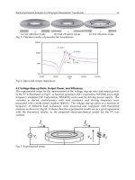
Fig. 4. Signaling by G
q
. Activated Gα
q
activates PLCβ (PLC). PLCβ then hydrolyzes phosphatidylinositol to form two second
messengers, DAG and IP
3
. The binding of IP
3
to the IP
3
receptor on the ER stimulates calcium efflux from the ER to increase
intracellular calcium. DAG activates PKC. PKC can then stimulate transcription by phosphorylation of transcription factors. Tyro-
sine kinase–linked receptors activate PLCγ to produce DAG and IP
3
as well.
Chapter 3 / Second-Messenger Systems 41
activity by phosphorylation of target enzymes, and like
PKA, PKC can modify gene transcription by regulating
phosphorylation of transcription factors. PKC is acti-
vated by the class of compounds known as phorbol
esters that were originally described for their ability to
promote tumor growth. One phorbol ester that potently
stimulates PKC activity is 12-O-tetradecanoylphorbol-
13-acetate (TPA or PMA). It was initially shown that
TPA could activate gene transcription through a DNA
sequence element known as the AP-1-binding site. Iso-
lation of the transcription factors that bound to AP-1 led
to the isolation of Jun and Fos, which bind to the AP-1
site as hetero- or homodimers to regulate transcription.
Thus, hormones that signal through G
q
regulate gene
transcription through DAG, which activates PKC, lead-
ing to phosphorylation of jun and fos. PKC, like PKA,
can also regulate receptor activity by directly phospho-
rylating ion channels and seven-transmembrane recep-
tors.
3. SIGNALING THROUGH RECEPTORS
LINKED TO TYROSINE KINASES
OR SERINE/THREONINE KINASES
The second major signaling pathway involves cas-
cades of phosphorylation events. These pathways can
be divided into those that commence with a tyrosine
phosphorylation event and those that commence with a
serine/threonine phosphorylation event. These path-
ways are similar in that they are a series of protein-
binding and/or phosphorylation events. There are two
primary mechanism by which the binding of hormone to
its receptor causes signal propagation. In the first mecha-
nism, hormone binding triggers receptor autophos-
phorylation via an intrinsic receptor kinase. Receptor
phosphorylation then allows binding of additional pro-
teins that recognize the phosphotyrosines. The EGFR
uses this pathway. In the second mechanism, hormone
binding triggers a receptor conformational change that
stimulates binding of a second protein to the receptor.
One important way in which hormone binding to the
receptor triggers conformational change is by causing
receptor dimerization. Examples of this are the GH and
PRL receptors. These are discussed in greater detail in
Section 3.2.
3.1. Signaling Through Receptors
With Intrinsic Tyrosine Kinase Activity
(EGF, Insulin, Insulin-like Growth Factor-1)
Hormones and growth factors that signal through
receptors with intrinsic tyrosine kinase activity include
the EGFR, the vascular endothelial growth factor recep-
tor, and the insulin receptor. Binding of ligand to the
receptor stimulates the receptor’s intrinsic tyrosine
kinase, resulting in autophosphorylation (i.e., the recep-
tor phosphorylates itself), which then induces binding
of the next signaling protein or effector protein. Within
this category there are differences depending on recep-
tor structure. Prototype signaling mechanisms are dis-
cussed below.
3.1.1. EGFR S
IGNALING
The EGFR is a single-transmembrane receptor that
binds EGF as a monomer. EGF binding causes a change
in conformation that induces dimerization with a second
EGF- EGFR complex. Dimerization of the EGFR com-
plexes activates the EGFR’s intrinsic tyrosine kinase,
and each receptor in the dimer transphosphorylates the
other receptor at multiple tyrosine residues. These
phosphotyrosines then serve as docking sites for src
homology 2 (SH2) domain proteins. SH2 domains are
conserved regions of approx 100 amino acids that serve
to target proteins to phosphotyrosines. Depending on
the amino acids adjacent to the phosphotyrosine, differ-
ent SH2 domain proteins will have different affinities
for the phosphotyrosine residue. Thus, depending on
which tyrosine residues are phosphorylated, and the
sequences surrounding those tyrosines, different pro-
teins will dock on the ligand-activated receptor. This
provides specificity of effector action and the ability for
multiple proteins to dock on a single receptor. The bind-
ing of the SH2 domain protein to the receptor propa-
gates signals by a number of mechanisms including 1
bringing an effector molecule to the membrane where it
is next to its target molecule, 2 binding that triggers a
conformational change that can activate endogenous
enzymatic activity in the SH2 proteins (e.g., kinase ac-
tivity), and 3 binding that can position the SH2 protein
so that it can be phosphorylated and activated. The
EGFR employs these mechanisms as follows.
As shown in Fig. 5, the binding of EGF to its receptor
activates the MAPK pathway, PLCγ, phosphatidylinos-
itol 3-kinase (PI3K), and transcription factors. Many
growth factors use pathways similar to EGF, so it is
important to consider the multiple pathways of EGF sig-
nal transduction. As previously described, Ras is a small
G protein with GTPase activity like Rho. When the
EGFR is phosphorylated, the SH2 domain protein GRB-
2 (growth factor receptor–binding protein-2) binds to
the receptor and then binds through its SH3 domain to a
guanine nucleotide exchange factor (GEF), which acti-
vates RAS by stimulating the exchange of GDP for GTP
by RAS. The GEF that binds to the EGFR is known as
SOS, or “son of sevenless,” because of its homology to
the drosophila protein) (Fig. 6). This brings SOS close
to the membrane and in close proximity to Ras, which is
anchored in the membrane. SOS then converts ras-GDP
42 Part II / Hormone Secretion and Action
into the active ras-GTP form. In some systems, SOS
does not bind directly to GRB-2, but an intermediate
adapter protein, Shc, is recruited, which then binds SOS.
Ras-GTP then activates Raf kinase, which activates
MAPK kinase, which activates MAPK, which phospho-
rylates the final effector proteins that regulate growth or
cellular metabolism. As always, there is important nega-
tive regulation, this time by GTPase-activating proteins
Fig. 5. Signaling by EGFR. Binding of EGF to its receptor causes dimerization of liganded receptors. Receptor dimerization causes
receptor autophosphorylation by activating the receptor’s intrinsic tyrosine kinase activity (shown in dark gray). SH2 domain proteins
such as GRB-2, PLCγ and PI3K then bind to the phosphotyrosine residues. This results in activation of the SH2 domain proteins by
either phosphorylation, localization, or both.
Fig. 6. The MAPK and Akt signaling cascades. Binding of EGF induces phosphorylation of the EGFR, which activates both the
MAPK signaling cascade and signaling by Akt. For MAPK activation, the GRB-2-SOS complex binds to the receptor, positioning
it near membrane-bound Ras-GDP, which is then activated. The activated Ras GTP activates Raf kinase, which activates MAPK
kinase, which activates MAPK which then activates the final effector proteins, many of which are transcription factors. Active Ras-
GTP is converted into inactive Ras-GDP by GAP. For Akt signaling, PI3K binds by the SH2 domain, is activated, and converts
membrane-bound PIP
2
to PIP
3
. PDK1 and Akt bind to PI3K through the Pleckstrin homology domain. This results in phosphorylation
to activate Akt, which then triggers cell proliferation by both growth pathways and inhibition of apoptosis. PTEN is a key negative
regulator that acts by dephosphorylating Akt.
Chapter 3 / Second-Messenger Systems 43
(GAPs) that increase the rate of hydrolysis of GTP bound
to RAS to convert RAS to the inactive state. Thus, the
GAPs are very similar to the RGS proteins that nega-
tively regulate G protein signaling by increasing the rate
of GTP hydrolysis by α-subunits.
There are in fact a number of parallel MAPK path-
ways with different MAPKs and MAPK kinases. Other
MAPK pathways include MEK kinase, which is equiva-
lent to MAPK kinase, and extracellular-regulated
kinase (ERK), which is equivalent to MAPK. Transcrip-
tional targets for ERK include the ELK and SAP tran-
scription factors. One important MAPK subtype is Jun
kinase, which activates the Jun transcription factors.
Specificity of these pathways comes in part from the
initial SH2 docking protein that binds to the tyrosine
kinase pathways and also from multiple inputs from
other proteins. MAPKs are, in turn, rapidly inactivated
by phosphatases.
The second major signaling pathway of tyrosine
kinase receptors such as the EGFR is through activation
of PLCγ. While PLCγ is activated by Gα
q
, PLCγ is an
SH2 domain protein. Thus, when EGF stimulates phos-
phorylation of the EGFR, PLCγ, through its SH2
domains, binds to phosphotyrosines in the EGFR. This
serves two purposes: first, it brings PLCγ close to the
membrane adjacent to phosphatidyl inositols; and, sec-
ond, it allows the EGFR to phosphorylate PLCγ. Phos-
phorylation activates PLCγ resulting in hydrolysis of
phosphatidylinositol to IP
3
and DAG. Thus, tyrosine
kinase–linked receptors, like G
q
-linked receptors, also
signal through IP
3
and DAG.
The third major pathway by which the EGFR sig-
nals is by activation of other enzymes of which PI3K
is one of the most important. PI3K phosphorylates
phosphoinositols such as phosphatidylinositol-4,5-
bisphosphate (PIP
2
) in the 3 position to create phos-
phatidylinositol-3,4,5-trisphosphate (PIP
3
). These
phosphoinositols remain membrane bound. The kinase
Akt then binds to PIP
3
through a sequence known as
the Pleckstrin homology domain. The kinase PDK1
then binds to the Akt and PIP
3
also through the
Pleckstrin domain and activates Akt by phosphoryla-
tion. Phosphorylated Akt then stimulates cell growth
both by inhibiting apoptosis through the BAD pathway
and by stimulating growth. Growth stimulation pro-
ceeds in part through the phosphorylation of mTOR,
leading to activation of protein translation. Negative
regulation is provided by the phosphatase PTEN,
which dephosphorylates PIP
3
. PTEN, because of its
ability to counter the growth stimulatory effects of Akt,
is an important tumor suppressor. Finally, the EGFR
can also directly activate some nuclear transcription
factors by phosphorylation.
The EGFR has been discussed in depth because it
serves as a model for most other tyrosine kinase recep-
tors. The key concept is that ligand binding induces
autophosphorylation and SH2 proteins then bind to
phosphotyrosines to activate multiple signaling mecha-
nisms. Specificity is achieved in that different SH2 pro-
teins recognize different phosphotyrosines.
3.1.2. S
IGNALING BY INSULIN
AND
INSULIN-LIKE GROWTH FACTOR RECEPTORS
The signal transduction mechanism employed by
the insulin receptor is a variation of that employed by
the EGFR (Fig. 7). Binding of insulin to the insulin
receptor (a heterotetramer composed of two α-subunits
and two β-subunits), like binding of EGF to its recep-
tor, triggers receptor autophosphorylation. However,
the insulin receptor does not signal by directly binding
SH2 domain proteins. Rather, ligand-induced receptor
autophosphorylation stimulates binding of bridging
proteins called insulin receptor substrate (IRS) pro-
teins (IRS1–4). Four IRSs have been described to date,
though IRS1 and IRS2 play the key role in insulin sig-
naling. IRSs bind to the insulin receptor and are phos-
phorylated, and then multiple SH2 proteins bind in turn
to the IRSs. Just as EGF-induced signaling depends on
which SH2 domain proteins bind to the EGFR, insulin
signaling depends on which SH2 proteins bind to the
IRS. Examples of proteins that bind to IRSs include
GRB-2 and PI3K. GRB-2 then activates the Ras path-
way and PI3K activates Akt as discussed above. Akt
and PI3K then play key roles in activating glycogen
Fig. 7. Signaling by insulin receptor. Binding of insulin to its
receptor causes autophosphorylation. This stimulates binding of
the IRS protein, which is then phosphorylated by the insulin
receptor. SH2 proteins such as GRB-2 and PI3K then bind to the
IRS and signal as described for the EGFR. The binding of PI3K
to the IRS plays a key role in stimulating glucose entry into cells.
44 Part II / Hormone Secretion and Action
synthesis and glucose transport into the cell. IRSs do
not bind to the insulin receptor via SH2 domains but,
rather, appear to utilize Pleckstrin homology domains
and phosphotyrosine-binding domains, though the
exact details are yet to be determined.
3.2. Signaling Through Receptors
That Signal Through Ligand-Induced
Binding of Tyrosine Kinases (GH, PRL)
The GH and PRL receptors belong to a large super-
family of receptors that include the cytokine receptors
for interleukin-2 (IL-2), IL-3, IL-4, IL-5, IL-6, IL-7,
IL-9, IL-11, IL-12, erythropoietin, granulocyte
macrophage colony-stimulating factor, interferon-β
(IFN-β), IFN-γ, and CNTF. Many of these receptors
are heterodimers consisting of an α-ligand-binding
subunit and a β-signaling subunit. However, the GH
and PRL receptors have single subunits that contain
both the ligand-binding and signaling domains. The
receptors in this family lack intrinsic tyrosine kinase
activity. Instead, these receptors associate with kinases
belonging to the JAK kinase family. Ligand binding to
the receptor induces receptor dimerization bringing
two JAK kinases in close apposition, which results in
activation of the associated JAK kinases by reciprocal
phosphorylation (Fig. 8). The JAK kinases then phos-
phorylate target proteins and signaling commences.
The name JAK kinase is short for Janus kinase; Janus
is the ancient Roman god of gates and doorways who
is depicted with two faces, one looking outward, and
one looking inward (it has also been claimed that JAK
stands for Just Another Kinase). There is a family of
JAK kinases and different receptors associate with
different kinases. At the present time, four members of
the family have been described: Jak1, Jak2, Jak3, and
tyk2. The different kinases phosphorylate different
targets to achieve signaling specificity. For example,
the PRL and GH receptors bind Jak2, the IL-2 and IL-
4 receptors bind Jak 1 and Jak3, and the IFN receptors
bind tyk2.
Fig. 8. Signaling by GH receptor (GHR). GH causes receptor dimerization by binding to two receptors. This brings two Jak kinases
that are bound to the GHR into close apposition and allows each Jak kinase to phosphorylate the other and the reciprocal GHR
(transphosphorylation). Stat proteins then bind through SH2 domains to the Jak kinases and are phosphorylated. The phosphorylated
STAT proteins then form homo- or heterodimers, translocate to the nucleus, and stimulate gene transcription.
Chapter 3 / Second-Messenger Systems 45
The activated JAK kinases phosphorylate the signal
transduction and activation of transcription (STAT)
proteins among others. Seven STAT proteins have
been described to date, though there are likely more
members of this important gene family. STAT proteins
contain an SH2 domain and a single conserved tyrosine
residue that is phosphorylated in response to ligand
binding. Phosphorylation of STAT releases the STAT
from the receptor, and the SH2 domains in the STAT
allow them to form as homodimers or as heterodimers
with other STATs or with unrelated proteins (Fig. 8).
The dimerized STATs can then bind to DNA to stimu-
late transcription. For example, IFN-α stimulates gene
transcription by activation of Stat1 and Stat2, which
heterodimerize and bind to DNA. Similarly, CNTF or
IL-6 results in binding of Stat1 and Stat3 heterodimers
to DNA. A key question remaining to be clarified is,
How is exact signal specificity achieved? There are
more receptors and ligands than JAK kinases and
STATs. Specificity may reside in the time course of
activation (reflecting the balance between kinases and
phosphatases), which STATs are activated, phospho-
rylation status of other proteins, and the binding of
other transcriptional regulators elsewhere in the gene.
Negative regulation results both from STAT-induced
transcription of negative regulators and from phos-
phatases (SHP-1) that dephosphorylate STATs.
3.3. Signaling Through Receptors With
Intrinsic Serine/Threonine Kinase Activity
(Activin, Inhibin, Transforming Growth Factor-
β
)
Receptors with intrinsic serine/threonine kinase
activity form a large family of receptors. These recep-
tors include the transforming growth factor-β (TGF-β),
activin, inhibin, and bone morphogenic proteins. Sig-
naling for TGF-β is best characterized and serves as a
model for the signal transduction mechanism of serine/
threonine kinase– linked receptors (Fig. 9). TGF-β binds
to a type II receptor dimmer, which then recruits a type
I receptor dimer. The type II receptor then phosphory-
lates the type I receptor, which results in the recruitment
of Smad proteins, which are the signaling intermediates
of the TGF-β receptor. First, Smad2 or Smad3 binds to
the TGF-β receptor. Second, the Smad is phosphory-
lated, disassociates from the receptor, and dimerizes
with Smad4. Third the Smad2/3-Smad4 heterodimer
translocates to the nucleus and stimulates gene tran-
scription. Negative regulation is achieved by inhibitory
Fig. 9. Signaling by TGF-β receptor. Binding of TGF-β to the type II receptor recruits the type I receptor, which is then phospho-
rylated. This triggers binding of a Smad protein, which is phosphorylated, dimerizes with a second Smad, and translocates to the
nucleus to stimulate transcription.
46 Part II / Hormone Secretion and Action
Smads (Smad6, Smad7) which can dimerize with the
Smad2 or Smad3 or bind to the TGF-β receptor to pre-
vent signaling.
4. SIGNALING THROUGH NITRIC OXIDE
AND THROUGH RECEPTORS
LINKED TO GUANYLATE CYCLASE
4.1. Signaling Through Nitric Oxide and
Soluble Guanylate Cyclase
Nitric oxide (NO) is one of the more recently charac-
terized signaling molecules. Knowledge of this signal-
ing pathway arose in part from the discovery that NO is
the active metabolite of nitroglycerin and other nitrates
used for vasodilation. NO is synthesized by oxidation of
the amidine nitrogen of arginine through the actions of
the enzyme NO synthase (NOS) (Fig. 10). Study of the
role of NO has been greatly facilitated by substituted
arginine analogs such as L-NAM, which act as potent
NOS inhibitors. Because NO has a short half-life, is not
stored, and is released immediately on synthesis, NO
release reflects regulation of NOS. There are three
major forms of NOS: an inducible form present in
macrophage, a brain-specific form, and an endothelium-
specific form. The brain and endothelial forms are acti-
vated by calcium and calcium- calmodulin complexes.
The primary signaling mechanism of NO appears to be
through cyclic guanosine 5´-monophosphate (cGMP).
NO binds specifically to a soluble guanylate cyclase
(GC) to stimulate the formation of cGMP. CGMP, in
turn, activates ion channels and also activates a cGMP-
activated protein kinase (PKG) that can then activate
enzymes and signal similarly to PKC and PKA. The
soluble GC that acts as the NO receptor is a heterodimer
of Mr = 151,000. However, activation of GC likely does
not explain all of NO’s actions, and other NO signal
transduction mechanisms remain to be determined. NO
likely plays an important role in signaling by sensory
neurotransmission mediated by neuropeptides such as
substance P, vasoactive intestinal peptide, and soma-
tostatin that increase intracellular calcium.
4.2. Hormones That Signal Through
Membrane-Bound GC (Natriuretic Peptides)
The action of the atrial natriuretic peptides is medi-
ated by a membrane-bound form of GC. There are three
natriuretic peptides: ANP, BNP, and CNP. ANP and
BNP bind to GC A (GC-A), and CNP binds to guanylate
cyclase B (GC-B). There is a third natriuretic peptide
receptor that binds all three peptides. This receptor has
been thought to be primarily a clearance receptor, but
recent studies suggest that it may also have independent
signal transduction properties. GC-A and GC-B are
single-transmembrane domain receptors with an extra-
cellular ligand-binding domain, a transmembrane
domain, and an intracellular catalytic (GC) domain.
Binding of natriuretic peptide to GC-A or GC-B acti-
vates the receptors’ GC activity, thus stimulating the
formation of cGMP. cGMP then signals as discussed
above. A third type of membrane-bound GC (GC-C) has
also been described in the gastrointestinal tract and kid-
ney. The endogenous ligand of this cyclase may be the
small peptide guanylin.
5. SIGNALING THROUGH
LIGAND-GATED ION CHANNELS
(ACETYLCHOLINE, SEROTONIN)
Although serotonin (5-hydroxytryptamine [5-HT
1
])
and acetylcholine (ACh) are most typically thought of
as neurotransmitters, they also function as autocrine and
paracrine hormones. Serotonin is secreted by pulmo-
nary and gut neuroendocrine cells and ACh by lung air-
way epithelium. The nicotinic ACh receptors (nAChR)
and the serotonin 5-HT
3
receptors are receptors that
belong to the family of ligand-gated ion channels. As
shown in Fig. 11, binding of the ligand allows calcium
or sodium to enter the cell. Depending on the subunit
composition, the selectivity for sodium or calcium can
vary significantly. Primary signaling is by calcium,
which signals by diverse mechanism. Changes in cell
potential can open voltage-sensitive calcium channels
(VSCCs) to allow more calcium entry to amplify the
initial signal. The elevated calcium can then signal
through CAM kinase II, which activates the MAPK, Akt
pathways, and adenylyl cyclase pathways. Calcium can
also activate CAM kinase kinase directly, which further
activates Akt. A second important signaling route for
calcium is activation of the Ras signaling pathways
through mechanisms that involve the EGFR and Pyk2
kinase.
Fig. 10. Formation of NO. NOS and NADPH catalyze the oxida-
tion of arginine to citrulline and NO.
Chapter 3 / Second-Messenger Systems 47
6. CROSS TALK
BETWEEN SIGNALING SYSTEMS
As might be imagined, given the complexity and
multiplicity of the signaling systems described in this
chapter, there is considerable opportunity for cross talk
between signal transduction systems. Although signal-
ing systems in this chapter have been discussed as if
isolated, it is important to realize that in the cell there
is abundant cross activation. For example, multiple hor-
mones can activate the same kinases, and the same
kinase can, in turn, phosphorylate targets in more than
one signaling pathway. Conversely, one hormone can
activate multiple signaling pathways. Thus, signal
transduction should not be considered a linear pathway
but, rather, a network of activation, and signaling
events represent the summation of activation. Equally
important is the time course of activation as reflected
by the half-life of second messengers and the balance
between phosphorylation and dephosphorylation.
Cross talk can be at the level of the receptor, second
messenger, signaling protein, or transcription factor
activation. CREB, e.g., as well as being activated by
cAMP, is activated by PKC, Akt, MAPK, and CAM
kinase II, making it an important integrator of multiple
signaling pathways.
7. DISEASES ASSOCIATED
WITH ALTERED SIGNAL TRANSDUCTION
As might be expected, given the diverse mecha-
nisms and multiple effector molecules, there are a
number of disease entities associated with signal trans-
duction. A few examples are highlighted here, and
more are described elsewhere in this book.
7.1. Oncogenes and Tumor Suppressors
Given the relation between signal transduction and
growth, it is not surprising that mutations in signal trans-
duction molecules can lead to unregulated growth and
tumorigenesis. Genes that when mutated can cause
transformation are called oncogenes (the normal
unmutated gene is a protooncogene). Alterations in re-
ceptor structure can lead to constitutive activation and
constant stimulation of the signaling cascade. An ex-
ample of this includes the neu oncogene, a point muta-
tion of the EGFR, which leads to rat neuroblastoma and
the trk oncogene, a truncation of the nerve growth factor
receptor, which occurs in human colon carcinomas.
Mutations of the transcription factors jun and fos result
in oncogenes carried by avian and murine retroviruses.
Similarly, other avian retroviruses carry mutated forms
of the tyrosine kinases ras and src. Loss of genes that
shut off signaling pathways such as PTEN also results in
tumors. This is discussed further in Chapter 19.
7.2. Alteration of G Protein Function
7.2.1. PERTUSSIS AND CHOLERA TOXIN
Pertussis and cholera toxin are two toxins of major
clinical importance that achieve their actions in part by
interacting with G protein α-subunits. Cholera toxin
causes adenosine 5´-diphosphate ribosylation of the
α-subunit of G
s
. This has the effect of inhibiting the
α-subunit’s GTPase activity, thus “locking” the sub-
unit in its active GTP-bound conformation, which
increases its ability to activate adenylyl cyclase and
results in increased levels of cAMP. Increased levels
of cAMP in the intestinal epithelial cells causes fluid
secretion throughout the intestinal tract and the mas-
sive diarrhea that characterizes cholera. Pertussis toxin
causes ADP ribosylation of the α-subunit of G
i
. This
results in uncoupling of the G protein from the receptor
and leads to constitutive activation of adenylyl cyclase
and increased levels of cAMP.
7.2.2. T
YPE 1 PSEUDOHYPOPARATHYROIDISM
Type I pseudohypoparathyroidism (PHP), also
known as Albright’s hereditary osteodystrophy (AHO),
is a genetic disorder caused by defects in Gα
s
. AHO is
characterized by a distinctive phenotype of short stat-
ure, round face, obesity, shortened metacarpals, and
subcutaneous ossification. In examining kindreds of
type I PHP, multiple defects in Gα
s
have been described.
These include point mutations, frame shifts, and splic-
ing mutations that all produce decreased levels of Gα
s
.
This results in decreased responsiveness to PTH, which
Fig. 11. Signaling by nAChR, a ligand-gated ion channel. The
binding of Ach allows calcium or sodium to flow through the
channel. Calcium and sodium activate VSCCs and calcium, in
turn, can signal through multiple mechanisms. These include
activation of CAM kinase II, CAM kinase kinase, Ras, adenylyl
cyclase, PI3 kinase, Akt kinase, MAPKs, Pyk2 kinase, and the
EGFR.
48 Part II / Hormone Secretion and Action
signals through G
s
and, hence, the appearance of appar-
ent hypoparathyroidism. As would be expected, given
that G
s
mediates signaling for multiple other hormones,
patients with PHP exhibit multiple hormone resistance
and a variety of cell types have lowered levels of
adenylyl cyclase. As well as the hallmark symptoms
associated with PTH resistance, patients with AHO fre-
quently exhibit hypothyroidism and hypogonadism.
PHP is discussed further in another chapter.
7.3. Alterations in cAMP-Induced
Gene Transcription (RTS)
RTS is a well-defined syndrome with facial abnor-
malities, broad thumbs, broad big toes, and mental retar-
dation. It has recently been discovered that RTS is
caused by genetic defects in CBP. Kindreds of RTS
have chromosomal break points, microdeletions, or
point mutations in the CPB gene. The disease occurs in
patients heterozygous for the mutation. Because CPB
mediates the ability of cAMP and CREB to stimulate
gene transcription, mutations in CPB will interfere with
a large number of target genes. How this results in the
specific syndrome remains to be determined.
7.4. Alterations in cGMP Signaling
(Heat-Stable Enterotoxin)
Some strains of pathogenic bacteria produce a heat-
stable enterotoxin. These toxins are a major cause of
diarrhea in humans and animals and are a major cause of
infant mortality in developing countries. Patients typi-
cally present with a watery diarrhea and no fever. These
toxins act by binding to the membrane-bound forms of
GC to increase cGMP. The increased cGMP appears to
cause the diarrhea. There are two forms of heat-stable
enterotoxin: STa and STb. STa binds to GC-C which is
found in the intestinal mucosa. The exact mechanism by
which STa activates GC remains to be determined. Some
of the effects of STa may also be mediated by cGMP
activation of PKA.
SELECTED READING
Cabrera-Vera TM, Vanhauwe J, Thomas TO, Medkova M,
Preininger A, Mazzoni MR, Hamm HE. Insights into G protein
structure, function, and regulation. Endocr Rev 2003;24:
765–781.
Cross MJ, Dixelius J, Matsumoto T, Claesson-Welsh L. VEGF-
receptor signal transduction. Trends Biochem Sci 2003;28:
488–494.
Hollinger S, Hepler JR. Cellular regulation of RGS proteins: modu-
lators and integrators of G protein signaling. Pharmacol Rev
2002;54:527–559.
Kohout TA, Lefkowitz RJ. Regulation of G protein–coupled recep-
tor kinases and arrestins during receptor desensitization. Mol
Pharmacol 2003;63:9–18.
Kopperud R, Krakstad C, Selheim F, Doskeland SO. cAMP effector
mechanisms: novel twists for an ‘old’ signaling system. FEBS
Lett 2003;546:121–126.
Mayr B, Montminy M. Transcriptional regulation by the phospho-
rylation-dependent factor CREB. Nat Rev Mol Cell Biol 2001;2:
599–609.
McManus KJ, Hendzel MJ. CBP, a transcriptional coactivator and
acetyltransferase. Biochem Cell Biol 2001;79:253–266.
Petrij F, Giles RH, Dauwerse HG, Saris JJ, Hennekam RC, Masuno
M, Tommerup N, van Ommen GJ, Goodman RH, Peters DJ.
Rubinstein-Taybi syndrome caused by mutations in the transcrip-
tional co-activator CBP. Nature 1995;376:348–351.
Proskocil BJ, Sekhon HS, Jia Y, Savchenko V, Blakely RD,
Lindstrom J, Spindel ER. Acetylcholine is an autocrine or
paracrine hormone synthesized and secreted by airway bronchial
epithelial cells. Endocrinology 2004;145:2498–2506.
Spiegel AM, Weinstein LS. Inherited diseases involving g proteins
and g protein-coupled receptors. Annu Rev Med 2004;55:27–39.
Sutherland EW. Studies on the mechanism of hormone action. Sci-
ence 1972;177:401–408.
Ten Dijke P, Goumans MJ, Itoh F, Itoh S. Regulation of cell prolif-
eration by Smad proteins. J Cell Physiol 2002;191:1–16.
West AE, Chen WG, Dalva MB, Dolmetsch RE, Kornhauser JM,
Shaywitz AJ, Takasu MA, Tao X, Greenberg ME. Calcium regu-
lation of neuronal gene expression. Proc Natl Acad Sci USA
2001;98:11,024–11,031.
White MF. IRS proteins and the common path to diabetes. Am J
Physiol Endocrinol Metab 2002;283:E413–E422.
Chapter 4 / Steroid Hormones 49
49
From: Endocrinology: Basic and Clinical Principles, Second Edition
(S. Melmed and P. M. Conn, eds.) © Humana Press Inc., Totowa, NJ
4
Steroid Hormones
Derek V. Henley, PhD, Jonathan Lindzey, PhD,
and Kenneth S. Korach, PhD
CONTENTS
INTRODUCTION
STEROID HORMONE SYNTHESIS
MECHANISMS OF STEROID HORMONE ACTION
STEROIDS AND DEVELOPMENT
STEROIDS AND NORMAL PHYSIOLOGY
STEROIDS AND PATHOPHYSIOLOGY
CONCLUSION
1. INTRODUCTION
Steroids are lipophilic molecules used as chemical
messengers by organisms ranging in complexity from
water mold to humans. In vertebrates, steroids act on a
wide range of tissues and influence many aspects of
biology including sexual differentiation, reproductive
physiology, osmoregulation, and intermediate metabo-
lism. Major sites of steroid synthesis and secretion
include the ovaries, testes, adrenals, and placenta.
Based on the distance of a target site from the site of
synthesis and secretion, steroid hormones can be clas-
sified as either endocrine (distant target tissue),
paracrine (neighboring cells), or autocrine (same cell)
factors. When secreted into the environment, steroids
can also act as pheromones by conveying information
to other organisms.
Owing to the pervasive effects of steroids in verte-
brate biology, a number of pathologic states can occur
because of problems related to steroid hormone action
(see Section 6). These disease states include cancer,
steroid insensitivity, and abnormal steroid synthesis.
The purpose of this chapter is to provide an overview
of steroid synthesis, steroid hormone effects in normal
physiology, molecular and biochemical mechanisms
of action of steroid hormones, and pathologic states
related to steroid hormone action.
2. STEROID HORMONE SYNTHESIS
Steroid hormones are lipid molecules derived from
a common cholesterol precursor (Cholestane, C27).
There are four major classes of steroid hormones:
progestins, androgens, estrogens, and corticoids,
which contain 21, 19, 18, and 21 carbons, respectively.
Steroid hormones are synthesized by dehydrogenases
and cytochrome P450 enzymes, which catalyze hydro-
xylation and dehydroxylation-oxidation reactions.
Eukaryotic cytochromes P450 are membrane-bound
enzymes expressed in either the inner mitochondrial or
endoplasmic reticulum membranes of steroid-synthe-
sizing tissues. A common and important rate-limiting
step for the synthesis of all steroid hormones is cleav-
age of the side chain from cholesterol (C27) to yield
pregnenolone (C21), the common branch point for
synthesis of progestins, corticoids, androgens, and,
hence, estrogens (Fig. 1). Expression of the side-chain
cleavage enzyme cytochrome P450scc (cytP450scc),
50 Part II / Hormone Secretion and Action
which converts cholesterol to pregnenolone, is one of
the unique features of steroidogenic cells that partici-
pate in de novo steroid synthesis.
In vertebrates, the synthesis and secretion of gonadal
and adrenal steroid hormones are regulated by tropic
hormones from the anterior pituitary such as follicle-
stimulating hormone (FSH), luteinizing hormone (LH),
and adrenocorticotropic hormone (ACTH). Mineralo-
corticoids are also regulated by ion concentrations and
circulating levels of angiotensin II. Common regulatory
Fig. 1. (A) Synthetic pathways and structures of major progestins and corticoids found in humans. Major enzymes involved in the
synthesis are in boldface.
Chapter 4 / Steroid Hormones 51
Fig. 1. (B) Synthetic pathways and structures of major androgens and estrogens found in humans. Major enzymes involved in the
synthesis are in boldface.
mechanisms for steroid synthesis and release are nega-
tive feedback loops in which elevated circulating levels
of steroids suppress production of tropic hormones by
acting at specific sites in the brain and the anterior pitu-
itary. The complex interplay among different compo-
nents of the hypothalamic-pituitary-gonad (HPG)/adre-
nal (HPA) axes is an important feature of endocrine
physiology and is discussed in Section 5.
52 Part II / Hormone Secretion and Action
2.1. Synthesis of Progesterone
Pregnenolone serves as a principal precursor to all
the other steroid hormones synthesized by the ovary,
testes, or adrenals. It appears that the rate-limiting step
for the synthesis of progesterone is side-chain cleavage
of cholesterol by P450scc. Pregnenolone is then con-
verted into progesterone by 3β-hydroxysteroid dehy-
drogenase (3β-HSD). Thus, deficiencies in either
P450scc or 3β-HSD have profound effects on the syn-
thesis of all steroids.
In the ovary, progesterone is produced at all stages of
follicular development as an intermediate for androgen
and estrogen synthesis but becomes a primary secretory
product during the peri- and postovulatory (luteal)
phases. The synthesis of progesterone is under the con-
trol of FSH during the early stages of folliculogenesis
and, following acquisition of LH receptors, becomes
sensitive to LH later in the ovarian cycle. The synthesis
of progesterone by the corpus luteum is stimulated dur-
ing early pregnancy by increasing levels of chorionic
gonadotropin. In addition, the placenta secretes high lev-
els of progesterone during pregnancy, although a differ-
ent isozyme of 3β-HSD is involved in the synthesis.
2.2. Synthesis of Androgen
Androgens are synthesized and secreted primarily by
the Leydig cells of the testes, thecal cells of the ovary,
and cells in the reticularis region of the adrenals. In most
tetrapod vertebrates, testosterone is the dominant circu-
lating androgen. Testicular synthesis and secretion of
testosterone is stimulated by circulating LH, which
upregulates the amount of 17α-hydroxylase:C-17,20-
lyase, a rate-limiting enzyme for conversion of C21 into
C19 steroids. Once taken up by target tissues, testoster-
one can be reduced by 5α-reductase to yield a more
active androgen metabolite, 5α-dihydrotestosterone
(5α-DHT). Testosterone and androstenedione can also
be converted into estrogens such as 17β-estradiol (E
2
)
or estrone through a process termed aromatization.
Aromatization is carried out by a cytochromeP450
aromatase enzyme that is expressed in the granulosa
cells of the ovary, Leydig cells of the testes, and many
other tissues including the placenta, brain, pituitary,
liver, and adipose tissue. Indeed, many of the effects of
circulating testosterone are owing to conversion into
either 5α-DHT or E
2
within target tissues.
2.3. Synthesis of Estrogen
Estrogens and progestins are synthesized and secreted
primarily by maturing follicles, corpora lutea of ova-
ries, and the placenta during pregnancy. The predomi-
nant estrogen secreted is E
2
and the predominant
progestin is progesterone. The profile of the synthesis of
estrogen changes during the course of folliculogenesis
during which, under the influence of LH, the thecal cells
synthesize and secrete androstenedione and testoster-
one, which diffuse across the basement membrane and
are subsequently aromatized to estrone and E
2
, respec-
tively, by the granulosa cells. The level of aromatase
and, hence, estrogens produced in the granulosa cells is
under the control of FSH during midfollicular phases.
Later in the cycle, the follicle/corpora lutea express
greater numbers of LH receptors and LH begins to regu-
late E
2
production. During pregnancy, the placenta uti-
lizes androgen precursors from the fetal adrenal gland
and secretes large amounts of E
2
. In addition, in male
vertebrates, many target tissues such as pituitary cells
and hypothalamic neurons convert circulating testoster-
one into E
2
.
2.4. Synthesis of Corticoid
Corticoids are divided into gluco- and mineralocor-
ticoid hormones. The predominant human glucocorti-
coid, cortisol, is synthesized in the zona fasciculata of
the adrenal cortex. The synthesis of cortisol involves
hydroxylations of progesterone at the 17α, 21 (CYP21),
and 11β (CYP11B1) positions. The synthesis of cortisol
is under the control of an anterior pituitary hormone,
ACTH, and a negative feedback mechanism in which
elevated cortisol suppresses the release of ACTH (see
Section 5.2).
The dominant human mineralocorticoid is aldoster-
one, which is produced in the zona glomerulosa of the
adrenal. The synthesis of aldosterone involves the syn-
thesis of corticosterone and subsequent hydroxylation and
oxidation at C18 to yield aldosterone. The synthesis of
aldosterone is regulated directly by levels of potassium,
and indirectly by the effects of sodium levels and blood
volume on levels of angiotensin II (see Section 5.2).
2.5. Serum-Binding Proteins
Following synthesis, steroids are transported to their
target tissues through the bloodstream. The hydropho-
bic nature of steroid hormones results in low water solu-
bility; therefore, transport proteins, known as
serum-binding proteins, help transport steroid hormones
to their target tissues. This transport is accomplished
through the binding of steroid hormones to a specific
high-affinity ligand-binding domain (LBD) within the
serum-binding proteins. Five serum-binding proteins
have been identified: corticosteroid-binding globulin,
retinol-binding protein, sex hormone–binding globulin
(SHBG), thyroxine-binding globulin, and vitamin D–
binding protein. As indicated by their respective names,
each serum-binding protein preferentially binds a
unique class of steroid hormones.
Chapter 4 / Steroid Hormones 53
Recent studies have suggested that serum-binding pro-
teins may serve more dynamic roles beyond steroid hor-
mone transport. SHBG, e.g., has been shown to play a
role in cell membrane–associated signal transduction
through the second-messenger cyclic adenosine mono-
phosphate (cAMP) and protein kinase A (PKA). In addi-
tion, cell-surface SHBG receptors have been identified in
tissues such as the breast, testis, and prostate, further
supporting a role for SHBG in cell signaling.
3. MECHANISMS OF STEROID
HORMONE ACTION
The effects of steroids are typically slow in relation to
the rapid time courses for the effects of second-messen-
ger-mediated peptide hormones. This is owing both to the
signal amplification inherent to second-messenger cas-
cades and to the slower changes in gene transcription and
translation exerted by steroids (genomic effects). Early
experiments confirmed these paths of nuclear hormone
action by utilizing protein and RNA synthesis inhibitors
such as cycloheximide and actinomycin D, respectively.
Though most characterized effects of nuclear hormones
are mediated via nuclear receptors and genomic path-
ways, there are examples of very rapid, “nongenomic”
effects of steroids that appear to be owing to membrane-
mediated effects. In addition, alternative mechanisms of
nuclear hormone receptor (NHR) activation include
ligand-independent activation and genomic activation
independent of a hormone-responsive element.
3.1. Genomic Mechanisms
of Steroid Action
The basic genomic mechanisms of steroid action hold
relatively constant across different target tissues and
different classes of nuclear hormones despite the wide
diversity in target tissues and the responses elicited. In
the absence of hormone, estrogen receptor (ER) and
progesterone receptor (PR) are principally localized
in the nucleus, and glucocorticoid receptor (GR) and
androgen receptor (AR) are located in the cytoplasm.
Current dogma holds that steroid hormones move pas-
sively from the circulation and interstitial spaces across
cell membranes and bind to and activate NHR proteins.
The activated NHR-ligand complex then associates with
members of a class of signal modulators termed
coregulator proteins. The NHR-ligand-coregulator
complex binds to specific DNA sequences termed hor-
mone response elements (HREs) that are associated with
promoter regions involved in regulating gene transcrip-
tion. Most ligand-bound NHR complexes bind to DNA
as homodimers, although some NHRs, including vita-
min D and orphan receptors, can bind to DNA as heter-
odimers with other receptors such as the retinoid X
receptor. Binding of the activated NHR-ligand com-
plexes to an HRE is thought to position the activated
NHR so that transactivation domains of the NHR inter-
act with proteins comprising the transcriptional com-
plex bound to a promoter and, hence, stimulate or inhibit
rates of transcription.
HREs are a family of highly related DNA palin-
dromic repeats. The estrogen, COUP factor, thyroid
hormone, and retinoic acid receptors share highly
homologous consensus response elements, and GR,
AR, PR, and mineralocortoid receptor (MR) share very
similar and, in some cases, identical elements. The high
degree of homology between and within these two
groups of HREs is also reflected in the high degree of
homology between protein sequences of the DNA-
binding domains (DBD) of the various receptors. This
would seem to create a problem with specificity of
hormone action but, as seen in Table 1, mutation of two
nucleotides is sufficient to alter a consensus estrogen
response element (ERE) into a consensus androgen
response element. In addition, as other nonconsensus
elements are characterized more light is shed on the
nature of NHR-specific interactions with the genome.
Table 1
Hormone Response Elements
a
Type of response element Sequence Gene Species
• Estrogen GGTCAcagTGACC vitA2 Xenopus
GGTCAcggTGGCC PS2 Human
GGTCAnnnTGACC Consensus
• Androgen AGAACAgcaAGTGCT PSA Human
• Progesterone AGTACGtgaTGTTCT C(3) Rat
• Glucocorticoid AGA/GACAnnnTGTA/CCC/T Consensus
• Mineralocorticoid
a
Sequence of some characterized response elements for ERs vs ARs, PRs, and corticoid receptors are
given. Also provided are consensus sequences for an ERE and a GRE (GRE consensus sequence is
identical to a PRE and an ARE). Italicized nucleotides demonstrate potential sites for mutation that can
convert one class of 4 to another.
54 Part II / Hormone Secretion and Action
The different classes of steroid hormones are all
present in the circulation, and their respective levels
vary with the different physiologic states of the organ-
ism. In addition, many target cells express multiple
classes of NHR. This presents the organism with the
problem of how to activate a specific gene by a specific
steroid hormone. Specificity of steroid hormone– acti-
vated gene expression lies in (1) hormone-specific bind-
ing by the receptor, (2) DNA-specific binding exhibited
by the different types of steroid receptors, and (3) con-
trol of access of steroid receptors to genes through dif-
ferential organization of chromatin in the many different
target cells and tissues. Many of the hormone insensitiv-
ity syndromes stem from mutations that alter steroid- or
DNA-binding characteristics of the NHR.
As a whole, NHR proteins are a highly conserved
group of “ligand-dependent” nuclear transcription fac-
tors (Fig. 2). NHRs are modular in nature and can be
broken down into different functional domains such as
transactivating domains, DBD, and LBD. Among the
different classes of NHRs—AR, PR, ER, GR, and MR,
the DBD is the most highly conserved region followed
by the LBD and then the amino-terminal transactivating
domain. The following discussion of different func-
tional domains focuses on the ER, but many of the char-
acteristics hold true for other NHR types.
3.2. Structure of ER Gene and Protein
Two forms of the ER have been identified, ERα and
ERβ, that are coded for by separate genes located on
separate chromosomes. Both ER proteins contain modu-
lar functional domain structures characteristic of the
steroid hormone nuclear receptor superfamily. The ER
proteins contain six functional domains that are termed
A/B, C, D, E, and F domains. These domains have been
found to possess the following functions: ligand-inde-
pendent activation function (A/B), DNA binding (C),
ligand binding (E), nuclear localization (D), and dimer-
ization and ligand-dependent activation function (E)
(Fig. 3). The ERα and ERβ proteins share a high degree
of homology within their DBDs and LBDs, 97 and 60%,
respectively, which results in both receptors binding to
the same EREs and exhibiting a similar binding affinity
for most endogenous and exogenous ER ligands. The
modular nature of the different functional domains and
the interdependency of these domains means that splice
variants of NHR mRNAs can produce altered proteins
that behave in appreciably different fashions from the
full-length NHR. The importance of these variants in
normal physiology is still under investigation, but splice
variants may play a role in disease states such as the
progression from steroid-dependent to -independent
cancer (see Section 6.1).
Fig. 2. Mechanisms of nuclear hormone action. E
2
and ER-mediated biologic effects occur through multiple pathways. 1. In the classic
ligand-dependent pathway, E
2
diffuses across the cell membrane and binds to ER, causing dissociation of heat-shock proteins and
allowing the activated ligand-ER complex to recruit transcriptional coactivators and bind to an ERE, resulting in the up- or
downregulation of gene transcription. 2. Ligand-independent ER activation occurs following growth factor (GF) stimulation of
kinase pathways that phosphorylate the ER. 3. E
2
-ER complexes can transactivate genes in an ERE-independent manner through
association with other DNA-bound transcription factors. 4. E
2
can exert rapid effects on a cell through nongenomic actions that occur
at the cell surface.
Chapter 4 / Steroid Hormones 55
3.2.1. LIGAND-BINDING DOMAIN
The LBDs (domain E) of ERα and ERβ consist of
251 and 245 amino acids, respectively, and are coded
for by exons 5–9. The LBD forms a large hydrophobic
pocket that exhibits specific, high-affinity binding for
estradiol (k
d
~ 0.1 nM). Binding of estrogens to this re-
gion produces a conformational change in the ER that
allows for the recruitment of transcriptional coregulators
and subsequent transcriptional activation or suppres-
sion of target genes. Based on studies in which removal
of the LBD results in a constitutively active or “ligand-
independent” ER, it is possible that the LBD functions
as a repressor of a transcription factor that would nor-
mally be constitutively active. Indeed, a constitutively
active exon 5 splice variant of ERα has been detected in
some human breast cancers. Finally, it appears that E
2
binding to the LBD of the ER is not always necessary for
ER-mediated genomic actions. Recent evidence has
shown ligand-independent ER activation of target genes
owing to growth factor activation of kinase signaling
pathways.
3.2.2. DNA-B
INDING DOMAIN
The DBD exhibits specific binding for sequences of
DNA termed EREs. This region is highly conserved and
contains two “zinc finger” motifs, each of which con-
tains cysteine residues that bind zinc. The first zinc fin-
ger dictates sequence-specific interactions with DNA,
and the second appears to dictate the spacing require-
ments between the arms of the palindrome. These fin-
gers are critical for DNA binding but the surrounding
amino acids also influence binding. The canonical
element is a palindrome inverted repeat (GGTCA
nnnTGACC) although deviations from this consensus
sequence are quite common (see Table 1). The ER binds
to the DNA sequence as a dimer with one receptor
molecule contacting each 5-bp inverted repeat. Binding
of the ER-ligand complex to an ERE sequence positions
the ligand-activated ER and associated coactivators
where they can interact with the basal transcription com-
plexes and influence the rate of gene transcription. In
addition to ERE-mediated gene expression, recent evi-
dence indicates that the ERs are capable of transacti-
vating genes whose promoters lack a functional ERE
through protein-protein interactions with other DNA-
bound transcription factors, such as Fos and Jun, at
AP-1-binding sites. The result of the ER association is
a tethering of the ER to DNA and an upregulation of
gene expression via an ERE-independent mechanism.
3.2.3. T
RANSCRIPTION ACTIVATION FUNCTIONS
ERα contains two regions known to possess tran-
scriptional activation functions, activation function-1
(AF-1) and AF-2, located in the A/B and E domains,
respectively. Depending on the cell type and target
genes, AF-1 and AF-2 can act independently or in con-
cert. For instance, removal of AF-1 has no effect on E
2
induction of a reporter construct containing the
vitellogenin ERE, whereas the same AF-1 deficient ERα
has only 20% of the wild-type induction of a PS2-ERE.
As mentioned earlier, removal of the LBD (containing
AF-2) can lead to a constitutively active ERα. Interest-
ingly, this constitutive activity may require phosphory-
lation and activation by second messengers. Studies
using AF-1 and AF-2 truncated ERα have demonstrated
that AF-1 responds to growth factors that act via second
messengers such as cAMP or to mitogen-activated pro-
tein kinase (MAPK) signaling pathway activation,
whereas AF-2 is E
2
(ligand) dependent. Thus, the ER is
actually a nuclear transcription factor that responds to
both steroid and second-messenger signaling pathways.
In contrast to the well-characterized activation domains
of ERα, the roles of the homologous regions of ERβ
have not been clearly defined with respect to transcrip-
tional activity. “Ligand-independent” or second-mes-
senger activation of transcriptional activity has also been
demonstrated for AR and PR, suggesting that this may
be an important and conserved mechanism for physi-
ologic activation of steroid receptors.
The transcriptional activation functions of AF-1 and
AF-2 are mediated through transcriptional coregulators,
proteins that provide the link between ligand-activated,
DNA-bound receptors and the general transcriptional
machinery. The conformational change induced by
agonist binding to the ER allows coregulators to inter-
act primarily with AF-2 sites on the receptor; however,
interaction with AF-1 sites does occur. Many different
coregulators have been identified that interact with the
ligand-bound ER, including the p160 family members
Fig. 3. Protein structure of ERs. Major functional domains of the
mouse ERs and the homology between ERα and ERβ with re-
spect to these domains is shown.
56 Part II / Hormone Secretion and Action
SRC-1, GRIP1, and AIB1, p68 helicase, and CBP/p300.
The p160 family of coregulators contains characteristic
α-helical LXXLL motifs that are involved in AF recog-
nition and binding.
3.2.4. D
IMERIZATION
Most data indicate that NHRs act as homodimers,
although some data suggest possible effects by NHR
monomers. The region of the protein responsible for
dimerization of the mouse ER overlaps with steroid-
binding function and spans amino acids 501–522. These
amino acids form an amphipathic, helical structure with
an imperfect heptad repeat of hydrophobic amino acids
reminiscent of the leucine zippers found in the JUN/
FOS and CREB families of transcription factors. Muta-
tions of amino acids in this hydrophobic stretch have
proven that this area is critical for dimerization, steroid
binding, and, hence, transactivation. The dimerization
function is critical for the effects of NHR homodimers
but may also play a role in the formation of hetero-
dimers between NHRs and other transcription factors.
Heterodimers consisting of ERα and ERβ, as well as
heterodimers of ERα and SP1 proteins, have been shown
to regulate expression of genes such as c-FOS and trans-
forming growth factor-α. Thus, the dimerization func-
tion is critical for the effects of NHR homodimers
but also plays a role in the formation of heterodimers
between NHRs and other transcription factors with simi-
lar dimerization domains.
3.2.5. N
UCLEAR LOCALIZATION SIGNAL
NHRs and many other transcription factors possess a
segment of amino acids that targets the proteins to the
cell nucleus. These stretches of amino acids tend to be
basic and have been termed the nuclear localization
signal (NLS). It appears that the NLS is located between
amino acids 250 and 270 of the ERα, a region that shares
homology with the nuclear localization domains of the
glucocorticoid and progesterone receptors. The NLS for
ERβ has yet to be characterized.
3.3. Nongenomic Mechanisms
of Steroid Action
Although steroids typically act through the classic
genomic mechanism, a process that takes several min-
utes to hours for effects to be seen, steroids are also
capable of eliciting rapid biologic effects within sec-
onds to minutes after administration through nonge-
nomic mechanisms. Nongenomic steroid action results
in the rapid activation of a variety of cell-signaling
molecules, including MAPKs, adenylyl cyclase, and
PKA and PKC. Rapid responses to estrogen have been
observed in granulosa cells, endometrial cells, and
oocytes, all of which exhibit increased intracellular cal-
cium concentrations shortly, if not immediately, after
E
2
exposure. Other estrogen-mediated nongenomic
mechanisms have been observed in spermatozoa, breast
cells, nerve cells, and vascular tissues. In addition,
nongenomic mechanisms have been described for
progesterone, androgens, glucocorticoids, and miner-
alocorticoids. Current research is under way to deter-
mine whether these nongenomic steroid mechanisms
are owing to receptor-independent events at the plasma
membrane, nonsteroid associated membrane receptors,
or membrane-bound NHRs.
4. STEROIDS AND DEVELOPMENT
Scientists have known for years that in utero and
neonatal exposure to steroids are critical for sexual dif-
ferentiation of the brain and peripheral reproductive
structures. A guiding concept for the study of develop-
mental actions of steroidal effects is the organization-
activation hypothesis. Stated simply, prenatal or
neonatal exposure to steroid hormones organizes or
alters differentiation of the phenotype such that hor-
monal exposure in adulthood is more likely to activate
a particular response. A corollary of this rule is that the
initial exposures must fall within certain critical peri-
ods of sensitivity. These critical periods typically occur
during the fetal, neonatal, and pubertal stages.
Steroids affect development of organs and tissues
through both induction and inhibition of growth. Inhibi-
tion occurs via active cell death, a process termed
apoptosis. Apoptosis is an active process requiring pro-
tein synthesis and resulting in chromatin condensation,
degradation of chromatin in a characteristic segmented
manner that produces an observable “ladder” pattern,
and development of apoptotic bodies.
4.1. Stromal-Mesenchymal Interactions
A recurring theme in development of steroid-depen-
dent glandular tissues is the importance of stromal-
mesenchymal tissue induction. In this scheme, the fate
of undifferentiated epithelium is determined by the
underlying mesenchyme with which it comes into con-
tact. For instance, undifferentiated epithelium com-
bined with prostatic or integumental mesenchyme
develops a phenotype dictated by the type of mesen-
chyme. In the case of hormone-directed morphogen-
esis such as in the prostate or breast, hormonal
influences on the glandular epithelium can occur either
directly on epithelial cells or indirectly via inductive
influences of the mesenchyme. Recent experiments
demonstrate that epithelium can also influence the
underlying mesenchyme, indicating a bidirectional
epithelial-mesenchymal interaction.
Chapter 4 / Steroid Hormones 57
4.2. Secondary Sex Structures
In the developing mammalian embryo, gonadal sex
is determined by genotype. In turn, the embryonic
gonads secrete hormones that, coupled with maternal
hormones, determine the early hormonal milieu to
which secondary sex structures are exposed and, hence,
dictate development of male or female phenotype.
Dogma holds that mammals possess a default system
such that embryos develop a female phenotype in the
absence of any gonadal steroid hormones. In males, as
the developing testes begin to develop sex cords, the
testes secrete Müllerian-inhibiting substance (MIS) and
testosterone. The MIS induces ipsilateral regression of
the Müllerian ducts, which prevents development of
Müllerian derivatives such as the uterus and fallopian
tubes. Elevated testosterone stimulates development of
Wolffian derivatives such as epididymis, vas deferens,
and seminal vesicles. Differentiation of external geni-
talia and accessory glands (such as the prostate) from
the genital tubercle, scrotal folds, and urogenital sinus
requires 5α-DHT. This is illustrated by 5α-reductase-
deficient males who have normal Wolffian derivatives
but have feminized external genitalia despite the pres-
ence of testosterone (see Section 6.2).
Although it is true that external genitalia and internal
reproductive structures of genotypic females are grossly
feminized without the influence of gonadal steroids, it
is clear that steroidal effects are needed for complete
and functional differentiation of some structures such as
the uterus and breast. For instance, gene-targeted mice
lacking functional ERα (αERKO) have uteri that pos-
sess all the normal tissue types and structures but are
hypoplastic. In addition, in wild-type females, exposure
to estrogens and progestins is required for differentia-
tion of the nipple and mesenchyme surrounding the
epithelium of breast tissue. Estrogen and progesterone
also increase alveolar formation and branching of mam-
mary ducts during mammary gland development.
4.3. Sexual Behavior and Sexual
Dimorphisms of Brain
Sex behavior in most adult vertebrates is dependent
on (1) organizational effects of hormones early in devel-
opment, and (2) activational effects of circulating ste-
roids in the adult. In many species, in utero and neonatal
hormone exposures alter adult patterns of sexual behav-
iors. Historically, this observation led to the assumption
that at some organizational level the brains of males and
females must be morphologically or functionally dis-
tinct in order to favor female- or male-typical behaviors.
In the case of the rat, sexually dimorphic nuclei have
been found in the central nervous system (CNS). Male
rats possess enlarged sexually dimorphic nuclei in the
medial preoptic area of the hypothalamus and in the
spinal cord. The development of these nuclei and subse-
quent function in adult males are androgen dependent;
androgen ablation during early critical periods of differ-
entiation leads to smaller, female-typical nuclei and also
decreases in male-typical copulatory behavior. In rats,
the effects of testicular testoterone on the sexually di-
morphic nuclei of the medial preoptic area appear to be
predominantly through aromatization to E
2
; treatment
with E
2
mimics the effect of testosterone, and the use of
an aromatase inhibitor can prevent masculinization of
sexually dimorphic nuclei. Similar steroid-dependent
dimorphisms are found in the CNSs of gerbils, voles,
songbirds, lizards, and fish. These dimorphisms may be
present as differences in gross volume, cell number; cell
size, dendritic arborization, and levels of expression of
enzymes, neurotransmitters, neuropeptides, or recep-
tors. Sexual dimorphisms in humans have also been
reported in the anterior hypothalamus (AH), preoptic
area (POA), and anterior commissure, although there
are some conflicting data.
4.4. Steroids and Bone
Bone cells express ER, AR, and PR and the develop-
ment and maintenance of bone structure is regulated by
estrogens and androgens. Pubertal surges in estrogens
and androgens initiate growth spurts including long bone
growth, primarily mediated by increased insulin-like
growth factor-1, and, subsequently, cessation of bone
growth through epiphyseal closure. In adults, E
2
main-
tains bone mass and mineralization. The importance of
the effects of E
2
on bone growth and development is
manifest in individuals lacking in E
2
action. For instance,
a human male patient lacking functional ERα exhibits
continued bone growth, decreased bone density, and
absence of epiphyseal closure (see Section 6.3). In addi-
tion, the absence of E
2
owing to either ovariectomy or
menopause contributes to osteoporosis whereas exogen-
ous E
2
helps ameliorate this condition. Excess produc-
tion of cortisol results in a loss of bone mass (osteopenia).
4.5. Steroids and Liver
Liver cells express ERs and ARs which regulate pro-
duction of secreted proteins and steroid-metabolizing
enzymes. In humans, the liver synthesizes and secretes
into the bloodstream a plasma protein termed SHBG.
This protein serves to sequester and prevent steroids
from being metabolized and/or cleared from the blood-
stream. SHBG binds DHT with high affinity (k
d
~ 0.5
nM) and testosterone and E
2
with approx 5- and 15-fold
lower affinity, respectively. Estrogens stimulate
whereas androgens inhibit the synthesis and secretion of
hepatic SHBG.