Obstructive Sleep Apnea Diagnosis and Treatment - part 3 doc
Bạn đang xem bản rút gọn của tài liệu. Xem và tải ngay bản đầy đủ của tài liệu tại đây (615.78 KB, 47 trang )
80 Patel and Schwab
is complex. To predict the success of oral appliances, investigators are beginning
to use upper airway imaging techniques to access the size and position of the
upper airway (77).
Upper Airway Surgery (See Also Chapter 11)
There are several surgical options for sleep apnea patients including UPPP (tonsil-
lectomy and removal of the uvula, distal margin of soft palate, and any excessive
tissue), uvulopalatopharyngo-glossoplasty (UPPGP—combines UPPP with limited
resection of the tongue), transpalatal advancement pharnyngoplasty (TPAP—resec-
tion of the posterior hard palate with advancement of the soft palate to enlarge the
retropalatal airway), sliding genioplasty or genioglossus advancement (advancing
the tongue forward by displacing its attachment to the genial tubercle forward),
hyoid advancement (displacement of the hyoid bone forward to enlarge the retro-
glossal airway), and maxillary-mandibular advancement (forward displacement of
the maxillae and mandible to advance the soft tissue structures) (162). Typically,
surgical options to treat sleep apnea are invasive and may require a staged approach.
Since the upper airway obstruction may not be at one site, selecting the appropriate
sleep apnea patient and a suitable surgical approach is important.
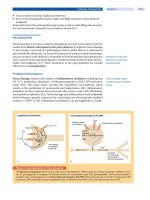
Surgical selection may be achieved by examining data from clinical, fiberop-
tic, and radiologic sources. The Müller maneuver (voluntary inspiration against a
closed mouth and obstructed nares) permits visualization of the airway structures
during a simulated apneic event and has been used to identify surgical candidates
(78). CT and MRI can also be employed to provide detailed information about struc-
tural dimensions during wakefulness and sleep (28,163) and may predict surgical
outcome (70).
UPPP, the most common upper airway surgical procedure, was introduced in
1981 and although there have been many studies in OSA patients examining this
surgical technique its failure rate exceeds 50% (162). UPPP only corrects one
vulnerable upper airway site, the retropalatal pharynx. Patients with retropalatal
obstruction have been shown to have a 52% success rate with UPPP whereas patients
with retroglossal obstruction have a 5% success rate with UPPP (164). CT and MRI
studies have demonstrated that UPPP results in enlargement of the airway only in
the operated area (162). Upper airway narrowing in the unresected portion of the
soft palate post-UPPP is a recognized problem and likely explains the limited
efficacy of UPPP. A further issue, highlighted by a study of LAUP, is that anatomical
improvements in the airway postsurgery, as documented by videoendoscopy
measurements during wakefulness, are not necessarily indicative or predictive of
objective improvements in apnea severity during sleep (75).
Patients with craniofacial abnormalities should be considered for surgical
techniques such as mandibular and/or maxillary advancement and sliding genio-
plasty (24). Cephalometry and nasopharyngoscopy have shown that maxilloman-
dibular advancement increases upper airway caliber in the retroglossal and
retropalatal regions by physically expanding the skeletal boundaries of the upper
airway (165). Maxillomandibular advancement is reported as the most effective
surgical treatment for sleep apnea with success rates between 75% and 100% (165).
Bariatric Surgery (See Also Chapter 13)
Bariatric surgery has the potential for improving patients with sleep apnea sec-
ondary to weight loss (166). Although significant weight loss is expected after
Upper Airway Imaging 81
bariatric surgery, limited data exist regarding the effect of gastric surgery on OSA
(167). That significant improvement occurs in the AHI (greater than 50% decrease)
even in the long-term is promising; however, large-scale studies examining
polysomnography pre- and postgastric bypass surgery need to be performed
(167,168). Furthermore, it is necessary to re-evaluate after surgery for the presence
of persistent sleep apnea requiring CPAP treatment. Currently, no data are
available regarding the anatomic changes in the upper airway associated with
this surgery.
CONCLUSIONS
Upper airway imaging techniques employed to study the human upper airway
have significantly advanced our understanding of OSA. Important determinants of
airway geometry have been identified: volume of tongue, lateral pharyngeal wall
thickness, and total amount of soft tissue surrounding the airway. The sleep apneic
airway has been characterized as an elliptical or circular shape that is oriented in the
anteroposterior axis. Static imaging studies have shown that soft tissue and cranio-
facial structures are influenced by important factors such as body mass, neck
circumference, gender, and genetics. The effects of sleep apnea treatments have also
been clarified through imaging techniques. Upper airway imaging is not routinely
indicated in the assessment of a sleep apnea patient. If CPAP therapy is efficacious,
then upper airway imaging is not warranted. However, imaging does have role in
the preoperative (and postoperative) evaluation of patients undergoing upper
airway surgery to characterize the airway geometry. The likelihood of success of
UPPP is related to the site of airway obstruction and this can be assessed by MRI
(preferably three-dimensional) or by nasopharyngoscopy with the Müller maneuver.
Imaging should also be considered when utilizing an oral appliance to determine if
upper airway caliber increases with the appliance (especially if the AHI does not
improve). Upper airway imaging studies have provided important new insights
into the pathogenesis, genetics, and treatment of OSA.
REFERENCES
1. Horner RL. Motor control of the pharyngeal musculature and implications for the
pathogenesis of obstructive sleep apnea. Sleep 1996; 19(10):827–853.
2. Hudgel DW. Variable site of airway narrowing among obstructive sleep apnea patients.
J Appl Physiol 1986; 61(4):1403–1409.
3. Schwartz AR, Smith PL, Wise RA, Gold AR, Permutt S. Induction of upper airway occlusion in
sleeping individuals with subatmospheric nasal pressure. J Appl Physiol 1988; 64(2):535–542.
4. Suratt PM, Wilhoit SC, Cooper K. Induction of airway collapse with subatmospheric
pressure in awake patients with sleep apnea. J Appl Physiol 1984; 57(1):140–146.
5. Kuna ST, Smickley JS, Vanoye CR. Respiratory-related pharyngeal constrictor muscle
activity in normal human adults. Am J Respir Crit Care Med 1997; 155(6):1991–1999.
6. Mathew OP. Upper airway negative-pressure effects on respiratory activity of upper
airway muscles. J Appl Physiol 1984; 56(2):500–505.
7. Mezzanotte WS, Tangel DJ, White DP. Waking genioglossal electromyogram in sleep
apnea patients versus normal controls (a neuromuscular compensatory mechanism).
J Clin Invest 1992; 89(5):1571–1579.
8. Stanchina ML, Malhotra A, Fogel RB, et al. Genioglossus muscle responsiveness to
chemical and mechanical stimuli during non-rapid eye movement sleep. Am J Respir
Crit Care Med 2002; 165(7):945–949.
9. Wheatley JR, White DP. The influence of sleep on pharyngeal reflexes. Sleep 1993;
16(suppl 8):S87–S89.
82 Patel and Schwab
10. Wiegand DA, Latz B, Zwillich CW, Wiegand L. Upper airway resistance and geniohyoid
muscle activity in normal men during wakefulness and sleep. J Appl Physiol 1990;
69(4):1252–1261.
11. Fogel RB, Malhotra A, Pillar G, et al. Genioglossal activation in patients with obstructive
sleep apnea versus control subjects. Mechanisms of muscle control. Am J Respir Crit
Care Med 2001; 164(11):2025–2030.
12. Armstrong JJ, Leigh MS, Sampson DD, Walsh JH, Hillman DR, Eastwood PR.
Quantitative upper airway imaging with anatomic optical coherence tomography. Am
J Respir Crit Care Med 2006; 173(2):226–233.
13. Abbey NC, Block AJ, Green D, Mancuso A, Hellard DW. Measurement of pharyngeal
volume by digitized magnetic resonance imaging. Effect of nasal continuous positive
airway pressure. Am Rev Respir Dis 1989; 140(3):717–723.
14. Ciscar MA, Juan G, Martinez V, et al. Magnetic resonance imaging of the pharynx in
OSA patients and healthy subjects. Eur Respir J 2001; 17(1):79–86.
15. Do KL, Ferreyra H, Healy JF, Davidson TM. Does tongue size differ between patients
with and without sleep-disordered breathing? Laryngoscope 2000; 110(9):1552–1555.
16. Jager L, Gunther E, Gauger J, Reiser M. Fluoroscopic MR of the pharynx in patients
with obstructive sleep apnea. AJNR Am J Neuroradiol 1998; 19(7):1205–1214.
17. Malhotra A, Huang Y, Fogel RB, et al. The male predisposition to pharyngeal collapse:
importance of airway length. Am J Respir Crit Care Med 2002; 166(10):1388–1395.
18. Rodenstein DO, Dooms G, Thomas Y, et al. Pharyngeal shape and dimensions in healthy
subjects, snorers, and patients with obstructive sleep apnoea. Thorax 1990; 45(10):
722–727.
19. Ryan CF, Lowe AA, Li D, Fleetham JA. Magnetic resonance imaging of the upper airway
in obstructive sleep apnea before and after chronic nasal continuous positive airway
pressure therapy. Am Rev Respir Dis 1991; 144(4):939–944.
20. Schoenberg SO, Floemer F, Kroeger H, Hoffmann A, Bock M, Knopp MV. Combined
assessment of obstructive sleep apnea syndrome with dynamic MRI and parallel EEG
registration: initial results. Invest Radiol 2000; 35(4):267–276.
21. Schwab RJ. Imaging for the snoring and sleep apnea patient. Dent Clin North Am 2001;
45(4):759–796.
22. Schwab RJ, Goldberg AN. Upper airway assessment: radiographic and other imaging
techniques. Otolaryngol Clin North Am 1998; 31(6):931–968.
23. Schwab RJ, Gupta KB, Gefter WB, Metzger LJ, Hoffman EA, Pack AI. Upper airway and soft
tissue anatomy in normal subjects and patients with sleep-disordered breathing. Significance
of the lateral pharyngeal walls. Am J Respir Crit Care Med 1995; 152(5 Pt 1):1673–1689.
24. Shelton KE, Woodson H, Gay S, Suratt PM. Pharyngeal fat in obstructive sleep apnea.
Am Rev Respir Dis 1993; 148(2):462–466.
25. Suto Y, Matsuo T, Kato T, et al. Evaluation of the pharyngeal airway in patients with
sleep apnea: value of ultrafast MR imaging. AJR Am J Roentgenol 1993; 160(2):311–314.
26. Schwab RJ, Pack AI, Gupta KB, et al. Upper airway and soft tissue structural changes induced
by CPAP in normal subjects. Am J Respir Crit Care Med 1996; 154(4 Pt 1):1106–1116.
27. Shelton KE, Gay SB, Hollowell DE, Woodson H, Suratt PM. Mandible enclosure of upper
airway and weight in obstructive sleep apnea. Am Rev Respir Dis 1993; 148(1):195–200.
28. Schwab RJ. Upper airway imaging. Clin Chest Med 1998; 19(1):33–54.
29. Schwab RJ, Pasirstein M, Pierson R, et al. Identification of upper airway anatomic risk
factors for obstructive sleep apnea with volumetric magnetic resonance imaging. Am J
Respir Crit Care Med 2003; 168(5):522–530.
30. Li HY, Chen NH, Wang CR, Shu YH, Wang PC. Use of 3-dimensional computed tomog-
raphy scan to evaluate upper airway patency for patients undergoing sleep-disordered
breathing surgery. Otolaryngol Head Neck Surg 2003; 129(4):336–342.
31. Ryan CF, Lowe AA, Li D, Fleetham JA. Three-dimensional upper airway computed
tomography in obstructive sleep apnea. A prospective study in patients treated by
uvulopalatopharyngoplasty. Am Rev Respir Dis 1991; 144(2):428–432.
32. Aksoz T, Akan H, Celebi M, Sakan BB. Does the oropharyngeal fat tissue influence the
oropharyngeal airway in snorers? Dynamic CT study. Korean J Radiol 2004; 5(2):102–106.
33. Akan H, Aksoz T, Belet U, Sesen T. Dynamic upper airway soft-tissue and caliber changes
in healthy subjects and snoring patients. AJNR Am J Neuroradiol 2004; 25(10):1846–1850.
Upper Airway Imaging 83
34. Burger CD, Stanson AW, Daniels BK, Sheedy PF, II, Shepard JW, Jr. Fast-CT evaluation
of the effect of lung volume on upper airway size and function in normal men. Am Rev
Respir Dis 1992; 146(2):335–339.
35. Burger CD, Stanson AW, Sheedy PF, II, Daniels BK, Shepard JW, Jr. Fast-computed
tomography evaluation of age-related changes in upper airway structure and function
in normal men. Am Rev Respir Dis 1992; 145(4 Pt 1):846–852.
36. Caballero P, Alvarez-Sala R, Garcia-Rio F, et al. CT in the evaluation of the upper airway
in healthy subjects and in patients with obstructive sleep apnea syndrome. Chest 1998;
113(1):111–116.
37. Ell SR, Jolles H, Galvin JR. Cine CT demonstration of nonfixed upper airway obstruction.
AJR Am J Roentgenol 1986; 146(4):669–677.
38. Ell SR, Jolles H, Keyes WD, Galvin JR. Cine CT technique for dynamic airway studies.
AJR Am J Roentgenol 1985; 145(1):35–36.
39. Galvin JR, Rooholamini SA, Stanford W. Obstructive sleep apnea: diagnosis with ultrafast
CT. Radiology 1989; 171(3):775–778.
40. Haponik EF, Smith PL, Bohlman ME, Allen RP, Goldman SM, Bleecker ER. Computerized
tomography in obstructive sleep apnea. Correlation of airway size with physiology
during sleep and wakefulness. Am Rev Respir Dis 1983; 127(2):221–226.
41. Horner RL, Shea SA, McIvor J, Guz A. Pharyngeal size and shape during wakefulness
and sleep in patients with obstructive sleep apnoea. Q J Med 1989; 72(268):719–735.
42. Kuna ST, Bedi DG, Ryckman C. Effect of nasal airway positive pressure on upper airway
size and configuration. Am Rev Respir Dis 1988; 138(4):969–975.
43. Schwab RJ, Gefter WB, Hoffman EA, Gupta KB, Pack AI. Dynamic upper airway imaging
during awake respiration in normal subjects and patients with sleep disordered breathing.
Am Rev Respir Dis 1993; 148(5):1385–1400.
44. Shepard JW, Jr, Garrison M, Vas W. Upper airway distensibility and collapsibility in
patients with obstructive sleep apnea. Chest 1990; 98(1):84–91.
45. Shepard JW, Jr, Stanson AW, Sheedy PF, Westbrook PR. Fast-CT evaluation of the upper
airway during wakefulness in patients with obstructive sleep apnea. Prog Clin Biol Res
1990; 345:273–279; discussion 280–282.
46. Stanford W, Galvin J, Rooholamini M. Effects of awake tidal breathing, swallowing,
nasal breathing, oral breathing and the Muller and Valsalva maneuvers on the dimen-
sions of the upper airway. Evaluation by ultrafast computerized tomography. Chest
1988; 94(1):149–154.
47. Stauffer JL, Zwillich CW, Cadieux RJ, et al. Pharyngeal size and resistance in obstructive
sleep apnea. Am Rev Respir Dis 1987; 136(3):623–627.
48. Stein MG, Gamsu G, de Geer G, Golden JA, Crumley RL, Webb WR. Cine CT in obstructive
sleep apnea. AJR Am J Roentgenol 1987; 148(6):1069–1074.
49. Shepard JW, Jr, Thawley SE. Evaluation of the upper airway by computerized tomogra-
phy in patients undergoing uvulopalatopharyngoplasty for obstructive sleep apnea.
Am Rev Respir Dis 1989; 140(3):711–716.
50. Fleetham JA. Upper airway imaging in relation to obstructive sleep apnea. Clin Chest
Med 1992; 13(3):399–416.
51. Davies RJ, Ali NJ, Stradling JR. Neck circumference and other clinical features in the
diagnosis of the obstructive sleep apnoea syndrome. Thorax 1992; 47(2):101–105.
52. Brown IG, Zamel N, Hoffstein V. Pharyngeal cross-sectional area in normal men and
women. J Appl Physiol 1986; 61(3):890–895.
53. Strobel RJ, Rosen RC. Obesity and weight loss in obstructive sleep apnea: a critical
review. Sleep 1996; 19(2):104–115.
54. Ingman T, Nieminen T, Hurmerinta K. Cephalometric comparison of pharyngeal
changes in subjects with upper airway resistance syndrome or obstructive sleep apnoea
in upright and supine positions. Eur J Orthod 2004; 26(3):321–326.
55. Pae EK, Lowe AA, Fleetham JA. A role of pharyngeal length in obstructive sleep apnea
patients. Am J Orthod Dentofacial Orthop 1997; 111(1):12–17.
56. Ferguson KA, Ono T, Lowe AA, Ryan CF, Fleetham JA. The relationship between obesity
and craniofacial structure in obstructive sleep apnea. Chest 1995; 108(2):375–381.
57. Bacon WH, Turlot JC, Krieger J, Stierle JL. Cephalometric evaluation of pharyngeal obstruc-
tive factors in patients with sleep apneas syndrome. Angle Orthod 1990; 60(2):115–122.
84 Patel and Schwab
58. deBerry-Borowiecki B, Kukwa A, Blanks RH. Cephalometric analysis for diagnosis and
treatment of obstructive sleep apnea. Laryngoscope 1988; 98(2):226–234.
59. Shepard JW, Jr, Gefter WB, Guilleminault C, et al. Evaluation of the upper airway in
patients with obstructive sleep apnea. Sleep 1991; 14(4):361–371.
60. Nelson S, Hans M. Contribution of craniofacial risk factors in increasing apneic activity
among obese and nonobese habitual snorers. Chest 1997; 111(1):154–162.
61. Mayer P, Pepin JL, Bettega G, et al. Relationship between body mass index, age and upper
airway measurements in snorers and sleep apnoea patients. Eur Respir J 1996; 9(9):1801–1809.
62. Tsuiki S, Lowe AA, Almeida FR, Kawahata N, Fleetham JA. Effects of mandibular
advancement on airway curvature and obstructive sleep apnoea severity. Eur Respir
J 2004; 23(2):263–268.
63. Pae EK, Lowe AA, Sasaki K, Price C, Tsuchiya M, Fleetham JA. A cephalometric and
electromyographic study of upper airway structures in the upright and supine posi-
tions. Am J Orthod Dentofacial Orthop 1994; 106(1):52–59.
64. Bacon WH, Krieger J, Turlot JC, Stierle JL. Craniofacial characteristics in patients with
obstructive sleep apneas syndrome. Cleft Palate J 1988; 25(4):374–378.
65. Bradley TD, Brown IG, Grossman RF, et al. Pharyngeal size in snorers, nonsnorers, and
patients with obstructive sleep apnea. N Engl J Med 1986; 315(21):1327–1331.
66. Hoffstein V, Zamel N, Phillipson EA. Lung volume dependence of pharyngeal cross-
sectional area in patients with obstructive sleep apnea. Am Rev Respir Dis 1984;
130(2):175–178.
67. Rivlin J, Hoffstein V, Kalbfleisch J, McNicholas W, Zamel N, Bryan AC. Upper airway
morphology in patients with idiopathic obstructive sleep apnea. Am Rev Respir Dis
1984; 129(3):355–360.
68. Martin SE, Mathur R, Marshall I, Douglas NJ. The effect of age, sex, obesity and posture
on upper airway size. Eur Respir J 1997; 10(9):2087–2090.
69. Isono S, Remmers JE, Tanaka A, Sho Y, Sato J, Nishino T. Anatomy of pharynx in patients
with obstructive sleep apnea and in normal subjects. J Appl Physiol 1997; 82(4):1319–1326.
70. Launois SH, Feroah TR, Campbell WN, et al. Site of pharyngeal narrowing predicts out-
come of surgery for obstructive sleep apnea. Am Rev Respir Dis 1993; 147(1):182–189.
71. Badr MS, Toiber F, Skatrud JB, Dempsey J. Pharyngeal narrowing/occlusion during
central sleep apnea. J Appl Physiol 1995; 78(5):1806–1815.
72. Morrell MJ, Arabi Y, Zahn B, Badr MS. Progressive retropalatal narrowing preceding
obstructive apnea. Am J Respir Crit Care Med 1998; 158(6):1974–1981.
73. Woodson BT, Wooten MR. Comparison of upper-airway evaluations during wakeful-
ness and sleep. Laryngoscope 1994; 104(7):821–828.
74. Schwartz AR, Gold AR, Schubert N, et al. Effect of weight loss on upper airway collaps-
ibility in obstructive sleep apnea. Am Rev Respir Dis 1991; 144(3 Pt 1):494–498.
75. Ryan CF, Love LL. Unpredictable results of laser assisted uvulopalatoplasty in the treat-
ment of obstructive sleep apnoea. Thorax 2000; 55(5):399–404.
76. Isono S, Tanaka A, Sho Y, Konno A, Nishino T. Advancement of the mandible improves
velopharyngeal airway patency. J Appl Physiol 1995; 79(6):2132–2138.
77. Johal A, Battagel JM, Kotecha BT. Sleep nasendoscopy: a diagnostic tool for predicting
treatment success with mandibular advancement splints in obstructive sleep apnoea.
Eur J Orthod 2005; 27(6):607–614.
78. Sher AE, Thorpy MJ, Shprintzen RJ, Spielman AJ, Burack B, McGregor PA. Predictive
value of Muller maneuver in selection of patients for uvulopalatopharyngoplasty.
Laryngoscope 1985; 95(12):1483–1487.
79. Crumley RL, Stein M, Gamsu G, Golden J, Dermon S. Determination of obstructive site
in obstructive sleep apnea. Laryngoscope 1987; 97(3 Pt 1):301–308.
80. Hsu PP, Han HN, Chan YH, et al. Quantitative computer-assisted digital-imaging upper
airway analysis for obstructive sleep apnoea. Clin Otolaryngol Allied Sci 2004; 29(5):522–529.
81. Hsu PP, Tan BY, Chan YH, Tay HN, Lu PK, Blair RL. Clinical predictors in obstructive
sleep apnea patients with computer-assisted quantitative videoendoscopic upper
airway analysis. Laryngoscope 2004; 114(5):791–799.
82. Bohlman ME, Haponik EF, Smith PL, Allen RP, Bleecker ER, Goldman SM. CT demon-
stration of pharyngeal narrowing in adult obstructive sleep apnea. AJR Am J Roentgenol
1983; 140(3):543–548.
Upper Airway Imaging 85
83. Schwab RJ. Pro: sleep apnea is an anatomic disorder. Am J Respir Crit Care Med 2003;
168(3):270–271; discussion 273.
84. Schwab RJ, Gefter WB, Pack AI, Hoffman EA. Dynamic imaging of the upper airway
during respiration in normal subjects. J Appl Physiol 1993; 74(4);1504–1514.
85. Bhattacharyya N, Blake SP, Fried MP. Assessment of the airway in obstructive sleep
apnea syndrome with 3-dimensional airway computed tomography. Otolaryngol Head
Neck Surg 2000; 123(4):444–449.
86. Lyberg T, Krogstad O, Djupesland G. Cephalometric analysis in patients with obstructive
sleep apnoea syndrome. I. Skeletal morphology. J Laryngol Otol 1989; 103(3):287–292.
87. Lyberg T, Krogstad O, Djupesland G. Cephalometric analysis in patients with obstructive
sleep apnoea syndrome: II. Soft tissue morphology. J Laryngol Otol 1989; 103(3):293–297.
88. Partinen M, Guilleminault C, Quera-Salva MA, Jamieson A. Obstructive sleep apnea
and cephalometric roentgenograms. The role of anatomic upper airway abnormalities
in the definition of abnormal breathing during sleep. Chest 1988; 93(6):1199–1205.
89. Pracharktam N, Hans MG, Strohl KP, Redline S. Upright and supine cephalometric
evaluation of obstructive sleep apnea syndrome and snoring subjects. Angle Orthod
1994; 64(1):63–73.
90. Riley R, Guilleminault C, Herran J, Powell N. Cephalometric analyses and flow-volume
loops in obstructive sleep apnea patients. Sleep 1983; 6(4):303–311.
91. Lowe AA, Gionhaku N, Takeuchi K, Fleetham JA. Three-dimensional CT reconstruc-
tions of tongue and airway in adult subjects with obstructive sleep apnea. Am J Orthod
Dentofacial Orthop 1986; 90(5):364–374.
92. Leiter JC. Upper airway shape: Is it important in the pathogenesis of obstructive sleep
apnea? Am J Respir Crit Care Med 1996; 153(3):894–898.
93. Riha RL, Brander P, Vennelle M, Douglas NJ. A cephalometric comparison of patients
with the sleep apnea/hypopnea syndrome and their siblings. Sleep 2005;
28(3):315–320.
94. Sforza E, Bacon W, Weiss T, Thibault A, Petiau C, Krieger J. Upper airway collapsibility
and cephalometric variables in patients with obstructive sleep apnea. Am J Respir Crit
Care Med 2000; 161(2 Pt 1):347–352.
95. Verin E, Tardif C, Buffet X, et al. Comparison between anatomy and resistance of upper
airway in normal subjects, snorers and OSAS patients. Respir Physiol 2002; 129(3):
335–343.
96. Young JW, McDonald JP. An investigation into the relationship between the severity of
obstructive sleep apnoea/hypopnoea syndrome and the vertical position of the hyoid
bone. Surgeon 2004; 2(3);145–151.
97. Bliwise DL, Feldman DE, Bliwise NG, et al. Risk factors for sleep disordered breathing
in heterogeneous geriatric populations. J Am Geriatr Soc 1987; 35(2):132–141.
98. Young T, Palta M, Dempsey J, Skatrud J, Weber S, Badr S. The occurrence of sleep-disordered
breathing among middle-aged adults. N Engl J Med 1993; 328(17):1230–1235.
99. Vgontzas AN, Papanicolaou DA, Bixler EO, et al. Sleep apnea and daytime sleepiness
and fatigue: relation to visceral obesity, insulin resistance, and hypercytokinemia. J Clin
Endocrinol Metab 2000; 85(3):1151–1158.
100. Young T, Shahar E, Nieto FJ, et al. Predictors of sleep-disordered breathing in community-
dwelling adults: the Sleep Heart Health Study. Arch Intern Med 2002; 162(8):893–900.
101. Davies RJ, Stradling JR. The relationship between neck circumference, radiographic pharyn-
geal anatomy, and the obstructive sleep apnoea syndrome. Eur Respir J 1990; 3(5):509–514.
102. Mortimore IL, Marshall I, Wraith PK, Sellar RJ, Douglas NJ. Neck and total body fat
deposition in nonobese and obese patients with sleep apnea compared with that in
control subjects. Am J Respir Crit Care Med 1998; 157(1):280–283.
103. Horner RL, Mohiaddin RH, Lowell DG, et al. Sites and sizes of fat deposits around the
pharynx in obese patients with obstructive sleep apnoea and weight matched controls.
Eur Respir J 1989; 2(7):613–622.
104. Brown IG, Bradley TD, Phillipson EA, Zamel N, Hoffstein V. Pharyngeal compliance in
snoring subjects with and without obstructive sleep apnea. Am Rev Respir Dis 1985;
132(2):211–215.
105. Stauffer JL, Buick MK, Bixler EO, et al. Morphology of the uvula in obstructive sleep
apnea. Am Rev Respir Dis 1989; 140(3):724–728.
86 Patel and Schwab
106. Hill JO, Sparling PB, Shields TW, Heller PA. Effects of exercise and food restriction on
body composition and metabolic rate in obese women. Am J Clin Nutr 1987; 46(4):
622–630.
107. Wadden TA, Foster GD, Letizia KA, Mullen JL. Long-term effects of dieting on resting
metabolic rate in obese outpatients. JAMA 1990; 264(6):707–711.
108. Series F, Cote C, Simoneau JA, et al. Physiologic, metabolic, and muscle fiber type
characteristics of musculus uvulae in sleep apnea hypopnea syndrome and in snorers.
J Clin Invest 1995; 95(1):20–25.
109. Foster GD, Wadden TA, Mullen JL, et al. Resting energy expenditure, body composition,
and excess weight in the obese. Metabolism 1988; 37(5):467–472.
110. Schotland HM, Insko EK, Schwab RJ. Quantitative magnetic resonance imaging
demonstrates alterations of the lingual musculature in obstructive sleep apnea. Sleep
1999; 22(5):605–613.
111. Stradling JR, Crosby JH. Predictors and prevalence of obstructive sleep apnoea and
snoring in 1001 middle aged men. Thorax 1991; 46(2):85–90.
112. Stradling JR, Crosby JH, Payne CD. Self reported snoring and daytime sleepiness in
men aged 35–65 years. Thorax 1991; 46(11):807–810.
113. Kimoff RJ, Sforza E, Champagne V, Ofiara L, Gendron D. Upper airway sensation in
snoring and obstructive sleep apnea. Am J Respir Crit Care Med 2001; 164(2):250–255.
114. Boyd JH, Petrof BJ, Hamid Q, Fraser R, Kimoff RJ. Upper airway muscle inflammation
and denervation changes in obstructive sleep apnea. Am J Respir Crit Care Med 2004;
170(5):541–546.
115. Petrof BJ, Pack AI, Kelly AM, Eby J, Hendricks JC. Pharyngeal myopathy of loaded
upper airway in dogs with sleep apnea. J Appl Physiol 1994; 76(4):1746–1752.
116. Bixler EO, Vgontzas AN, Lin HM, et al. Prevalence of sleep-disordered breathing in
women: effects of gender. Am J Respir Crit Care Med 2001; 163(3 Pt 1):608–613.
117. Brooks LJ, Strohl KP. Size and mechanical properties of the pharynx in healthy men and
women. Am Rev Respir Dis 1992; 146(6):1394–1397.
118. Dancey DR, Hanly PJ, Soong C, Lee B, Shepard J, Jr, Hoffstein V. Gender differences in
sleep apnea: the role of neck circumference. Chest 2003; 123(5):1544–1550.
119. Guilleminault C, Partinen M, Hollman K, Powell N, Stoohs R. Familial aggregates in
obstructive sleep apnea syndrome. Chest 1995; 107(6):1545–1551.
120. Legato MJ. Gender-specific aspects of obesity. Int J Fertil Womens Med 1997;
42(3):184–197.
121. Millman RP, Carlisle CC, McGarvey ST, Eveloff SE, Levinson PD. Body fat distribution
and sleep apnea severity in women. Chest 1995; 107(2):362–366.
122. Whittle AT, Marshall I, Mortimore IL, Wraith PK, Sellar RJ, Douglas NJ. Neck soft tissue
and fat distribution: comparison between normal men and women by magnetic reso-
nance imaging. Thorax 1999; 54(4):323–328.
123. Mohsenin V. Gender differences in the expression of sleep-disordered breathing: role of
upper airway dimensions. Chest 2001; 120(5):1442–1447.
124. Rowley JA, Sanders CS, Zahn BR, Badr MS. Gender differences in upper airway
compliance during NREM sleep: role of neck circumference. J Appl Physiol 2002;
92(6):2535–2541.
125. Guilleminault C, Quera-Salva MA, Partinen M, Jamieson A. Women and the obstructive
sleep apnea syndrome. Chest 1988; 93(1):104–109.
126. Johnson MW, Anch AM, Remmers JE. Induction of the obstructive sleep apnea syn-
drome in a woman by exogenous androgen administration. Am Rev Respir Dis 1984;
129(6):1023–1025.
127. Palmer LJ, Redline S. Genomic approaches to understanding obstructive sleep apnea.
Respir Physiol Neurobiol 2003; 135(2–3):187–205.
128. Redline S, Tishler PV, Tosteson TD, et al. The familial aggregation of obstructive sleep
apnea. Am J Respir Crit Care Med 1995; 151(3 Pt 1):682–687.
129. Buxbaum SG, Elston RC, Tishler PV, Redline S. Genetics of the apnea hypopnea index
in Caucasians and African Americans: I. Segregation analysis. Genet Epidemiol 2002;
22(3):243–253.
130. Palmer LJ, Buxbaum SG, Larkin E, et al. A whole-genome scan for obstructive sleep
apnea and obesity. Am J Hum Genet 2003; 72(2):340–350.
Upper Airway Imaging 87
131. Palmer LJ, Buxbaum SG, Larkin EK, et al. Whole genome scan for obstructive sleep
apnea and obesity in African-American families. Am J Respir Crit Care Med 2004;
169(12):1314–1321.
132. Schwab RJ, Pasirstein M, Kaplan L, et al. Family aggregation of upper airway soft tissue
structures in normal subjects and patients with sleep apnea. Am J Respir Crit Care Med
2006; 173(4):453–463.
133. Mathur R, Douglas NJ. Family studies in patients with the sleep apnea-hypopnea
syndrome. Ann Intern Med 1995; 122(3):174–178.
134. Will MJ, Ester MS, Ramirez SG, Tiner BD, McAnear JT, Epstein L. Comparison of cephalometric
analysis with ethnicity in obstructive sleep apnea syndrome. Sleep 1995; 18(10):873–875.
135. Lam B, Ip MS, Tench E, Ryan CF. Craniofacial profile in Asian and white subjects with
obstructive sleep apnoea. Thorax 2005; 60(6):504–510.
136. Li KK, Kushida C, Powell NB, Riley RW, Guilleminault C. Obstructive sleep apnea
syndrome: a comparison between Far-East Asian and white men. Laryngoscope 2000;
110(10 Pt 1):1689–1693.
137. Liu Y, Zeng X, Fu M, Huang X, Lowe AA. Effects of a mandibular repositioner on
obstructive sleep apnea. Am J Orthod Dentofacial Orthop 2000; 118(3):248–256.
138. Trudo FJ, Gefter WB, Welch KC, Gupta KB, Maislin G, Schwab RJ. State-related changes
in upper airway caliber and surrounding soft-tissue structures in normal subjects. Am J
Respir Crit Care Med 1998; 158(4):1259–1270.
139. Burger CD, Stanson AW, Daniels BK, Sheedy PF, II, Shepard JW, Jr. Fast-computed
tomographic evaluation of the effect of route of breathing on upper airway size and
function in normal men. Chest 1993; 103(4):1032–1037.
140. Ritter CT, Trudo FJ, Goldberg AN, Welch KC, Maislin G, Schwab RJ. Quantitative
evaluation of the upper airway during nasopharyngoscopy with the Muller maneuver.
Laryngoscope 1999; 109(6):954–963.
141. Welch KC, Ritter CT, Gefter WB, et al. Dynamic respiratory related upper airway imaging
during wakefulness in normal subjects and patients with sleep-disordered breathing
using MRI. Am J Respir Crit Care Med 1998; 157:A54.
142. Sanders MH, Kern N. Obstructive sleep apnea treated by independently adjusted
inspiratory and expiratory positive airway pressures via nasal mask. Physiologic and
clinical implications. Chest 1990; 98(2):317–324.
143. Sanders MH, Moore SE. Inspiratory and expiratory partitioning of airway resistance
during sleep in patients with sleep apnea. Am Rev Respir Dis 1983; 127(5):554–558.
144. Ritter CT, Trudo FJ, Goldberg AN, et al. Quantitative evaluation of the upper airway
changes in normals and apneics during Muller maneuver. Am J Respir Crit Care Med
1998; 157:A54.
145. Smith PL, Gold AR, Meyers DA, Haponik EF, Bleecker ER. Weight loss in mildly to
moderately obese patients with obstructive sleep apnea. Ann Intern Med 1985; 103(6 Pt
1):850–855.
146. Suratt PM, McTier RF, Findley LJ, Pohl SL, Wilhoit SC. Changes in breathing and the
pharynx after weight loss in obstructive sleep apnea. Chest 1987; 92(4):631–637.
147. Loube DI, Loube AA, Mitler MM. Weight loss for obstructive sleep apnea: the optimal
therapy for obese patients. J Am Diet Assoc 1994; 94(11):1291–1295.
148. Welch KC, Foster GD, Ritter CT, et al. A novel volumetric magnetic resonance imaging
paradigm to study upper airway anatomy. Sleep 2002; 25(5):532–542.
149. Brown IB, McClean PA, Boucher R, Zamel N, Hoffstein V. Changes in pharyngeal cross-
sectional area with posture and application of continuous positive airway pressure in
patients with obstructive sleep apnea. Am Rev Respir Dis 1987; 136(3):628–632.
150. Collop NA, Block AJ, Hellard D. The effect of nightly nasal CPAP treatment on underly-
ing obstructive sleep apnea and pharyngeal size. Chest 1991; 99(4):855–860.
151. Ferguson KA, Cartwright R, Rogers R, Schmidt-Nowara W. Oral appliances for snoring
and obstructive sleep apnea: a review. Sleep 2006; 29(2):244–262.
152. Ferguson KA, Love LL, Ryan CF. Effect of mandibular and tongue protrusion on upper
airway size during wakefulness. Am J Respir Crit Care Med 1997; 155(5):1748–1754.
153. Ishida M, Inoue Y, Suto Y, et al. Mechanism of action and therapeutic indication of pros-
thetic mandibular advancement in obstructive sleep apnea syndrome. Psychiatry Clin
Neurosci 1998; 52(2):227–229.
88 Patel and Schwab
154. Schmidt-Nowara WW, Meade TE, Hays MB. Treatment of snoring and obstructive sleep
apnea with a dental orthosis. Chest 1991; 99(6):1378–1385.
155. Johal A, Battagel JM. An investigation into the changes in airway dimension and the
efficacy of mandibular advancement appliances in subjects with obstructive sleep
apnoea. Br J Orthod 1999; 26(3):205–210.
156. Liu Y, Park YC, Lowe AA, Fleetham JA. Supine cephalometric analyses of an adjustable
oral appliance used in the treatment of obstructive sleep apnea. Sleep Breath 2000;
4(2):59–66.
157. Tsuiki S, Lowe AA, Almeida FR, Fleetham JA. Effects of an anteriorly titrated mandibu-
lar position on awake airway and obstructive sleep apnea severity. Am J Orthod
Dentofacial Orthop 2004; 125(5):548–555.
158. Gale DJ, Sawyer RH, Woodcock A, Stone P, Thompson R, O’Brien K. Do oral appliances
enlarge the airway in patients with obstructive sleep apnoea? A prospective computer-
ized tomographic study. Eur J Orthod 2000; 22(2):159–168.
159. Cobo J, Canut JA, Carlos F, Vijande M, Llamas JM. Changes in the upper airway of
patients who wear a modified functional appliance to treat obstructive sleep apnea. Int
J Adult Orthodon Orthognath Surg 1995; 10(1):53–57.
160. Bonham PE, Currier GF, Orr WC, Othman J, Nanda RS. The effect of a modified func-
tional appliance on obstructive sleep apnea. Am J Orthod Dentofacial Orthop 1988;
94(5):384–392.
161. Ryan CF, Love LL, Peat D, Fleetham JA, Lowe AA. Mandibular advancement oral appli-
ance therapy for obstructive sleep apnoea: effect on awake calibre of the velopharynx.
Thorax 1999; 54(11):972–977.
162. Sher AE. Upper airway surgery for obstructive sleep apnea. Sleep Med Rev 2002;
6(3):195–212.
163. Welch KC, Goldberg AN, Trudo FJ, et al. Upper anatomic changes with magnetic reso-
nance imaging in uvulopalatopharyngoplasty patients. Am J Respir Crit Care Med
1997; 155:A938.
164. Sher AE, Schechtman KB, Piccirillo JF. The efficacy of surgical modifications of the upper
airway in adults with obstructive sleep apnea syndrome. Sleep 1996; 19(2):156–177.
165. Li KK. Surgical therapy for obstructive sleep apnea syndrome. Semin Respir Crit Care
Med 2005; 26(1):80–88.
166. Schirmer B, Watts SH. Laparoscopic bariatric surgery. Surg Endosc 2003; 17(12):
1875–1878.
167. Verse T. Bariatric surgery for obstructive sleep apnea. Chest 2005; 128(2):485–487.
168. Dixon JB, Schachter LM, O’Brien PE. Polysomnography before and after weight loss in
obese patients with severe sleep apnea. Int J Obes (Lond) 2005; 29(9):1048–1054.
89
Alertness and Sleepiness Assessment
Max Hirshkowitz
Sleep Center, VA Medical Center and Departments of Medicine and Psychiatry,
Baylor College of Medicine, Houston, Texas, U.S.A.
INTRODUCTION
Excessive sleepiness represents a major, albeit poorly recognized public safety and
health problem (1). Countless motor vehicle and work-related accidents directly
result from sleepiness. Sleepiness contributes to such accidents via inattention,
response slowing, or unexpected lapses into sleep. Sleepiness is the normal physio-
logical consequence of sleep loss, sleep disruption, or diminished sleep integrity.
Sleepiness can also arise from central nervous system alterations produced by brain
lesions, medications, or disease. Severe sleepiness is the hallmark symptom of
several sleep disorders, including narcolepsy, obstructive sleep apnea, behaviorally
induced insufficient sleep syndrome, and idiopathic hypersomnia with or without
long sleep time (2). Excessive sleepiness may occur secondary to psychiatric,
neurological, medical, and substance abuse conditions. Therefore, a careful
evaluation of sleepiness is both clinically relevant and important. Results of such
evaluation must be interpreted within the context of sleep schedule, napping, diet,
comorbid illnesses, and concurrent medication.
As a hypothalamic physiologically motivated state, sleepiness may be viewed
as an appetite. This appetite promotes a behavioral action designed to alleviate a
“drive” state. Thus, in response to hunger we eat, in response to thirst we drink, and
in response to sleepiness we sleep. Sleepiness, however, has an additional layer of
complexity in as much as it is the net balance between physiological systems
promoting sleep and other systems promoting wakefulness. The two-factor model,
as proposed by Borbély (3) posits increasing sleepiness in response to sustained
wakefulness (Factor S) and fluctuating sleepiness in response to an internal biological
clock (Factor C). At least one current model views the circadian (C) factor as an
alerting signal opposing the wakefulness-driven rising sleep load. The alerting
signal is further countered by oscillating melatonin levels (that provide the brain an
internal signal for darkness) but melatonin itself can be suppressed by bright light.
These interwoven systems governing sleepiness and alertness are further compli-
cated by autonomic nervous system (ANS) influences. ANS sympathetic activation
can increase alertness and reduce sleepiness. Thus, sleepiness may be viewed as a
composite of at least three (and maybe more) physiological systems. Consequently,
it is easy to appreciate the difficulty encountered when attempts are made to measure
it as a unitary phenomenon.
When we ask, “how sleepy are you?” are we asking (i) how do you feel?
(ii) how quickly could you fall asleep? or (iii) how difficult would it be for you to
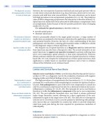
remain awake? To better delineate issues, Carskadon and Dement (4) proposed
characterizing sleepiness as introspective, physiological, and manifest. This
approach provides a potentially useful framework for understanding measurement
similarities and differences (Fig. 1). “Introspective sleepiness” indexes an individual’s
self-assessment of their internal state, or more simply, how they feel. By contrast,
5
90 Hirshkowitz
“physiological sleepiness” can be thought of as the underlying biologic drive to
sleep indexed by the amount of time it takes to fall asleep, given the opportunity.
Finally, “manifest sleepiness” reflects an individual’s inability to volitionally remain
awake. This state can be indexed by behavioral signs of sleepiness or sleep onset
(eye closure, head bobbing, snoring) or by performance deficit on a wide variety of
psychomotor and cognitive tasks. Although introspective, physiological, and manifest
sleepiness levels may stem from a common source, tests assessing sleepiness in these
different realms cannot be used interchangeably. Furthermore, attempts to use these
measures interchangeably miss the importance of the differences between them.
At extreme ends of the spectrum, sleepiness measures may be concordant.
That is, a male soldier who has remained awake continuously for 48 hours, when
asked at 4:00 .. if he is sleepy will most likely respond affirmatively. If provided
the opportunity to lie down on a comfortable bed in a dark, quiet room he would
probably fall asleep rapidly (in 5 minutes, or less). Furthermore, if he sits down in a
comfortable chair in a nonstimulating environment (a dark, quiet, and warm room),
he may be unable to remain awake (unless provided strong coffee or other
stimulants). In such a circumstance, introspective, physiological, and manifest
sleepiness measures would all agree. Conversely, a woman on vacation who has
caught up on her sleep to the point that she spontaneously awakens in the morning
and feels alert all day will typically not fall asleep at 7:00 .., even if she lays down
on a comfortable bed. Nor does she have any difficulty remaining awake sitting in a
comfortable chair in a darkened, quiet environment. Thus, at this other end of the
spectrum, there is a convergence in measures indicating full alertness.
It is the state in between full alertness and maximal sleepiness that provide a
challenge for understanding test measurement. For example, if a couple has stayed up
all night (24 hours) and is watching the sunrise at 7:00 .. they may not feel sleepy.
However, 2.5 hours ago at 4:30 .., they could barely rally enough to stay awake, but
that feeling of overwhelming sleepiness has dissipated. Nonetheless, if given the
opportunity to lie down in a comfortable bed in a dark and quiet room, they would
fall asleep instantly (unless they have been drinking coffee all night or taking stimu-
lants). Depending on their individual ability to maintain alertness, they may be able
to stay awake for more than 20 minutes if seated in a dark and quiet room. Thus, the
introspective and manifest measures of sleepiness are negative, while the physiological
measure is positive. Furthermore, even though they are now awake longer, 2.5 hours
ago, introspective and physiological measures were positive but manifest sleepiness
level was not. This illustrates the importance of understanding the differences between
measurements and not generalizing results from one to another.
FIGURE 1 Venn diagram for introspective,
physiological, and manifest sleepiness.
Alertness and Sleepiness Assessment 91
INTROSPECTIVE SLEEPINESS
Overview
There is an assortment of instruments available to assess introspective sleepiness
(Table 1). These questionnaires are all self-administered and request the individual
either to make a prediction about their behavior, estimate what they have done in
the recent past, or to assess how they feel “right now” with respect to one or another
descriptor. The instruments asking for self-report “right now” fall into a general
category of testing called “momentary assessment.” Such instruments are sensitive
to oscillation occurring over the course of a day and can be extremely useful in
scientific investigations. By contrast, assessment of how one felt during “the past
month” provide more global estimates that may be more useful clinically.
Introspective sleepiness evaluation instruments rely on self-report. Thus, all
of the advantages and disadvantages inherent in self-reported information apply.
In one sense, when it comes to rating how sleepy someone is, there is no one in a
better position to have knowledge than the person in question. Furthermore, when
it comes to indicating how one feels, the person is the only one who can render an
accurate judgment. However, the resultant index is inherently subjective. Sleepiness
reduces self-awareness and has been shown to interfere with the ability to accurately
judge internal states (i.e., it produces alexithymia). Self-reported sleepiness tends to
follow an adaptation curve such that high levels over a long duration may regress
toward the mean. This may be related to a resetting of an individual’s reference
point over time, develop from memory impairment, or both. Moreover, some
individuals are minimizers or may even be in denial. They will report low values for
everything, including sleepiness. By contrast, others are augmenters and generally
provide high scores and extreme values, regardless of the specifics in question.
Finally, responses are directly controlled by the individuals. If they have an agenda,
they can further that agenda. For example, if an individual wants his motor vehicle
driver license reinstated, he is unlikely to affirm excessive sleepiness when asked.
Epworth Sleepiness Scale
Clinically, the most widely used test for sleepiness is probably the Epworth sleepiness
scale (ESS). The popularity of ESS stems in part from its brevity. This, in conjunction
with open access for use, its simplicity, and validation studies, has cemented its
place in the clinical arena. Developed by Murray Johns (5) at the Epworth Hospital
in Melbourne, Australia, the ESS is an eight-item, validated questionnaire asking for
a self-reported expectation of “dozing” in different situations. The response set for
chance of dozing is: (0) none, (1) slight, (2) moderate, or (3) high. The situations
asked about on ESS are: (1) sitting and reading, (2) watching television, (3) sitting
and inactive in a public place (e.g., a theater or a meeting), (4) as a passenger in a car
TABLE 1 Instruments Used to Assess Introspective Sleepiness
Test name Abbreviation Creator
Epworth sleepiness scale ESS Johns
Stanford sleepiness scale SSS Hoddes et al.
Karolinska sleepiness scale KSS Akerstedt
Pictorial sleepiness scale PSS Maldonado
Profile of mood states POMS McNair
Visual analog scales VAS Assorted versions
Side effect checklists SEC Assorted versions
92 Hirshkowitz
for an hour without a break, (5) lying down to rest in the afternoon when
circumstances permit, (6) sitting and talking to someone, (7) sitting quietly after a
lunch without alcohol, and (8) in a car, while stopped for a few minutes in traffic.
Johns conducted reliability and validity studies on patients with sleep apnea and
medical student control subjects (6). In Johns’ study, student control subjects had a
mean ESS total score of 7.6. Adult respondents in our hospital (942 patients waiting
at outpatient dermatology, audiology, and ophthalmology clinics) had a mean ESS
of 8.1 compared to a mean of 5.2 found from 1120 healthy people attending health
fairs or community health lectures (7). Based on these data, we categorize ESS score
0 to 8 as normal, 9 to 12 as mild, 13 to 16 as moderate, and greater than 16 as severe.
ESS is a popular treatment outcome measure for assessing the benefit of positive
airway pressure for treating sleep apnea (8), and an ESS score above 10 is often used
to indicate significant sleepiness.
Stanford Sleepiness Scale
The Stanford sleepiness scale (SSS) is a momentary assessment scale developed by
Hoddes et al. (9) in the early 1970s. It is very brief and easy to use. It can be adminis-
tered repeatedly at short intervals (e.g., hourly) and is sensitive to sleepiness as it
waxes and wanes over the course of a day in response to circadian factors. It is also
responsive to sleep deprivation; however, normative data do not exist. For many
years, SSS was the standard measure of introspective sleepiness. It produces a single
score chosen by the individual to reflect or best describe how they feel. Choices are:
(1) feeling active and vital, alert, wide awake, (2) functioning at a high level but not
at peak; able to concentrate, (3) relaxed, awake, responsive, not at full alertness, (4) a
little foggy, not at peak; let down, (5) fogginess, beginning to lose interest in remaining
awake; slowed down, (6) sleepiness, prefer to be lying down, fighting sleep, woozy,
and (7) almost in a reverie, sleep onset soon, lost struggle to remain awake.
Karolinska Sleepiness Scale
The Karolinska sleepiness scale (KSS) has been gaining popularity for use in research
studies over the past five years. It consists of a nine-point scale that ranges from
(1) very alert to (9) very sleepy, great effort to stay awake or fighting sleep. Any
score of 7 or above is considered pathological. The KSS has been used to assess
sleepiness in drug trials, and in aircrews, oil-rig workers, train engineers, and profes-
sional drivers. Its brevity combined with now proven sensitivity to expected changes
is bringing KSS onto equal footing with the SSS. Furthermore, KSS has been vali-
dated against electrophysiologic (EEG) and behavioral parameters (10).
Pictorial Sleepiness Scale
Maldonado et al. (11) sought to develop a nonverbal sleepiness scale that could be
used to test young children or poorly educated adults. Subject groups were asked to
rank in order seven cartoon faces designed to depict different sleepiness levels.
Results were used to transform rankings into linear measures that eliminated two
faces. A new subject group ranked the remaining five cartoons and a scale was
constructed. The scale correlated significantly with KSS and SSS when tested in
groups of normal control adults, sleep apnea patients, shift workers, and school
children. The authors envision using this scale clinically and for research. It remains
to be seen whether this scale will gain popularity.
Alertness and Sleepiness Assessment 93
Profile of Mood States
As the name implies, the profile of mood states (POMS) was originally designed to
assess mood (12). However, over the years POMS gained significant popularity
among sleep researchers. Ironically, early versions of the test included a dimension
for sleepiness that was eliminated as part of psychometric test purification (because
it was not an independent factor). Sleepiness loads several subscales, including
Vigor (negative), Confusion, and Fatigue. The Confusion scale appears to be responsive
to severe sleepiness while the Vigor scale may be more responsive to partial sleep
deprivation (13). Similarly, subscales of the Medical Outcomes Study Short Form-36
(SF-36) have been used to assess sleepiness, particularly in drug trials.
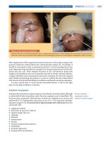
Visual Analog Scales and Side Effect Checklists
Any number of visual analog scales and side effect checklists are available to assess
sleepiness (Fig. 2). These tests are popular because of their simplicity, ease of administra-
tion, and face validity. Formal validation studies have not been conducted on these
tests. However, these types of tests are generally sensitive to changes within an
individual in response to an intervention. For example, sleepiness reliably increases
in response to sleep deprivation and decreases in response to stimulant administra-
tion. Nonetheless, absolute values of the scores are difficult to compare meaning-
fully across subjects. This limits these tests’ utility for clinical purposes.
PHYSIOLOGICAL SLEEPINESS
Overview
The three most common methods for indexing physiological sleepiness use: (i) the
multiple sleep latency test (MSLT), (ii) pupillometry, and (iii) quantitative electroen-
cephalography (EEG) analysis. Measuring sleepiness using physiological indices
provides the clinician an objective technique that does not have the disadvantages
inherently associated with self-report. Falling asleep represents an involuntary
process. Therefore, appraising the biological substrate either during this process or
under conditions conducive to falling asleep can minimize psychological, psychiatric,
and intentional confounds. However, one should be aware that the results derived
from such testing are not necessarily above manipulation by a test subject. In as much
as the test procedure relies on cooperation, there is room for intentional alteration,
within limits. For example, individuals attempting to prove they are not sleepy may
engage in a mental arithmetic task when asked to close their eyes and relax during an
FIGURE 2 Example of a visual analog
scale.
94 Hirshkowitz
EEG baseline sample. Physiological measures may be more reliable for demonstrating
sleepiness than proving alertness (i.e., if a positive test is one that affirms sleepiness,
physiological tests are more prone to false negative than false positive results).
Multiple Sleep Latency Test
MSLT provides a direct, objective, quantitative measure indexing sleepiness. It is
generally thought that nonsleepy individuals cannot make themselves fall asleep.
Thus, if a positive MSLT is one that indicates sleepiness, then false-positive test
liability is minimal. The MSLT consists of a series of nap opportunities (4–6)
presented across the day. The series begins approximately two hours after the morning
awaking from an in-laboratory polysomnographic study and continues with
successive trials at two-hour intervals (14). Testing includes polysomnographic
sleep monitoring during the nap opportunities. Recordings include: (1) left or right
central EEG (C3 or C4), (2) left or right occipital EEG (O1 or O2), (3) left horizontal
or oblique electrooculogram (EOG), (4) right horizontal or oblique EOG, (5) sub-
mentalis (chin) electromyogram (EMG), (6) electrocardiogram (ECG), (7) respiratory
flow (if needed), and (8) respiratory sounds (if needed). Subjects may not remain in
bed between test sessions and should wear normal street clothing. Rooms must be
dark and quiet during nap opportunities. Two MSLT protocols have evolved, one
for clinical use and one for research. In the clinical protocol, a subject that falls asleep
on a nap opportunity is allowed an additional 15 minutes of sleep to see whether
rapid eye movement (REM) sleep occurs. The presence of REM sleep on two or
more naps is used to confirm narcolepsy. In the research protocol, sleep accumulation
is minimized. The test session is terminated when either (i) unequivocal sleep occurs
(one epoch of stage 2, 3, 4, or REM) or (ii) three successive epochs of stage 1 sleep
occur. In both clinical or research protocols, the nap opportunity is terminated after
20 minutes if sleep onset fails to occur. At the beginning of each nap opportunity, the
subject is instructed to “let yourself fall asleep” or “do not resist falling asleep.” The
speed with which an individual falls asleep (defined by polysomnographic criteria)
is used to index the level of sleepiness. Changes in sleep latency associated with age,
circadian factors, sleep deprivation, sleep extension, and sleep disorders have been
described (15–18). The MSLT provides a sensitive index for evaluating response to
treatment and is a favorite outcome measure in drug trials (19–21) and other treat-
ment outcome studies (8). Therefore, the MSLT should not be conducted after a
night of profoundly disturbed sleep, during drug withdrawal, or while a patient is
under the influence of sedating medication. The American Academy of Sleep
Medicine (AASM) Standards of Practice Committee published new practice
parameters for the clinical use of MSLT (22). Recommendations were based on a
comprehensive evidence-based medicine literature review; however, expert consensus
was used to bridge areas where data were insufficient (23). Recommendations are
summarized in Table 2.
Pupillometry
In a dark room, an individual’s pupils dilate in order to allow more light to enter the
eye. This process is part of dark adaptation. However, when a person is sleepy and
begins to fall asleep, the pupils constrict in response to the ANS increasing parasym-
pathetic tone associated with sleep. Thus, recording changes in pupil size can physi-
ologically index sleep tendency (24,25). Thus, a wide-awake individual’s pupils will
dilate and remain dilated over time in a dark room. By contrast, an individual with
Alertness and Sleepiness Assessment 95
excessive sleepiness will not maintain pupil dilation as he or she drifts from alert-
ness to drowsiness to sleep. Pupillometry provides an objective method for recording
changes in the pupil and has been used to study narcolepsy (26). Pupillometry has
also been correlated with MSLT and SSS indices (27). Changes occurring within
subjects are sensitive and reliable; however, comparisons across subjects can be
difficult. Additionally, normative data are not available.
Quantitative Electroencephalographic Analysis
It would seem obvious that the EEG should change as sleepiness increases. With
widely available computerized waveform analysis one would expect there to be a
recognizable marker in brain activity. EEG delta activity seems a strong candidate
and was found to increase in response to sleep deprivation (28). Other EEG markers
have also been reported (29). It is also classic knowledge that EEG alpha disappears
at sleep onset (first noted by Hans Berger almost 100 years ago). Furthermore, just
before alpha activity disappears, its frequency decreases and its amplitude increases.
Santamaria and Chiappa (30) filled an entire book with illustrations of the EEG of
drowsiness. Nonetheless, the interlocking piece reliably connecting EEG and
sleepiness has been elusive. Even simple observation by a trained observer remains
more sensitive than the most sophisticated measures for predicting sleepiness (31)
and sleepiness-related performance deficits (32).
MANIFEST SLEEPINESS
Overview
Measurements of manifest sleepiness are actually designed to index the point at
which alertness fails. An individual may be very sleepy but nonetheless can maintain
wakefulness. However, at some point, even the most heroic attempts to stay awake
are to no avail and it is at this point the individual lapses into sleep. There is usually
some response slowing and lapsing even before frank sleep onset occurs. For many
years the notion of “functional deafferentation” was considered; however, most data
point toward inattention and slowing of information processing. Another controversial
issue involves the concept of microsleep (brief sleep episodes lasting 5–15 seconds).
TABLE 2 Summary of American Academy of Sleep Medicine Standards of Practice
Committee Recommendations for Clinical Use of the Multiple Sleep Latency Test
The MSLT is indicated as part of the clinical evaluation for patients with suspected narcolepsy
(two or more SOREMPs has a 0.78 sensitivity and a 0.93 specificity for diagnosing
narcolepsy).
The MSLT may be helpful for clinical assessment of patients with suspected idiopathic
hypersomnia.
The MSLT is not indicated for routine evaluation of obstructive sleep apnea, insomnia, circadian
rhythm disorders, or dyssomnia associated with medical, psychiatric, or neurological disorders
(other than narcolepsy and idiopathic hypersomnia).
The MSLT is not indicated for routine re-evaluation of patients with sleep apnea treated with
positive airway pressure therapy.
Repeat MSLT may be indicated when:
Study conditions were inappropriate during initial testing
Results are ambiguous
Earlier MSLT did not confirm narcolepsy.
Abbreviations: MSLT, multiple sleep latency test; SOREMP, sleep-onset REM period.
96 Hirshkowitz
Industrial psychology and human factors research attempting to design better man–
machine interfaces (ergonomics) avoided issues surrounding microsleep because an
equipment operator’s performance cannot be improved by rearranging a panel’s
switches, keyboards, and indicator lights if he or she has fallen asleep. Another issue
concerning microsleep involves the locus of its generation. Attempts to externalize
the cause of microsleep have led to blaming rural motor vehicle accidents on things
such as “highway hypnosis.” The problem, however, is internal. The driver is sleepy
and when sleepiness reaches a threshold that exceeds the alertness system’s ability to
offset, manifest sleepiness occurs and sleep onset soon follows.
Observation and Observer Scales
Yawning has long been a traditional sign of sleepiness. Simple observation of eyelid
closure, wandering direction of gaze, and eyes rolling upward are among the strongest
indicators of sleepiness. Head nodding with eyes closed strongly suggests sleep
onset. These observable events testify to manifest sleepiness. Some of the earliest
attempts to index sleepiness involved the development of observer scales, one of
which was used to grade individuals undergoing prolonged sleep deprivation.
Studies conducted at Walter Reed Hospital in the 1960s employed the “Cognitive
Disorganization Scale” (33). This five-point observer rating scale ranges from (1)
slowing of mental processes, some difficulty thinking of words (no undue interfer-
ence with normal communication) all the way to (5) rambling incoherent speech for
brief periods, with failure to recognize errors, unable to straighten out jumble of
incoherent thoughts when challenged.
Maintenance of Wakefulness Test
The maintenance of wakefulness test (MWT) is procedurally similar to the MSLT.
The major differences are: (i) the person being tested is told to “attempt to remain
awake” at the beginning of each test session, (ii) the individual is seated rather than
laying down in bed, (iii) each test session is 40 minutes in duration, and (iv) poly-
somnography the night before testing is not required. Thus, the MWT is used to
assess an individual’s capability to not be overwhelmed by sleepiness. In a sense,
this test is gauging the strength of the wakefulness system (34). If the wakefulness
system fails, sleepiness becomes manifest. In the MWT there is no other task than
remaining awake and concurrent EEG–EOG–EMG monitoring is conducted to
verify success or failure (35,36). In some ways the MWT is a simulation of sedentary
inactivity in a nonstimulating environment. Like the MSLT, there are four to six
sessions, scheduled at two-hour intervals beginning approximately two hours after
awakening from the previous night’s sleep. Until standardization at 40 minutes
recommended by the AASM Standards of Practice Committee, individual test ses-
sion durations on the MWT varied from 20 to 60 minutes in different studies. The
MWT has proven useful as an outcome measure in clinical trials (37) and is recog-
nized for evaluating noncommercial pilot relicensing after sleep apnea treatment (38).
Practice guidelines for MWT were developed along with those for the MSLT. MWT
testing is indicated for evaluating individuals whose inability to remain alert
constitutes a safety hazard. MWT testing is also recommended to determine
treatment response in patients with narcolepsy (or idiopathic hypersomnia).
The practice parameter emphasizes the critical importance of clinical judgment
and stresses the fact that normal MWT values do not guarantee safety.
Recommendations are summarized in Table 3.
Alertness and Sleepiness Assessment 97
Vigilance and Performance Tests
Impaired vigilance and performance decrements accompany sleepiness and an
increase in response to sleep deprivation. Inattention, cognitive slowing, and lapses
represent manifestations of sleepiness. Experimenter-paced, monotonous, time-
keeping tasks with low target presentation rates generally fall into the category of
“vigilance tests.” Tests with higher throughput and greater intrinsic stimulation are
classed as “performance tests.” Vigilance tests do not require much skill, are less
sensitive to educational level, and have little in the way of learning curves com-
pared to performance tests. Landmark studies of sleep deprivation pioneered the
use of vigilance and performance testing to assess sleepiness (39,40). These studies
are collectively known as the Walter Reed experiments because they were conducted
at Walter Reed Army Hospital. Results indicated that time-on-task, response slowing,
and response lapsing were essential factors in sustained attention tasks [for an excellent
review, see Dinges and Kribbs (41)]. Several vigilance and performance tests are in
standard use in sleepiness research (42–44). Perhaps, the most popular is the psy-
chomotor vigilance test (PVT) developed by David Dinges et al. at the University of
Pennsylvania (45,46). This hand-held device measures response speed and accuracy
in a signal detection algorithm. The PVT has been cross-validated with the SSS and
MSLT and data are reported from normal controls, sleep-deprived individuals, and
sleep disorder patient groups. Another test system that has been used in several
studies is the Oxford sleep resistance (OSLER) test (47,48). The OSLER test
consists of four 40-minute-long trials during which there are multiple signal presen-
tations. The subject is instructed to respond to each signal with a simple button
press. Trials are ended after 40 minutes or after a failure to respond (which is thought
to indicate sleep onset).
CONCLUSIONS
Sleepiness is a cardinal symptom for some sleep disorders. Consequently, assessing
sleepiness and alertness is a crucial part of a clinical evaluation. There is an assort-
ment of techniques available to measure sleepiness. Sleepiness, however, represents
an interaction of physiological systems that increase sleep tendency and other
systems opposing sleep drive. These complementary factors provide a dynamic
equilibrium that oscillates over the course of a day depending on homeostatic sleep
debt and circadian phase. Furthermore, the internal state individuals interpret as indi-
cating sleepiness can be influenced by other factors. The net result is that different
TABLE 3 Summary of American Academy of Sleep Medicine Standards of Practice
Committee Recommendations for Clinical Use of the Maintenance of Wakefulness Test
The 40-minute MWT protocol should be used.
MWT is indicated for assessing an individual’s ability to remain awake when sleepiness
constitutes a public or personal safety issue.
MWT may be used to assess response to treatment.
On a 40-minute MWT, 59% of normal subjects remain awake the entire time across all four trials.
On the 40-minute MWT
A sleep latency less than eight minutes is abnormal
A sleep latency between 8 and 40 minutes is of unknown significance
The mean (± SD) sleep latency is 30.4 (± 11.2)
The upper 95% confidence interval is 40 minutes.
Abbreviation: MWT, maintenance of wakefulness test.
98 Hirshkowitz
measures may not correlate well with one another in some individuals.
Understanding the differences between introspective, physiological, and manifest
sleepiness metrics is useful to interpret results. The ESS is a widely used self-report
instrument in general clinical practice. By contrast, the MWT is a recommended
objective evaluation technique when personal and public safety concerns arise.
This test measures the ability to sustain wakefulness in a soporific environment.
Recommendations for the use of the MSLT and the MWT are summarized in this
chapter. Unfortunately, no simple blood test or assay is available for indexing sleep-
iness. The difficulty in measuring sleepiness poses a significant challenge in the
regulatory arena. Nonetheless, sleepiness is a serious, noncommunicable, poten-
tially life-threatening condition.
REFERENCES
1. National Commission on Sleep Disorders Research: Wake up America: A national sleep
alert. Executive Summary and Executive Report, Report of the National Commission on
Sleep Disorders Research. Washington, DC, National Institutes of Health, U.S.
Government Printing Office, 1993; 1:45.
2. American Academy of Sleep Medicine. The International Classification of Sleep
Disorders, revised: diagnostic and coding manual. 2nd ed. Rochester: American Academy
of Sleep Medicine, 2004.
3. Borbély AA, Achermann P. Concepts and models of sleep regulation: an overview. J Sleep
Res 1992; 1:63.
4. Carskadon MA, Dement WC. The multiple sleep latency test: what does it measure?
Sleep 1982; 5:S67–S72.
5. Johns MW. A new method for measuring daytime sleepiness: the Epworth Sleepiness
Scale. Sleep 1991; 14:540–545.
6. Johns MW. Reliability and factor analysis of the Epworth Sleepiness Scale. Sleep 1992;
15:376–381.
7. Sharafkhaneh A, Hirshkowitz M. Contextual factors and perceived self-reported sleepiness:
a preliminary report. Sleep Med 2003; 4:327–331.
8. Patel SR, White DP, Malhotra A, Stanchina ML, Ayas NT. Continuous positive airway
pressure therapy for treating sleepiness in a diverse population with obstructive sleep
apnea: results of a meta-analysis. Arch Intern Med 2003; 163(5):565–571.
9. Hoddes E, Zarcone V, Smythe H, et al. Quantification of sleepiness: a new approach.
Psychophysiology 1973; 10:431–436.
10. Kaida K, Takahashi M, Akerstedt T, et al. Validation of the Karolinska sleepiness scale
against performance and EEG variables. Clin Neurophysiol 2006; 117(7):1574–1581.
11. Maldonado CC, Bentley AJ, Mitchell D. A pictorial sleepiness scale based on cartoon
faces. Sleep 2004; 27(3):541–548.
12. McNair DM, Lorr M, Droppleman LF. Manual of the Profile of Mood States. San Diego,
California: Educational and Industrial Testing Service, 1992.
13. Horne J. Dimensions to sleepiness. In: Monk TM, ed. Sleep, Sleepiness and Performance.
Chichester, England: John Wiley, 1991:169–196.
14. Carskadon MA, Dement WC, Mitler MM, et al. Guidelines for the multiple sleep latency
test (MSLT): a standard measure of sleepiness. Sleep 1986; 9:519–524.
15. Carskadon MA, Dement WC. Cumulative effects of sleep restriction on daytime sleepiness.
Psychophysiology 1981; 18:107–113.
16. Carskadon MA, Dement WC. Daytime sleepiness: quantification of behavioral state.
Neurosci Biobehav Rev 1987; 11:307–317.
17. Mitler MM, Nelson S, Hajdukovic R. Narcolepsy: diagnosis, treatment, and management.
Psychiatr Clin North Am 1987; 10:593–606.
18. Richardson GS, Carskadon MA, Orav EJ, et al. Circadian variation in sleep tendency in
elderly and young adult subjects. Sleep 1982; 5:S82–S94.
19. Lamphere J, Roehrs T, Wittig R, et al. Recovery of alertness after CPAP in apnea. Chest
1989; 96:1364–1367.
Alertness and Sleepiness Assessment 99
20. U.S. Narcolepsy Study Group (USMNSG). Randomized trial of for the treatment of
pathological somnolence in narcolepsy. Ann Neurol 1998; 43:88–97.
21. Zorick F, Roehrs T, Conway W, et al. Effects of uvulopalatopharyngoplasty on the day-
time sleepiness associated with sleep apnea syndrome. Bull Eur Physiopathol Respir
1983; 19:600–603.
22. Standards of Practice Committee of the American Academy of Sleep Medicine. Practice
parameters for clinical use of the multiple sleep latency test and the maintenance of
wakefulness test. Sleep 2005; 28:113–121.
23. A Review by the MSLT and MWT Task Force of the Standards of Practice Committee of
the American Academy of Sleep Medicine. The clinical use of the MSLT and MWT. Sleep
2005; 28:123–144.
24. Schmidt HS, Fortin L. Electronic pupillography in disorders of arousal. In: Guilleminault
C, ed. Sleeping and Waking Disorders: Indications and Techniques. Menlo Park, California:
Addison-Wesley, 1982:127–143.
25. Morad Y, Lemberg H, Yofe N, Dagan Y. Pupillography as an objective indicator of fatigue.
Curr Eye Res 2000; 21:535–542.
26. Yoss RE, Moyer NJ, Ogle KN. The pupillogram and narcolepsy: a method to measure
decreased levels of wakefulness. Neurology 1969; 19:921–928.
27. Danker-Hopfe H, Kraemer S, Dorn H, et al. Time-of-day variations in different measures
of sleepiness (MSLT, pupillography, and SSS) and their interrelations. Psychophysiology
2001; 38:828–835.
28. Borbely AA, Baumann F, Brandeis D, et al. Sleep deprivation: effect on sleep stages
and EEG power density in man. Electroencephalogr Clin Neurophysiol 1981;
51:483–493.
29. Hasan J, Hirvonen K, Varri A, et al. Validation of computer analyzed polygraphic patterns
during drowsiness and sleep onset. Electroencephalogr Clin Neurophysiol 1993; 87:117–127.
30. Santamaria J, Chiappa KH. The EEG of Drowsiness. New York: Demos, 1987.
31. Mitler MM, Miller JC, Lipsitz JJ, et al. The sleep of long-haul truck drivers. N Engl J Med
1997; 337:755–761.
32. Dinges DF, Mallis MM. Managing fatigue by drowsiness detection: can technological
promises be realized? Third International Conference on Fatigue in Transportation:
Coping with the 24-Hour Society. Fremantle, Western Australia, February 9–13, 1998.
33. Morris GO, Williams HL, Lubin A. Misperception and disorientation during sleep depri-
vation. Arch Gen Psychiat 1960; 2:247–254.
34. Sangal RB, Thomas L, Mitler MM. Disorders of excessive sleepiness: treatment improves
ability to stay awake but does not reduce sleepiness. Chest 1992; 102:699–703.
35. Doghramji K, Mitler MM, Sangal RB, et al. A normative study of the Maintenance of
Wakefulness Test (MWT). Electroencephalogr Clin Neurophysiol 1997; 103:554–562.
36. Mitler MM, Miller JC. Methods of testing for sleepiness. Behav Med 1996; 21:171–183.
37. Mitler MM, Gujavarty KS, Browman CP. Maintenance of wakefulness test: a polysomno-
graphic technique for evaluation of treatment efficacy in patients with excessive somnolence.
Electroencephalogr Clin Neurophysiol 1982; 53:658–661.
38. Federal Aviation Administration (FAA). Sleep Apnea Evaluation Specifications. Federal
Aviation Administration specification letter dated October 6, 1992. U.S. Department of
Transportation, 1992.
39. Lubin A. Performance under sleep loss and fatigue. In: Kety SS, Evarts EV, Williams HL, eds.
Sleep and Altered States of Consciousness. Baltimore, MD: Williams and Wilkins;
1967:506–513.
40. Williams HL, Lubin A, Goodnow JJ. Impaired performance and acute sleep loss. Psychol
Monogr 1959; 73(Pt 14):1–26.
41. Dinges DF, Kribbs NB. Performing while sleepy: effects of experimentally induced
sleepiness. In: Monk TM, ed. Sleep, Sleepiness and Performance. Chichester, England:
John Wiley, 1991:97–128.
42. Wilkinson RT. Sleep deprivation: performance tests for partial and selective sleep deprivation.
In: Abt LA, Reiss BF, eds. Progress in Clinical Psychology. Vol. 3. London: Grune & Stratton,
1968:28–43.
43. Hirshkowitz M, De La Cueva L, Herman JH. The multiple vigilance test. Behav Res
Methods Instrum Comput 1993; 25:272–275.
100 Hirshkowitz
44. Findley L, Unverzagt M, Guchu R, et al. Vigilance and auto-mobile accidents in patients
with sleep apnea or narcolepsy. Chest 1995; 108:619–624.
45. Kribbs NB, Pack AI, Kline LR, et al. Effects of one night without nasal CPAP treatment on
sleep and sleepiness in patients with obstructive sleep apnea. Am Rev Respir Dis 1993;
147:1162–1168.
46. Doran SM, Van Dongen HP, Dinges DF. Sustained attention performance during sleep
deprivation: evidence of state instability. Arch Ital Biol 2001; 139:253–267.
47. Bennett LS, Stradling JR, Davies RJ. A behavioural test to assess daytime sleepiness in
obstructive sleep apnoea. J Sleep Res 1997; 6:142–145.
48. Priest B, Brichard C, Aubert G, et al. Microsleep during a simplified maintenance of wakefulness
test: a validation study of the OSLER test. Am J Respir Crit Care Med 2001; 163:1619–1625.
101
Section II: Treatment
Continuous Positive Airway Pressure
Peter R. Buchanan and Ronald R. Grunstein
Royal Prince Alfred Hospital, Woolcock Institute of Medical Research and
The University of Sydney, Sydney, Australia
INTRODUCTION
Continuous positive airway pressure (CPAP), usually nasally applied, is the estab-
lished treatment for moderate-to-severe obstructive sleep apnea (OSA) (1). Nasal
CPAP therapy for sleep apnea was first described in 1981 (2). Although there was
initial skepticism of its efficacy and concern regarding its potential adverse effects
on breathing (3,4), there was also early recognition of the importance of having a
treatment that could essentially prevent disordered breathing during sleep in OSA
patients. This is in contrast to the efficacy of other alternatives available, including
partial or variable response to surgery (5). By 1985, more than 100 patients were
using this therapy on a regular basis (6). Over the past 20 years, the evidence base
supporting the use of CPAP has improved both in quantity and quality, driven at
least in part by the demands of government funding authorities and health main-
tenance organizations and the availability of industry sponsorship with the increas-
ing commercial success of companies selling CPAP equipment (7).
There are methodological problems designing studies to assess and validate a
mechanical device such as CPAP, compared with those required for medications.
Performing true double-blind randomized controlled trials (RCTs) of CPAP
treatment or variants are technically and logistically difficult. “Sham CPAP” by its
nature will have less efficacy on unavoidably observable variables such as snoring
or apnea with consequent difficulties to truly “blind” study participants. Due to the
requisite modification to the equipment, it is also quite difficult to effectively
blind a CPAP therapist or doctor involved in such studies compared with pharma-
ceutical trials involving placebo medications. Also the advent of automatically
titrating CPAP devices (see Chapter 8) has major implications for the delivery of
healthcare to patients with sleep apnea and for the traditional sleep laboratory–
patient relationship.
CPAP is currently the “gold standard” treatment for moderate-to-severe
OSA because of its demonstrated efficacy. Even so, many patients do not use it, or
use it irregularly, reducing the delivered effectiveness of the therapy. Comparative,
double-blind intention-to-treat trials in all degrees of OSA severity are needed to
delineate treatment pathways in this condition. Currently, studies focusing on com-
parative treatments and ways in which there are better effectiveness of CPAP,
including timely and economical initiation of therapy, are forming the next phase in
the historical development of this treatment modality. Although there are tremen-
dous interest and active research in potential pharmacotherapy for OSA, the absence
of any currently available viable pharmacological therapy for sleep apnea (8–10)
suggests that CPAP will remain the appropriate therapy standard in the foreseeable
future for OSA of more than mild degree.
6
102 Buchanan and Grunstein
CONTINUOUS POSITIVE AIRWAY PRESSURE
Mode of Action
The concept of CPAP in managing respiratory failure is relatively old (11). However,
the original experiments using CPAP in sleep apnea followed from the notion that
closure of the oropharynx in OSA results from an imbalance of the forces (12)
that normally keep the upper airway open. In the first description of CPAP use for treat-
ment of OSA in 1981 (2), it was suggested that nasal CPAP acts as a pneumatic splint
to prevent collapse of the pharyngeal airway, by elevating the pressure in the
oropharyngeal airway and reversing the transmural pressure gradient across the
pharyngeal airway (Fig. 1). This notion has been subsequently confirmed by a
number of studies which either demonstrate the “pneumatic splint” effect by endo-
scopic or other imaging, or show that CPAP does not increase upper airway muscle
activity by reflex mechanisms (13). Detailed magnetic resonance imaging has
confirmed that CPAP increases airway volume and airway area, and reduces lateral
pharyngeal wall thickness and upper airway edema secondary to chronic vibration
and occlusion of the airway (14). The apparatus providing the pressure at the nasal
FIGURE 1 Mechanism of upper
airway occlusion in obstructive
sleep apnea and its prevention by
continuous positive airway pres-
sure: “pneumatic splint” effect.
Source: From Ref. 2.
Continuous Positive Airway Pressure 103
airway must have the “capacity” to maintain any given pressure during inspiration.
Early embodiments of the technology failed to respond rapidly to the airflow chal-
lenge at early inspiration, with an attendant pressure drop and consequently requir-
ing a higher “set” pressure to compensate for such drops. The simplest CPAP
systems involve an air blower with sufficient pressure-flow characteristics to pro-
vide CPAP while also accommodating both a fixed resistive leak in the system (typi-
cally adjacent to the mask) as well as unintentional leaks at the patient/delivery
interface (Fig. 2). Such contemporary devices are generally microprocessor-con-
trolled in order to meet rapid changes in the flow demands presented by the patient
as well as the dynamics of variable leak often seen in clinical use of CPAP.
Continuous Positive Airway Pressure and Central Apnea
Regardless of the mechanism, nasal CPAP has been documented to be effective in elimi-
nating both mixed and obstructive apneas (15). Some central apneas (see Chapter 19),
particularly those observed in patients with predominantly obstructive events, are
also eliminated by nasal CPAP (15). Clearly, some central apneas are associated with
increased upper airway resistance and it could be argued that it is better to consider
apnea classification as being CPAP responsive or CPAP nonresponsive. CPAP may
also be effective in controlling central apneas associated with cardiac failure (see
section: Continuous Positive Airway Pressure and Cardiac Failure). In some
instances, CPAP alone will not control severe mixed central and obstructive apneas
and adjunctive entrainment of low concentrations of carbon dioxide, with CPAP, or
the use of other noninvasive ventilatory techniques, may further reduce the respira-
tory disturbance index (RDI) in such individuals (16).
PRACTICAL ASPECTS OF TREATMENT
Originally, most patients commenced CPAP under supervision, usually in a hospital-
based sleep laboratory. The purposes of this supervision included ensuring that the
patient was appropriately educated about the therapy, to select the best interface
(mask) for the individual, and determine the adequacy of CPAP across the night.
Such observation also allowed an evaluation of the immediate acceptance of or
problems with the therapy. Economic pressures within health systems however
FIGURE 2 Subject wearing nasal con-
tinuous positive airway pressure.
104 Buchanan and Grunstein
have challenged this approach. Alternative nonlaboratory-based approaches to
initiating CPAP are being applied in numerous health systems. For example,
in 2004, throughout New Zealand nearly all (> 90% in some centers) CPAP initiation
was implemented via either an attended in-laboratory split-night or unattended
home auto-CPAP titration (Neill A, personal communication). Similarly, in the
United Kingdom full in-laboratory CPAP titration studies are not routinely under-
taken in many centers. Economic drivers have been of major importance in the
adoption of these practices in these and other health services. As part of the drive
toward economic rationalization, health authorities expect some evidence base for
clinical CPAP titration strategies. Some such evidence has been accumulated; how-
ever, findings are somewhat contradictory.
Irrespective of the location or method of CPAP titration, there is a clear demand
for proper patient assessment (e.g., does the patient have awake respiratory failure
or marked hypoxemia in sleep?), which in turn requires specific physician training
and experience. Until recently there was no evidence for the safety and efficacy of
CPAP titration outside of a medically supervised process (17). Current evidence
supports the use of trained technologists to provide patient education, technical
aspects of titration, and follow-up. However, data from studies of small patient
samples have challenged the presumption of close medical supervision during the
initiation of therapy. Clearly this area requires further major research focus before a
consensus may be derived (18–20).
The First Night
Sleeping with a nasal mask applied to the face, along with feeling the pressure
sensation of CPAP, although not necessarily uncomfortable, are certainly novel
experiences for most patients. Physician explanation, video programs, and mask
“acclimatization” sessions prior to commencing CPAP are routine in many centers.
Although the benefits of these approaches have not been fully scientifically evalu-
ated, it would seem obvious that patient education about CPAP will reduce anxiety
and improve acceptance. Current evidence provides some support for the benefit of
more intensive patient education in CPAP usage (17,21). Thus, patient exposure to
CPAP actually may begin prior to the first full night of therapy.
On the first night of treatment, it is important to ensure that the CPAP level that
is identified as most therapeutically effective is sufficient not only to prevent apnea
and oxyhaemoglobin desaturation (Fig. 3) but also to prevent respiratory-related
arousals in all sleep stages and postures of sleep. Thus, simple apnea prevention is
not the sole endpoint of CPAP titration. It is important to ensure that the airflow-
CPAP pressure measurement is competent so as to avoid residual partial airway
obstruction (7). An abnormal or technically challenged tracing (e.g., amplifier satu-
ration, clipped signals) presents an opportunity for failure in detection of flow limi-
tation and snoring. It is important to treat residual flow limitation as it may indicate
upper airway obstruction, potentially causing arousal (22). Studies have empha-
sized the importance of proper airflow measurement in CPAP titration using pres-
sure-based airflow transducers rather than thermistors or other more indirect
airflow measures (23). Proper airflow measurement could help determine the optimal
CPAP level by providing insights regarding the etiology of arousals; whether they
are related to respiratory events (respiratory-related arousals) and if increasing
pressure has a beneficial effect on sleep continuity. Although acute (one-night) studies
suggest that flow limitation correction may be the preferred endpoint of CPAP